Neural repetition suppression: evidence for perceptual expectation in object-selective regions
8
ORIGINAL RESEARCH ARTICLE published: 17 April 2014 doi: 10.3389/fnhum.2014.00225 Neural repetition suppression: evidence for perceptual expectation in object-selective regions Lisa Mayrhauser 1 *, Jürgen Bergmann 2 , Julia Crone 1,2 and Martin Kronbichler 1,2 1 Centre for Neurocognitive Research, University of Salzburg, Salzburg, Austria 2 Neuroscience Institute, Christian-Doppler Clinic, Paracelsus Medical University, Salzburg, Austria Edited by: Christopher Summerfield, Oxford University, UK Reviewed by: Tobias Egner, Duke University, USA Floris P. De Lange, Radboud University Nijmegen, Netherlands *Correspondence: Lisa Mayrhauser, Centre for Neurocognitive Research, University of Salzburg, Hellbrunnerstraße 34, 5020, Salzburg, Austria e-mail: [email protected] It is an established finding that neuronal activity is decreased for repeated stimuli. Recent studies revealed that repetition suppression (RS) effects are altered by manipulating the probability with which stimuli are repeated. RS for faces is more pronounced when the probability of repetition is high than when it is low. This response pattern is interpreted with reference to the predictive coding (PC) account, which assumes that RS is influenced by top-down expectations. Recent findings challenge the generality of PC accounts of RS by showing repetition probability does not modulate RS for other visual stimuli than faces. However, a number of findings on visual processing are in line with PC. Thus, the influence of repetition probability on RS effects during object processing requires careful reinvestigations. In the present fMRI study, object pictures were presented in a high (75%) or low (25%) repetition probability context. We found increased RS in the high-probability context compared to the low-probability context in the left lateral occipital complex (LOC). The dorsal-caudal and the ventral-anterior subdivisions of the LOC revealed similar neuronal responses. These results indicate that repetition probability effects can be found for other visual objects than faces and provide evidence in favor of the PC account. Keywords: fMRI, lateral occipital complex, object processing, predictive coding, repetition suppression INTRODUCTION Repetition suppression (RS) is commonly defined as a dimin- ished neural activation that results from the repeated presen- tation of a stimulus (Henson, 2003). In functional magnetic resonance imaging (fMRI), this pattern becomes manifest as the reduced blood oxygen-level-dependent (BOLD) response elicited by a repeated stimulus, also called fMRI adaptation (Grill- Spector and Malach, 2001; for a recent review see also Segaert et al., 2013). Critically, its underlying neuronal mechanisms have been discussed as either arising from neuronal fatique (Grill- Spector et al., 2006), neuronal sharpening (Martens and Gruber, 2012), or neuronal facilitation (Grill-Spector et al., 2006), accord- ing to which RS is a relatively automatic result of bottom-up mechanisms. By contrast, the predictive coding (PC) model emphasizes the role of top-down influences on RS. According to this approach, information about a stimulus (e.g., an object) flows in a hierar- chical manner from lower to higher cortical layers while expecta- tions, which are built upon prior object regularities, are top-down backward influences which modulate the processing of the cur- rent object (Friston, 2005). Bottom-up flow of information and top-down expectations are then compared at each level of hier- archical processing. From this perspective, RS occurs due to a correct prediction of the upcoming stimulus, that is, the cur- rently processed object matches the expectation. Therefore, RS effects reflect a smaller prediction error for expected stimuli, that is, decreased activation for repeated stimuli. This model was recently put forward by the finding that a manipulation of expectations alters RS effects (Summerfield et al., 2008, 2011). In these studies, the probability with which pictures of faces were repeated altered between blocks, thus inducing a rel- atively high expectation of repetitions in high-probability blocks (75% repetitions) and a rather weak expectation of repetitions in low-probability blocks (25% repetitions). RS effects were more pronounced when repetition was expected compared to when it was less expected. These perceptual expectation effects cannot be explained by bottom-up mechanisms alone. Findings challenging the PC account of RS revealed that RS effects during object processing in monkey inferior temporal cortex and human lateral occipital complex (LOC) were not mod- ulated by repetition probability(Kaliukhovich and Vogels, 2011; Kovács et al., 2013; respectively), thus questioning the general- ity of top-down effects in visual perception. However, the notion that perceptual expectations about objects should not affect RS effects would be surprising, since prior investigations indicate that top-down effects modulate the processing of visual stimuli at each level of hierarchical processing. To illustrate, the results of Cardin et al. (2011) suggest that learnt regularities, which are related to increased activation in anterior visual and frontal areas, gener- ate top-down signals. Additional support for the validity of PC in object perception is provided by recent findings indicating that RS is driven by changes in top-down effects in body-sensitive net- works (Ewbank et al., 2011) and that top-down expectations and surprise effects account for neuronal responses in the FFA and Frontiers in Human Neuroscience www.frontiersin.org April 2014 | Volume 8 | Article 225 | 1 HUMAN NEUROSCIENCE
Transcript of Neural repetition suppression: evidence for perceptual expectation in object-selective regions

ORIGINAL RESEARCH ARTICLEpublished: 17 April 2014
doi: 10.3389/fnhum.2014.00225
Neural repetition suppression: evidence for perceptualexpectation in object-selective regionsLisa Mayrhauser1*, Jürgen Bergmann2, Julia Crone1,2 and Martin Kronbichler1,2
1 Centre for Neurocognitive Research, University of Salzburg, Salzburg, Austria2 Neuroscience Institute, Christian-Doppler Clinic, Paracelsus Medical University, Salzburg, Austria
Edited by:
Christopher Summerfield, OxfordUniversity, UK
Reviewed by:
Tobias Egner, Duke University, USAFloris P. De Lange, RadboudUniversity Nijmegen, Netherlands
*Correspondence:
Lisa Mayrhauser, Centre forNeurocognitive Research, Universityof Salzburg, Hellbrunnerstraße 34,5020, Salzburg, Austriae-mail: [email protected]
It is an established finding that neuronal activity is decreased for repeated stimuli. Recentstudies revealed that repetition suppression (RS) effects are altered by manipulating theprobability with which stimuli are repeated. RS for faces is more pronounced when theprobability of repetition is high than when it is low. This response pattern is interpretedwith reference to the predictive coding (PC) account, which assumes that RS is influencedby top-down expectations. Recent findings challenge the generality of PC accounts ofRS by showing repetition probability does not modulate RS for other visual stimuli thanfaces. However, a number of findings on visual processing are in line with PC. Thus,the influence of repetition probability on RS effects during object processing requirescareful reinvestigations. In the present fMRI study, object pictures were presented ina high (75%) or low (25%) repetition probability context. We found increased RS inthe high-probability context compared to the low-probability context in the left lateraloccipital complex (LOC). The dorsal-caudal and the ventral-anterior subdivisions of theLOC revealed similar neuronal responses. These results indicate that repetition probabilityeffects can be found for other visual objects than faces and provide evidence in favor ofthe PC account.
Keywords: fMRI, lateral occipital complex, object processing, predictive coding, repetition suppression
INTRODUCTIONRepetition suppression (RS) is commonly defined as a dimin-ished neural activation that results from the repeated presen-tation of a stimulus (Henson, 2003). In functional magneticresonance imaging (fMRI), this pattern becomes manifest asthe reduced blood oxygen-level-dependent (BOLD) responseelicited by a repeated stimulus, also called fMRI adaptation (Grill-Spector and Malach, 2001; for a recent review see also Segaertet al., 2013). Critically, its underlying neuronal mechanisms havebeen discussed as either arising from neuronal fatique (Grill-Spector et al., 2006), neuronal sharpening (Martens and Gruber,2012), or neuronal facilitation (Grill-Spector et al., 2006), accord-ing to which RS is a relatively automatic result of bottom-upmechanisms.
By contrast, the predictive coding (PC) model emphasizes therole of top-down influences on RS. According to this approach,information about a stimulus (e.g., an object) flows in a hierar-chical manner from lower to higher cortical layers while expecta-tions, which are built upon prior object regularities, are top-downbackward influences which modulate the processing of the cur-rent object (Friston, 2005). Bottom-up flow of information andtop-down expectations are then compared at each level of hier-archical processing. From this perspective, RS occurs due to acorrect prediction of the upcoming stimulus, that is, the cur-rently processed object matches the expectation. Therefore, RSeffects reflect a smaller prediction error for expected stimuli, thatis, decreased activation for repeated stimuli.
This model was recently put forward by the finding that amanipulation of expectations alters RS effects (Summerfield et al.,2008, 2011). In these studies, the probability with which picturesof faces were repeated altered between blocks, thus inducing a rel-atively high expectation of repetitions in high-probability blocks(75% repetitions) and a rather weak expectation of repetitions inlow-probability blocks (25% repetitions). RS effects were morepronounced when repetition was expected compared to when itwas less expected. These perceptual expectation effects cannot beexplained by bottom-up mechanisms alone.
Findings challenging the PC account of RS revealed that RSeffects during object processing in monkey inferior temporalcortex and human lateral occipital complex (LOC) were not mod-ulated by repetition probability(Kaliukhovich and Vogels, 2011;Kovács et al., 2013; respectively), thus questioning the general-ity of top-down effects in visual perception. However, the notionthat perceptual expectations about objects should not affect RSeffects would be surprising, since prior investigations indicate thattop-down effects modulate the processing of visual stimuli at eachlevel of hierarchical processing. To illustrate, the results of Cardinet al. (2011) suggest that learnt regularities, which are related toincreased activation in anterior visual and frontal areas, gener-ate top-down signals. Additional support for the validity of PC inobject perception is provided by recent findings indicating thatRS is driven by changes in top-down effects in body-sensitive net-works (Ewbank et al., 2011) and that top-down expectations andsurprise effects account for neuronal responses in the FFA and
Frontiers in Human Neuroscience www.frontiersin.org April 2014 | Volume 8 | Article 225 | 1
HUMAN NEUROSCIENCE

Mayrhauser et al. Expectational effects during object processing
PPA while pictures of faces and houses were processed (Egneret al., 2010).
Resolving the ambiguity whether RS during object perceptionis modulated by top-down effects is critical for a validation of thePC account beyond face processing. To address this, we investi-gated whether RS effects are modulated by repetition probabilityduring object processing. Similar to prior studies of Summerfieldet al. (2008, 2011) we performed an fMRI experiment whereparticipants were presented with pictures of objects in either ahigh- or low-repetition probability context. Compared to theprior investigation of Kovács et al. (2013), who failed to detectRS modulation during object processing in humans, we aim tostrengthen the contrast between high- and low expectation con-texts by raising the probability with which pictures are repeatedin the high-probability context to a higher level (75% repeti-tions compared to 60% in the study of Kovács et al., 2013).Furthermore, we assess RS effects in a larger sample in order toincrease the chances to detect effects of repetition probability.
According to the PC approach, we expect that repeated stim-uli in the high-probability context elicit a pronounced decreasein activation in the LOC, since the correct prediction of arepeated object facilitates its processing. Repeated objects are lessexpected in the low-probability context and thus RS effects shouldbe less pronounced. To test the additional assumption of PCthat the influence of object expectations is restricted to object-selective regions, we investigate RS effects in the FFA and theParahippocampal Place Area (PPA), which are relatively dedicatedto the processing of face and place stimuli, respectively. If this isthe case, we expect RS effects not to be modulated by repetitionprobability in these regions. Although some accounts assume thatvisual representations are widely distributed in the visual path-way (e.g., Haxby et al., 2001), many studies show that some visualregions (including the FFA and the PPA) respond much strongerto one specific stimulus category compared to most other visualstimuli (Epstein et al., 1999; Kanwisher and Yovel, 2006).
METHODSPARTICIPANTSNineteen undergraduate students (11 female) from the Universityof Salzburg (age 18–30 years) participated in the present study.All participants had normal or corrected-to-normal vision andreported no history of neurological or psychiatric disease. Theyprovided written informed consent and were remunerated forparticipation with structural images of their brains on CDand payment. All methods conform to the Code of Ethics ofthe World Medical Association (Declaration of Helsinki). Theinstitutional guidelines of the University of Salzburg (Statutesof the University of Salzburg—see https://online.uni-salzburg.at/plus_online/wbMitteilungsblaetter.display?pNr=98160) statein § 163 (1) that ethical approval is necessary for research onhuman subjects if it affects the physical or psychological integrity,the right for privacy or other important rights or interests ofthe subjects or their dependents. In § 163 (2) it is stated that itis the responsibility of the PI to decide, whether (1) applies to astudy or not. Therefore, we did not seek ethical approval for thisstudy. Since it was non-invasive and performed on healthy adultvolunteers who gave their informed consent to participate, (1)
did not apply. Data was processed in anonymized/deidentifiedform. Upon arrival at the lab, participants were assigned a subjectID (v001, v002, etc.) which was used throughout the study.
STIMULI AND DESIGNThe majority of stimuli (examples are depicted in Figure 1) weremonochrome object line-drawings from a standardized corpus(Szekely et al., 2004). Additional pictures downloaded from thepublic domain of the World Wide Web were matched in size andluminance to the pictures of the corpus. Since we were specifi-cally interested in whether objects reveal perceptual expectationeffects, the stimulus material does not include any scene andpeople drawings.
Stimuli were presented centrally on a white backgroundthrough the scanner bore onto a mirror (at a distance of approx-imately 80 cm), which reflected the image to the participant. Twoconsecutive scan sessions consisted of 12 epochs each. Within asession, epochs alternated between high and low probability con-text. Runs were initiated and closed by a screen depicting thewords “start” and “end,” respectively. Both screens were presentedfor 800 ms. The time lag between epochs was 1200 ms. Withinan epoch, stimuli were presented in 20 successive pairs of pic-tures and each participant attended 480 pairs in total. Both thefirst and the second picture were presented for 250 ms (Figure 1).They were separated by a blank screen for a jittered time intervalof 485–515 ms within a pair and a jittered inter-stimulus intervalof 2000–4000 ms between pairs. Similar to prior investigations onRS (e.g., Summerfield et al., 2008), each stimulus pair was treatedas compound trial. In epochs with a low probability of an imagebeing repeated within a pair (low-probability context), imageswere either the same (25% of trials) or different (75% of trials).In the high-probability context, images were the same in 75%of trials. Participants were not explicitly told that the repetitionprobability was manipulated across epochs. To maintain theirattention, participants had to indicate (via button box) blurredpictures which occurred on 20% of trials. Target pictures occurredequally often as first or second stimulus and were excluded fromanalyses.
LOCALIZER TASKSubsequent to the main task, 16 out of 19 subjects performed astandard localizer task to define the LOC, the FFA, and the PPA.During the localizer task, participants passively viewed pictures ofobjects, faces, buildings, words, and scrambled objects. Each stim-ulus category was presented in six separate blocks (6 pictures perblock resulting in 36 pictures for each condition). Each picturewas presented for 800 ms, followed by a jittered inter-stimulusinterval (1770–1830 ms) displaying a blank screen. Importantly,stimuli differed from those used in the main task.
IMAGE ACQUISITION AND DATA ANALYSISFunctional imaging data were acquired with a Siemens MagnetomTrio 3 Tesla scanner (Siemens AG, Erlangen, Germany) equippedwith a 12-channel head-coil. Functional images sensitive to BOLDcontrast were acquired with a T2∗ weighted gradient echo EPIsequence (TR 2000 ms, TE 30 ms, matrix 64 × 64 mm, FOV192 mm, flip angle 70◦). Thirty-six slices with a slice thickness
Frontiers in Human Neuroscience www.frontiersin.org April 2014 | Volume 8 | Article 225 | 2

Mayrhauser et al. Expectational effects during object processing
FIGURE 1 | Objects are presented in successive pairs separated by a
blank screen. Pairs either comprise the same object (repetition trials) or twodifferent objects (alternation trial). Subjects screen stimuli for blurred objects
(target trials), occurring on 20% of trials. Targets are presented in either thehigh-probability (75% probability of stimulus repetition) or the low-probabilityblock (25% probability of stimulus repetition).
of 3 mm and a slice gap of 0.3 mm were acquired within theTR. Scanning proceeded in two sessions with 526 scans per ses-sion. Six dummy scans were acquired at the beginning of eachfunctional run before stimulus presentation started. Additionally,a gradient echo field map (TR 488 ms, TE 1 = 4.49 ms, TE 2= 6.95 ms) and a high resolution (1 × 1× 1.2 mm) structuralscan with a T1 weighted MPRAGE sequence were acquired fromeach participant.
For preprocessing and statistical analysis, SPM8 software(http://www.fil.ion.ucl.ac.uk/spm/), running in a MATLAB 7.6environment (Mathworks Inc., Natick MA, USA), was used.Functional images were realigned, unwarped and corrected forgeometric distortions using the fieldmap of each participant andslice time corrected. The high resolution structural T1weightedimage of each participant was processed and normalized withthe VBM8 toolbox (http://dbm.neuro.uni-jena.de/vbm8) usingdefault settings, each structural image was segmented into graymatter, white matter and CSF and denoised, then each image waswarped into MNI space by registering it to the DARTEL tem-plate provided by the VBM8 toolbox via the high-dimensionalDARTEL (Ashburner, 2007) registration algorithm. Based onthese steps, a skull stripped version of each image in native spacewas created.
To normalize functional images into MNI space, the func-tional images were coregistered to the skull stripped structuralimage and the parameters from the DARTEL registration wereused to warp the functional images, which were resampledto 3 × 3× 3 mm voxels and smoothed with a 6 mm FWHMGaussian kernel.
Statistical analysis was performed with GLM two staged mixedeffects model. In the subject-specific first level model, each con-dition was modeled by convolving box-car functions (duration= 1 s) at its onsets (i.e., start of each compound trial) with SPM8′scanonical hemodynamic response function (as in Summerfieldet al., 2008). Target trials and start and end messages were mod-eled as separate events of no interest, the model also includedthe six motion parameters as regressors of no interest. Parameterestimates for each condition were calculated via these first levelgeneral linear models (GLM), using a temporal high-pass filter
(cut-off 128 s) to remove low-frequency drifts and modelingtemporal autocorrelation across scans with an AR (1) process(Friston et al., 2002). For the voxel-based group analyses con-trasts for effects were calculated at the first level and used forsecond level analyses using one-sample t-tests for each effect ofinterest. A threshold of p = 0.001 (uncorrected) with a mini-mum extent of 10 voxels was used for these exploratory groupanalyses.
ROI ANALYSISThe following Regions of interest (ROIs) were defined based onvoxel-based group analyses of the localizer task by using specifict-contrasts: the LOC (object drawings > scrambled stimuli), theFFA (faces > scrambled stimuli) and the PPA (buildings > scram-bled stimuli). To define the LOC, the PPA, and the right FFA,we used a voxel-level threshold of p < 0.001 (uncorrected) anda FWE correction of p < 0.05 for cluster extent. Since no FFAin the left hemisphere could be defined with this corrected clus-ter threshold, we used a liberal voxel-level threshold of p < 0.01,uncorrected. To facilitate the comparison of results to the dataof Kovács et al. (2013), we split the LOC in two smaller ROIs[i.e., the lateral occipital (LO) and the posterior fusiform (PFs)region] according to the following criteria: all LOC voxels ante-rior to y = −63 and inferior to z = −13 were assigned to the PFsROI, the remaining voxels formed the LO ROI (see Sayres andGrill-Spector, 2006).
These definitions led to the following locations in MNI spaceand sizes for the ROIs: left LOC −42, −76, −2 (331 voxels)including the left LO −33, −82, −11 (303 voxels) and the left PFs−42, −49, −17 (28 voxels), the right LOC 45, −82, −5 (308 vox-els) including right LO 39, −73, −14 (286 voxels) and right PFs39, −61, −11 (22 voxels), left PPA −24, −46, −14 (130 voxels),right PPA 27, −46, −20 (239 voxels), left FFA −33, −49, −26 (19voxels) and right FFA 39, −55, −20 (176 voxels). The ROIs for theFFA and PPA are depicted in Supplementary Figures A.1 and A.2,respectively.
The coordinates of these ROI closely correspond to thelocation of these regions in previous studies (e.g., Kanwisher et al.,1997; Sayres and Grill-Spector, 2006; Baldassano et al., 2013).
Frontiers in Human Neuroscience www.frontiersin.org April 2014 | Volume 8 | Article 225 | 3
Mayrhauser et al. Expectational effects during object processing
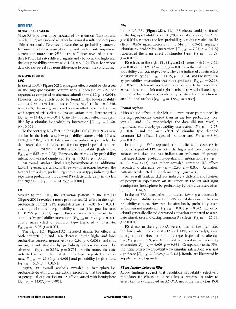
RESULTSBEHAVIORAL RESULTSSince RS is known to be modulated by attention (Larsson andSmith, 2012) we assessed whether behavioral results indicate pos-sible attentional differences between the two probability contexts.In general, hit rates were at ceiling and participants respondedcorrectly in more than 95% of trials. T-tests revealed that nei-ther RT nor hit rates differed significantly between the high- andthe low-probability context (t < 1.28, p > 0.2). Thus, behavioraldata did not reveal apparent differences between the conditions.
IMAGING RESULTSLOCIn the left LOC (Figure 2C1), strong RS effects could be observedin the high-probability context with a decrease of 21% forrepeated as compared to alternate stimuli (t = 6.19, p < 0.001).However, no RS effects could be found in the low-probabilitycontext (1% activation increase for repeated trials; t = 0.246,p = 0.808). Formally, we found a main effect of stimulus type,with repeated trials eliciting less activation than alternate trials[F(1,18) = 15.45, p = 0.001]. Critically, this main effect was qual-ified by a stimulus-by-probability interaction [F(1, 18) = 21.68,p < 0.001].
To the contrary, RS effects in the right LOC (Figure 2C2) weresimilar in the high- and low-probability context with 15 and14% (t > 2.87, p < 0.01) decrease in activation, respectively. Thedata revealed a main effect of stimulus type [repeated < alter-nate, F(1, 18) = 20.97, p < 0.001] and of probability [high > low,F(1, 18) = 5.53, p = 0.03]. However, the stimulus-by-probabilityinteraction was not significant [F(1, 18) = 0.148, p = 0.705].
An overall analysis (including hemisphere as an additionalfactor) revealed a significant three-way interaction between thefactors hemisphere, probability, and stimulus type, indicating thatrepetition probability modulated RS effects differently in the leftand right LOC [F(1, 18) = 14.76, p = 0.001].
LOSimilar to the LOC, the activation pattern in the left LO(Figure 2D1) revealed a more pronounced RS effect in the high-probability context (21% signal decrease, t = 6.09, p < 0.001)as compared to the low-probability context (1% signal increase,t = 0.256, p = 0.801). Again, the data were characterized by astimulus-by-probability interaction [F(1, 18) = 19.77, p < 0.001]and a main effect of stimulus type [repeated < alternate,F(1, 18) = 15.05, p = 0.001].
The right LO (Figure 2D2) revealed similar RS effects inboth contexts [15 and 14% decrease in the high- and low-probability context, respectively (t > 2.96, p < 0.008)] and thusno significant stimulus-by probability interaction could beobserved [F(1, 18) = 0.129, p = 0.724]. Furthermore, the dataindicated a main effect of stimulus type [repeated < alter-nate, F(1, 18) = 21.69, p < 0.001] and probability [high > low,F(1, 18) = 5.77, p = 0.027].
Again, an overall analysis revealed a hemisphere-by-probability-by-stimulus interaction, indicating that the influenceof perceptual expectation on RS effects varied with hemisphere[F(1, 18) = 14.07, p = 0.001].
PFsIn the left PFs (Figure 2E1), high RS effects could be foundin the high-probability context (28% signal decrease, t = 6.09,p < 0.001), whereas the low-probability context revealed no RSeffects (0,4% signal increase, t = 0.044, p = 0.965). Again, astimulus-by-probability interaction [F(1, 18) = 7.28, p = 0.015]superseded the main effect of stimulus type [F(1, 18) = 11.29,p = 0.003].
RS effects in the right PFs (Figure 2E2) were 14% (t = 2.63,p = 0.017) and 12% (t = 1.86, p = 0.079) in the high- and low-probability context, respectively. The data indicated a main effectfor stimulus type [F(1, 18) = 11.19, p = 0.004] and the stimulus-by-probability interaction was not significant [F(1, 18) = 0.296,p = 0.593]. Different modulations of RS effects by perceptualexpectations in the left and right hemisphere was indicated by asignificant hemisphere-by-probability-by-stimulus interaction inan additional analysis [F(1, 18) = 4.95, p = 0.039].
Control regionsAlthough RS effects in the left FFA were more pronounced inthe high-probability context than in the low-probability con-text (21 and 11%, respectively), the data did not reveal asignificant stimulus-by-probability interaction [F(1, 18) = 3.57,p = 0.075] and the main effect of stimulus type denotedcommon RS effects [repeated < alternate, F(1, 18) = 9.86,p = 0.006].
In the right FFA, repeated stimuli elicited a decrease inresponse signal of 14% in both, the high- and low-probabilitycontext and thus did not indicate an influence of percep-tual expectation [probability-by-stimulus interaction, F(1, 18) =0.112, p = 0.752], but rather revealed common RS effects[repeated < alternate, F(1, 18) = 12.63, p = 0.002]. Activationpatterns are depicted in Supplementary Figure A.3.
An overall analysis did not indicate a different modulationof perceptual expectation on RS effects in the left and righthemisphere [hemisphere-by-probability-by-stimulus interaction,F(1, 18) = 1.14, p = 0.3].
In the left PPA, repeated stimuli caused 12% signal decrease inthe high-probability context and 22% signal decrease in the low-probability context. However, the stimulus-by-probability inter-action was not significant [F(1, 18) = 0.838, p = 0.372]. Repeatedstimuli generally elicited decreased activation compared to alter-nate stimuli thus indicating common RS effects [F(1, 18) = 20.08,p < 0.001].
RS effects in the right PPA were similar in the high- andthe low-probability context (12 and 14%, respectively), indi-cating a main effect of stimulus type [repeated < alterna-tive, F(1, 18) = 19.99, p < 0.001] and no stimulus-by-probabilityinteraction [F(1, 18) = 0.008, p = 0.931]. Comparably to the FFA,the hemisphere-by-probability-by-stimulus interaction was notsignificant [F(1, 18) = 0.639, p = 0.435]. Results are illustrated inSupplementary Figure A.4.
RS modulation between ROIsAbove findings suggest that repetition probability selectivelymodulates RS effects in object-selective regions. In order toassess this, we conducted an ANOVA including the factors ROI
Frontiers in Human Neuroscience www.frontiersin.org April 2014 | Volume 8 | Article 225 | 4

Mayrhauser et al. Expectational effects during object processing
FIGURE 2 | (A,B) shows the location of the LOC (sum of red and greenclusters) as identified in the functional localizer. The subregions LO and PFsare depicted as green and red regions, respectively. (C–E), Contrast
estimates in the left and right hemisphere in the LOC (C-1, C-2) the LO (D-1,D-2) and the PFs (E-1, E-2). Error bars indicate two standard deviations of themean. ∗p < 0.05; ∗∗p < 0.01; ∗∗∗p < 0.001 (post-hoc comparisons).
Frontiers in Human Neuroscience www.frontiersin.org April 2014 | Volume 8 | Article 225 | 5

Mayrhauser et al. Expectational effects during object processing
(LOC, FFA, and PPA), probability, stimulus, and hemisphere.The analysis revealed a significant four-way interaction thusshowing that RS modulation varies significantly between ROIs[F(1, 18) = 18.55, p < 0.001]. Given that repetition probabilityeffects were left-lateralized in above results, we conducted twoadditional ANOVAs (i.e., for the left and right hemisphere) toexamine the influence of ROIs in more detail. The findings werein line with our prior analysis, indicating that RS modulationdiffered between ROIs (probability-by-stimulus-by-ROI interac-tion) in the left hemisphere [F(1, 18) = 11.05, p = 0.001] but notin the right hemisphere [F(1, 18) = 0.114, p = 0.893].
Whole brain analysisRepeated stimuli exhibited decreased activation compared tonew stimuli in the left and right inferior lateral occipital cor-tex (Figure 3). Further regions indicating decreased activationwere localized in the left occipital pole and in the right occip-ital fusiform gyrus. Notably, there was no significant decreasein activation due to stimulus repetition beyond visual regionsthat survived a threshold of p < 0.001 (uncorrected; 10 voxelextent). We found a significant stimulus-by-probability interac-tion in bilateral LOC, which exhibited a similar activation patternas the ROI analyses (p < 0.001, uncorrected; 10 voxel extent). Areversed pattern, that is, increased activation for repeated com-pared to alternative stimuli in the high-probability context, wasfound in the right middle frontal gyrus and the right frontalpole. The exact coordinates of regions exhibiting a main effect orinteractions are listed in Supplementary Table 1.
DISCUSSIONThe objective of the current investigation was to examine whetherstimulus repetition probability modulates RS effects during visualobject processing in the LOC. According to the PC account,repetition probability modulates the expectation of a repetition,which in turn influences RS effects in object-selective regions.Critically, investigations on perceptual expectation modulationsduring object processing revealed no influence of repetition prob-ability on RS effects (Kaliukhovich and Vogels, 2011; Kovácset al., 2013), thus challenging the role of top-down effects on RS.To the contrary, the current examination indicates modulatoryeffects of perceptual expectation on RS during object process-ing in the LOC. To illustrate, pronounced RS effects could befound in the high-probability context (i.e., 21% signal decrease)whereas activation levels of repeated and alternate stimuli in thelow-probability context do not differ in the current investigation.
FIGURE 3 | Activation clusters revealed by the whole brain analyses.
Regions that elicited decreased activation for repeated trials are illustratedin red. Green spots mark clusters where RS effects were modulated byrepetition probability (i.e., interaction). All clusters were extracted at athreshold of p < 0.001 (uncorrected, with a minimum extent of 10 voxels).
These findings take issue with the study of Kovács et al. (2013)which indicates that RS effects during object processing in theLO are not modulated by expectations. Of note, we examined RSeffects in the whole LOC whereas Kovács et al. (2013) investigatedRS effects in the caudal-dorsal subdivision of the LOC, the LO.One could argue that extracting activation from the whole LOCmay average across two regions (i.e., the LO and the PFs) whichmay consist of heterogeneous and functionally divergent subre-gions also with respect to RS effects (Grill-Spector et al., 1999).Thus, we analyzed the neural response in the two subdivisions ofthe LOC separately (the coordinates of the LO extracted in thecurrent study correspond to the cluster of Kovács et al., 2013).Notably, both the LO and the PFs indicate a modulation of RSeffects by repetition probability in a similar manner as the wholeLOC. Thus, undeliberate averaging across functionally divergentregions is not a valid explanation why the current data revealedan effect of perceptual expectations whereas Kovács et al. (2013)failed to find such an effect.
An alternative explanation may be that the diverging extentof repetition probability modulations causes the opposed results.To illustrate, in the study of Kovács et al. (2013), perceptualexpectations were induced by 60% repetition probability in thehigh-probability block and 20% repetition probability in the low-probability block. To the contrary, in the current experiment (andin prior investigations on perceptual expectation as well; e.g.,Summerfield et al., 2008, 2011) repetition probability was 75 and25% in the high- and low-probability context, respectively. Toillustrate, the difference in repetition probability between high-and low-probability blocks is 40% in the study of Kovács et al.(2013) compared to 50% in the current investigation. Therefore,the repetition probability manipulation of Kovács et al. (2013)possibly underruns a critical difference which is needed to reli-ably indicate modulatory effects of perceptual expectation forobjects. Furthermore, Kovács et al. (2013) omit reporting analysisof reaction times. Thus, a definite conclusion whether attentionor vigilance varied as a function of experimental manipulation ishardly possible by merely interpreting hit rates.
Noteworthy, using the same experimental design, the authorsfound modulatory effects of perceptual expectation for face stim-uli, thus partly excluding different repetition probabilities asa potential explanation. They argue that repetition probabilitymodulation is stronger for face selective neurons as comparedto neurons preferring non-face objects. This seems plausiblesince it has been shown that face processing is a highly special-ized mechanism that is capable of even fine-grained differences(Tovée, 1998). A higher sensibility for faces may explain whyperceptual expectation modulation on a lower level of repeti-tion probability distinction (40%) becomes visible for face stimulibut not for object stimuli. Vice versa, one may speculate that agreater difference in repetition probability should result in mea-surable modulatory effects on RS effects during object processing.Critically, this is exactly what we found.
Similar to the study of Kaliukhovich and Vogels (2011) ,Kovácset al. (2013) could not find effects of repetition probability onRS. However, comparisons between their and the current studymust be drawn carefully since the studies differ in a few aspects.First, Kaliukhovich and Vogels (2011) investigated RS effects in
Frontiers in Human Neuroscience www.frontiersin.org April 2014 | Volume 8 | Article 225 | 6

Mayrhauser et al. Expectational effects during object processing
a different species (i.e., monkeys). Second, the authors examinedelectric potentials in the extracellular space, whereas the currentstudy investigated the hemodynamic response succeeding neuralactivation. However, in a recent electroencephalography study,Summerfield et al. (2011) reported effects of repetition proba-bility. This rules out the possibility that investigating perceptualexpectation by means of electric potentials may be less feasible.One possible explanation for the absence of perceptual expecta-tion effects in their study may be that the authors implementeda fixation task. In this task, monkeys were solely rewarded whenthey remained fixated on a red cross. Accordingly, it seems rea-sonable to hypothesize that the monkeys attended the fixationcross stronger than the actual stimulus material. This is insofarimportant, as Larsson and Smith (2012) found that repetitionprobability modulates RS effects only under conditions of sus-tained attention toward stimuli. Critically, the modulatory effectof repetition probability vanishes when attention is diverted away.Thus, the selection of an appropriate task may be crucial forthe detection of perceptual expectation effects in future studies.Note that differences in species and task cannot (fully) accountfor the absence of modulatory effects since their results resemblethe fMRI investigation on RS effects in humans of Kovács et al.(2013).
As already mentioned, Kovács et al. (2013) and Larsson andSmith (2012) reported influences of top-down expectations onRS effects in the LO. Critically, since perceptual expectation wasconstantly found for face but not for object stimuli, Kovács et al.(2013) assumed that perceptual expectation effects are dedicatedto face processing. Importantly, the current findings demonstratethat perceptual expectation effects on repetition suppression arenot restricted to faces. These results are well in line with the PCaccount, according to which top-down expectations are a gen-eral phenomenon in (visual) perception (Summerfield and Egner,2009). These expectations, which are supposed to feed back infor-mations and to facilitate information processing (Bar et al., 2001,2006; Bar, 2003), are a core assumption of the PC account.
An interesting finding in the current investigation was that this(apparent) top-down modulation was lateralized. Specifically, RSeffects in the left LOC were modulated by repetition probability(thus indicating influences of expectation), whereas the ROIs inthe right hemisphere revealed no such effect. As yet, the litera-ture is sparse with regard to the effect of repetition probabilityon neural activation and much less is known about possible lat-erality effects in the context of RS. One speculative explanationmight be that participants verbally code the stimuli depicted onscreen. More specifically, participants may encode their expecta-tion of a forthcoming target in the high-probability context byinternally verbalizing the expected object. Accordingly, internalverbalization could account for the left lateralized finding of per-ceptual expectation effects since this hemisphere is strongly linkedwith language processes (Vigneau et al., 2006). However, this sug-gestion is highly speculative and must await more fine-grainedinvestigations in future studies.
Besides our main objective, we investigated whether top-downexpectations selectively affect regions that process a current object(i.e., the LOC) or whether expectations influence a broaderrange of regions in the ventral visual pathway (e.g., the FFA and
PPA). Here, the response signal in the FFA and the PPA revealscommon RS effects which are not affected by repetition proba-bility. This suggests that top-down expectations reflect a ratherspecific mechanism which modulated RS effects in the LOC, butnot in the FFA and the PPA.
As expected, an exploratory whole brain analysis revealedcommon RS effects along the ventral visual pathway. In addi-tion, these RS effects varied with stimulus probability in a similarmanner as in the region of interest analyses. Interestingly, frontalregions like the right middle frontal gyrus and the right frontalpole revealed the reversed activation pattern with repeated stimulieliciting higher activation than alternative stimuli [i.e., repeti-tion enhancement (RE)]. Although the co-existence of RS and REseems challenging, the PC account provides a reasonable frame-work for this finding. Accordingly, perception requires both, theforming of expectations indexed by repetition enhancement andprediction error signals that indicate the accuracy of the predic-tion. The previous study by De Gardelle et al. (2013) supportsthis notion by showing that RE and RS could be found simulta-neously, even within the same functional network. Furthermore,RE and RS units could consistently be separated across scan-ner runs and functional connectivity analyses revealed that REvoxels were relatively more connected to higher visual process-ing areas whereas RS voxels were connected with lower visualregions. These findings are in line with the current patternthat repeated compared to alternative stimuli revealed decreasedactivation in the LOC, thus possibly reflecting a decreased pre-diction error signal, whereas RE effects in higher levels of theprocessing hierarchy (i.e., frontal regions) might index the devel-opment of predictions which then modulate object processingtop-down. However, these findings require future investigations(and replications) since the current voxel-based whole brain anal-ysis used an uncorrected threshold and has to be seen as anexploratory analysis. Notably, probability and interaction effectsmust be interpreted with caution due to the present studydesign.
Taken together, the present study provides further evidencein favor of the PC model. We found that repetition probabilitymodulates RS effects in the LOC during visual object process-ing, indicating that the influence of perceptual expectations is notrestricted to face perception. These results are consistent with agrowing body of studies indicating that the PC model is capableof explaining a broader range of perceptual processes well beyondface processing (e.g., Todorovic et al., 2011; Andics et al., 2013).
ACKNOWLEDGMENTSThis work was funded by grants provided to Martin Kronbichlerby the Austrian Science Fund (FWF grant number: P 23219-B18) and the Scientific Funds of the Paracelsus Medical University(grant number: E-10/12/062-KRO).We would like to thank EvaReiter for help with data acquisition. Correspondence should beaddressed to [email protected].
SUPPLEMENTARY MATERIALThe Supplementary Material for this article can be foundonline at: http://www.frontiersin.org/journal/10.3389/fnhum.
2014.00225/abstract
Frontiers in Human Neuroscience www.frontiersin.org April 2014 | Volume 8 | Article 225 | 7

Mayrhauser et al. Expectational effects during object processing
REFERENCESAndics, A., Gál, V., Vicsi, K., Rudas, G., and Vidnyánszky, Z. (2013). FMRI repeti-
tion suppression for voices is modulated by stimulus expectations. Neuroimage69, 277–283. doi: 10.1016/j.neuroimage.2012.12.033
Ashburner, J. (2007). A fast diffeomorphic image registration algorithm.Neuroimage 38, 95–113. doi: 10.1016/j.neuroimage.2007.07.007
Baldassano, C., Beck, D. M., and Fei-Fei, L. (2013). Differential connectiv-ity within the Parahippocampal Place Area. Neuroimage 75, 228–237. doi:10.1016/j.neuroimage.2013.02.073
Bar, M. (2003). A cortical mechanism for triggering top-down facilita-tion in visual object recognition. J. Cogn. Neurosci. 15, 600–609. doi:10.1162/089892903321662976
Bar, M., Kassam, K., Ghuman, A., Boshyan, J., Schmid, A., Dale, A., et al. (2006).Top-down facilitation of visual recognition. Proc. Natl. Acad. Sci. U.S.A. 103,449–454. doi: 10.1073/pnas.0507062103
Bar, M., Tootell, R. B. H., Schacter, D. L., Greve, D. N., Fischl, B., and Mendola,J. D. (2001). Cortical mechanisms specific to explicit visual object recognition.Neuron 29, 529–535. doi: 10.1016/S0896-6273(01)00224-0
Cardin, V., Friston, K. J., and Zeki, S. (2011). Top-down modulations in thevisual form pathway revealed with dynamic causal modeling. Cereb. Cortex 21,550–562. doi: 10.1093/cercor/bhq122
De Gardelle, V., Waszczuk, M., Egner, T., and Summerfield, C. (2013). Concurrentrepetition enhancement and suppression responses in extrastriate visual cortex.Cereb. Cortex 23, 2235–2244. doi: 10.1093/cercor/bhs211
Egner, T., Monti, J. M., and Summerfield, C. (2010). Expectation and surprisedetermine neural population responses in the ventral visual stream. J. Neurosci.30, 16601–16608. doi: 10.1523/JNEUROSCI.2770-10.2010
Epstein, R., Harris, A., Stanley, D., and Kanwisher, N. (1999). The parahippocam-pal place area: recognition, navigation, or encoding? Neuron 23, 115–125. doi:10.1016/S0896-6273(00)80758-8
Ewbank, M. P., Lawson, R. P., Henson, R. N., Rowe, J. B., Passamonti, L., andCalder, A. J. (2011). Changes in “top-down” connectivity underlie repetitionsuppression in the ventral visual pathway. J. Neurosci. 31, 5635–5642. doi:10.1523/JNEUROSCI.5013-10.2011
Friston, K. J. (2005). A theory of cortical responses. Philos. Trans. R. Soc. B Biol. Sci.360, 815–836. doi: 10.1098/rstb.2005.1622
Friston, K. J., Glaser, D. E., Henson, R. N., Kiebel, S., Phillips, C., and Ashburner,J. (2002). Classical and Bayesian inference in neuroimaging: applications.Neuroimage 16, 484–512. doi: 10.1006/nimg.2002.1091
Grill-Spector, K., Henson, R., and Martin, A. (2006). Repetition and the brain:neural models of stimulus-specific effects. Trends Cogn. Sci. 10, 14–23. doi:10.1016/j.tics.2005.11.006
Grill-Spector, K., Kushnir, T., Edelman, S., Avidan, G., Itzchak, Y., and Malach, R.(1999). Differential processing of objects under various viewing conditions inthe human lateral occipital complex. Neuron 24, 187–203. doi: 10.1016/S0896-6273(00)80832-6
Grill-Spector, K., and Malach, R. (2001). fMR-adaptation: a tool for studying thefunctional properties of human cortical neurons. Acta Psychol. 107, 293–321.doi: 10.1016/S0001-6918(01)00019-1
Haxby, J. V., Gobbini, M. I., Furey, M. L., Ishai, A., Schouten, J. L., and Pietrini,P. (2001). Distributed and overlapping representations of faces and objects inventral temporal cortex. Science 293, 2425–2430. doi: 10.1126/science.1063736
Henson, R. N. (2003). Neuroimaging studies of priming. Prog. Neurobiol. 70, 53–81.doi: 10.1016/S0301-0082(03)00086-8
Kaliukhovich, D., and Vogels, R. (2011). Stimulus repetition probability does notaffect repetition suppression in macaque inferior temporal cortex. Cereb. Cortex21, 1547–1558. doi: 10.1093/cercor/bhq207
Kanwisher, N., McDermott, J., and Chun, M. (1997). The fusiform face area: a mod-ule in human extrastriate cortex specialized for face perception. J. Neurosci. 17,4302–4311.
Kanwisher, N., and Yovel, G. (2006). The fusiform face area: a cortical regionspecialized for the perception of faces. Philos. Trans. R. Soc. B Biol. Sci. 361,2109–2128. doi: 10.1098/rstb.2006.1934
Kovács, G., Kaiser, D., Kaliukhovich, D. A., Vidnyánszky, Z., and Vogels, R. (2013).Repetition probability does not affect fMRI repetition suppression for objects.J. Neurosci. 33, 9805–9812. doi: 10.1523/JNEUROSCI.3423-12.2013
Larsson, J., and Smith, A. (2012). fMRI repetition suppression: neuronal adapta-tion or stimulus expectation? Cereb. Cortex 22, 567–576. doi: 10.1093/cercor/bhr119
Martens, U., and Gruber, T. (2012). Sharpening and formation: two distinct neu-ronal mechanisms of repetition priming. Eur. J. Neurosci. 36, 2989–2995. doi:10.1111/j.1460-9568.2012.08222.x
Sayres, R., and Grill-Spector, K. (2006). Object-selective cortex exhibitsperformance-independent repetition suppression. J. Neurophysiol. 95,995–1007. doi: 10.1152/jn.00500.2005
Segaert, K., Weber, K., de Lange, F., Petersson, K., and Hagoort, P. (2013). The sup-pression of repetition enhancement: a review of fMRI studies. Neuropsychologia51, 59–66. doi: 10.1016/j.neuropsychologia.2012.11.006
Summerfield, C., and Egner, T. (2009). Expectation (and attention) in visualcognition. Cell 13, 403–409. doi: 10.1016/j.tics.2009.06.003
Summerfield, C., Trittschuh, E., Monti, J., Mesulam, M., and Egner, T. (2008).Neural repetition suppression reflects fulfilled perceptual expectations. Nat.Neurosci. 11, 1004–1006. doi: 10.1038/nn.2163
Summerfield, C., Wyart, V., Johnen, V., and de Gardelle, V. (2011). Humanscalp electroencephalography reveals that repetition suppression varies withexpectation. Front. Hum. Neurosci. 5:67. doi: 10.3389/fnhum.2011.00067
Szekely, A., Jacobsen, T., D’Amico, S., Devescovi, A., Andonova, E., Herron, D., et al.(2004). A new on-line resource for psycholinguistic studies. J. Mem. Lang. 51,247–250. doi: 10.1016/j.jml.2004.03.002
Todorovic, A., van Ede, F., Maris, E., and de Lange, F. P. (2011). Prior expectationmediates neural adaptation to repeated sounds in the auditory cortex: an MEGstudy. J. Neurosci. 31, 9118–9123. doi: 10.1523/JNEUROSCI.1425-11.2011
Tovée, M. J. (1998). Face processing: getting by with a little help from its friends.Curr. Biol. 8, 317–320. doi: 10.1016/S0960-9822(98)70197-6
Vigneau, M., Beaucousin, V., Herve, P. Y., Duffau, H., Crivello, F., Houde,O., et al. (2006). Meta-analyzing left hemisphere language areas: phonol-ogy, semantics, and sentence processing. Neuroimage 30, 1414–1432. doi:10.1016/j.neuroimage.2005.11.002
Conflict of Interest Statement: The authors declare that the research was con-ducted in the absence of any commercial or financial relationships that could beconstrued as a potential conflict of interest.
Received: 11 March 2014; accepted: 31 March 2014; published online: 17 April 2014.Citation: Mayrhauser L, Bergmann J, Crone J and Kronbichler M (2014) Neuralrepetition suppression: evidence for perceptual expectation in object-selective regions.Front. Hum. Neurosci. 8:225. doi: 10.3389/fnhum.2014.00225This article was submitted to the journal Frontiers in Human Neuroscience.Copyright © 2014 Mayrhauser, Bergmann, Crone and Kronbichler. This is an open-access article distributed under the terms of the Creative Commons Attribution License(CC BY). The use, distribution or reproduction in other forums is permitted, providedthe original author(s) or licensor are credited and that the original publication in thisjournal is cited, in accordance with accepted academic practice. No use, distribution orreproduction is permitted which does not comply with these terms.
Frontiers in Human Neuroscience www.frontiersin.org April 2014 | Volume 8 | Article 225 | 8