Neural crest cells and patterning of the mammalian dentition
Upload
independentCategory
view
0download
0
Neuron, Vol. 35, 657–669, August 15, 2002, Copyright 2002 by Cell Press
Neural Crest Stem Cells Persist in the Adult Gutbut Undergo Changes in Self-Renewal, NeuronalSubtype Potential, and Factor Responsiveness
the adult CNS are sometimes thought of as left-overfrom fetal development or as deriving from the life-longself-renewal of fetal neural stem cells (reviewed by Mor-rison et al., 1997). These views assume that the impor-tant properties of neural stem cells, such as develop-
Genevieve M. Kruger, Jack T. Mosher,Suzanne Bixby, Nancy Joseph,Toshihide Iwashita, and Sean J. Morrison1
Howard Hughes Medical InstituteDepartment of Internal Medicine andDepartment of Cell and Developmental Biology mental potential and self-renewal capacity, do not
change between fetal and postnatal life. Even assumingUniversity of Michigan1500 E. Medical Center Drive that postnatal neural stem cells are derived from fetal
neural stem cells, it is possible that fetal stem cellsAnn Arbor, Michigan 48109undergo developmental changes that give rise to pheno-typically and functionally distinct populations of postna-tal neural stem cells. Adult hematopoietic stem cells areSummarymultipotent and self-renewing (Morrison et al., 1995b)but exhibit less self-renewal potential than fetal liverWe found neural crest stem cells (NCSCs) in the adult
gut. Postnatal gut NCSCs were isolated by flow-cytome- hematopoietic stem cells (Lansdorp et al., 1993; Mor-rison et al., 1995a) and lose the ability to make certaintry and compared to fetal gut NCSCs. They self-renewed
extensively in culture but less than fetal gut NCSCs. subtypes of lymphocytes that arise only during fetaldevelopment (Hayakawa et al., 1985; Ikuta et al., 1990;Postnatal gut NCSCs made neurons that expressed a
variety of neurotransmitters but lost the ability to make Kantor et al., 1992). This raises the question of whetherpostnatal neural stem cells undergo similar changes.certain subtypes of neurons that are generated during
fetal development. Postnatal gut NCSCs also differed Postmigratory rat NCSCs persist into late gestationby self-renewing within peripheral nerves and the gutin their responsiveness to lineage determination fac-
tors, affecting cell fate determination in vivo and possi- (Morrison et al., 1999; Bixby et al., 2002). At embryonicday (E)14.5, sciatic nerve NCSCs can be prospectivelybly explaining their reduced neuronal subtype poten-
tial. These perinatal changes in gut NCSCs parallel identified and isolated by flow-cytometry as p75�P0�
cells, and gut NCSCs can be isolated as p75��4� cells.perinatal changes in hematopoietic stem cells, sug-
gesting that stem cells in different tissues undergo Seventy to eighty percent of single cells in each popula-tion self-renew and form multilineage colonies in culturesimilar developmental transitions. The persistence of
NCSCs in the adult PNS opens up new possibilities that contain neurons, glia, and myofibroblasts (Morrisonet al., 1999, 2000a; Bixby et al., 2002). In this study, wefor regeneration after injury or disease.have discovered that NCSCs do persist in the adult gutand that these cells undergo temporal changes in self-Introductionrenewal potential and neuronal subtype potential. Post-natal gut NCSCs gave rise to neurons and glia in vitroMost of the neurons and glia of the peripheral nervous
system (PNS) arise during fetal development from the and in vivo but, in contrast to E14.5 gut NCSCs, gener-ated mainly glia upon transplantation into developingneural crest, a heterogeneous collection of progenitors
that migrates out of the neural tube in midgestation peripheral nerve. The reduced neuronal subtype poten-tial and gliogenic bias of postnatal gut NCSCs may be(LeDouarin, 1986). Migrating neural crest cells, which
include neural crest stem cells (NCSCs), undergo pro- caused by perinatal changes in their responsiveness tolineage determination factors.gressive restrictions in developmental potential (Barof-
fio et al., 1991) and terminally differentiate soon afterreaching postmigratory sites. The postnatal PNS was Resultsthought to lack stem cells.
Like the PNS, the adult CNS was once thought to lack Multipotent Progenitors Persist Postnatallystem cells. However, multipotent neural progenitors and in the Gutneurogenesis do persist in certain regions of the adult We have so far been unable to identify any multipotentCNS (Reynolds and Weiss, 1992; Palmer et al., 1997; progenitors from the postnatal sciatic nerve (data notJohansson et al., 1999; Doetsch et al., 1999; Altman, shown), but we have found multipotent progenitors in the1969; Eriksson et al., 1998; Gould et al., 1999). Stem cells postnatal gut. Postnatal day (P)5 to P15 rat gut was disso-from the adult CNS have recently been prospectively ciated into single cell suspensions and plated in cultureidentified and purified by flow-cytometry, creating the at clonal density. Clonal density allowed individual founderpossibility of studying their properties as they exist in cells to form spatially distinct colonies so that the devel-vivo (Rietze et al., 2001). The discovery of stem cells in opmental potential of the founder cells could be as-the adult CNS raises the question of whether stem cells sessed based on colony composition. After 14 daysalso persist in the adult PNS. in culture, we consistently observed the formation of
It has not been clear whether neural stem cells un- multilineage colonies containing neurons, glia, and myo-dergo perinatal developmental changes. Stem cells in fibroblasts (Figure 1) that resembled colonies formed
by embryonic NCSCs (Stemple and Anderson, 1992;Morrison et al., 1999; Bixby et al., 2002), though they1Correspondence: [email protected]
Neuron658
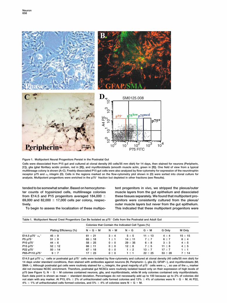
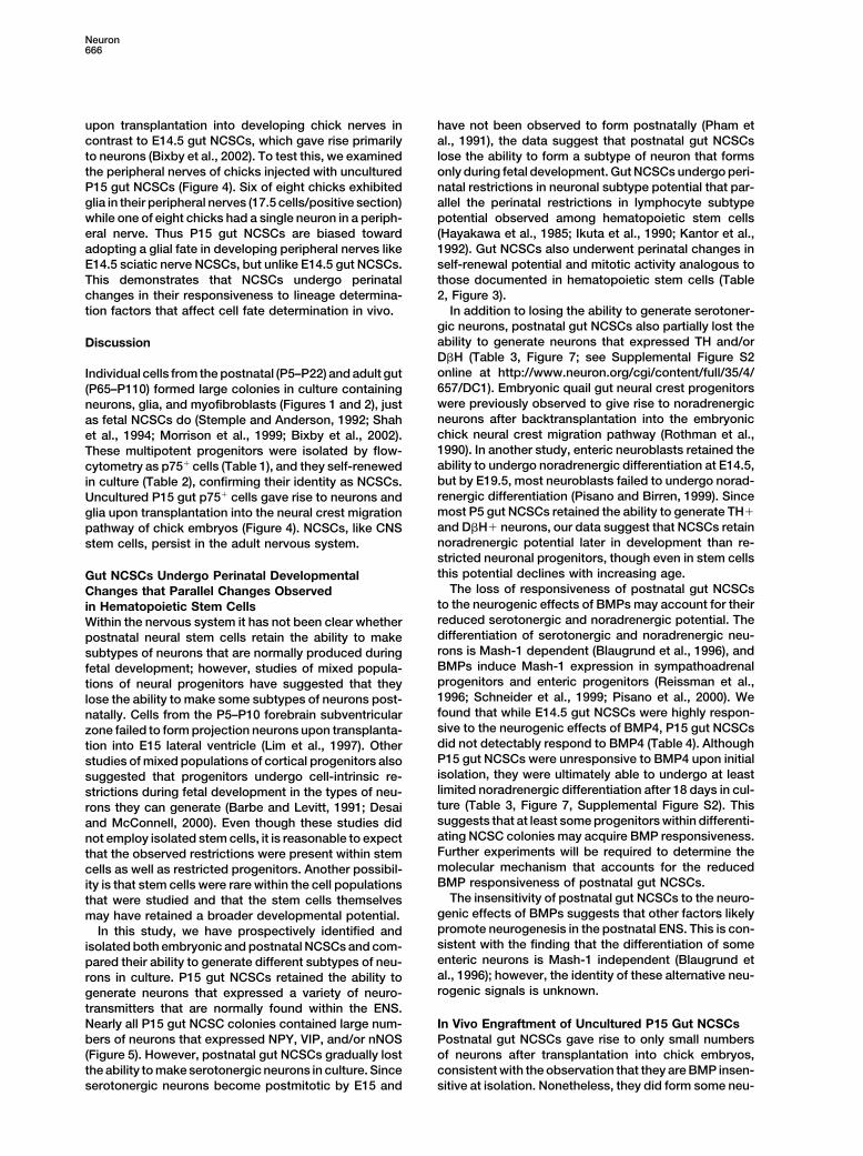
Figure 1. Multipotent Neural Progenitors Persist in the Postnatal Gut
Cells were dissociated from P15 gut and cultured at clonal density (40 cells/35 mm dish) for 14 days, then stained for neurons (Peripherin,[C]), glia (glial fibrillary acidic protein, red in [B]), and myofibroblasts (smooth muscle actin, green in [B]). One field of view from a typicalmultilineage colony is shown (A–C). Freshly dissociated P15 gut cells were also analyzed by flow-cytometry for expression of the neurotrophinreceptor p75 and �4 integrin (D). Cells in the regions marked on the flow-cytometry plot shown in (D) were sorted into clonal culture foranalysis. Multipotent progenitors were enriched in the p75� fraction but depleted in other fractions (see Results).
tended to be somewhat smaller. Based on hemocytome- tent progenitors in vivo, we stripped the plexus/outermuscle layers from the gut epithelium and dissociatedter counts of trypsinized cells, multilineage colonies
from E14.5 and P15 progenitors averaged 184,000 � these tissues separately. We found that multipotent pro-genitors were consistently cultured from the plexus/69,000 and 82,000 � 17,000 cells per colony, respec-
tively. outer muscle layers but never from the gut epithelium.This indicated that these multipotent progenitors wereTo begin to assess the localization of these multipo-
Table 1. Multipotent Neural Crest Progenitors Can Be Isolated as p75� Cells from the Postnatal and Adult Gut
Colonies that Contain the Indicated Cell Types (%)
Plating Efficiency (%) N � G � M N � M N � G G � M G Only M Only
E14.5 p75� �4� 45 � 9 61 � 21 3 � 4 5 � 5 11 � 13 4 � 4 15 � 15
P5 p75� 51 � 9 65 � 18 1 � 1 14 � 11 7 � 7 6 � 7 6 � 4P10 p75� 44 � 6 58 � 25 0 � 0 29 � 35 6 � 8 3 � 3 4 � 5P15 p75� 52 � 12 66 � 11 0 � 0 12 � 8 7 � 5 11 � 8 4 � 5P22 p75� 50 � 14 67 � 18 0 � 0 1 � 2 13 � 7 17 � 7 1 � 1P65–P110 p75� 9 � 7 17 � 23 0 � 0 1 � 1 22 � 25 53 � 28 7 � 14
E14.5 gut p75��4� cells or postnatal gut p75� cells were isolated by flow-cytometry and cultured at clonal density (40 cells/35 mm dish) for
14 days under standard conditions, then stained with antibodies against neurons (N; Peripherin�), glia (G; GFAP�), and myofibroblasts (M;SMA�). Although postnatal gut cells were routinely stained for �4 integrin, the great majority of p75� cells were �4
�, so use of the �4 markerdid not increase NCSC enrichment. Therefore, postnatal gut NCSCs were routinely isolated based only on their expression of high levels ofp75 (see Figure 1). N � G � M colonies contained neurons, glia, and myofibroblasts, while M only colonies contained only myofibroblasts.Each data point is shown as mean � standard deviation. Percentages do not necessarily add up to 100 because up to 2% of colonies didnot stain with any marker. At P15, 6% � 2% of unfractionated cells formed colonies and 13% � 4% of colonies were N � G � M. At P22,4% � 1% of unfractionated cells formed colonies, and 5% � 4% of colonies were N � G � M.
Neural Crest Stem Cells in the Adult Gut659
localized in vivo to the submucosal plexus, myentericplexus, or outer muscle layers of the gut. All subsequentexperiments on postnatal gut progenitors were per-formed using cells dissociated from the plexus/outermuscle layers.
Multipotent progenitors were infrequent among theunfractionated cells obtained from the preparations ofdissociated P15 plexus/outer muscle layers. Only 0.7%of dissociated (but unfractionated) P15 cells survivedand formed multilineage colonies in culture. Since p75has consistently been observed to mark NCSCs in thegut and other tissues (Stemple and Anderson, 1992;Morrison et al., 1999; Lo and Anderson, 1995; Bixby etal., 2002), we tested whether these multipotent progeni-tors were enriched within the p75� fraction of cells. Asin the E14.5 gut, we found that multipotent progenitorswere enriched among cells with high levels of p75 ex-pression (see Figure 1D for a flow-cytometric profile ofp75 expression). Although p75� and p75low cells formedno multilineage colonies and p75med cells gave rise tofew multilineage colonies, p75� cells were highly en-riched for multipotent progenitors. An average of fifty-two percent of p75� cells survived to form colonies inculture, and 66% of these colonies were multilineage(Table 1). Interestingly, although E14.5 gut NCSCs arep75��4� (Bixby et al., 2002), Figure 1D shows that p75�
cells were largely negative for �4 integrin by P15. Bystaining sections of P15 gut with an antibody againstp75, we found that p75� cells localized to the myentericand submucosal plexi (see Supplemental Figure S1 on-line at http://www.neuron.org/cgi/content/full/35/4/657/DC1).
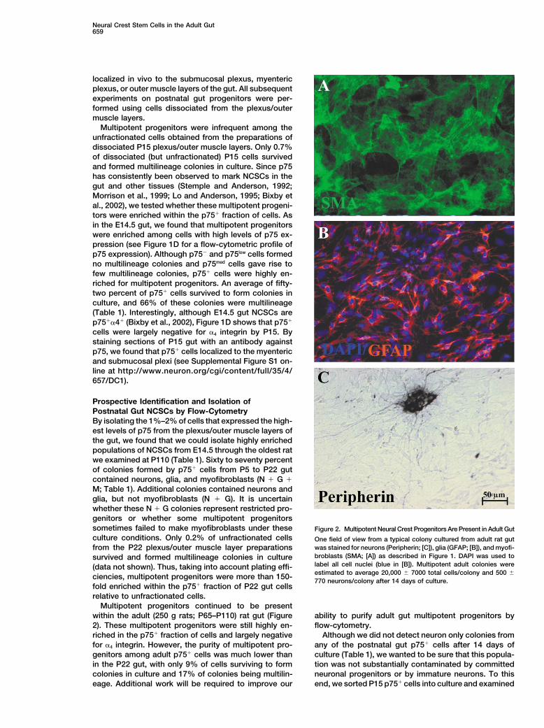
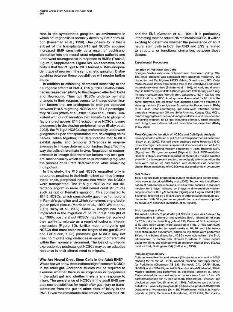
Prospective Identification and Isolation ofPostnatal Gut NCSCs by Flow-CytometryBy isolating the 1%–2% of cells that expressed the high-est levels of p75 from the plexus/outer muscle layers ofthe gut, we found that we could isolate highly enrichedpopulations of NCSCs from E14.5 through the oldest ratwe examined at P110 (Table 1). Sixty to seventy percentof colonies formed by p75� cells from P5 to P22 gutcontained neurons, glia, and myofibroblasts (N � G �M; Table 1). Additional colonies contained neurons andglia, but not myofibroblasts (N � G). It is uncertainwhether these N � G colonies represent restricted pro-genitors or whether some multipotent progenitorssometimes failed to make myofibroblasts under these Figure 2. Multipotent Neural Crest Progenitors Are Present in Adult Gutculture conditions. Only 0.2% of unfractionated cells One field of view from a typical colony cultured from adult rat gut
was stained for neurons (Peripherin; [C]), glia (GFAP; [B]), and myofi-from the P22 plexus/outer muscle layer preparationsbroblasts (SMA; [A]) as described in Figure 1. DAPI was used tosurvived and formed multilineage colonies in culturelabel all cell nuclei (blue in [B]). Multipotent adult colonies were(data not shown). Thus, taking into account plating effi-estimated to average 20,000 � 7000 total cells/colony and 500 �ciencies, multipotent progenitors were more than 150-770 neurons/colony after 14 days of culture.
fold enriched within the p75� fraction of P22 gut cellsrelative to unfractionated cells.
Multipotent progenitors continued to be presentwithin the adult (250 g rats; P65–P110) rat gut (Figure ability to purify adult gut multipotent progenitors by
flow-cytometry.2). These multipotent progenitors were still highly en-riched in the p75� fraction of cells and largely negative Although we did not detect neuron only colonies from
any of the postnatal gut p75� cells after 14 days offor �4 integrin. However, the purity of multipotent pro-genitors among adult p75� cells was much lower than culture (Table 1), we wanted to be sure that this popula-
tion was not substantially contaminated by committedin the P22 gut, with only 9% of cells surviving to formcolonies in culture and 17% of colonies being multilin- neuronal progenitors or by immature neurons. To this
end, we sorted P15 p75� cells into culture and examinedeage. Additional work will be required to improve our
Neuron660
Table 2. Postnatal Gut NCSCs Self-Renew in Culture But the Extent of Self-Renewal Declines Significantly with Increasing Age
Subclones Per Multipotent Founder Cell
Population Total N � G � M N � G G � M G Only M Only
E14.5 gut p75��4� 845 a 730 � 459 a 81 � 151 27 � 67 2 � 5 a 5 � 10
P5 gut p75� 311 b 146 � 90 b 15 � 5 52 � 23 96 � 107 b 2 � 3P15 gut p75� 386 b 143 � 143 b 29 � 50 69 � 50 142 � 100 b 3 � 7P22 gut p75� 360 b 70 � 132 b 22 � 35 24 � 31 242 � 100 c 2 � 4Adult gut p75� 366 35 � 49 3 � 3 110 � 155 193 � 230 24 � 34
Single gut progenitors were deposited into individual wells of 96-well plates. After 8 (E14–P22) or 15 (adult) days in culture, individual colonieswere subcloned into secondary cultures at clonal density. Secondary clones were cultured for 14 days and stained with antibodies to identifyneurons (N), glia (G), and myofibroblasts (M) as described in Table 1. P22 gut NCSCs gave rise to significantly fewer total subclones andmultipotent subclones but significantly more glial only subclones than E14.5 NCSCs (p � 0.05). Significantly different statistics are followedby different letters, except for adult subclones, which were not compared because they were subcloned after 15 days in culture.
the colonies 4 days later. 2.3% of colonies contained rise to multipotent daughter cells while others did not.It is not yet clear whether this heterogeneity reflects thea single neuron, and 0.6% of colonies contained two
neurons. No neuron only colonies contained more than subcloning of restricted progenitor colonies or whethersome multipotent adult progenitors may fail to de-two neurons. We also sorted P15 p75� cells into culture
and stained these cells 17 hr later for the early neuronal tectably self-renew in this assay. Nonetheless, at leastsome of the adult multipotent progenitors self-renewed,marker neuron-specific (class III) �-tubulin (TuJ1 anti-
body). On average, 3.9% � 5% of P15 gut p75� cells producing an average of 35 multipotent subclones perfounder cell (Table 2).expressed �-tubulin. This suggests that around 4% of
P15 p75� cells are immature neurons or committed neu- The 8–15 day culture assay represents one way ofquantitating self-renewal potential, but it does not esti-ronal progenitors with the ability to divide once in
culture. mate the maximum self-renewal capacity of NCSCs. Byculturing the postnatal gut NCSCs for longer periods oftime, they give rise to larger numbers of multipotentPostnatal Gut Multipotent Neural Crestdaughter cells.Progenitors Self-Renew in Culture
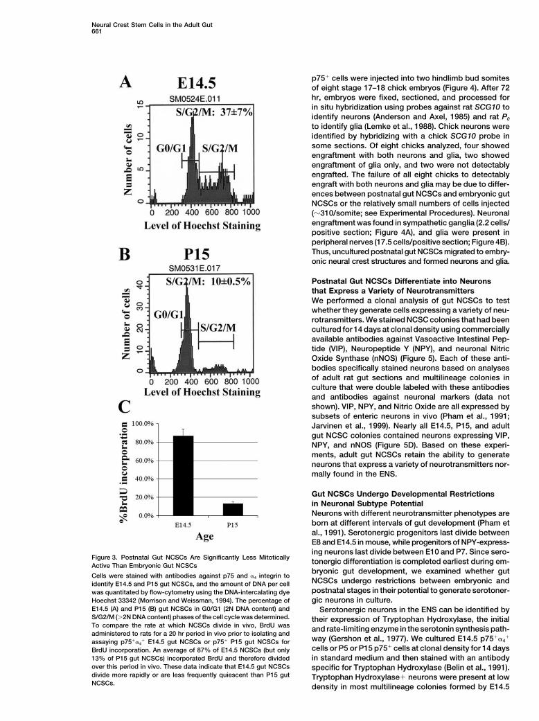
The self-renewal capacity of the gut multipotent progen-Postnatal Gut NCSCs Are Less Mitotically Activeitors was assayed by depositing single p75� cells intothan E14 Gut NCSCsindividual wells of 96-well plates by flow-cytometry andIn the hematopoietic system and the adult CNS, stemthen culturing for 8 days under standard conditionscells go from being highly mitotic during fetal develop-(Morrison et al., 1999). Multipotent colonies were thenment (Morrison et al., 1995a; Cai et al., 1997) to beingtrypsinized and subcloned into secondary cultures atrelatively quiescent in adults (Morshead et al., 1994;clonal density as previously described (Morrison et al.,Cheshier et al., 1999; Johansson et al., 1999). Since this1999). Almost all multipotent primary colonies gave riseis the first identification of NCSCs in the postnatal PNS,to multipotent daughter colonies (20/20 at E14.5, 11/11we used our ability to prospectively identify the gutat P5, 16/18 at P15, and 6/6 at P22). Since the multipo-NCSCs to ask whether their cell-cycle distributiontent progenitors from postnatal gut were p75�, formedchanged perinatally. The cell-cycle distribution of uncul-colonies similar to embryonic NCSC colonies, appearedtured NCSCs was assayed at isolation by flow-cytome-to localize to the myenteric or submucosal plexi, andtry, using Hoechst 33342 staining to determine the DNAhad the capacity to self-renew, we conclude that theycontent of individual cells (Figure 3). E14.5 gut p75��4�are postnatal NCSCs.NCSCs included 37% � 7% of cells in S/G2/M phasesAlthough the postnatal gut NCSCs consistently self-of the cell cycle (�2N DNA content), as compared torenewed in culture, the extent of self-renewal quantita-only 26% � 2% of P5 p75� cells (data not shown) andtively declined with increasing age. Each E14.5 gut10% � 0.5% of P15 p75� cells (Figure 3; p � 0.01).NCSC produced an average of 730 multipotent daughterWhen BrdU was administered for a 20 hr period in vivocolonies (845 total daughter colonies) subclonable afterprior to isolation of NCSCs, 87% � 7% of E14.5 gut8 days in culture, while P22 gut NCSCs produced anp75��4� cells incorporated BrdU while only 13.1% �average of only 70 multipotent daughter colonies (3602.5% of P15 gut p75� cells incorporated BrdU. Thus,total daughter colonies) under the same conditions (p �while nearly all E14.5 gut NCSCs divided at least once0.002; Table 2). NCSC self-renewal did not decline uni-in a 20 hr period in vivo, only 13% of P15 gut p75� cellsformly over time as some NCSCs at P15 and P22 self-divided in the same period. Either P15 gut NCSCs haverenewed at a rate comparable to what was observeda much longer cell-cycle time than E14.5 gut NCSCs,among E14.5 NCSCs. For example, one P15 NCSC gaveor a significant percentage of gut NCSCs are quiescentrise to 370 multipotent daughter cells, while a P22 NCSCat P15.gave rise to 335 multipotent daughter cells.
Because of the impurity of multipotent progenitors inthe adult p75� population, we cultured single adult gut NCSCs from the P15 Gut Differentiate
into Neurons and Glia In Vivocells for 15 days prior to subcloning in an attempt todistinguish multipotent colonies from restricted colonies To test the ability of the P15 gut NCSCs to generate
neurons and glia in vivo, freshly isolated, unculturedbased on size. Some of the large adult colonies gave
Neural Crest Stem Cells in the Adult Gut661
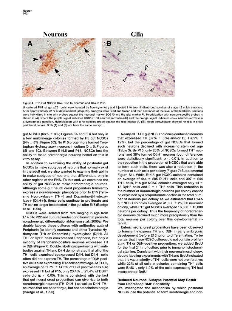
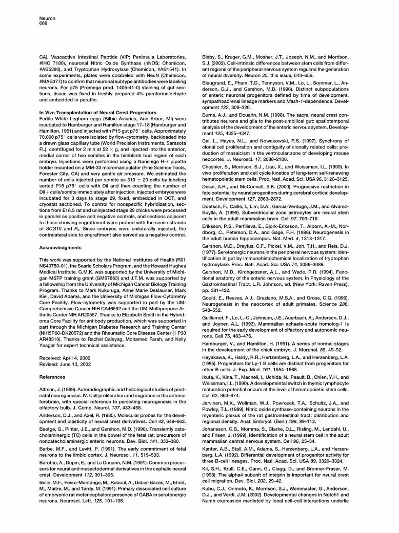
p75� cells were injected into two hindlimb bud somitesof eight stage 17–18 chick embryos (Figure 4). After 72hr, embryos were fixed, sectioned, and processed forin situ hybridization using probes against rat SCG10 toidentify neurons (Anderson and Axel, 1985) and rat P0
to identify glia (Lemke et al., 1988). Chick neurons wereidentified by hybridizing with a chick SCG10 probe insome sections. Of eight chicks analyzed, four showedengraftment with both neurons and glia, two showedengraftment of glia only, and two were not detectablyengrafted. The failure of all eight chicks to detectablyengraft with both neurons and glia may be due to differ-ences between postnatal gut NCSCs and embryonic gutNCSCs or the relatively small numbers of cells injected(�310/somite; see Experimental Procedures). Neuronalengraftment was found in sympathetic ganglia (2.2 cells/positive section; Figure 4A), and glia were present inperipheral nerves (17.5 cells/positive section; Figure 4B).Thus, uncultured postnatal gut NCSCs migrated to embry-onic neural crest structures and formed neurons and glia.
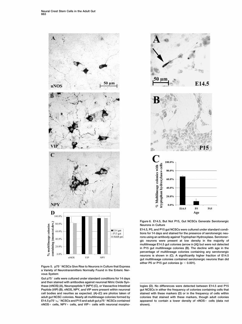
Postnatal Gut NCSCs Differentiate into Neuronsthat Express a Variety of NeurotransmittersWe performed a clonal analysis of gut NCSCs to testwhether they generate cells expressing a variety of neu-rotransmitters. We stained NCSC colonies that had beencultured for 14 days at clonal density using commerciallyavailable antibodies against Vasoactive Intestinal Pep-tide (VIP), Neuropeptide Y (NPY), and neuronal NitricOxide Synthase (nNOS) (Figure 5). Each of these anti-bodies specifically stained neurons based on analysesof adult rat gut sections and multilineage colonies inculture that were double labeled with these antibodiesand antibodies against neuronal markers (data notshown). VIP, NPY, and Nitric Oxide are all expressed bysubsets of enteric neurons in vivo (Pham et al., 1991;Jarvinen et al., 1999). Nearly all E14.5, P15, and adultgut NCSC colonies contained neurons expressing VIP,NPY, and nNOS (Figure 5D). Based on these experi-ments, adult gut NCSCs retain the ability to generateneurons that express a variety of neurotransmitters nor-mally found in the ENS.
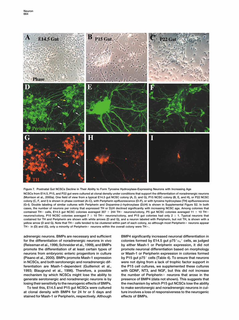
Gut NCSCs Undergo Developmental Restrictionsin Neuronal Subtype PotentialNeurons with different neurotransmitter phenotypes areborn at different intervals of gut development (Pham etal., 1991). Serotonergic progenitors last divide betweenE8 and E14.5 in mouse, while progenitors of NPY-express-ing neurons last divide between E10 and P7. Since sero-
Figure 3. Postnatal Gut NCSCs Are Significantly Less Mitotically tonergic differentiation is completed earliest during em-Active Than Embryonic Gut NCSCsbryonic gut development, we examined whether gut
Cells were stained with antibodies against p75 and �4 integrin toNCSCs undergo restrictions between embryonic andidentify E14.5 and P15 gut NCSCs, and the amount of DNA per cellpostnatal stages in their potential to generate serotoner-was quantitated by flow-cytometry using the DNA-intercalating dyegic neurons in culture.Hoechst 33342 (Morrison and Weissman, 1994). The percentage of
E14.5 (A) and P15 (B) gut NCSCs in G0/G1 (2N DNA content) and Serotonergic neurons in the ENS can be identified byS/G2/M (�2N DNA content) phases of the cell cycle was determined. their expression of Tryptophan Hydroxylase, the initialTo compare the rate at which NCSCs divide in vivo, BrdU was and rate-limiting enzyme in the serotonin synthesis path-administered to rats for a 20 hr period in vivo prior to isolating and
way (Gershon et al., 1977). We cultured E14.5 p75��4�
assaying p75��4� E14.5 gut NCSCs or p75� P15 gut NCSCs for
cells or P5 or P15 p75� cells at clonal density for 14 daysBrdU incorporation. An average of 87% of E14.5 NCSCs (but onlyin standard medium and then stained with an antibody13% of P15 gut NCSCs) incorporated BrdU and therefore divided
over this period in vivo. These data indicate that E14.5 gut NCSCs specific for Tryptophan Hydroxylase (Belin et al., 1991).divide more rapidly or are less frequently quiescent than P15 gut Tryptophan Hydroxylase� neurons were present at lowNCSCs. density in most multilineage colonies formed by E14.5
Neuron662
Figure 4. P15 Gut NCSCs Give Rise to Neurons and Glia In Vivo
Uncultured P15 rat gut p75� cells were isolated by flow-cytometry and injected into two hindlimb bud somites of stage 18 chick embryos.After approximately 72 hr of development (stage 29), embryos were fixed and frozen and then sectioned at the level of the hindlimb. Sectionswere hybridized in situ with probes against the neuronal marker SCG10 and the glial marker P0. Hybridization with neuron-specific probes isshown in (A), where the purple signal indicates SCG10� rat neurons (arrowheads) and the orange signal indicates chick neurons (arrows) ina sympathetic ganglion. Hybridization with a rat-specific probe against the glial marker P0 ([B], open arrowheads) showed rat glia in chickperipheral nerves. Both (A) and (B) are from the same embryo.
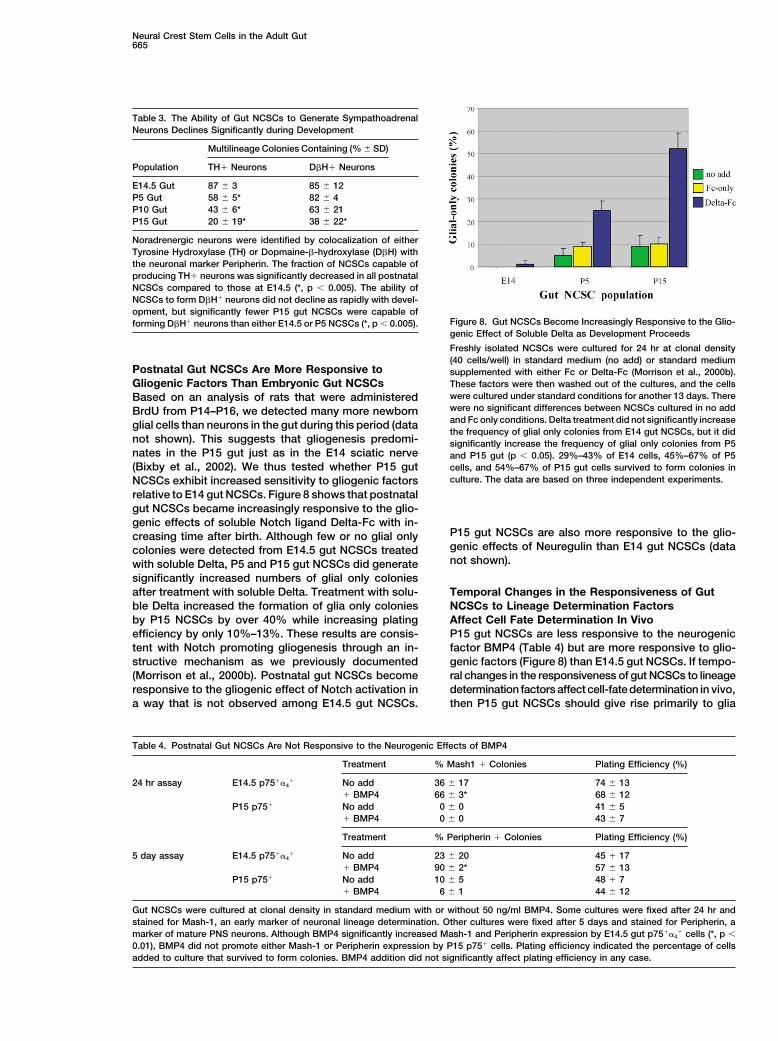
gut NCSCs (66% � 3%; Figures 6A and 6C) but only in Nearly all E14.5 gut NCSC colonies contained neuronsthat expressed TH (87% � 3%) and/or D�H (85% �a few multilineage colonies formed by P5 gut NCSCs12%), but the percentage of gut NCSCs that formed(9% � 5%; Figure 6C). No P15 progenitors formed Tryp-such neurons declined with increasing stem cell agetophan Hydroxylase� neurons in culture (0 � 0; Figures(Table 3). By P15, only 20% of NCSCs formed TH� neu-6B and 6C). Between E14.5 and P15, NCSCs lost therons, and 38% formed D�H� neurons (both differencesability to make serotonergic neurons based on this inwere statistically significant; p � 0.01). In addition tovitro assay.the reduction in the proportion of NCSCs that were ableIn addition to examining the ability of postnatal gutto form such cells, there was also a reduction in theNCSCs to make subtypes of neurons that normally existnumber of such cells per colony (Figure 7; Supplementalin the adult gut, we also wanted to examine their abilityFigure S1). While E14.5 gut NCSC colonies containedto make subtypes of neurons that differentiate only inan average of 444 � 285 D�H� cells and 307 � 244other regions of the PNS. To this end, we examined theTH� cells, P15 gut NCSC colonies averaged only 14 �ability of gut NCSCs to make noradrenergic neurons.13 D�H� cells and 2 � 1 TH� cells. This reduction inAlthough some gut neural crest progenitors transientlythe number of noradrenegic neurons per colony cannotexpress a noradrenergic phenotype prior to E15 (Tyro-be explained by a proportionate decline in the total num-sine Hydroxylase� [TH�] and Dopamine-�-hydroxy-ber of neurons per colony as we estimated that E14.5lase� [D�H�]), these cells continue to proliferate andgut NCSC colonies averaged 41,000 � 25,000 neurons/TH can no longer be detected in the gut after E15 (Baetgecolony, while P15 gut NCSCs averaged 16,000 � 13,000et al., 1990).neurons per colony. Thus the frequency of noradrener-NCSCs were isolated from rats ranging in age fromgic neurons declined much more precipitously than the
E14.5 to P22 and cultured under conditions that promotetotal neurons per colony over this developmental in-
noradrenergic differentiation (Morrison et al., 2000a). We terval.double labeled these cultures with antibodies against Enteric neural crest progenitors have been observedPeripherin (to identify neurons) and either Tyrosine Hy- to transiently express TH and D�H in early embryonicdroxylase (TH) or Dopamine-�-hydroxylase (D�H). All development (before E15) prior to differentiating. To beTH� or D�H� cells coexpressed Peripherin, but only a certain that these NCSC cultures did not contain prolifer-minority of Peripherin-positive neurons expressed TH ating TH or D�H-positive progenitors, we added BrdUor D�H (Figure 7). Double labeling experiments with anti- for the final 24 hr of culture prior to immunohistochemi-bodies against TH and D�H demonstrated that all of the cal staining. Consistent with their neuronal morphology,TH� cells examined coexpressed D�H, but D�H� cells double labeling experiments with TH and BrdU indicatedoften did not express TH. The percentage of D�H posi- that the vast majority of TH� cells were not proliferative:tive cells also expressing TH declined with age. At E14.5, while 22% of all cells in colonies containing TH� cellsan average of 51.7% � 14.5% of D�H positive cells also were BrdU�, only 1.9% of the cells expressing TH hadexpressed TH but at P15, only 23.4% � 21.4% of DBH� incorporated BrdU.cells did (p � 0.05). This is consistent with the factthat gut neural crest progenitors can give rise to both Reduced Neuronal Subtype Potential May Resultnoradrenergic neurons (TH�D�H�) as well as D�H�TH� from Decreased BMP Sensitivityneurons that are peptidergic, but not catecholaminergic We investigated the mechanism by which postnatal
NCSCs lose the potential to form serotonergic and nor-(Baetge et al., 1990).
Neural Crest Stem Cells in the Adult Gut663
Figure 6. E14.5, But Not P15, Gut NCSCs Generate SerotonergicNeurons in Culture
E14.5, P5, and P15 gut NCSCs were cultured under standard condi-tions for 14 days and stained for the presence of serotonergic neu-rons using an antibody against Tryptophan Hydroxylase. Serotoner-gic neurons were present at low density in the majority ofmultilineage E14.5 gut colonies (arrow in [A]) but were not detectedin P15 gut multilineage colonies (B). The decline with age in thepercentage of multilineage colonies containing any serotonergicneurons is shown in (C). A significantly higher fraction of E14.5gut multilineage colonies contained serotonergic neurons than dideither P5 or P15 gut colonies (p � 0.001).
Figure 5. p75� NCSCs Give Rise to Neurons in Culture that Expressa Variety of Neurotransmitters Normally Found in the Enteric Ner-vous System
Gut p75� cells were cultured under standard conditions for 14 daysand then stained with antibodies against neuronal Nitric Oxide Syn-thase (nNOS) (A), Neuropeptide Y (NPY) (C), or Vasoactive Intestinal logies (D). No differences were detected between E14.5 and P15Peptide (VIP) (B). nNOS, NPY, and VIP were present within neuronal gut NCSCs in either the frequency of colonies containing cells thatcell bodies and neurites as expected. (A)–(C) are photos taken of stained with these markers (D) or in the frequency of cells withinadult gut NCSC colonies. Nearly all multilineage colonies formed by colonies that stained with these markers, though adult coloniesE14.5 p75��4
� NCSCs and P15 and adult gut p75� NCSCs contained appeared to contain a lower density of nNOS� cells (data notnNOS� cells, NPY� cells, and VIP� cells with neuronal morpho- shown).
Neuron664
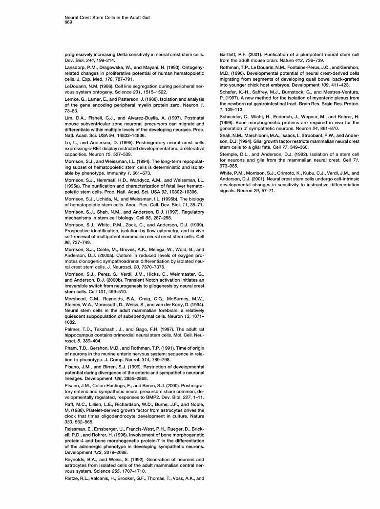
Figure 7. Postnatal Gut NCSCs Decline in Their Ability to Form Tyrosine Hydroxylase-Expressing Neurons with Increasing Age
NCSCs from E14.5, P15, and P22 gut were cultured at clonal density under conditions that support the differentiation of noradrenergic neurons(Morrison et al., 2000a). One field of view from a typical E14.5 gut NCSC colony (A, D, and G), P15 NCSC colony (B, E, and H), or P22 NCSCcolony (C, F, and I) is shown in phase contrast (A–C), with Peripherin epifluorescence (D–F), or with tyrosine hydroxylase (TH) epifluorescence(G–I). Double labeling of similar cultures with Peripherin and Dopamine-�-hydroxylase (D�H) is shown in Supplemental Figure S2. In bothcases, the number of neurons per colony that expressed TH or D�H declined significantly with increasing NCSC age. Among colonies thatcontained TH� cells, E14.5 gut NCSC colonies averaged 307 � 244 TH� neurons/colony, P5 gut NCSC colonies averaged 11 � 10 TH�
neurons/colony, P10 NCSC colonies averaged 7 � 10 TH� neurons/colony, and P15 gut colonies had only 2 � 1. Typical neurons thatcostained for TH and Peripherin are shown with white arrows (D and G), and a neuron labeled with Peripherin, but not TH, is shown with ayellow arrow (D and G). Note that TH� cells tended to be clustered within part of each colony, so although most Peripherin� neurons appearTH� in (D) and (G), only a minority of Peripherin� neurons within the overall colony were TH�.
adrenergic neurons. BMPs are necessary and sufficient BMP4 significantly increased neuronal differentiation incolonies formed by E14.5 gut p75��4
� cells, as judgedfor the differentiation of noradrenergic neurons in vivo(Reissman et al., 1996; Schneider et al., 1999), and BMPs by either Mash-1 or Peripherin expression, it did not
promote neuronal differentiation based on morphologypromote the differentiation of at least certain types ofneurons from embryonic enteric progenitors in culture or Mash-1 or Peripherin expression in colonies formed
by P15 gut p75� cells (Table 4). To ensure that neurons(Pisano et al., 2000). BMPs promote Mash-1 expressionin NCSCs, and both serotonergic and noradrenergic dif- were not dying from a lack of trophic factor support in
the P15 cell cultures, we supplemented these culturesferentiation are Mash-1-dependent (Guillemot et al.,1993; Blaugrund et al., 1996). Therefore, a possible with GDNF, NT3, and NGF, but this did not increase
the number of Peripherin� neurons that arose in themechanism by which NCSCs might lose the ability togenerate serotonergic and noradrenergic neurons is by presence of BMP4 (data not shown). This suggests that
the mechanism by which P15 gut NCSCs lose the abilitylosing their sensitivity to the neurogenic effects of BMPs.To test this, E14.5 and P15 gut NCSCs were cultured to make serotonergic and noradrenergic neurons in cul-
ture involves a loss of responsiveness to the neurogenicat clonal density with BMP4 for 24 hr or 5 days andstained for Mash-1 or Peripherin, respectively. Although effects of BMPs.
Neural Crest Stem Cells in the Adult Gut665
Table 3. The Ability of Gut NCSCs to Generate SympathoadrenalNeurons Declines Significantly during Development
Multilineage Colonies Containing (% � SD)
Population TH� Neurons D�H� Neurons
E14.5 Gut 87 � 3 85 � 12P5 Gut 58 � 5* 82 � 4P10 Gut 43 � 6* 63 � 21P15 Gut 20 � 19* 38 � 22*
Noradrenergic neurons were identified by colocalization of eitherTyrosine Hydroxylase (TH) or Dopmaine-�-hydroxylase (D�H) withthe neuronal marker Peripherin. The fraction of NCSCs capable ofproducing TH� neurons was significantly decreased in all postnatalNCSCs compared to those at E14.5 (*, p � 0.005). The ability ofNCSCs to form D�H� neurons did not decline as rapidly with devel-opment, but significantly fewer P15 gut NCSCs were capable of
Figure 8. Gut NCSCs Become Increasingly Responsive to the Glio-forming D�H� neurons than either E14.5 or P5 NCSCs (*, p � 0.005).genic Effect of Soluble Delta as Development Proceeds
Freshly isolated NCSCs were cultured for 24 hr at clonal density(40 cells/well) in standard medium (no add) or standard medium
Postnatal Gut NCSCs Are More Responsive to supplemented with either Fc or Delta-Fc (Morrison et al., 2000b).Gliogenic Factors Than Embryonic Gut NCSCs These factors were then washed out of the cultures, and the cells
were cultured under standard conditions for another 13 days. ThereBased on an analysis of rats that were administeredwere no significant differences between NCSCs cultured in no addBrdU from P14–P16, we detected many more newbornand Fc only conditions. Delta treatment did not significantly increaseglial cells than neurons in the gut during this period (datathe frequency of glial only colonies from E14 gut NCSCs, but it did
not shown). This suggests that gliogenesis predomi- significantly increase the frequency of glial only colonies from P5nates in the P15 gut just as in the E14 sciatic nerve and P15 gut (p � 0.05). 29%–43% of E14 cells, 45%–67% of P5(Bixby et al., 2002). We thus tested whether P15 gut cells, and 54%–67% of P15 gut cells survived to form colonies in
culture. The data are based on three independent experiments.NCSCs exhibit increased sensitivity to gliogenic factorsrelative to E14 gut NCSCs. Figure 8 shows that postnatalgut NCSCs became increasingly responsive to the glio-genic effects of soluble Notch ligand Delta-Fc with in-
P15 gut NCSCs are also more responsive to the glio-creasing time after birth. Although few or no glial onlygenic effects of Neuregulin than E14 gut NCSCs (datacolonies were detected from E14.5 gut NCSCs treatednot shown).with soluble Delta, P5 and P15 gut NCSCs did generate
significantly increased numbers of glial only coloniesafter treatment with soluble Delta. Treatment with solu- Temporal Changes in the Responsiveness of Gut
NCSCs to Lineage Determination Factorsble Delta increased the formation of glia only coloniesby P15 NCSCs by over 40% while increasing plating Affect Cell Fate Determination In Vivo
P15 gut NCSCs are less responsive to the neurogenicefficiency by only 10%–13%. These results are consis-tent with Notch promoting gliogenesis through an in- factor BMP4 (Table 4) but are more responsive to glio-
genic factors (Figure 8) than E14.5 gut NCSCs. If tempo-structive mechanism as we previously documented(Morrison et al., 2000b). Postnatal gut NCSCs become ral changes in the responsiveness of gut NCSCs to lineage
determination factors affect cell-fate determination in vivo,responsive to the gliogenic effect of Notch activation ina way that is not observed among E14.5 gut NCSCs. then P15 gut NCSCs should give rise primarily to glia
Table 4. Postnatal Gut NCSCs Are Not Responsive to the Neurogenic Effects of BMP4
Treatment % Mash1 � Colonies Plating Efficiency (%)
24 hr assay E14.5 p75��4� No add 36 � 17 74 � 13
� BMP4 66 � 3* 68 � 12P15 p75� No add 0 � 0 41 � 5
� BMP4 0 � 0 43 � 7
Treatment % Peripherin � Colonies Plating Efficiency (%)
5 day assay E14.5 p75��4� No add 23 � 20 45 � 17
� BMP4 90 � 2* 57 � 13P15 p75� No add 10 � 5 48 � 7
� BMP4 6 � 1 44 � 12
Gut NCSCs were cultured at clonal density in standard medium with or without 50 ng/ml BMP4. Some cultures were fixed after 24 hr andstained for Mash-1, an early marker of neuronal lineage determination. Other cultures were fixed after 5 days and stained for Peripherin, amarker of mature PNS neurons. Although BMP4 significantly increased Mash-1 and Peripherin expression by E14.5 gut p75��4
� cells (*, p �
0.01), BMP4 did not promote either Mash-1 or Peripherin expression by P15 p75� cells. Plating efficiency indicated the percentage of cellsadded to culture that survived to form colonies. BMP4 addition did not significantly affect plating efficiency in any case.
Neuron666
upon transplantation into developing chick nerves in have not been observed to form postnatally (Pham etal., 1991), the data suggest that postnatal gut NCSCscontrast to E14.5 gut NCSCs, which gave rise primarily
to neurons (Bixby et al., 2002). To test this, we examined lose the ability to form a subtype of neuron that formsonly during fetal development. Gut NCSCs undergo peri-the peripheral nerves of chicks injected with uncultured
P15 gut NCSCs (Figure 4). Six of eight chicks exhibited natal restrictions in neuronal subtype potential that par-allel the perinatal restrictions in lymphocyte subtypeglia in their peripheral nerves (17.5 cells/positive section)
while one of eight chicks had a single neuron in a periph- potential observed among hematopoietic stem cells(Hayakawa et al., 1985; Ikuta et al., 1990; Kantor et al.,eral nerve. Thus P15 gut NCSCs are biased toward
adopting a glial fate in developing peripheral nerves like 1992). Gut NCSCs also underwent perinatal changes inself-renewal potential and mitotic activity analogous toE14.5 sciatic nerve NCSCs, but unlike E14.5 gut NCSCs.
This demonstrates that NCSCs undergo perinatal those documented in hematopoietic stem cells (Table2, Figure 3).changes in their responsiveness to lineage determina-
tion factors that affect cell fate determination in vivo. In addition to losing the ability to generate serotoner-gic neurons, postnatal gut NCSCs also partially lost theability to generate neurons that expressed TH and/orDiscussionD�H (Table 3, Figure 7; see Supplemental Figure S2online at http://www.neuron.org/cgi/content/full/35/4/Individual cells from the postnatal (P5–P22) and adult gut657/DC1). Embryonic quail gut neural crest progenitors(P65–P110) formed large colonies in culture containingwere previously observed to give rise to noradrenergicneurons, glia, and myofibroblasts (Figures 1 and 2), justneurons after backtransplantation into the embryonicas fetal NCSCs do (Stemple and Anderson, 1992; Shahchick neural crest migration pathway (Rothman et al.,et al., 1994; Morrison et al., 1999; Bixby et al., 2002).1990). In another study, enteric neuroblasts retained theThese multipotent progenitors were isolated by flow-ability to undergo noradrenergic differentiation at E14.5,cytometry as p75� cells (Table 1), and they self-renewedbut by E19.5, most neuroblasts failed to undergo norad-in culture (Table 2), confirming their identity as NCSCs.renergic differentiation (Pisano and Birren, 1999). SinceUncultured P15 gut p75� cells gave rise to neurons andmost P5 gut NCSCs retained the ability to generate TH�glia upon transplantation into the neural crest migrationand D�H� neurons, our data suggest that NCSCs retainpathway of chick embryos (Figure 4). NCSCs, like CNSnoradrenergic potential later in development than re-stem cells, persist in the adult nervous system.stricted neuronal progenitors, though even in stem cellsthis potential declines with increasing age.Gut NCSCs Undergo Perinatal Developmental
The loss of responsiveness of postnatal gut NCSCsChanges that Parallel Changes Observedto the neurogenic effects of BMPs may account for theirin Hematopoietic Stem Cellsreduced serotonergic and noradrenergic potential. TheWithin the nervous system it has not been clear whetherdifferentiation of serotonergic and noradrenergic neu-postnatal neural stem cells retain the ability to makerons is Mash-1 dependent (Blaugrund et al., 1996), andsubtypes of neurons that are normally produced duringBMPs induce Mash-1 expression in sympathoadrenalfetal development; however, studies of mixed popula-progenitors and enteric progenitors (Reissman et al.,tions of neural progenitors have suggested that they1996; Schneider et al., 1999; Pisano et al., 2000). Welose the ability to make some subtypes of neurons post-found that while E14.5 gut NCSCs were highly respon-natally. Cells from the P5–P10 forebrain subventricularsive to the neurogenic effects of BMP4, P15 gut NCSCszone failed to form projection neurons upon transplanta-did not detectably respond to BMP4 (Table 4). Althoughtion into E15 lateral ventricle (Lim et al., 1997). OtherP15 gut NCSCs were unresponsive to BMP4 upon initialstudies of mixed populations of cortical progenitors alsoisolation, they were ultimately able to undergo at leastsuggested that progenitors undergo cell-intrinsic re-limited noradrenergic differentiation after 18 days in cul-strictions during fetal development in the types of neu-ture (Table 3, Figure 7, Supplemental Figure S2). Thisrons they can generate (Barbe and Levitt, 1991; Desaisuggests that at least some progenitors within differenti-and McConnell, 2000). Even though these studies didating NCSC colonies may acquire BMP responsiveness.not employ isolated stem cells, it is reasonable to expectFurther experiments will be required to determine thethat the observed restrictions were present within stemmolecular mechanism that accounts for the reducedcells as well as restricted progenitors. Another possibil-BMP responsiveness of postnatal gut NCSCs.ity is that stem cells were rare within the cell populations
The insensitivity of postnatal gut NCSCs to the neuro-that were studied and that the stem cells themselvesgenic effects of BMPs suggests that other factors likelymay have retained a broader developmental potential.promote neurogenesis in the postnatal ENS. This is con-In this study, we have prospectively identified andsistent with the finding that the differentiation of someisolated both embryonic and postnatal NCSCs and com-enteric neurons is Mash-1 independent (Blaugrund etpared their ability to generate different subtypes of neu-al., 1996); however, the identity of these alternative neu-rons in culture. P15 gut NCSCs retained the ability torogenic signals is unknown.generate neurons that expressed a variety of neuro-
transmitters that are normally found within the ENS.Nearly all P15 gut NCSC colonies contained large num- In Vivo Engraftment of Uncultured P15 Gut NCSCs
Postnatal gut NCSCs gave rise to only small numbersbers of neurons that expressed NPY, VIP, and/or nNOS(Figure 5). However, postnatal gut NCSCs gradually lost of neurons after transplantation into chick embryos,
consistent with the observation that they are BMP insen-the ability to make serotonergic neurons in culture. Sinceserotonergic neurons become postmitotic by E15 and sitive at isolation. Nonetheless, they did form some neu-
Neural Crest Stem Cells in the Adult Gut667
rons in the sympathetic ganglion, an environment in and the ENS (Gershon et al., 1994), it is particularlyinteresting that the adult ENS maintains NCSCs. It will bewhich neurogenesis is normally driven by BMP stimula-
tion (Reissman et al., 1996). One possibility is that a exciting to determine whether the persistence of adultneural stem cells in both the CNS and ENS is relatedsubset of the transplanted P15 gut NCSCs acquired
increased BMP sensitivity as a result of backtrans- to structural or functional similarities between thesetissues.plantation into the neural crest migration pathway and
underwent neurogenesis in response to BMPs (Table 3,Experimental ProceduresFigure 7, Supplemental Figure S2). An alternative possi-
bility is that the P15 gut NCSCs formed a BMP-indepen-Isolation of Postnatal Gut Cellsdent type of neuron in the sympathetic ganglion. Distin-Sprague-Dawley rats were obtained from Simonsen (Gilroy, CA).
guishing between these possibilities will require further The small intestine was separated from attached mesentery andstudy. placed in cold Ca, Mg-free HBSS (Gibco, Grand Island, NY). Outer
muscle/plexus layers were peeled free of the underlying epitheliumIn addition to exhibiting decreased sensitivity to theas previously described (Schafer et al., 1997), minced, and dissoci-neurogenic effects of BMP4, P15 gut NCSCs also exhib-ated in 0.025% trypsin/EDTA (Gibco product 25300-054) plus 1 mg/ited increased sensitivity to the gliogenic effects of Deltaml type 4 collagenase (Worthington, Lakewood, NJ) in Ca, Mg-freeand Neuregulin. Thus gut NCSCs undergo perinatalHBSS for 8 min at 37C. Adult gut was dissociated for 20 min in the
changes in their responsiveness to lineage determina- same enzymes. The digestion was quenched with two volumes oftion factors that are analogous to changes observed staining medium (for recipe see Experimental Procedures in Bixby
et al., 2002). After centrifuging, gut cells were triturated, filteredbetween E10.5 migrating NCSCs and E14.5 postmigra-through a nylon screen (45 m, Sefar America, Kansas City, MO) totory NCSCs (White et al., 2001; Kubu et al., 2002). Con-remove aggregates of cells and undigested tissue, and resuspendedsistent with our observation that sensitivity to gliogenicin staining medium. E14.5 gut, including stomach, small intestine,factors predisposes E14.5 sciatic nerve NCSCs towardand hindgut, were dissected and dissociated as described (Bixby
gliogenesis in developing peripheral nerve (Bixby et al., et al., 2002).2002), the P15 gut NCSCs also preferentially underwentgliogenesis upon transplantation into developing chick Flow-Cytometric Isolation of NCSCs and Cell-Cycle Analysis
Flow cytometric isolation of gut NCSCs was performed as describednerves. Taken together, the data indicate that NCSCs(Bixby et al., 2002). For cell cycle analyses using Hoechst 33342,exhibit spatial and temporal differences in respon-dissociated gut cells were suspended at a concentration of 1–2 �siveness to lineage determination factors that affect the106 cells/ml in staining medium containing 4 g/ml Hoechst 33342
way the cells differentiate in vivo. Regulation of respon- (Sigma) and 50 g/ml verapamil (Sigma) to block MDR-mediatedsiveness to lineage determination factors may be a gen- Hoechst efflux. Cells were incubated for 45 min at 37C and agitatederal mechanism by which stem cells intrinsically regulate every 5–10 min to prevent settling. Immediately after incubation, the
cells were put on ice and stained with antibodies as describedthe process of cell fate determination while remainingabove. Hoechst staining of NCSCs was assayed by flow-cytometry.multipotent.
In this study, the P15 gut NCSCs engrafted only inCell Culturestructures proximal to the hindlimb bud somites (sympa-Tissue culture plate preparation, culture medium, and culture condi-
thetic chain, peripheral nerves) into which the NCSCs tions were as described (Bixby et al., 2002). To promote the differen-were transplanted. The P15 gut NCSCs did not de- tiation of noradrenergic neurons, NCSCs were cultured in standardtectably engraft in more distal neural crest structures medium for 6 days, followed by 6 days in differentiation medium
supplemented with 5 M forskolin (Sigma) and 1 ng/ml BMP4 (R&Dsuch as gut or Remak’s ganglion. This contrasted toSystems), followed by a final 6 days in differentiation medium sup-E14.5 NCSCs, which consistently gave rise to neuronsplemented with 50 ng/ml nerve growth factor and neurotrophin-3in Remak’s ganglion and which sometimes engrafted inas previously described (Morrison et al., 2000a).
gut or pelvic plexus (Morrison et al., 1999; White et al.,2001; Bixby et al., 2002). Since �4 integrin has been BrdU Labeling In Vivoimplicated in the migration of neural crest cells (Kil et The mitotic activity of postnatal gut NCSCs in vivo was assayed byal., 1998), postnatal gut NCSCs may have lost some of administering 5�-bromo-2�-deoxyuridine (BrdU, Sigma) to rat pups
for 20 hr prior to dissecting guts at P15. Doses of BrdU equivalenttheir ability to migrate as a result of losing �4 integrinto 50 g/g body weight were dissolved in 1 mL D-PBS with 0.007expression (Figure 1D). Unlike most embryonic gutM NaOH and injected intraperitoneally at 20, 16, and 2 hr beforeNCSCs that must colonize the length of the gut (Burnsdissection. In one experiment, additional injections were performed
and LeDouarin, 1998) postnatal gut NCSCs may not 18 and 14 hr before dissection. NCSCs were isolated from the BrdUneed to migrate long distances in order to differentiate administered or control rats, allowed to adhere to tissue culturewithin their normal environment. The loss of �4 integrin plates for 18 hr, and stained with an antibody against BrdU (Caltag
product IU-4, Burlingame CA) (Raff et al., 1988).expression by postnatal gut NCSCs may be an adaptiveresponse to their altered need to migrate.
ImmunocytochemistryCultures were fixed in acid ethanol (5% glacial acetic acid in 100%
Why Are Neural Crest Stem Cells in the Adult ENS? ethanol) for 20 min at �20C, washed, blocked, and triply labeledWe do not yet know the functional significance of NCSCs for Peripherin (Chemicon AB1530; Temecula CA), GFAP (Sigma
G-3893), and � SMA (Sigma A-2547) as described (Shah et al., 1994).in the adult gut. Additional studies will be required toMash-1 staining was performed as described (Shah et al., 1994).examine whether there is neurogenesis or gliogenesisPlates stained for neuronal subtype markers were fixed in fresh 4%in the adult gut and whether there is any response toparaformaldehyde for 10 min at room temperature, washed, andinjury. The persistence of NCSCs in the adult ENS cre-blocked as described (Shah et al., 1994). Antibodies were obtained
ates new possibilities for repair after gut injury or trans- as follows: Tyrosine Hydroxylase (TH) (Chemicon, product #MAB5280),plantation from the gut to other sites of injury in the Dopamine-�-hydroxylase (D�H; BD PharMingen, #556313), Neuro-
peptide Y (NPY; Peninsula Laboratories, #IHC 7161, San Carlos,PNS. Given the remarkable similarities between the CNS
Neuron668
CA), Vasoactive Intestinal Peptide (VIP; Peninsula Laboratories, Bixby, S., Kruger, G.M., Mosher, J.T., Joseph, N.M., and Morrison,S.J. (2002). Cell-intrinsic differences between stem cells from differ-#IHC 7180), neuronal Nitric Oxide Synthase (nNOS; Chemicon,
#AB5380), and Tryptophan Hydroxylase (Chemicon, #AB1541). In ent regions of the peripheral nervous system regulate the generationof neural diversity. Neuron 35, this issue, 643–656.some experiments, plates were colabeled with NeuN (Chemicon,
#MAB377) to confirm that neuronal subtype antibodies were labeling Blaugrund, E., Pham, T.D., Tennyson, V.M., Lo, L., Sommer, L., An-neurons. For p75 (Promega prod. 1405-41-0) staining of gut sec- derson, D.J., and Gershon, M.D. (1996). Distinct subpopulationstions, tissue was fixed in freshly prepared 4% paraformaldehyde of enteric neuronal progenitors defined by time of development,and embedded in paraffin. sympathoadrenal lineage markers and Mash-1-dependence. Devel-
opment 122, 309–320.In Vivo Transplantation of Neural Crest Progenitors Burns, A.J., and Douarin, N.M. (1998). The sacral neural crest con-Fertile White Leghorn eggs (Bilbie Aviaries, Ann Arbor, MI) were tributes neurons and glia to the post-umbilical gut: spatiotemporalincubated to Hamburger and Hamilton stage 17–18 (Hamburger and analysis of the development of the enteric nervous system. Develop-Hamilton, 1951) and injected with P15 gut p75� cells. Approximately ment 125, 4335–4347.70,000 p75� cells were isolated by flow-cytometry, backloaded into
Cai, L., Hayes, N.L., and Nowakowski, R.S. (1997). Synchrony ofa drawn glass capillary tube (World Precision Instruments, Sarasotaclonal cell proliferation and contiguity of clonally related cells: pro-FL), centrifuged for 2 min at 52 � g, and injected into the anterior,duction of mosaicism in the ventricular zone of developing mousemedial corner of two somites in the himblimb bud region of eachneocortex. J. Neurosci. 17, 2088–2100.embryo. Injections were performed using a Narishige H-7 pipetteCheshier, S., Morrison, S.J., Liao, X., and Weissman, I.L. (1999). Inholder mounted on a MM-33 micromanipulator (Fine Science Tools,vivo proliferation and cell cycle kinetics of long-term self-renewingForester City, CA) and very gentle air pressure. We estimated thehematopoietic stem cells. Proc. Natl. Acad. Sci. USA 96, 3120–3125.number of cells injected per somite as 310 � 20 cells by labeling
sorted P15 p75� cells with DiI and then counting the number of Desai, A.R., and McConnell, S.K. (2000). Progressive restriction inDiI� cells/somite immediately after injection. Injected embryos were fate potential by neural progenitors during cerebral cortical develop-incubated for 3 days to stage 29, fixed, embedded in OCT, and ment. Development 127, 2863–2872.cryostat sectioned. To control for nonspecific hybridization, sec- Doetsch, F., Caille, I., Lim, D.A., Garcia-Verdugo, J.M., and Alvarez-tions from E16.5 rat and uninjected stage 29 chicks were processed Buylla, A. (1999). Subventricular zone astrocytes are neural stemin parallel as positive and negative controls, and sections adjacent cells in the adult mammalian brain. Cell 97, 703–716.to those showing engraftment were probed with the sense strands
Eriksson, P.S., Perfilieva, E., Bjork-Eriksson, T., Alborn, A.-M., Nor-of SCG10 and P0. Since embryos were unilaterally injected, thedborg, C., Peterson, D.A., and Gage, F.H. (1998). Neurogenesis incontralateral side to engraftment also served as a negative control.the adult human hippocampus. Nat. Med. 4, 1313–1317.
Gershon, M.D., Dreyfus, C.F., Pickel, V.M., Joh, T.H., and Reis, D.J.Acknowledgments(1977). Serotonergic neurons in the peripheral nervous system: iden-tification in gut by immunohistochemical localization of tryptophanThis work was supported by the National Institutes of Health (R01hydroxylase. Proc. Natl. Acad. Sci. USA 74, 3086–3089.NS40750-01), the Searle Scholars Program, and the Howard Hughes
Medical Institute. G.M.K. was supported by the University of Michi- Gershon, M.D., Kirchgessner, A.L., and Wade, P.R. (1994). Func-tional anatomy of the enteric nervous system. In Physiology of thegan MSTP training grant (GM07863) and J.T.M. was supported by
a fellowship from the University of Michigan Cancer Biology Training Gastrointestinal Tract, L.R. Johnson, ed. (New York: Raven Press),pp. 381–422.Program. Thanks to Mark Kukuruga, Anne Marie Deslaurier, Mark
Kiel, David Adams, and the University of Michigan Flow-Cytometry Gould, E., Reeves, A.J., Graziano, M.S.A., and Gross, C.G. (1999).Core Facility. Flow-cytometry was supported in part by the UM- Neurogenesis in the neocortex of adult primates. Science 286,Comprehensive Cancer NIH CA46592 and the UM-Multipurpose Ar- 548–552.thritis Center NIH AR20557. Thanks to Elizabeth Smith in the Hybrid-
Guillemot, F., Lo, L.-C., Johnson, J.E., Auerbach, A., Anderson, D.J.,oma Core Facility for antibody production, which was supported in
and Joyner, A.L. (1993). Mammalian achaete-scute homolog-1 ispart through the Michigan Diabetes Research and Training Center
required for the early development of olfactory and autonomic neu-(NIH5P60-DK20572) and the Rheumatic Core Disease Center (1 P30
rons. Cell 75, 463–476.AR48310). Thanks to Rachel Calayag, Mohamed Farah, and Kelly
Hamburger, V., and Hamilton, H. (1951). A series of normal stagesYeager for expert technical assistance.in the development of the chick embryo. J. Morphol. 88, 49–92.
Hayakawa, K., Hardy, R.R., Hertzenberg, L.A., and Herzenberg, L.A.Received: April 4, 2002(1985). Progenitors for Ly-1 B cells are distinct from progenitors forRevised: June 13, 2002other B cells. J. Exp. Med. 161, 1554–1568.
References Ikuta, K., Kina, T., Macneil, I., Uchida, N., Peault, B., Chien, Y.H., andWeissman, I.L. (1990). A developmental switch in thymic lymphocytematuration potential occurs at the level of hematopoietic stem cells.Altman, J. (1969). Autoradiographic and histological studies of post-
natal neurogenesis. IV. Cell proliferation and migration in the anterior Cell 62, 863–874.forebrain, with special reference to persisting neurogenesis in the Jarvinen, M.K., Wollman, W.J., Powrozek, T.A., Schultz, J.A., andolfactory bulb. J. Comp. Neurol. 137, 433–458. Powley, T.L. (1999). Nitric oxide synthase-containing neurons in the
myenteric plexus of the rat gastrointestinal tract: distribution andAnderson, D.J., and Axel, R. (1985). Molecular probes for the devel-opment and plasticity of neural crest derivatives. Cell 42, 649–662. regional density. Anat. Embryol. (Berl.) 199, 99–112.
Johansson, C.B., Momma, S., Clarke, D.L., Risling, M., Lendahl, U.,Baetge, G., Pintar, J.E., and Gershon, M.D. (1990). Transiently cate-cholaminergic (TC) cells in the bowel of the fetal rat: precursors of and Frisen, J. (1999). Identification of a neural stem cell in the adult
mammalian central nervous system. Cell 96, 25–34.noncatecholaminergic enteric neurons. Dev. Biol. 141, 353–380.
Barbe, M.F., and Levitt, P. (1991). The early commitment of fetal Kantor, A.B., Stall, A.M., Adams, S., Herzenberg, L.A., and Herzen-berg, L.A. (1992). Differential development of progenitor activity forneurons to the limbic cortex. J. Neurosci. 11, 519–533.three B-cell lineages. Proc. Natl. Acad. Sci. USA 89, 3320–3324.Baroffio, A., Dupin, E., and Le Douarin, N.M. (1991). Common precur-
sors for neural and mesectodermal derivatives in the cephalic neural Kil, S.H., Krull, C.E., Cann, G., Clegg, D., and Bronner-Fraser, M.(1998). The alpha4 subunit of integrin is important for neural crestcrest. Development 112, 301–305.cell migration. Dev. Biol. 202, 29–42.Belin, M.F., Fevre-Montange, M., Reboul, A., Didier-Bazes, M., Ehret,
M., Maitre, M., and Tardy, M. (1991). Primary dissociated cell culture Kubu, C.J., Orimoto, K., Morrison, S.J., Weinmaster, G., Anderson,D.J., and Verdi, J.M. (2002). Developmental changes in Notch1 andof embryonic rat metencephalon: presence of GABA in serotonergic
neurons. Neurosci. Lett. 125, 101–106. Numb expression mediated by local cell-cell interactions underlie
Neural Crest Stem Cells in the Adult Gut669
progressively increasing Delta sensitivity in neural crest stem cells. Bartlett, P.F. (2001). Purification of a pluripotent neural stem cellfrom the adult mouse brain. Nature 412, 736–739.Dev. Biol. 244, 199–214.
Lansdorp, P.M., Dragowska, W., and Mayani, H. (1993). Ontogeny- Rothman, T.P., Le Douarin, N.M., Fontaine-Perus, J.C., and Gershon,M.D. (1990). Developmental potential of neural crest-derived cellsrelated changes in proliferative potential of human hematopoietic
cells. J. Exp. Med. 178, 787–791. migrating from segments of developing quail bowel back-graftedinto younger chick host embryos. Development 109, 411–423.LeDouarin, N.M. (1986). Cell line segregation during peripheral ner-
vous system ontogeny. Science 231, 1515–1522. Schafer, K.-H., Saffrey, M.J., Burnstock, G., and Mestres-Ventura,P. (1997). A new method for the isolation of myenteric plexus fromLemke, G., Lamar, E., and Patterson, J. (1988). Isolation and analysisthe newborn rat gastrointestinal tract. Brain Res. Brain Res. Protoc.of the gene encoding peripheral myelin protein zero. Neuron 1,1, 109–113.73–83.Schneider, C., Wicht, H., Enderich, J., Wegner, M., and Rohrer, H.Lim, D.A., Fishell, G.J., and Alvarez-Buylla, A. (1997). Postnatal(1999). Bone morphogenetic proteins are required in vivo for themouse subventricular zone neuronal precursors can migrate andgeneration of sympathetic neurons. Neuron 24, 861–870.differentiate within multiple levels of the developing neuraxis. Proc.
Natl. Acad. Sci. USA 94, 14832–14836. Shah, N.M., Marchionni, M.A., Isaacs, I., Stroobant, P.W., and Ander-son, D.J. (1994). Glial growth factor restricts mammalian neural crestLo, L., and Anderson, D. (1995). Postmigratory neural crest cellsstem cells to a glial fate. Cell 77, 349–360.expressing c-RET display restricted developmental and proliferative
capacities. Neuron 15, 527–539. Stemple, D.L., and Anderson, D.J. (1992). Isolation of a stem cellfor neurons and glia from the mammalian neural crest. Cell 71,Morrison, S.J., and Weissman, I.L. (1994). The long-term repopulat-973–985.ing subset of hematopoietic stem cells is deterministic and isolat-
able by phenotype. Immunity 1, 661–673. White, P.M., Morrison, S.J., Orimoto, K., Kubu, C.J., Verdi, J.M., andAnderson, D.J. (2001). Neural crest stem cells undergo cell-intrinsicMorrison, S.J., Hemmati, H.D., Wandycz, A.M., and Weissman, I.L.developmental changes in sensitivity to instructive differentiation(1995a). The purification and characterization of fetal liver hemato-signals. Neuron 29, 57–71.poietic stem cells. Proc. Natl. Acad. Sci. USA 92, 10302–10306.
Morrison, S.J., Uchida, N., and Weissman, I.L. (1995b). The biologyof hematopoietic stem cells. Annu. Rev. Cell. Dev. Biol. 11, 35–71.
Morrison, S.J., Shah, N.M., and Anderson, D.J. (1997). Regulatorymechanisms in stem cell biology. Cell 88, 287–298.
Morrison, S.J., White, P.M., Zock, C., and Anderson, D.J. (1999).Prospective identification, isolation by flow cytometry, and in vivoself-renewal of multipotent mammalian neural crest stem cells. Cell96, 737–749.
Morrison, S.J., Csete, M., Groves, A.K., Melega, W., Wold, B., andAnderson, D.J. (2000a). Culture in reduced levels of oxygen pro-motes clonogenic sympathoadrenal differentiation by isolated neu-ral crest stem cells. J. Neurosci. 20, 7370–7376.
Morrison, S.J., Perez, S., Verdi, J.M., Hicks, C., Weinmaster, G.,and Anderson, D.J. (2000b). Transient Notch activation initiates anirreversible switch from neurogenesis to gliogenesis by neural creststem cells. Cell 101, 499–510.
Morshead, C.M., Reynolds, B.A., Craig, C.G., McBurney, M.W.,Staines, W.A., Morassutti, D., Weiss, S., and van der Kooy, D. (1994).Neural stem cells in the adult mammalian forebrain: a relativelyquiescent subpopulation of subependymal cells. Neuron 13, 1071–1082.
Palmer, T.D., Takahashi, J., and Gage, F.H. (1997). The adult rathippocampus contains primordial neural stem cells. Mol. Cell. Neu-rosci. 8, 389–404.
Pham, T.D., Gershon, M.D., and Rothman, T.P. (1991). Time of originof neurons in the murine enteric nervous system: sequence in rela-tion to phenotype. J. Comp. Neurol. 314, 789–798.
Pisano, J.M., and Birren, S.J. (1999). Restriction of developmentalpotential during divergence of the enteric and sympathetic neuronallineages. Development 126, 2855–2868.
Pisano, J.M., Colon-Hastings, F., and Birren, S.J. (2000). Postmigra-tory enteric and sympathetic neural precursors share common, de-velopmentally regulated, responses to BMP2. Dev. Biol. 227, 1–11.
Raff, M.C., Lillien, L.E., Richardson, W.D., Burne, J.F., and Noble,M. (1988). Platelet-derived growth factor from astrocytes drives theclock that times oligodendrocyte development in culture. Nature333, 562–565.
Reissman, E., Ernsberger, U., Francis-West, P.H., Rueger, D., Brick-ell, P.D., and Rohrer, H. (1996). Involvement of bone morphogeneticprotein-4 and bone morphogenetic protein-7 in the differentiationof the adrenergic phenotype in developing sympathetic neurons.Development 122, 2079–2088.
Reynolds, B.A., and Weiss, S. (1992). Generation of neurons andastrocytes from isolated cells of the adult mammalian central ner-vous system. Science 255, 1707–1710.
Rietze, R.L., Valcanis, H., Brooker, G.F., Thomas, T., Voss, A.K., and
Copyright © 2022 FDOKUMEN