Vesicles Bearing Toxoplasma Apicoplast Membrane Proteins Persist Following Loss of the Relict...
14
Vesicles Bearing Toxoplasma Apicoplast Membrane Proteins Persist Following Loss of the Relict Plastid or Golgi Body Disruption Anne Bouchut 1¤ , Jennifer A. Geiger 1,2 , Amy E. DeRocher 1 , Marilyn Parsons 1,2 * 1 Seattle Biomedical Research Institute, Seattle, WA, United States of America, 2 Dept. of Global Health, University of Washington, Seattle, WA, United States of America Abstract Toxoplasma gondii and malaria parasites contain a unique and essential relict plastid called the apicoplast. Most apicoplast proteins are encoded in the nucleus and are transported to the organelle via the endoplasmic reticulum (ER). Three trafficking routes have been proposed for apicoplast membrane proteins: (i) vesicular trafficking from the ER to the Golgi and then to the apicoplast, (ii) contiguity between the ER membrane and the apicoplast allowing direct flow of proteins, and (iii) vesicular transport directly from the ER to the apicoplast. Previously, we identified a set of membrane proteins of the T. gondii apicoplast which were also detected in large vesicles near the organelle. Data presented here show that the large vesicles bearing apicoplast membrane proteins are not the major carriers of luminal proteins. The vesicles continue to appear in parasites which have lost their plastid due to mis-segregation, indicating that the vesicles are not derived from the apicoplast. To test for a role of the Golgi body in vesicle formation, parasites were treated with brefeldin A or transiently transfected with a dominant-negative mutant of Sar1, a GTPase required for ER to Golgi trafficking. The immunofluorescence patterns showed little change. These findings were confirmed using stable transfectants, which expressed the toxic dominant-negative sar1 following Cre-loxP mediated promoter juxtaposition. Our data support the hypothesis that the large vesicles do not mediate the trafficking of luminal proteins to the apicoplast. The results further show that the large vesicles bearing apicoplast membrane proteins continue to be observed in the absence of Golgi and plastid function. These data raise the possibility that the apicoplast proteome is generated by two novel ER to plastid trafficking pathways, plus the small set of proteins encoded by the apicoplast genome. Citation: Bouchut A, Geiger JA, DeRocher AE, Parsons M (2014) Vesicles Bearing Toxoplasma Apicoplast Membrane Proteins Persist Following Loss of the Relict Plastid or Golgi Body Disruption. PLoS ONE 9(11): e112096. doi:10.1371/journal.pone.0112096 Editor: Stuart Alexander Ralph, University of Melbourne, Australia Received April 25, 2012; Accepted October 13, 2014; Published November 4, 2014 Copyright: ß 2014 Bouchut et al. This is an open-access article distributed under the terms of the Creative Commons Attribution License, which permits unrestricted use, distribution, and reproduction in any medium, provided the original author and source are credited. Funding: JAG was supported by a training grant from the National Institute of Allergy and Infectious Diseases (Grant T32AI007509). The work was supported by a grant from the National Institutes of Health R01AI50506. The funders had no role in study design, data collection and analysis, decision to publish, or preparation of the manuscript. Competing Interests: The authors have declared that no competing interests exist. * Email: [email protected] ¤ Current address: Department of Pharmacology & Toxicology, Indiana University School of Medicine, Indianapolis, IN, United States of America Introduction Toxoplasma gondii is an obligate intracellular protozoan parasite belonging to the phylum Apicomplexa, which also includes the malaria parasite Plasmodium falciparum. T. gondii, one of the most successful known parasites, infects one-third of the world’s human population and its ability to differentiate into quiescent bradyzoite cysts leads to lifelong persistence [1,2]. While infection of immunocompetent hosts is often asymptomatic, T. gondii has been recognized as a major pathogen of immunocom- promised patients, i.e. transplant recipients or those with HIV/ AIDS, as well as being vertically transmitted to the fetus from recently infected mothers. Indeed, T. gondii is the causative agent of both toxoplasmic encephalitis, the most common cause of focal brain lesions in people with HIV/AIDS, and congenital toxoplas- mosis, a leading cause of neurological birth defects in children. Insights leading to new therapeutic options are needed since available drugs can have serious side effects. Among the characteristics of many Apicomplexa is the presence of a unique organelle, the apicoplast. It is a non-photosynthetic plastid acquired by secondary endosymbiosis from an alga, i.e., a secondary plastid. The apicoplast is the site of several anabolic pathways including iron-sulfur cluster biosynthesis, lipoic acid synthesis [3], part of the heme biosynthesis pathway [4], type II fatty acid synthesis [5], and the non-mevalonate pathway of isoprenoid biosynthesis [6,7]. The last pathway is found in apicoplasts of all species, is essential to the parasites (although not necessarily in all developmental stages), and is absent from animal hosts. Interference with apicoplast DNA replication [8] or translation [9,10], as well as inhibition of certain apicoplast metabolic functions [5,6], is lethal for T. gondii and Plasmodium. Thus the organelle is a potential target for the development of novel drugs. The apicoplast’s small genome (35 kb) encodes primarily RNAs and proteins important for the propagation of the organelle. Hence most plastid functions are fulfilled by numerous nucleus- encoded proteins. These are typically imported into the apicoplast lumen by virtue of an N-terminal bipartite sequence which is composed of a signal peptide and an adjacent transit peptide (signal+transit, S+T). Proteins destined for the lumen of the apicoplast, such as acyl carrier protein (ACP), are first imported PLOS ONE | www.plosone.org 1 November 2014 | Volume 9 | Issue 11 | e112096
-
Upload
independent -
Category
Documents
-
view
0 -
download
0
Transcript of Vesicles Bearing Toxoplasma Apicoplast Membrane Proteins Persist Following Loss of the Relict...

Vesicles Bearing Toxoplasma Apicoplast MembraneProteins Persist Following Loss of the Relict Plastid orGolgi Body DisruptionAnne Bouchut1¤, Jennifer A. Geiger1,2, Amy E. DeRocher1, Marilyn Parsons1,2*
1 Seattle Biomedical Research Institute, Seattle, WA, United States of America, 2 Dept. of Global Health, University of Washington, Seattle, WA, United States of America
Abstract
Toxoplasma gondii and malaria parasites contain a unique and essential relict plastid called the apicoplast. Most apicoplastproteins are encoded in the nucleus and are transported to the organelle via the endoplasmic reticulum (ER). Threetrafficking routes have been proposed for apicoplast membrane proteins: (i) vesicular trafficking from the ER to the Golgiand then to the apicoplast, (ii) contiguity between the ER membrane and the apicoplast allowing direct flow of proteins,and (iii) vesicular transport directly from the ER to the apicoplast. Previously, we identified a set of membrane proteins of theT. gondii apicoplast which were also detected in large vesicles near the organelle. Data presented here show that the largevesicles bearing apicoplast membrane proteins are not the major carriers of luminal proteins. The vesicles continue toappear in parasites which have lost their plastid due to mis-segregation, indicating that the vesicles are not derived from theapicoplast. To test for a role of the Golgi body in vesicle formation, parasites were treated with brefeldin A or transientlytransfected with a dominant-negative mutant of Sar1, a GTPase required for ER to Golgi trafficking. Theimmunofluorescence patterns showed little change. These findings were confirmed using stable transfectants, whichexpressed the toxic dominant-negative sar1 following Cre-loxP mediated promoter juxtaposition. Our data support thehypothesis that the large vesicles do not mediate the trafficking of luminal proteins to the apicoplast. The results furthershow that the large vesicles bearing apicoplast membrane proteins continue to be observed in the absence of Golgi andplastid function. These data raise the possibility that the apicoplast proteome is generated by two novel ER to plastidtrafficking pathways, plus the small set of proteins encoded by the apicoplast genome.
Citation: Bouchut A, Geiger JA, DeRocher AE, Parsons M (2014) Vesicles Bearing Toxoplasma Apicoplast Membrane Proteins Persist Following Loss of the RelictPlastid or Golgi Body Disruption. PLoS ONE 9(11): e112096. doi:10.1371/journal.pone.0112096
Editor: Stuart Alexander Ralph, University of Melbourne, Australia
Received April 25, 2012; Accepted October 13, 2014; Published November 4, 2014
Copyright: � 2014 Bouchut et al. This is an open-access article distributed under the terms of the Creative Commons Attribution License, which permitsunrestricted use, distribution, and reproduction in any medium, provided the original author and source are credited.
Funding: JAG was supported by a training grant from the National Institute of Allergy and Infectious Diseases (Grant T32AI007509). The work was supported by agrant from the National Institutes of Health R01AI50506. The funders had no role in study design, data collection and analysis, decision to publish, or preparationof the manuscript.
Competing Interests: The authors have declared that no competing interests exist.
* Email: [email protected]
¤ Current address: Department of Pharmacology & Toxicology, Indiana University School of Medicine, Indianapolis, IN, United States of America
Introduction
Toxoplasma gondii is an obligate intracellular protozoan
parasite belonging to the phylum Apicomplexa, which also
includes the malaria parasite Plasmodium falciparum. T. gondii,one of the most successful known parasites, infects one-third of the
world’s human population and its ability to differentiate into
quiescent bradyzoite cysts leads to lifelong persistence [1,2]. While
infection of immunocompetent hosts is often asymptomatic, T.gondii has been recognized as a major pathogen of immunocom-
promised patients, i.e. transplant recipients or those with HIV/
AIDS, as well as being vertically transmitted to the fetus from
recently infected mothers. Indeed, T. gondii is the causative agent
of both toxoplasmic encephalitis, the most common cause of focal
brain lesions in people with HIV/AIDS, and congenital toxoplas-
mosis, a leading cause of neurological birth defects in children.
Insights leading to new therapeutic options are needed since
available drugs can have serious side effects.
Among the characteristics of many Apicomplexa is the presence
of a unique organelle, the apicoplast. It is a non-photosynthetic
plastid acquired by secondary endosymbiosis from an alga, i.e., a
secondary plastid. The apicoplast is the site of several anabolic
pathways including iron-sulfur cluster biosynthesis, lipoic acid
synthesis [3], part of the heme biosynthesis pathway [4], type II
fatty acid synthesis [5], and the non-mevalonate pathway of
isoprenoid biosynthesis [6,7]. The last pathway is found in
apicoplasts of all species, is essential to the parasites (although
not necessarily in all developmental stages), and is absent from
animal hosts. Interference with apicoplast DNA replication [8] or
translation [9,10], as well as inhibition of certain apicoplast
metabolic functions [5,6], is lethal for T. gondii and Plasmodium.
Thus the organelle is a potential target for the development of
novel drugs.
The apicoplast’s small genome (35 kb) encodes primarily RNAs
and proteins important for the propagation of the organelle.
Hence most plastid functions are fulfilled by numerous nucleus-
encoded proteins. These are typically imported into the apicoplast
lumen by virtue of an N-terminal bipartite sequence which is
composed of a signal peptide and an adjacent transit peptide
(signal+transit, S+T). Proteins destined for the lumen of the
apicoplast, such as acyl carrier protein (ACP), are first imported
PLOS ONE | www.plosone.org 1 November 2014 | Volume 9 | Issue 11 | e112096
into the endoplasmic reticulum (ER), where the signal sequence is
cleaved and then transferred to the apicoplast via an unknown
mechanism. There the transit peptide is removed. Several lines of
evidence indicate that targeting of apicoplast luminal proteins
bypasses the Golgi body. First, it is resistant to the Golgi inhibitor
brefeldin A (BFA, which blocks COPI coat assembly leading to
collapse of Golgi body to the ER) [11,12]. Second, targeting is
resistant to low temperature treatment which disrupts ER to Golgi
trafficking [11]. Finally, apicoplast luminal proteins bearing a
sequence that mediates retrieval of proteins from the Golgi back to
the ER were still localized to the apicoplast, indicating that the
protein did not encounter the Golgi-localized retrieval receptor
[11,12].
The apicoplast is a product of secondary endosymbiosis and, as
such, is surrounded by four membranes. The inner two are
presumably homologous to chloroplast membranes; the third or
periplastid membrane likely originated from the algal plasma
membrane and the outer membrane is thought to have arisen
from an endocytic membrane from the apicomplexan progenitor.
Proteins residing in the membranes or intermembrane spaces of
the apicoplast appear to fall into two categories: those that possess
S+T targeting sequences and those that do not. Examples of those
which bear a S+T sequence include Tic20 [13] and Tic22 [14,15]
(components of the import apparatus) and PfiTPT (a transporter
of the P. falciparum apicoplast innermost membrane) [16], as well
as some proteins of the periplastid space or membrane such as
components related to the ER-associated degradation machinery
(e.g., Ufd1 and Der1-related proteins) [14,17–19]. Those that lack
a canonical targeting sequence include two non-luminal proteins
recently identified in a systematic screen [20], as well as three
membrane-associated proteins that have been examined in some
detail: the protease FtsH1 [21,22], the thioredoxin ATrx1 [23],
and the transporter APT1 [24,25]. FtsH1 is a 1250 aa protease
with a single transmembrane domain that is processed at both N-
and C-termini. ATrx1 is a peripheral membrane protein that
possesses a signal anchor sequence and is N-terminally processed,
removing the anchor. In contrast, APT1 is a polytopic membrane
protein that undergoes no detectable processing. In addition to
being present at the apicoplast, immunoelectron microscopy has
detected each of these proteins in large vesicles (Vap), which are
recognized as apparent vesicles and tubules in immunofluores-
cence analysis (IFA) [22–24]. Vap are not artifacts of overexpres-
sion since they can be seen in untransfected cells using a
monoclonal antibody (mAb) to ATrx1 [23]. The apicoplast
division cycle has been described by Streipen et al. [26]. In stage
1, the round plastid is associated with the centriole (early G1). In
stage 2, the plastid becomes ovoid, remaining associated with the
now-duplicated centrioles. In stage 3, it elongates and approaches
the nucleus (late G1 to S), forming a U-shape in stage 4. By stage
5, the apicoplast has divided and mitosis is underway, and at stage
6, mitosis is complete. In stages 2–4 of the apicoplast division cycle
as described, Vap become prominent. In some immunoelectron
microscopy images, Vap are observed very close to or merging with
the apicoplast [23]. Others have shown that Vap and the apicoplast
bear phosphatidylinositol 3-phosphate (PI3P) and that overexpres-
sion of a PI3P binding protein leads to loss of the apicoplast [27].
Additionally, mutations in APT1 that block targeting to the
apicoplast also block APT1 recruitment into Vap. Based on these
findings, we [24] and others [27] have suggested that Vap are
transport vesicles, which serve to move at least a subset of
apicoplast membrane proteins to the organelle. However, it is
possible that the Vap represent degradation intermediates origi-
nating from the apicoplast or persistent structures originating from
the ER or Golgi body. Here, we will refer to this group of
apicoplast proteins found abundantly on Vap as ApV proteins.
Three hypothetical routes for ApV protein transport to the
apicoplast have been proposed: (i) transit in vesicles first to the
Golgi body and then to the apicoplast, following the typical route
for secretory proteins; (ii) direct flow from the ER to the apicoplast
via continuous membrane; (iii) transport in vesicles directly from
the ER to the apicoplast. Here we probe the relationship of the
Golgi body and Vap pharmacologically using BFA and genetically
via expression of a dominant-negative mutant of SAR1, the
GTPase that initiates ER to Golgi trafficking. Vap persisted despite
these treatments. These studies suggest that recruitment of ApV
proteins into Vap may be Golgi-independent. Additionally our
studies show that luminal proteins are largely absent from Vap.
Results
A luminal marker protein does not traffic via Vap
When expressed from their cognate promoters, FtsH1, ATrx1
and APT1 [22–24] have been observed in Vap, large electron
dense vesicles near the apicoplast, suggesting that these proteins
may travel to the apicoplast via a vesicle trafficking pathway. In
contrast, previous studies have not revealed vesicles bearing
luminal proteins. However, most of these studies employed
heterologous promoters with different timing of expression, raising
the possibility that some normal trafficking pathways might not be
readily detected. We hypothesized that luminal S+T bearing
proteins may travel to the plastid in Vap, and that these trafficking
intermediates could be detected when expression was driven by a
promoter of a gene encoding an apicoplast luminal protein (these
genes are coordinately expressed along with genes encoding
apicoplast membrane proteins [28]). For these studies we chose the
ACP promoter for the luminal marker. It drives expression with
the same temporal pattern as APT1, although the ACP mRNA
expression level is about 4-fold higher (Fig. S1) ([28] and
ToxoDB). The ATrx1 mRNA shows two closely spaced peaks,
the second of which corresponds to peak expression of ACP and
APT1. We carefully examined the staining pattern of a luminally
targeted Heteractis crispa red fluorescent protein tagged with V5
epitopes (S+TRed-V5) (Fig. 1). We used antibodies to the V5
epitope to enhance the signal as compared to intrinsic fluorescence
and to detect protein that had not yet matured to its fluorescent
form [29]. Vap were revealed by detection of epitope-tagged APT1
or ATrx1 expressed using their cognate promoters (see Methods
for description of tagged proteins). The populations examined
contained a mix of parasites at different stages of the apicoplast
division cycle ensuring detection of co-trafficking, should it exist
(for example in the analysis of APT1-HA and S+TRed-V5, the
populations averaged: 30% stage 1+2, 13% stage 3+4 and 7%
stage 5+6).S+TRed-V5 was prominently observed at the apicoplast as a dot
surrounded by the membrane proteins or in an elongated bead
and tubule appearance interspersed with the membrane proteins,
as previously detailed [24]. When we rescaled the signal to search
for dimly stained objects, no additional structures were revealed in
80% of the parasites (Fig. 1A). In 20% of parasites bearing Vap
(Fig. 1B) we observed faint perinuclear S+TRed-V5 staining,
characteristic of the ER, suggesting the presence of newly
synthesized S+TRed-V5. Commensurate with the ER staining,
this population was enriched for parasites earlier in the apicoplast
division cycle when plastid proteins are beginning to be
synthesized, since almost all were stage 1 or stage 2 (.95%).
These cells had occasional spots of slightly more concentrated
fluorescence signal and most of these coincided with foci staining
Vesicles Bearing Apicoplast Membrane Proteins
PLOS ONE | www.plosone.org 2 November 2014 | Volume 9 | Issue 11 | e112096

Figure 1. Vap are not major vehicles for luminal protein trafficking to the apicoplast. For IFA analysis here and elsewhere unless indicated,proteins were detected by mAbs directed against epitope tags followed by fluorochrome-coupled secondary antibodies as described in Methods. Inthis case, the apicoplast membrane proteins were detected anti-HA mAb was followed by FITC-coupled secondary antibodies and S+TRed-V5 wasdetected by anti-V5 mAb followed by Texas Red-coupled antibodies to bypass the need for maturation of the HcRed chromophore. Here, as in otherfigures, the color coding for merged images is indicated by the text color above the merged images, while dashed lines mark the outline of theparasite. In this experiment, the parasites co-expressed S+TRed-V5 driven by the ACP promoter and epitope-tagged ApV proteins APT1-HA or ATrx1-HA. A) IFA showing the pattern seen in about 80% of parasites with ATrx1-HA in Vap (arrows) near the apicoplast. One set of anti-V5 images is scalednormally and the second is scaled to detect fainter signals (the staining at the apicoplast is then saturated). No evidence of localization of the luminalmarker S+TRed-V5 with Vap was observed when scanning through the deconvolved planes. ‘‘H’’ marks a host cell nucleus. B) In the approximately 20%parasites with evident Vap, occasional regions staining for membrane-associated proteins (Vap, arrows) also showed a weak signal for the luminalmarker S+TRed-V5. Bar, 2 mm. C) Individual parasites with Vap as detected by the presence of ATrx1-HA were randomly chosen for quantitation ofATrx1-HA and S+TRed-V5 signals. The average fluorescence corresponding to each protein in a 90 pixel area covering either the apicoplast (AP),vesicles (Vap) or adjacent regions (control) was determined and plotted for each individual parasite (see Methods). The mean florescence signal seenin the parasite population is marked for each region analyzed (black lines). The raw fluorescence intensities for the adjacent regions averaged 12243fluorescence units for ATrx1-HA and 5785 for S+TRed-V5, very close to the average background of 12267 (anti-HA, green line) and 6577 (anti-V5, redline) for these channels in untransfected RH parasites on the same slide.doi:10.1371/journal.pone.0112096.g001
Vesicles Bearing Apicoplast Membrane Proteins
PLOS ONE | www.plosone.org 3 November 2014 | Volume 9 | Issue 11 | e112096

for APT1 or ATrx1. However, the converse was not true: the
majority of APT1+ or ATrx1+ Vap within these same cells did not
co-stain for the luminal protein.
We quantified our observations by examining the average signal
of the luminal marker protein S+TRed-V5 and the ApV protein
ATrx1 at the apicoplast, at Vap (as defined by ATrx1), and at
adjacent non-apicoplast control regions (Fig. 1C). Background
signals in each channel were determined by assessing average
fluorescence in co-cultured wild type RH parasites (see Materials
and Methods). We determined that the average ATrx1 andS+TRed-V5 signals at the apicoplast were 5 and 12.4 times that of
the RH background respectively while the signals for adjacent
regions were very close to background (1.0 and 0.9 times
background respectively). The mean ATrx1 signal corresponding
to Vap was about 50% of that seen at the apicoplast. In contrast, in
those same regions, the S+TRed-V5 signal represented a much
lower fraction of that seen at the apicoplast (10%), hovering at the
background level. This difference is not an artifact related to
differential stability of the two proteins, as pulse-chase analysis
showed they have similar half-lives (Fig. S2). These results suggest
that even though integral and peripheral membrane proteins can
sometimes co-localize outside the apicoplast to apparent Vap, Vap
are not the main mode of transit for the luminal marker proteinS+TRed-V5. This work does not rule out the trafficking of
apicoplast proteins via small vesicles which would not be resolved
by deconvolution microscopy and could be spread over a large
area of the cell. If such vesicles are short-lived (rapidly fuse with
their destination membrane), it is doubtful that they would be
detected as a significant signal even as a ‘‘fuzz’’ above background.
Our previous findings showed that trafficking S+TRed-V5 from the
ER to the apicoplast was rapid, being mostly complete by
10 minutes [11]. This inability to detect luminal proteins contrasts
with the ready detection of other proteins of the non-luminal
apicoplast compartments in Vap [22,23]. For example, Tic22, a
protein of the innermost intermembrane space co-localized with
ATrx1 in these structures, as did Der1-ap, a protein of the
periplastid membrane (Fig. S3).
To further compare ApV proteins to luminal proteins, we
examined their localization in T. gondii lacking an apicoplast.
These parasites were generated by using a ‘‘poison’’ construct
described by He et al. [30], which encodes a chimeric protein
composed of an apicoplast targeting sequence fused to YFP
followed by the mature domain of the rhoptry protein Rop1
(S+TYFP-ROP1). In previous studies, it was shown that the
chimeric protein targets to the apicoplast and disrupts plastid
segregation, often resulting in parasitophorous vacuoles containing
one cell with a large plastid and several cells apparently lacking
apicoplast luminal proteins as well as the apicoplast genome [30].
The plasmid was transiently transfected into cells expressing a red
fluorescent luminal protein marker (S+TRed or S+TRed-V5) along
with tagged ApV proteins ATrx1 or FtsH1. Our analysis focused
on those vacuoles with strong expression of the chimeric protein in
only one parasite. There was a marked difference in the fate of the
luminal and ApV proteins in cells lacking an apicoplast (Fig. 2). As
expected, the apicoplast luminal marker partitioned with the
chimeric protein and these were either localized together typically
at the apicoplast (but occasionally at the residual body), or not
detected at all by intrinsic fluorescence. Using anti-V5 antibody to
visualize S+TRed-V5 prior to chromophore maturation addition-
ally revealed the protein in a faint ER-like pattern in some cells
(Fig. 2A, ‘‘enhanced’’), suggesting continued S+TRed-V5 produc-
tion. This pattern appeared to be somewhat more frequent in
parasites that lacked an apicoplast, although the difference from
control was not statistically significant. ATrx1 and FtsH1 on the
other hand accumulated in structures apical to the nucleus
(examples indicated by arrows), similar to the Vap seen in the cells
with an apicoplast (Fig. 2B, C). Quantitative analysis of progeny of
parasites expressing the chimeric construct showed that only about
20% stained for the luminal marker (Fig. 2D). In contrast almost
all parasites had Vap as revealed by ATrx1 or FtsH1 markers,
whether or not the vacuoles were positive for the chimeric protein.
These findings corroborate a previous study in which the
apicoplast was rapidly eliminated but Vap retained following
expression of a PI3P-binding protein [27]. Taken together, the
above data supports the possibility of two trafficking pathways: one
for luminal proteins and one for ApV proteins. Furthermore, the
similar abundance of Vap bearing ATrx1 and FtsH1 in cells with
and without an apicoplast indicates that Vap do not arise from
apicoplast.
Relationship of Vap to the Golgi bodyIt is unknown whether ApV proteins follow a Golgi-indepen-
dent pathway (similar to apicoplast luminal proteins) or whether
they transit the Golgi, as seen for other proteins that exit the ER. If
ApV proteins were to pass through the Golgi body, we might
detect their presence in the organelle. While previous immunoe-
lectron microscopy studies did not show evidence of localization of
these proteins to the Golgi [22–24], those studies were limited by
the number of relevant images analyzed. However, other studies
were able to detect colocalization of the microneme proteins
MIC2 and M2AP with the Golgi protein Rab51 as the former
transit the Golgi body [31]. Here, we looked for colocalization of
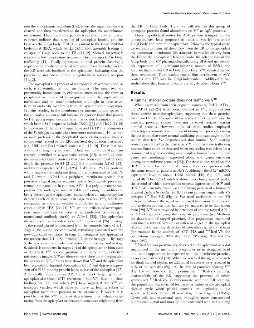
FtsH1 and the Golgi stacking protein, GRASP55 [32]. As seen in
earlier work [33], the apicoplast and Golgi body are usually in
close proximity. In some cells no overlap of the signal for the two
proteins was visible. However, we often observed closely abutting
or weak partial overlap of the two proteins and occasionally
stronger signal overlap (Fig. 3A). To assess the functional
significance of the overlap, we treated the intracellular parasites
with BFA to disrupt the Golgi body. The Golgi membrane marker
NST1 [25,34], was distributed back to the ER after addition of
BFA (Fig. 3B), demonstrating effective inhibition of ER-Golgi
transport, while the Golgi stacking protein GRASP55, which is
relatively resistant to BFA, maintained its position in the cell
(Fig. 3A) as seen by others [32]. The pattern of overlap between
FtsH1 and GRASP55 was maintained following BFA treatment
(Fig. 3C), indicating that the observed overlap is likely not
functional but rather reflects the closely juxtaposed positions of
the organelles. Thus these experiments provided no indication that
FtsH1 transiently inhabits the Golgi body.
ApV proteins could transit very rapidly through the Golgi body,
thus escaping steady state detection. We therefore examined in
detail the effect of BFA treatment on the presence of Vap. If Vap
represented ER to Golgi or Golgi to apicoplast intermediates, we
would expect that a block of Golgi function would inhibit their
formation. Although the Golgi marker NST1 relocalized to the
ER within 15 min of the application of BFA (not shown), the drug
might not affect the trafficking of previously formed Golgi to
apicoplast intermediates. Thus, we aimed to incubate the parasites
in drug as long as possible to allow pre-existing Vap to arrive at
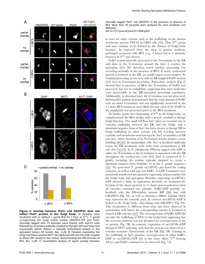
their destination while still allowing protein synthesis to generate
new Vap cargo. Protein synthesis, assessed by 35S-methionine
labeling of three proteins (FtsH1, the microneme protein MIC5,
and cytosolic GFP), continued robustly for 1.5 hour after
application of BFA, being very similar to the untreated control
(Fig. 4). Subsequently, protein synthesis dropped precipitously in
the BFA-treated parasites. Therefore we chose a 1.5 hour
treatment with BFA for our IFA studies.
Vesicles Bearing Apicoplast Membrane Proteins
PLOS ONE | www.plosone.org 4 November 2014 | Volume 9 | Issue 11 | e112096

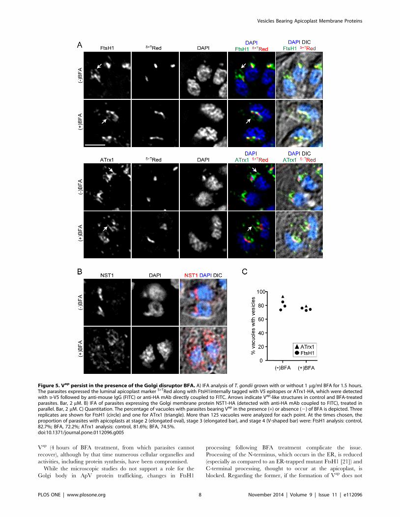
Intracellular T. gondii expressing either epitope-tagged FtsH1
or ATrx1 were incubated with or without BFA and analyzed by
IFA. We determined the proportion of vacuoles with parasites
showing vesicles bearing the tagged proteins in BFA treated and
control samples. Examples of such cells are shown in Fig. 5A and
the quantitative analysis is shown in Fig. 5C. In these experiments,
at the times chosen for analysis, a larger percentage of parasites
were at stages 2–4 of the apicoplast division cycle [26] as
compared to our previous studies [22,23], accounting for the
higher percentage of parasites bearing Vap. Parasites bearing
FtsH1 and ATrx1 marked vesicles were observed in both control
and BFA treated samples (Fig. 5A) even though the BFA treatment
triggered re-distribution of NST1 from the Golgi body to the ER
(Fig. 5B). After 1.5 hour in BFA, only a small reduction of the
proportion of vacuoles showing Vap was observed as compared to
the untreated control (Fig. 5C). Since protein synthesis continues
to be high within this period, newly synthesized ApV proteins must
have accumulated in many parasites. However, these markers
were not observed in a dispersed or perinuclear pattern as would
be expected if they were retained in the ER upon Golgi disruption,
Figure 2. Vap persist in parasites with plastid loss. T. gondii expressing the indicated tagged apicoplast proteins were transiently transfectedwith a plasmid encoding S+TYFP-ROP1 (chimera, endogenous fluorescence) to induce plastid mis-segregation. After 40 hours to allow for apicoplastloss through several cell divisions, the samples were subjected to IFA. Vacuoles with one or more parasites expressing the chimeric protein wereanalyzed. Individual cells and vacuoles are outlined with solid lines and dashed lines respectively. The markers are indicated above each panel. DIC,differential interference contrast, H indicates host cell nucleus. A) Loss of luminal marker in parasites expressing the ‘‘poison’’ chimera. S+TRed-V5 wasdetected with both anti-V5 mAb (followed by secondary antibody coupled to Dylight 649; panels labeled S+TRed-V5), and through intrinsicfluorescence (panels here and in B, C labeled S+TRed). The lower panels show enhanced scaling of S+TRed-V5 detected with anti-V5 to highlight faintER-like staining. Bar = 5 mM. B) Continued formation of Vap bearing FtsH1. FtsH1/S+TRed parasites were transiently transfected with the chimericconstruct (detected by endogenous fluorescence) and FtsH1 was detected with anti-V5 mAb (followed by secondary antibody coupled to DyLight649). Background of the S+TRed images in panels B and C were adjusted to correct for crossover fluorescence from the DyLight 649 fluorophore.Arrows indicate Vap-like staining in cells lacking an apicoplast. Vacuoles bearing transfected parasites (upper left) and untransfected parasites (lowerright) are shown. Bar = 5 mM. C) Continued formation of Vap bearing ATrx1. ATrx1/S+TRed expressing cells were transiently transfected with S+TROP1-YFP, which was detected by endogenous fluorescence. ATrx1 was detected with anti-HA mAb coupled to DyLight 649. Arrows indicate Vap-likestaining in cells lacking an apicoplast. Vacuoles bearing transfected parasites (upper left) and untransfected parasites (lower right) are shown.Parasites in the lower vacuole are in stage 1 of the organelle division cycle and therefore have few Vap. Bar = 5 mM. D) Quantitation of Vap inapicoplast-deficient parasites. ATrx1-4HA/S+TRed and FtsH1-V5233-HA/S+TRed expressing cell lines were transiently transfected with the chimericS+TYFP-ROP1 construct and vacuoles were scored for the presence or absence of YFP in at least one parasite (indicating expression of the chimericprotein in the original invading parasite). Individual parasites within each vacuole were then scored for the presence or absence of the luminalprotein S+TRed (detected by endogenous fluorescence) and the apicoplast membrane protein (detected by anti-HA or anti-V5 mAbs followed by anti-mouse IgG coupled to DyLight 649). The bar graph plots the percentage of cells bearing each marker protein in vacuoles derived from transfected(chimera+) and untransfected (chimera2) parasites. In the ATrx1 sample, 96 chimera+ and 128 chimera2 cells were counted; in the FtsH1 sample, 27chimera+ and 48 chimera2 cells were counted. These results are representative of three independent experiments.doi:10.1371/journal.pone.0112096.g002
Vesicles Bearing Apicoplast Membrane Proteins
PLOS ONE | www.plosone.org 5 November 2014 | Volume 9 | Issue 11 | e112096
as seen for other systems such as the trafficking of the plasma
membrane protein VSV-G in CHO cells [35]. Thus Vap persist
and may continue to be formed in the absence of Golgi body
function. As expected from the drop in protein synthesis,
prolonged treatment with BFA (e.g., 4 hours) led to a dramatic
decrease in Vap (not shown).
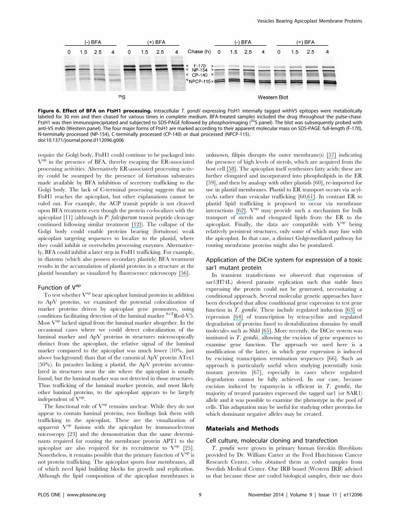
FtsH1 is proteolytically processed at the N-terminus in the ER
and then at the C-terminus around the time it reaches the
apicoplast [21]. We therefore tested whether processing was
occurring normally in the presence of BFA. If newly synthesized
protein is retained in the ER, we would expect near-complete N-
terminal processing as was seen with an ER-trapped FtsH1 mutant
[21], but no C-terminal processing. Pulse-chase analysis (Fig. 6)
showed that in presence of BFA, the N-terminus of FtsH1 was
processed, but not to completion, suggesting that some molecules
were inaccessible to the ER-associated processing machinery.
Additionally, as discussed later, the C-terminus was not processed.
Immunoblot analysis demonstrated that the total amount of FtsH1
with an intact C-terminus was not significantly increased in the
1.5 hour BFA treatment, most likely because most of the FtsH1 in
the population was processed prior to the BFA treatment.
To further probe the relationship of Vap to the Golgi body, we
complemented the BFA studies with a genetic method to disrupt
Golgi function. The small GTPase Sar1 plays an essential role in
vesicular trafficking between the ER and the Golgi, and a
dominant negative form of Sar1 has been shown to disrupt ER to
Golgi trafficking in other systems [36–39]. Cycling between
cytosolic and membrane-associated pools, Sar1 accumulates at ER
exit sites, where insertion of its N-terminal a-helix initiates vesicle
budding [40,41]. In mammalian cells, Sar1 is localized uniformly
across the ER membrane aside from some accumulation at ER
exit sites [42,43]. In P. falciparum, PfSar1p tagged with GFP at
either the N-terminus or the C-terminus is associated with the ER
throughout the erythrocytic cycle [44]. Sar1 is conserved in T.gondii, including the residue typically mutated to create a
dominant negative form (histidine 74 in the T. gondii sequence)
[45]. We generated T. gondii sar1(H74L) and fused the coding
sequence, as well as wild type (wt) SAR1, to GFP. Constructs were
transiently transfected into parasites expressing various markers for
the Golgi body and apicoplast. Parasites expressing sar1(H74L)-
GFP showed a delay in replication; therefore we examined the
location of the above proteins at 11 hours post-transfection when
all vacuoles contained one parasite. SAR1-GFP partially co-
localized with the ER-resident protein BiP [46] and with
GRASP55 (Fig. S4), as well as showing a diffuse staining which
may represent the cytosolic pool. In contrast sar1(H74L)-GFP is
locked at the Golgi body, colocalizing with GRASP55 (Fig. S4).
This localization is different from what has been observed in
mammalian cells where the corresponding mutant is localized at
clustered ER exit sites [47]. The overexpression of SAR1-GFP did
not alter the trafficking of NST1 to the Golgi body suggesting that
the secretory pathway was not disrupted by overexpression of the
wt protein (Fig. 7B). In contrast, expression of sar1(H74L)-GFP
disrupted NST1 targeting, such that the protein was observed in a
reticular structure characteristic of the ER (Fig. 7B). Turning to
the trafficking of ApV proteins, overexpression of either SAR1-
GFP or sar1(H74L)-GFP led to the same effect: Vap bearing
ATrx1 and FtsH1 continued to be observed (Fig. 7A)
Figure 3. Overlap between FtsH1 and GRASP55 does notreflect FtsH1 protein in the Golgi body. A) Parasites wereincubated with or without 1 mg/ml BFA for 1 hour at 37uC. T. gondiico-expressing the Golgi matrix marker GRASP55-YFP and FtsH1internally tagged with V5 epitopes were stained with anti-V5 mAbfollowed by secondary antibody coupled to DyLight 649 and Texas Redstreptavidin (which detects a naturally biotinylated protein in theapicoplast lumen, AP lumen). Bar, 2 mM. B) Parasites expressing theGolgi membrane protein NST1-HA (detected with anti-HA mAb coupledto Alexa 594) served as the control, demonstrating the effectiveness ofBFA. Bar, 2 mM. C) Quantitative analysis of signal overlap between
internally tagged FtsH1 and GRASP55 in the presence or absence ofBFA. More than 50 parasites were analyzed for each condition (seeMethods).doi:10.1371/journal.pone.0112096.g003
Vesicles Bearing Apicoplast Membrane Proteins
PLOS ONE | www.plosone.org 6 November 2014 | Volume 9 | Issue 11 | e112096
Conditional expression of dominant negative sar1 bypromoter juxtaposition
We were unable to obtain stable transfectants expressing
sar1(H74L)-GFP upon selection and so developed an approach
to conditionally overexpress the mutant or wt protein making use
of the di-CRE system [48] that was recently applied to T. gondii[49]. Constructs were prepared in which wt or mutant SAR1 fused
toYFP was separated from the TUBA promoter by a segment of
DNA encoding a red fluorescent protein flanked by loxP sites.
After stable transfection into parasites expressing two inactive
fragments of the Cre recombinase (DiCre) [49], assembly of
functional CRE was initiated by the addition of rapamycin (Fig.
S5A). This treatment should lead to the excision of the DNA
between the loxP sites and hence juxtaposition of the promoter
and the SAR1/sar1-YFP fusion genes. The fusion proteins
migrated according to the expected molecular mass on SDS-
PAGE (Fig. S5B), although the H74L mutant was expressed to
lower levels. Localization of the wt and mutant fusion proteins
corresponded to those seen in transient transfectants (Fig. S5D).
The percentage of parasites expressing the SAR1/sar1 fusion
proteins in clonal lines increased gradually over 24 hours, when
70–80% showed visible expression (not shown). We observed
cellular abnormalities such as lack of elongation of the inner
membrane complex and aberrant micronemes (as revealed by
Mic10) within 13 hours of rapamycin addition to induce
sar1(H64L); small cells began to appear by 16 hours and by
24 hours the majority of cells were shrunken (not shown). These
sar1(H64L)+parasites were lost upon cultivation (Fig. S4C). Hence
we aimed to use the earliest times possible for analysis to avoid
secondary affects.
At 8 hours after rapamycin treatment, in those cells where the
sar1(H74L) fusion protein was detected Golgi function was not yet
compromised, while at 11 hours disruption of NST1 localization
to the Golgi body was evident (Fig. S5D). We therefore examined
parasites for Vap 11 hours after rapamycin induction. Approxi-
mately 75% of those parasites expressing the wt SAR1 protein
showed Vap, while 60% of parasites expressing sar1(H74L) did
(Fig. 7C and Fig. S5E). This modest decrease in Vap in the
parasites expressing the dominant negative sar1 was paralleled by
an increase in parasites showing ER localization of ATrx1.
However, it is unclear whether ER retention of ATrx1 in this
population is a primary effect of Golgi disruption. Because of the
gradual onset of detectable expression and the pleiotropic effect of
Sar1 disruption, biochemical studies were not pursued.
Discussion
The work described here adds to the understanding of the
trafficking of apicoplast proteins in several ways. First, it
demonstrates that the luminal marker protein travels to the
apicoplast by routes largely independent of the pathway generat-
ing Vap. It shows that the presence, and possibly the formation of
Vap, does not require an intact Golgi body. Finally it supports
work suggesting that Vap are not derived from the apicoplast [27]
by revealing their continued presence several cell generations after
plastid loss. The persistence of Vap in such parasites indicates that
a retrograde pathway is not required for their formation.
Relationship of Vap to the Golgi bodyPrecedents exist for protein trafficking from the ER to
organelles but bypassing the Golgi body, such as pathways
generating the bounding membranes of lipid droplets [50,51] and
peroxisomes. However, in Euglena (which arose from a separate
evolutionary lineage from the apicoplast) and in Gonyaulax (which
is thought to be in the same lineage as the apicoplast), proteins
clearly transit the Golgi body to reach the secondary plastid [52–
54]. While a recent study suggested that a Plasmodium thioredoxin
peroxidase reaches the apicoplast via the Golgi body [55],
trafficking of most luminal proteins to the apicoplast appears to
be Golgi-independent [11,12]. We therefore assessed potential
Golgi involvement in Vap and membrane protein trafficking.
Although fluorescence microscopy images of T. gondii apicoplast
proteins and Golgi stacking protein GRASP55 often had partially
overlapping signals, we showed that such overlap is maintained
even when the Golgi body membranes and contents were
relocalized to the ER by treatment with BFA. Thus, this apparent
colocalization simply reflects the close juxtaposition of the
organelles and does not imply intersection of Vap proteins with
the Golgi during trafficking. To more directly test the role of the
Golgi body, we used both the chemical inhibitor BFA and the
genetic inhibitor sar1(H74L) to disrupt Golgi function. In both
cases, we observed Vap in the treated cells, indicating that they
either persist or continue to be formed when Golgi body function
is abrogated. Longer term Golgi disruption does lead to loss of
Figure 4. Protein synthesis during BFA treatment assessed by biosynthetic labeling of FtsH1, MIC5 and cytosolic GFP. Fibroblastmonolayers infected with T. gondii expressing FtsH1 tagged internally with V5 epitopes and a cytosolic GFP (,108) were pre-incubated with orwithout BFA (1 mg/ml) for the indicated times prior to being labeled with 35S-methionine/cysteine for 30 minutes. Samples were immunoprecipitatedwith anti-V5 mAb, anti-GFP, and anti-MIC5 before being separated on 7.5% (FtsH1) or 8–16% (GFP and MIC5) SDS-PAGE gels and transferred tonitrocellulose. The left panel shows phosphorimaging, the right panel shows the same lanes detected by Western blot. The four major forms of FtsH1are marked according to their apparent molecular mass on SDS-PAGE: full-length (F-170), N-terminally processed (NP-154), C-terminally processed(CP-140) or dual processed (NPCP-115). In a 30 min labeling, the first two forms predominate [21]. The precursor (p) and mature (m) forms of MIC5[75] are marked.doi:10.1371/journal.pone.0112096.g004
Vesicles Bearing Apicoplast Membrane Proteins
PLOS ONE | www.plosone.org 7 November 2014 | Volume 9 | Issue 11 | e112096
Vap (4 hours of BFA treatment, from which parasites cannot
recover), although by that time numerous cellular organelles and
activities, including protein synthesis, have been compromised.
While the microscopic studies do not support a role for the
Golgi body in ApV protein trafficking, changes in FtsH1
processing following BFA treatment complicate the issue.
Processing of the N-terminus, which occurs in the ER, is reduced
(especially as compared to an ER-trapped mutant FtsH1 [21]) and
C-terminal processing, thought to occur at the apicoplast, is
blocked. Regarding the former, if the formation of Vap does not
Figure 5. Vap persist in the presence of the Golgi disruptor BFA. A) IFA analysis of T. gondii grown with or without 1 mg/ml BFA for 1.5 hours.The parasites expressed the luminal apicoplast marker S+TRed along with FtsH1internally tagged with V5 epitopes or ATrx1-HA, which were detectedwith a-V5 followed by anti-mouse IgG (FITC) or anti-HA mAb directly coupled to FITC. Arrows indicate Vap-like structures in control and BFA-treatedparasites. Bar, 2 mM. B) IFA of parasites expressing the Golgi membrane protein NST1-HA (detected with anti-HA mAb coupled to FITC), treated inparallel. Bar, 2 mM. C) Quantitation. The percentage of vacuoles with parasites bearing Vap in the presence (+) or absence (2) of BFA is depicted. Threereplicates are shown for FtsH1 (circle) and one for ATrx1 (triangle). More than 125 vacuoles were analyzed for each point. At the times chosen, theproportion of parasites with apicoplasts at stage 2 (elongated oval), stage 3 (elongated bar), and stage 4 (V-shaped bar) were: FtsH1 analysis: control,82.7%; BFA, 72.2%; ATrx1 analysis: control, 81.6%; BFA, 74.5%.doi:10.1371/journal.pone.0112096.g005
Vesicles Bearing Apicoplast Membrane Proteins
PLOS ONE | www.plosone.org 8 November 2014 | Volume 9 | Issue 11 | e112096
require the Golgi body, FtsH1 could continue to be packaged into
Vap in the presence of BFA, thereby escaping the ER-associated
processing activities. Alternatively ER-associated processing activ-
ity could be swamped by the presence of fortuitous substrates
made available by BFA inhibition of secretory trafficking to the
Golgi body. The lack of C-terminal processing suggests that no
FtsH1 reaches the apicoplast, but other explanations cannot be
ruled out. For example, the ACP transit peptide is not cleaved
upon BFA treatment even though the protein co-localizes with the
apicoplast [11] (although in P. falciparum transit peptide cleavage
continued following similar treatment [12]). The collapse of the
Golgi body could enable proteins bearing (fortuitous) weak
apicoplast targeting sequences to localize to the plastid, where
they could inhibit or overwhelm processing enzymes. Alternative-
ly, BFA could inhibit a later step in FtsH1 trafficking. For example,
in diatoms (which also possess secondary plastids) BFA treatment
results in the accumulation of plastid proteins in a structure at the
plastid boundary as visualized by fluorescence microscopy [56].
Function of Vap
To test whether Vap bear apicoplast luminal proteins in addition
to ApV proteins, we examined the potential colocalization of
marker proteins driven by apicoplast gene promoters, using
conditions facilitating detection of the luminal marker S+TRed-V5.
Most Vap lacked signal from the luminal marker altogether. In the
occasional cases where we could detect colocalization of the
luminal marker and ApV proteins in structures microscopically
distinct from the apicoplast, the relative signal of the luminal
marker compared to the apicoplast was much lower (10%, just
above background) than that of the canonical ApV protein ATrx1
(50%). In parasites lacking a plastid, the ApV proteins accumu-
lated in structures near the site where the apicoplast is usually
found, but the luminal marker was not detected in those structures.
Thus trafficking of the luminal marker protein, and most likely
other luminal proteins, to the apicoplast appears to be largely
independent of Vap.
The functional role of Vap remains unclear. While they do not
appear to contain luminal proteins, two findings link them with
trafficking to the apicoplast. These are the visualization of
apparent Vap fusions with the apicoplast by immunoelectron
microscopy [23] and the demonstration that the same determi-
nants required for routing the membrane protein APT1 to the
apicoplast are also required for its recruitment to Vap [25].
Nonetheless, it remains possible that the primary function of Vap is
not protein trafficking. The apicoplast sports four membranes, all
of which need lipid building blocks for growth and replication.
Although the lipid composition of the apicoplast membranes is
unknown, filipin disrupts the outer membrane(s) [57] indicating
the presence of high levels of sterols, which are acquired from the
host cell [58]. The apicoplast itself synthesizes fatty acids; these are
further elongated and incorporated into phospholipids in the ER
[59], and then by analogy with other plastids [60], re-imported for
use in plastid membranes. Plastid to ER transport occurs via acyl-
coAs rather than vesicular trafficking [60,61]. In contrast ER to
plastid lipid trafficking is proposed to occur via membrane
interactions [62]. Vap may provide such a mechanism for bulk
transport of sterols and elongated lipids from the ER to the
apicoplast. Finally, the data are compatible with Vap being
relatively persistent structures, only some of which may fuse with
the apicoplast. In that case, a distinct Golgi-mediated pathway for
routing membrane proteins might also be postulated.
Application of the DiCre system for expression of a toxicsar1 mutant protein
In transient transfections we observed that expression of
sar1(H74L) slowed parasite replication such that stable lines
expressing the protein could not be generated, necessitating a
conditional approach. Several molecular genetic approaches have
been developed that allow conditional gene expression to test gene
function in T. gondii. These include regulated induction [63] or
repression [64] of transcription by tetracycline and regulated
degradation of proteins fused to destabilization domains by small
molecules such as Shld [65]. More recently, the DiCre system was
instituted in T. gondii, allowing the excision of gene sequences to
examine gene function. The approach we used here is a
modification of the latter, in which gene expression is induced
by excising transcription termination sequences [66]. Such an
approach is particularly useful when studying potentially toxic
mutant proteins [67], especially in cases where regulated
degradation cannot be fully achieved. In our case, because
excision induced by rapamycin is efficient in T. gondii, the
majority of treated parasites expressed the tagged sar1 (or SAR1)
allele and it was possible to examine the phenotype in the pool of
cells. This adaptation may be useful for studying other proteins for
which dominant negative alleles may be created.
Materials and Methods
Cell culture, molecular cloning and transfectionT. gondii were grown in primary human foreskin fibroblasts
provided by Dr. William Carter at the Fred Hutchinson Cancer
Research Center, who obtained them as coded samples from
Swedish Medical Center. Our IRB board (Western IRB) advised
us that because these are coded biological samples, their use does
Figure 6. Effect of BFA on FtsH1 processing. Intracellular T. gondii expressing FtsH1 internally tagged withV5 epitopes were metabolicallylabeled for 30 min and then chased for various times in complete medium. BFA-treated samples included the drug throughout the pulse-chase.FtsH1 was then immunoprecipitated and subjected to SDS-PAGE followed by phosphorimaging (35S panel). The blot was subsequently probed withanti-V5 mAb (Western panel). The four major forms of FtsH1 are marked according to their apparent molecular mass on SDS-PAGE: full-length (F-170),N-terminally processed (NP-154), C-terminally processed (CP-140) or dual processed (NPCP-115).doi:10.1371/journal.pone.0112096.g006
Vesicles Bearing Apicoplast Membrane Proteins
PLOS ONE | www.plosone.org 9 November 2014 | Volume 9 | Issue 11 | e112096

Figure 7. Expression of dominant negative sar1 does not eliminate Vap. A) T. gondii expressing S+TRed plus ATrx1-HA or FtsH1 internallytagged with V5 epitopes were transiently transfected with either wt SAR1-GFP or sar1(H74L)-GFP and analyzed by IFA for the localization of the twoApV protein. Epitope tagged proteins were detected by mAbs reactive with the epitope tags followed by secondary antibodies coupled to DyLight649. The fluorescent proteins were detected by endogenous fluorescence. Arrows point to Vap-like structures. Bar, 2 mM. B) Overexpression ofsar1(H74L) abrogates localization of NST1. SAR1 and sar1(H74L) constructs were transiently transfected into T. gondii expressing NST1-HA and thesamples analyzed as above. Note the reticular staining of NST1 following expression of the dominant negative protein. C) Vap are still present afterinduction of sar1(H74L) in stable transfectants. As described in Methods, parasites were stably transfected with constructs bearing sar1(H74L)-YFP orthe wt SAR1-YFP that was separated from a promoter by RFP flanked by loxP sequences. Addition of rapamycin led to excision of the RFP sequencethat separated and expression of the test proteins. After 11 hours YFP+ parasites were scored for the presence or absence of Vap using mAb 11G8which detects ATrx1 (followed by secondary antibody coupled to DyLight 350).doi:10.1371/journal.pone.0112096.g007
Vesicles Bearing Apicoplast Membrane Proteins
PLOS ONE | www.plosone.org 10 November 2014 | Volume 9 | Issue 11 | e112096
not constitute human subjects research. This is in agreement with
the U.S. Department of Health and Human Services (http://
www.hhs.gov/ohrp/sachrp/20110124attachmentatosecletter.html.
Among the strains used were RH and its corresponding HXGPRTdeletion strain [68], plus derivatives expressing apicoplast proteins C-
terminally tagged with four HA epitopes including ATrx1 (ToxoDB
v.7.2 TGME49_312110) [23], APT1 (TGME49_261070) [24], and
FtsH1 (TGME49_259260), which was additionally internally tagged
with two V5 epitopes at residue 233 (FtsH1-V5233-HA) [21]. Some
parasites also expressed Heteractis crispa red fluorescent protein
HcRed bearing the ACP (TGME49_264080) signal plus transit
sequences and tagged with V5 epitope (S+TRed-V5). The two V5 tags
(see [21] for sequence) were inserted into MunI and NdeI sites that
had been added by site directed mutagenesis using 59 CCCGA-
GAAGGCCAACCAATTGATACATATGTGACTGCAGCCC-
ACACAG 39 and 59 CTGTGTGGGCTGCAGTCACATATG-
TATCAATTGGTTGGCCTTCTCGGG 39. Expression of each of
these proteins was driven by its cognate promoter. Tic22
(TGME49_286050) [19] was amplified from T. gondii RH strain
cDNA using primers CTCAGATCTAAAATGGGCTTCAT-
CGCTCTCCG and GTGCCTAGGTGCTTGTCCTTGATC-
GTCGG, and cloned into a pGem shuttle vector. The relevant
region was excised with BglII and XbaI and cloned into the plasmid
pHX apt1:APT1-4HA that had been digested with BglII and AvrIIto remove the APT1 coding sequence. The product yielded Tic22 C-
terminally tagged with HA, with expression is driven by the APT1promoter, which has similar kinetics. Der1ap (Genbank FJ976520)
[19], was similarly amplified using primers GTGCCATGGAAA-
GAGGGGATTTTTTCTC and CACTCTAGAGCGTTTC-
CAACGGCGTCCTCG, cloned into a pGem shuttle vector and
then excised with NcoI and AvrII. The appropriate fragment was
cloned into pHX ATrx1:ATrx1-4HA that had been digested with the
same enzymes to remove the ATrx1 coding sequence. The resulting
construct encoded Der1ap C-terminally tagged with HA, with its
expression is driven by the ATrx1 promoter. GRASP55-HcRed was
generated by replacing the YFP tag in pCAT GRASP55-YFP with
the HcRed tag from pCAT ACP-HcRed, using AvrII and PstI. Other
markers include S+TRed (untagged), the Golgi membrane protein
NST1 (TGME49_267380) C-terminally tagged with HA [25], the
Golgi stacking protein GRASP55 (AF110267) fused to YFP [32,69] or
HcRed, and GFP used as a cytosolic marker. Expression of S+TRed
and NST1 were driven by the DHFR promoter and expression of
GRASP55-YFP, GRASP55-HcRed, and GFP was driven by the
TubA promoter. Plasmids were transfected into T. gondii by
electroporation and stable transfectants selected either with chloram-
phenicol or mycophenolic acid plus xanthine as previously described
[24], and clonal lines were isolated. Brefeldin A (Calbiochem) was
used at a final concentration of 1 mg/ml, previously shown to be
sufficient to block trafficking of the microneme protein MIC5.
The T. gondii small GTPase Sar1 (TGME49_215060) was
amplified from oligo-dT primed cDNA using 59 ATCGAGATC-
TAAAATGTTCGTCTTCAACTGGTTCTG 39 and 59 ATC-
GCCTAGGGTTGAGGAACTGAGACAACCAAC 39 and
cloned into pCAT GFP [70] cleaved with BglII and AvrII. Its
expression is driven by the TubA promoter (TubA mRNA
abundance and cell cycle kinetics are similar to that of SAR1).
The H74L mutant was generated by site-directed mutagenesis
using the pCAT Sar1-GFP plasmid as a template and primers: 59
TTCGATCTTGGGGGACTTGAAACAGCC 39 and 59 GGC-
TGTTTCAAGTCCCCCAAGATCGAA 39. An apicoplast ‘‘poi-
son’’ construct was kindly provided by Dr. Cynthia He. This
plasmid encodes an apicoplast-targeted fusion protein that also
contains sequences from the rhoptry protein Rhop1 (FNR-YFP-
ROP1), and is identical to that previously used by He et al. [71]
except that the apicoplast targeting sequence was derived from
ferredoxin reductase rather than ACP. These plasmids were
employed in transient transfections.
The plasmid ploxP-KillerRed-loxP-YFP, in which expression of
Killer Red was driven by the Tub8 promoter, was a gift from Drs.
Markus Meissner and Nicole Andenmatten [49]. It bears the
selectable marker HXGPRT. The plasmid and was modified by
inserting an XbaI restriction site upstream of YFP using the
oligonucleotides lox-Xba-YFP 59 CATTATACGAAGTTA-
TAAATCTAGAATGGTGAGTAAGGGCGAGGAG 39 and 59
CTCCTCGCCCTTACTCACCATTCTAGATTTATAACTT-
CGTATAATG 39. A segment of genomic DNA downstream of
the GRA3 locus was amplified from genomic DNA using primers
Gra3-tub8 f 59 ATTGGGTACCGGGCCCTACGGTCTCC-
TAGCTCCTTTG 39 and r 59 CGTCGAGGGGGGGCCGT-
GAGAATCGTAGGTGCAGGTG 39, and inserted into the
Apa1 site the plasmid by Gibson cloning. The coding regions of
SAR1 and sar1(H74L) were amplified from the above pCAT
plasmids using oligonucleotides P-lox-YFP-SAR1 f 59 CATTA-
TACGAAGTTATAAATCTAGAATGTTCGTCTTCAACTG-
GTTCTGG 39 and r 59 GCCCTTGCTCACCATTCTA-
GAGTTGAGAAACTGAGACAACCAACG 39 and inserted into
Xba1-digested plasmid using Gibson cloning. The SAR1 coding
regions in the plasmids were verified by sequencing.
For transfections, 50 mg of each plasmid (pGra3-loxP-Killer red
YFP vector and SAR1-YFP and sar1 (H74L)-YFP derivatives)
were digested with PaeI and transfected into RH DKU80DHXGPRT DiCre T. gondii and selected with mycophenolic
acid and xanthine. Clonal cell lines were isolated by limiting
dilution. Excision of the sequence separating the promoter from
the SAR1/sar1 CDS was induced by 50 nM rapamycin in 0.1%
DMSO. ATrx1 localization was categorized as plastid, plastid+ER
or plastid+Vap based on the localization within the majority of
parasites within a vacuole.
Pulse labeling, immunoprecipitation and immunoblotanalysis
Fibroblast monolayers bearing T. gondii (approximately 108)
were rinsed twice in medium lacking methionine and cysteine,
with or without BFA. Intracellular parasites were labeled for
30 min with 100 mCi/ml [35S] trans label (methionine and
cysteine, MP Biomedicals, Irvine, CA and Perkin Elmer) as
described, in the presence or absence of BFA [11]. Subsequently
the labeling medium was replaced with complete medium and the
incubation continued for the indicated times. The fibroblast layer
was then scraped from the flask and cells were pelleted by
centrifugation at 23006 g for 2 min. Pellets were lysed in 0.5 ml
lysis buffer (150 mM NaCl, 50 mM TrisHCl pH 7.5, 2 mM
EDTA, 1% NP-40, 0.25% deoxycholate, 1.7 mg/ml aprotinin,
5 mg/ml leupeptin, 1 mM pepstatin, 0.1 mM PMSF). FtsH1-
V5233-HA and S+TRed-V5 were immunoprecipitated using mouse
anti-V5 mAb (Invitrogen), and ATrx1-HA was immunoprecipi-
tated with anti-HA mAb. GFP and MIC5 were immunoprecip-
itated with anti-GFP B2 (Santa Cruz Biotechnology), and rabbit
anti-MIC5 (gift of Dr. Vern Carruthers) respectively. Immune
complexes were collected with Protein G coupled to magnetic
beads (Invitrogen). The washed immune complexes were separat-
ed by SDS-PAGE, transferred to nitrocellulose membranes.
Radiolabeled proteins were detected by phosphorimaging using
a Storm 860 (Molecular Dynamics).
Immunoprecipitated samples or total cell lysates (approximately
107 parasites) were used for immunoblot analyses. After blocking
in Odyssey block (LI-COR Biosciences), blots were probed with
mouse anti-V5 mAb at 0.5 mg/ml (Invitrogen), rabbit anti-GFP at
Vesicles Bearing Apicoplast Membrane Proteins
PLOS ONE | www.plosone.org 11 November 2014 | Volume 9 | Issue 11 | e112096
0.2 mg/ml (Invitrogen) and rabbit anti-MIC5 at a 1:10,000
dilution. This was followed by goat anti-mouse Ig coupled to
IRDye 800 (1:10,000, LI-COR) or goat anti-rabbit Ig coupled to
IRDye 680 (1:10,000, LI-COR). Membranes were scanned using
an Odyssey infrared imaging system (LI-COR) and analyzed using
the system software.
MicroscopyFor IFAs, parasites were grown overnight unless otherwise
noted within fibroblasts monolayers on coverslips. IFAs were
performed as described [24]. V5 tags were detected using mouse
anti-V5 mAb IgG2a at 1 mg/ml (Invitrogen), followed by goat
anti-mouse IgG2a FITC, goat anti-mouse IgG2a Texas Red,
(Southern Biology), or goat anti-mouse IgG DyLight 649 (Thermo
Scientific), all at 2 mg/ml. FtsH1-V5233-HA was always detected
using anti-V5 mAb. ATrx1 and APT1 were detected by virtue of
the HA tags, using FITC-coupled rat anti-HA mAb 3F10, 3 mg/
ml (Roche), or anti-HA mAb16B12 (Covance) followed by goat
anti-mouse IgG DyLight 649. ATrx1 was also detected by mAb
11G8 [72,73], a kind gift from Peter Bradley) followed by goat
anti-mouse IgG Dylight 350 (Thermo Scientific). Markers for the
apicoplast lumen included the naturally biotinylated apicoplast
luminal protein acetyl coA carboxylase revealed by Texas Red or
Alexa 680 coupled streptavidin (Invitrogen, 1 mg/ml) [74], andS+TRed or S+TRed-V5 [70]. Rabbit anti-MIC5 was used at a 1:500
dilution, rabbit anti-Trypanosoma brucei BiP (which cross-reacts
with T.gondii BiP, gift of Dr. Jay Bangs) at a 1:200 dilution [46]
and 4,6-diamidino-2-phenylindole (DAPI) was used to stain the
DNA. A Deltavision RT deconvolution microscope with an
Olympus UPlan/Apo 1006 1.35 NA objective was used to view
the slides. Images were deconvolved using softWoRx (version
3.5.1) using standard parameters and a conservative ratio
algorithm. Single deconvolved planes are shown except as
described below.
Image quantitationTo quantify relative fluorescence in vesicles or adjacent regions
versus in the apicoplast, the softWoRx ‘‘quick projection sum’’ tool
was used to generate a 2-D image showing the summed
fluorescence at each pixel from each plane of the 3-D image;
the resulting image was then converted to a TIFF file for further
analysis. Using Metamorph software, three ovals of 30 pixels each
were placed in each subcellular region analyzed: the apicoplast
region defined by S+TRed-V5 (Cy5 channel) and ATrx1-HA
(FITC channel), vesicle regions (defined by ATrx1 punctate
signal), and adjacent regions (areas next to ATrx1 vesicle regions).
Average fluorescence per oval in both FITC and Cy5 channels
was calculated and summed for each region (90 pixels) in each
parasite. Ten parasites from separate vacuoles were analyzed.
Background was calculated by placing all nine ovals on images of
the apical end of co-cultured RH parasites (which do not express
either tagged protein) and the corresponding average fluorescence
per 90 pixels was calculated for each channel, averaged over four
parasites.
To assess potential localization of ApV proteins in the Golgi
body, images from treated and non-treated samples were
scaled equally and colocalization of organellar markers was
examined in all image planes. Individual parasites were scored
as having abutting/partial overlap if regions of two or more
pixels wide in each channel overlapped in two or more focused
image planes.
Supporting Information
Figure S1 Cell cycle regulation of transcription of genesin this study. Quantitation of expression of the relevant genes
following post-thymidine block release of T. gondii RHTK+
parasites was obtained from ToxoDB, based on microarray data
from [28]. The time period covers approximately 1.66 cell cycles,
with internal daughter cells peaking at 4 and 12 hours. These data
show that the promoters for apicoplast reporters utilized in this
study have very similar temporal kinetics.
(TIF)
Figure S2 Similar half-life of S+TRed-V5 and ATrx1.Pulse-chase analysis was carried out as in Fig. 6, with S+TRed-V5
and ATrx1-HA co-expressed in the same parasite line. The
molecules were sequentially immunoprecipitated with mAbs
directed against the epitope tags and 35S-methionine labeled
proteins detected by phosphorimaging. For each antibody, the
lanes shown are from the same scan of the gel. Three main bands
are seen for ATrx1-HA, with the 90 kDa protein being a
precursor (p) to intermediate (i) and mature 65 kDa protein (m)
[23]. The subcellular location where processing occurs is not
known. The cleavage of the precursor (p) S+TRed-V5 to mature
form (m, 35 kDa) occurs within the apicoplast.
(TIF)
Figure S3 Tic22 and Der1ap inhabit Vap. Clonal lines
expressing the apicoplast luminal marker S+TRed and either
Tic22-HA or Der1-HA T. gondii within fibroblasts were processed
for IFA and stained for ATrx1 (using mAb 11G8 followed by anti-
mouse IgG coupled to DyLight 649) and for Tic22-HA or Der1-
HA (using rat anti-HA mAb coupled to FITC). Slides were co-
stained with DAPI. The apicoplast luminal marker S+TRed was
detected by endogenous fluorescence. Dotted lines indicate outline
of parasites within vacuole. A) Localization of Tic22-HA. Red,
ATrx1; green, Tic22-HA. Panel A9 shows images with enhanced
scaling to reveal Vap and a merge image showing DIC (grey),
DAPI (blue) Tic22 (green) S+TRed (orange) and ATrx1 (maroon).
Arrows indicate Vap containing both Tic22 and ATrx1. B)
Localization of Der1ap-HA. Red, ATrx1; green, Der1-HA. Panel
B9 shows images with enhanced scaling to reveal Vap and a merge
image showing DIC (grey), DAPI (blue) Der1 (green) S+TRed
(orange) and ATrx1 (maroon). Arrows indicate Vap containing
both Der1 and ATrx1.
(TIF)
Figure S4 Localization of SAR1-GFP and sar1(H74L)-GFP in T. gondii. The constructs were transiently transfected
into T. gondii expressing GRASP55-HcRed. After 11 hours, the
samples were fixed and subjected to IFA, comparing the
localization of SAR1 and sar1(H74L) (endogenous green fluores-
cence) to GRASP55 and BiP, an ER marker protein detected anti-
T. brucei BiP followed by anti-rabbit IgG coupled to Alexa 680.
Arrow marks colocalization of sar1(H74L)-GFP and GRASP55.
This particular cell has duplicated its Golgi body. ‘‘H’’ marks a
host cell nucleus. Bar, 2 mM.
(TIF)
Figure S5 Conditional expression of sar1(H74L)-YFP inT. gondii. A) Map of expression locus before (top) and after
(bottom) excision of the loxP-flanked red fluorescent protein
coding sequence which separates the promoter from the SAR1-
YFP fusion proteins. Translated segments are indicated by dark fill
with a line above.B) Western blot of protein from the parental RH
parasites, and 24 hour rapamycin induced sar1(H74L)-YFP and
SAR1-YFP parasites probed with anti-GFP. M, markers. The
fusion proteins migrated at the expected size (49 kDa). C) Parasites
Vesicles Bearing Apicoplast Membrane Proteins
PLOS ONE | www.plosone.org 12 November 2014 | Volume 9 | Issue 11 | e112096

expressing sar1(H74L) are lost upon cultivation. After rapamycin
mediated induction of expression (via excision of the RFP gene),
the percentage of vacuoles in each population with parasites
expressing YFP-tagged SAR1 or sar1(H74L) was monitored over
time. All parasites in a given vacuole showed the same expression
phenotype. Before excision (day 0) both parasite lines showed red
fluorescence only. For an intermediate period many parasites
expressed both yellow fluorescent protein and previously tran-
scribed and translated red fluorescent protein. Those parasites in
which expression of SAR1-YFP was induced continue to grow and
became YFP+/RFP2, whereas those expressing sar1(H74L)-YFP
did not survive and were outgrown by the minority population
that had not excised the RFP coding sequence (n.200 vacuoles
for each time point). D) The conditionally expressed mutant
sar1(H74L) disrupts the Golgi body. The SAR1/sar1 clonal
parasite lines were transiently transfected with NST1-HA and after
15 hours rapamycin was added. Parasites were analyzed 11 hours
later and representative examples are shown. Blind analysis
indicated that NST1-HA was localized to the Golgi body in
95% of parasites expressing SAR1-YFP, but was redistributed to
the ER in 81% of those parasites expressing sar1(H74L). E) Vap
persist in parasites expressing dominant negative sar1(H74L).
Expression of wt or mutant SAR1 was induced by the addition of
rapamycin and after 11 hours parasites were analyzed. Represen-
tative images are shown, detecting the ApV protein ATrx1 with
mAb 11G8 and the apicoplast lumen with streptavidin as
described in Methods.
(TIF)
Acknowledgments
We are grateful to Ms. Pashmi Vaney and Dr. Suzanne Scheele for
technical assistance. We particularly thank Drs. Nicole Andenmatten and
Markus Meissner for gifts of plasmids, and for making diCre Dku80 T.gondii available prior to publication.
Author Contributions
Conceived and designed the experiments: AB JAG ADR MP. Performed
the experiments: AB JAG ADR. Analyzed the data: AB JAG ADR MP.
Contributed reagents/materials/analysis tools: AB JAG ADR. Wrote the
paper: AB JAG ADR MP.
References
1. Tenter AM, Heckeroth AR, Weiss LM (2000) Toxoplasma gondii: from animals
to humans. Int J Parasitol 30: 1217–1258.
2. Dubey JP, Jones JL (2008) Toxoplasma gondii infection in humans and animals
in the United States. Int J Parasitol 38: 1257–1278.
3. Seeber F (2002) Biogenesis of iron-sulphur clusters in amitochondriate and
apicomplexan protists. Int J Parasitol 32: 1207.
4. Seeber F, Soldati-Favre D (2010) Metabolic pathways in the apicoplast of
apicomplexa. Int Rev Cell Mol Biol 281: 161–228.
5. Waller RF, Keeling PJ, Donald RGK, Striepen B, Handman E, et al. (1998)
Nuclear-encoded proteins target to the plastid in Toxoplasma gondii and
Plasmodium falciparum. Proc Natl Acad Sci USA 95: 12352–12357.
6. Jomaa H, Wiesner J, Sanderbrand S, Altincicek B, Weidemeyer C, et al. (1999)
Inhibitors of the nonmevalonate pathway of isoprenoid biosynthesis as
antimalerial drugs. Science 285: 1573–1576.
7. Ralph SA, van Dooren GG, Waller RF, Crawford MJ, Fraunholz MJ, et al.
(2004) Tropical infectious diseases: metabolic maps and functions of the
Plasmodium falciparum apicoplast. Nat Rev Microbiol 2: 203–216.
8. Fichera ME, Roos DS (1997) A plastid organelle as a drug target in
apicomplexan parasites. Nature 390: 407–409.
9. Budimulja AS, Syafruddin, Tapchaisri P, Wilairat P, Marzuki S (1997) The
sensitivity of Plasmodium protein synthesis to prokaryotic ribosomal inhibitors.
Mol Biochem Parasitol 84: 137–141.
10. McConkey GA, Rogers MJ, McCutchan TF (1997) Inhibition of Plasmodiumfalciparum protein synthesis. Targeting the plastid-like organelle with thios-
trepton. J Biol Chem 272: 2046–2049.
11. DeRocher A, Gilbert B, Feagin JE, Parsons M (2005) Dissection of brefeldin A-
sensitive and -insensitive steps in apicoplast protein targeting. J Cell Sci 118:
565–574.
12. Tonkin CJ, Struck NS, Mullin KA, Stimmler LM, McFadden GI (2006)
Evidence for Golgi-independent transport from the early secretory pathway to
the plastid in malaria parasites. Mol Microbiol 61: 614–630.
13. van Dooren GG, Tomova C, Agrawal S, Humbel BM, Striepen B (2008)
Toxoplasma gondii Tic20 is essential for apicoplast protein import. Proc Natl
Acad Sci USA 105: 13574–13579.
14. Kalanon M, Tonkin CJ, McFadden GI (2009) Characterization of two putative
protein translocation components in the apicoplast of Plasmodium falciparum.
Eukaryot Cell 8: 1146–1154.
15. Glaser S, van Dooren GG, Agrawal S, Brooks CF, McFadden GI, et al. (2012)
Tic22 is an essential chaperone required for protein import into the apicoplast.
J Biol Chem 287: 39505–39512.
16. Mullin KA, Lim L, Ralph SA, Spurck TP, Handman E, et al. (2006) Membrane
transporters in the relict plastid of malaria parasites. Proc Natl Acad Sci USA
103: 9572–9577.
17. Sommer MS, Gould SB, Lehmann P, Gruber A, Przyborski JM, et al. (2007)
Der1-mediated pre-protein import into the periplastid compartment of
chromalveolates? Mol Biol Evol 24: 918–928.
18. Spork S, Hiss JA, Mandel K, Sommer M, Kooij TW, et al. (2009) An unusual
ERAD-like complex is targeted to the apicoplast of Plasmodium falciparum.
Eukaryot Cell 8: 1134–1145.
19. Agrawal S, van Dooren GG, Beatty WL, Striepen B (2009) Genetic evidence
that an endosymbiont-derived ERAD system functions in import of apicoplast
proteins. J Biol Chem 284: 33683–33691.
20. Sheiner L, Demerly JL, Poulsen N, Beatty WL, Lucas O, et al. (2011) A
systematic screen to discover and analyze apicoplast proteins identifies a
conserved and essential protein import factor. PLoS Pathog 7: e1002392.
21. Karnataki A, DeRocher AE, Feagin JE, Parsons M (2009) Sequential processing
of the Toxoplasma apicoplast membrane protein FtsH1 in topologically distinct
domains during intracellular trafficking. Mol Biochem Parasitol 166: 126–133.
22. Karnataki A, DeRocher AE, Coppens I, Feagin JE, Parsons M (2007) A
membrane protease is targeted to the relict plastid of Toxoplasma via an internal
signal sequence. Traffic 8: 1543–1553.
23. DeRocher AE, Coppens I, Karnataki A, Gilbert LA, Rome ME, et al. (2008) A
thioredoxin family protein of the apicoplast periphery identifies abundant
candidate transport vesicles in Toxoplasma gondii. Eukaryot Cell 7: 1518–1529.
24. Karnataki A, DeRocher A, Coppens I, Nash C, Feagin JE, et al. (2007) Cell
cycle-regulated vesicular trafficking of Toxoplasma APT1, a protein localized to
multiple apicoplast membranes. Mol Microbiol 63: 1653–1668.
25. DeRocher AE, Karnataki A, Vaney P, Parsons M (2012) Apicoplast targeting of
a T. gondii transmembrane protein requires a cytosolic tyrosine-based motif.
Traffic 13: 694–704.
26. Striepen B, Crawford MJ, Shaw MK, Tilney LG, Seeber F, et al. (2000) The
plastid of Toxoplasma gondii is divided by association with the centrosomes.
J Cell Biol 151: 1423–1434.
27. Tawk L, Dubremetz JF, Montcourrier P, Chicanne G, Merezegue F, et al.
(2011) Phosphatidylinositol 3-monophosphate is involved in Toxoplasmaapicoplast biogenesis. PLoS Pathog 7: e1001286.
28. Behnke MS, Wootton JC, Lehmann MM, Radke JB, Lucas O, et al. (2010)
Coordinated progression through two subtranscriptomes underlies the tachy-
zoite cycle of Toxoplasma gondii. PLoS ONE 5: e12354.
29. Gurskaya NG, Fradkov AF, Terskikh A, Matz MV, Labas YA, et al. (2001)
GFP-like chromoproteins as a source of far-red fluorescent proteins. FEBS Lett
507: 16–20.
30. He CY, Shaw MK, Pletcher CH, Striepen B, Tilney LG, et al. (2001) A plastid
segregation defect in the protozoan parasite Toxoplasma gondii. EMBO J 20:
330–339.
31. Harper JM, Huynh MH, Coppens I, Parussini F, Moreno S, et al. (2006) A
cleavable propeptide influences Toxoplasma infection by facilitating the
trafficking and secretion of the TgMIC2-M2AP invasion complex. Mol Biol
Cell 17: 4551–4563.
32. Pelletier L, Stern CA, Pypaert M, Sheff D, Ngo HM, et al. (2002) Golgi
biogenesis in Toxoplasma gondii. Nature 418: 548–552.
33. Kohler S (2005) Multi-membrane-bound structures of Apicomplexa: I. the
architecture of the Toxoplasma gondii apicoplast. Parasitol Res 96: 258–272.
34. Caffaro CE, Koshy AA, Liu L, Zeiner GM, Hirschberg CB, et al. (2013) A
nucleotide sugar transporter involved in glycosylation of the toxoplasma tissue
cyst wall is required for efficient persistence of bradyzoites. PLoS Pathog 9:
e1003331.
35. Doms RW, Russ G, Yewdell JW (1989) Brefeldin A redistributes resident and
itinerant Golgi proteins to the endoplasmic reticulum. J Cell Biol 109: 61–72.
36. Sato K, Nakano A (2005) Dissection of COPII subunit-cargo assembly and
disassembly kinetics during Sar1p-GTP hydrolysis. Nat Struct Mol Biol 12: 167–
174.
37. Gorelick FS, Shugrue C (2001) Exiting the endoplasmic reticulum. Mol Cell
Endocrinol 177: 13–18.
Vesicles Bearing Apicoplast Membrane Proteins
PLOS ONE | www.plosone.org 13 November 2014 | Volume 9 | Issue 11 | e112096

38. Nakano A, Muramatsu M (1989) A novel GTP-binding protein, Sar1p, is
involved in transport from the endoplasmic reticulum to the Golgi apparatus.J Cell Biol 109: 2677–2691.
39. Barlowe C, Orci L, Yeung T, Hosobuchi M, Hamamoto S, et al. (1994) COPII:
a membrane coat formed by Sec proteins that drive vesicle budding from theendoplasmic reticulum. Cell 77: 895–907.
40. Forster R, Weiss M, Zimmermann T, Reynaud EG, Verissimo F, et al. (2006)Secretory cargo regulates the turnover of COPII subunits at single ER exit sites.
Curr Biol 16: 173–179.
41. Lee MC, Orci L, Hamamoto S, Futai E, Ravazzola M, et al. (2005) Sar1p N-terminal helix initiates membrane curvature and completes the fission of a
COPII vesicle. Cell 122: 605–617.42. Weissman JT, Plutner H, Balch WE (2001) The mammalian guanine nucleotide
exchange factor mSec12 is essential for activation of the Sar1 GTPase directingendoplasmic reticulum export. Traffic 2: 465–475.
43. Kuge O, Dascher C, Orci L, Rowe T, Amherdt M, et al. (1994) Sar1 promotes
vesicle budding from the endoplasmic reticulum but not Golgi compartments.J Cell Biol 125: 51–65.
44. Adisa A, Frankland S, Rug M, Jackson K, Maier AG, et al. (2007) Re-assessingthe locations of components of the classical vesicle-mediated trafficking
machinery in transfected Plasmodium falciparum. Int J Parasitol 37: 1127–
1141.45. Nakano A, Otsuka H, Yamagishi M, Yamamoto E, Kimura K, et al. (1994)
Mutational analysis of the Sar1 protein, a small GTPase which is essential forvesicular transport from the endoplasmic reticulum. J Biochem 116: 243–247.
46. Bangs JD, Uyetake L, Brickman MJ, Balber AE, Boothroyd JC (1993) Molecularcloning and cellular localization of a BiP homologue in Trypanosoma brucei.Divergent ER retention signals in a lower eukaryote. J Cell Sci 105: 1101–1113.
47. Ward TH, Polishchuk RS, Caplan S, Hirschberg K, Lippincott-Schwartz J(2001) Maintenance of Golgi structure and function depends on the integrity of
ER export. J Cell Biol 155: 557–570.48. Jullien N, Sampieri F, Enjalbert A, Herman JP (2003) Regulation of Cre
recombinase by ligand-induced complementation of inactive fragments. Nucleic
Acids Res 31: e131.49. Andenmatten N, Egarter S, Jackson AJ, Jullien N, Herman JP, et al. (2013)
Conditional genome engineering in Toxoplasma gondii uncovers alternativeinvasion mechanisms. Nat Methods 10: 125–127.
50. Farese RV, Jr., Walther TC (2009) Lipid droplets finally get a little R-E-S-P-E-C-T. Cell 139: 855–860.
51. Guo Y, Cordes KR, Farese RV, Jr., Walther TC (2009) Lipid droplets at a
glance. J Cell Sci 122: 749–752.52. Sulli C, Schwartzbach SD (1995) The polyprotein precursor to the Euglena
light-harvesting chlorophyl a/b-binding protein is transported to the Golgiapparatus prior to chloroplast import and polyprotein processing. J Biol Chem
270: 13084–13090.
53. Sulli C, Fang Z, Muchhal U, Schwartzbach SD (1999) Topology of Euglenachloroplast protein precursors within endoplasmic reticulum to Golgi to
chloroplast transport vesicles. J Biol Chem 274: 457–463.54. Nassoury N, Cappadocia M, Morse D (2003) Plastid ultrastructure defines the
protein import pathway in dinoflagellates. J Cell Sci 116: 2867–2874.55. Chaudhari R, Narayan A, Patankar S (2012) A novel trafficking pathway in
Plasmodium falciparum for the organellar localization of glutathione peroxidase-
like thioredoxin peroxidase. FEBS J 279: 3872–3888.56. Kilian O, Kroth PG (2005) Identification and characterization of a new
conserved motif within the presequence of proteins targeted into complexdiatom plastids. Plant J 41: 175–183.
57. Coppens I, Joiner KA (2003) Host but not parasite cholesterol controls
Toxoplasma cell entry by modulating organelle discharge. Mol Biol Cell 14:3804–3820.
58. Coppens I (2006) Contribution of host lipids to Toxoplasma pathogenesis. Cell
Microbiol 8: 1–9.59. Ramakrishnan S, Docampo MD, Macrae JI, Pujol FM, Brooks CF, et al. (2012)
The apicoplast and endoplasmic reticulum cooperate in fatty acid biosynthesis inthe apicomplexan parasite Toxoplasma gondii. J Biol Chem 287: 4957–4971.
60. Li-Beisson Y, Shorrosh B, Beisson F, Andersson MX, Arondel V, et al. (2013)
Acyl-lipid metabolism. Arabidopsis Book 11: e0161.61. Schnurr JA, Shockey JM, De Boer GJ, Browse JA (2002) Fatty Acid export from
the chloroplast. Molecular characterization of a major plastidial acyl-coenzyme asynthetase from Arabidopsis. Plant Physiol 129: 1700–1709.
62. Wang Z, Benning C (2012) Chloroplast lipid synthesis and lipid traffickingthrough ER-plastid membrane contact sites. Biochem Soc Trans 40: 457–463.
63. Meissner M, Brecht S, Bujard H, Soldati D (2001) Modulation of myosin A
expression by a newly established tetracycline repressor-based inducible systemin Toxoplasma gondii. Nucleic Acids Res 29: E115.
64. van Poppel NF, Welagen J, Duisters RF, Vermeulen AN, Schaap D (2006) Tightcontrol of transcription in Toxoplasma gondii using an alternative tet repressor.
Int J Parasitol 36: 443–452.
65. Herm-Gotz A, Agop-Nersesian C, Munter S, Grimley JS, Wandless TJ, et al.(2007) Rapid control of protein level in the apicomplexan Toxoplasma gondii.Nat Methods 4: 1003–1005.
66. Tsien JZ, Chen DF, Gerber D, Tom C, Mercer EH, et al. (1996) Subregion- and
cell type-restricted gene knockout in mouse brain. Cell 87: 1317–1326.67. Daher JP, Ying M, Banerjee R, McDonald RS, Hahn MD, et al. (2009)
Conditional transgenic mice expressing C-terminally truncated human alpha-
synuclein (alphaSyn119) exhibit reduced striatal dopamine without loss ofnigrostriatal pathway dopaminergic neurons. Mol Neurodegener 4: 34.
68. Donald RK, Carter D, Ullman B, Roos DS (1996) Insertional tagging, cloning,and expression of the Toxoplasma gondii hypoxanthine-xanthine-guanine
phosphoribosyltransferase gene. Use as a selectable marker for stable
transformation. J Biol Chem 271: 14010–14019.69. Shorter J, Watson R, Giannakou ME, Clarke M, Warren G, et al. (1999)
GRASP55, a second mammalian GRASP protein involved in the stacking ofGolgi cisternae in a cell-free system. EMBO J 18: 4949–4960.
70. DeRocher A, Hagen CB, Froehlich JE, Feagin JE, Parsons M (2000) Analysis oftargeting sequences demonstrates that trafficking to the Toxoplasma gondiiplastid branches off the secretory system. J Cell Sci 113: 3969–3977.
71. He CY, Striepen B, Pletcher CH, Murray JM, Roos DS (2001) Targeting andprocessing of nuclear-encoded apicoplast proteins in plastid segregation mutants
of Toxoplasma gondii. J Biol Chem 276: 28436–28442.72. Bradley PJ, Ward C, Cheng SJ, Alexander DL, Coller S, et al. (2005) Proteomic
analysis of rhoptry organelles reveals many novel constituents for host-parasite
interactions in Toxoplasma gondii. J Biol Chem 280: 34245–34258.73. Hehl AB, Lekutis C, Grigg ME, Bradley PJ, Dubremetz JF, et al. (2000)
Toxoplasma gondii homologue of plasmodium apical membrane antigen 1 isinvolved in invasion of host cells. Infect Immun 68: 7078–7086.
74. Jelenska J, Crawford MJ, Harb OS, Zuther E, Haselkorn R, et al. (2001)Subcellular localization of acetyl-CoA carboxylase in the apicomplexan parasite
Toxoplasma gondii. Proc Natl Acad Sci USA 98: 2723–2728.
75. Brydges SD, Sherman GD, Nockemann S, Loyens A, Daubener W, et al. (2000)Molecular characterization of TgMIC5, a proteolytically processed antigen
secreted from the micronemes of Toxoplasma gondii. Mol Biochem Parasitol111: 51–66.
Vesicles Bearing Apicoplast Membrane Proteins
PLOS ONE | www.plosone.org 14 November 2014 | Volume 9 | Issue 11 | e112096