Neocortical neuron morphology in Afrotheria: comparing the rock hyrax with the African elephant
10
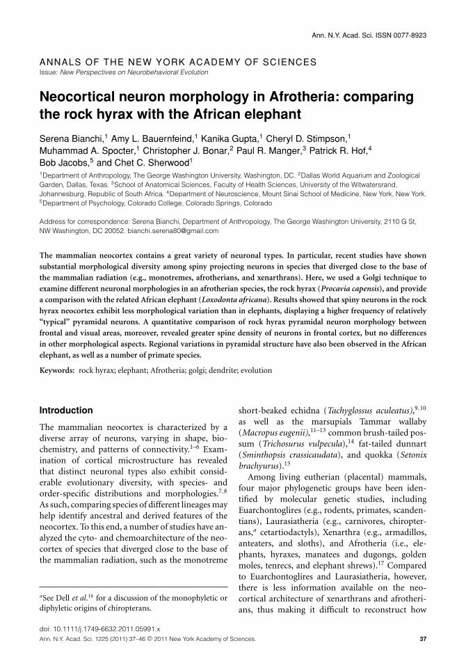
Ann. N.Y. Acad. Sci. ISSN 0077-8923 ANNALS OF THE NEW YORK ACADEMY OF SCIENCES Issue: New Perspectives on Neurobehavioral Evolution Neocortical neuron morphology in Afrotheria: comparing the rock hyrax with the African elephant Serena Bianchi, 1 Amy L. Bauernfeind, 1 Kanika Gupta, 1 Cheryl D. Stimpson, 1 Muhammad A. Spocter, 1 Christopher J. Bonar, 2 Paul R. Manger, 3 Patrick R. Hof, 4 Bob Jacobs, 5 and Chet C. Sherwood 1 1 Department of Anthropology, The George Washington University, Washington, DC. 2 Dallas World Aquarium and Zoological Garden, Dallas, Texas. 3 School of Anatomical Sciences, Faculty of Health Sciences, University of the Witwatersrand, Johannesburg, Republic of South Africa. 4 Department of Neuroscience, Mount Sinai School of Medicine, New York, New York. 5 Department of Psychology, Colorado College, Colorado Springs, Colorado Address for correspondence:Serena Bianchi, Department of Anthropology, The George Washington University, 2110 G St, NW Washington, DC 20052. [email protected] The mammalian neocortex contains a great variety of neuronal types. In particular, recent studies have shown substantial morphological diversity among spiny projecting neurons in species that diverged close to the base of the mammalian radiation (e.g., monotremes, afrotherians, and xenarthrans). Here, we used a Golgi technique to examine different neuronal morphologies in an afrotherian species, the rock hyrax (Procavia capensis), and provide a comparison with the related African elephant (Loxodonta africana). Results showed that spiny neurons in the rock hyrax neocortex exhibit less morphological variation than in elephants, displaying a higher frequency of relatively “typical” pyramidal neurons. A quantitative comparison of rock hyrax pyramidal neuron morphology between frontal and visual areas, moreover, revealed greater spine density of neurons in frontal cortex, but no differences in other morphological aspects. Regional variations in pyramidal structure have also been observed in the African elephant, as well as a number of primate species. Keywords: rock hyrax; elephant; Afrotheria; golgi; dendrite; evolution Introduction The mammalian neocortex is characterized by a diverse array of neurons, varying in shape, bio- chemistry, and patterns of connectivity. 1–6 Exam- ination of cortical microstructure has revealed that distinct neuronal types also exhibit consid- erable evolutionary diversity, with species- and order-specific distributions and morphologies. 7,8 As such, comparing species of different lineages may help identify ancestral and derived features of the neocortex. To this end, a number of studies have an- alyzed the cyto- and chemoarchitecture of the neo- cortex of species that diverged close to the base of the mammalian radiation, such as the monotreme a See Dell et al . 16 for a discussion of the monophyletic or diphyletic origins of chiropterans. short-beaked echidna (Tachyglossus aculeatus), 9,10 as well as the marsupials Tammar wallaby (Macropus eugenii), 11–13 common brush-tailed pos- sum (Trichosurus vulpecula), 14 fat-tailed dunnart (Sminthopsis crassicaudata), and quokka (Setonix brachyurus ). 15 Among living eutherian (placental) mammals, four major phylogenetic groups have been iden- tified by molecular genetic studies, including Euarchontoglires (e.g., rodents, primates, scanden- tians), Laurasiatheria (e.g., carnivores, chiropter- ans, a cetartiodactyls), Xenarthra (e.g., armadillos, anteaters, and sloths), and Afrotheria (i.e., ele- phants, hyraxes, manatees and dugongs, golden moles, tenrecs, and elephant shrews). 17 Compared to Euarchontoglires and Laurasiatheria, however, there is less information available on the neo- cortical architecture of xenarthrans and afrotheri- ans, thus making it difficult to reconstruct how doi: 10.1111/j.1749-6632.2011.05991.x Ann. N.Y. Acad. Sci. 1225 (2011) 37–46 c 2011 New York Academy of Sciences. 37
-
Upload
independent -
Category
Documents
-
view
1 -
download
0
Transcript of Neocortical neuron morphology in Afrotheria: comparing the rock hyrax with the African elephant
Ann. N.Y. Acad. Sci. ISSN 0077-8923
ANNALS OF THE NEW YORK ACADEMY OF SCIENCESIssue: New Perspectives on Neurobehavioral Evolution
Neocortical neuron morphology in Afrotheria: comparingthe rock hyrax with the African elephant
Serena Bianchi,1 Amy L. Bauernfeind,1 Kanika Gupta,1 Cheryl D. Stimpson,1
Muhammad A. Spocter,1 Christopher J. Bonar,2 Paul R. Manger,3 Patrick R. Hof,4
Bob Jacobs,5 and Chet C. Sherwood1
1Department of Anthropology, The George Washington University, Washington, DC. 2Dallas World Aquarium and ZoologicalGarden, Dallas, Texas. 3School of Anatomical Sciences, Faculty of Health Sciences, University of the Witwatersrand,Johannesburg, Republic of South Africa. 4Department of Neuroscience, Mount Sinai School of Medicine, New York, New York.5Department of Psychology, Colorado College, Colorado Springs, Colorado
Address for correspondence: Serena Bianchi, Department of Anthropology, The George Washington University, 2110 G St,NW Washington, DC 20052. [email protected]
The mammalian neocortex contains a great variety of neuronal types. In particular, recent studies have shownsubstantial morphological diversity among spiny projecting neurons in species that diverged close to the base ofthe mammalian radiation (e.g., monotremes, afrotherians, and xenarthrans). Here, we used a Golgi technique toexamine different neuronal morphologies in an afrotherian species, the rock hyrax (Procavia capensis), and providea comparison with the related African elephant (Loxodonta africana). Results showed that spiny neurons in the rockhyrax neocortex exhibit less morphological variation than in elephants, displaying a higher frequency of relatively“typical” pyramidal neurons. A quantitative comparison of rock hyrax pyramidal neuron morphology betweenfrontal and visual areas, moreover, revealed greater spine density of neurons in frontal cortex, but no differencesin other morphological aspects. Regional variations in pyramidal structure have also been observed in the Africanelephant, as well as a number of primate species.
Keywords: rock hyrax; elephant; Afrotheria; golgi; dendrite; evolution
Introduction
The mammalian neocortex is characterized by adiverse array of neurons, varying in shape, bio-chemistry, and patterns of connectivity.1–6 Exam-ination of cortical microstructure has revealedthat distinct neuronal types also exhibit consid-erable evolutionary diversity, with species- andorder-specific distributions and morphologies.7,8
As such, comparing species of different lineages mayhelp identify ancestral and derived features of theneocortex. To this end, a number of studies have an-alyzed the cyto- and chemoarchitecture of the neo-cortex of species that diverged close to the base ofthe mammalian radiation, such as the monotreme
aSee Dell et al.16 for a discussion of the monophyletic ordiphyletic origins of chiropterans.
short-beaked echidna (Tachyglossus aculeatus),9,10
as well as the marsupials Tammar wallaby(Macropus eugenii),11–13 common brush-tailed pos-sum (Trichosurus vulpecula),14 fat-tailed dunnart(Sminthopsis crassicaudata), and quokka (Setonixbrachyurus).15
Among living eutherian (placental) mammals,four major phylogenetic groups have been iden-tified by molecular genetic studies, includingEuarchontoglires (e.g., rodents, primates, scanden-tians), Laurasiatheria (e.g., carnivores, chiropter-ans,a cetartiodactyls), Xenarthra (e.g., armadillos,anteaters, and sloths), and Afrotheria (i.e., ele-phants, hyraxes, manatees and dugongs, goldenmoles, tenrecs, and elephant shrews).17 Comparedto Euarchontoglires and Laurasiatheria, however,there is less information available on the neo-cortical architecture of xenarthrans and afrotheri-ans, thus making it difficult to reconstruct how
doi: 10.1111/j.1749-6632.2011.05991.xAnn. N.Y. Acad. Sci. 1225 (2011) 37–46 c© 2011 New York Academy of Sciences. 37

Neuromorphology in Afrotheria Bianchi et al.
neocortical circuitry has changed in placental mam-mal evolution.
To address this limitation, a recent immunohis-tochemical analysis of the neocortex of several xe-narthrans and afrotherians has provided new dataon neuron types and distributions in these species.18
Taken together, these studies highlight substantialsimilarities across all mammals in the morphologyof nonprojection inhibitory interneurons (however,see De Felipe et al.2 for interspecies differences indouble-bouquet cells). A remarkable degree of vari-ation, however, has been observed among pyramidalcells, which exhibited a greater number of “atypical”features (e.g., inverted somata, widely bifurcatingapical dendrites) in monotremes, marsupials, xe-narthrans, and afrotherians compared to rodentsand primates.10,11,18
To elucidate the evolution of mammalian neu-romorphology, we examined the neocortex of anafrotherian species, the rock hyrax. As a memberof the order Hyracoidea, family Procaviidae, therock hyrax is a medium-sized (∼4 kg), herbivo-rous animal that inhabits sub-Saharan Africa andthe Middle East. Among mammals, the Afrothe-ria is a group of particular interest not only for itsbasal phylogenetic position—it diverged from othermammals about 100 million years ago17—but alsofor its striking diversity. Indeed, the Afrotheria iscomprised of species with diverse ecological niches,behavioral adaptations, and brain sizes, which rangefrom the ∼1.5 g brain of elephant shrews19 to the∼5 kg brain of elephants.20 Afrotherians, moreover,remain relatively underrepresented in the compara-tive neuroanatomical literature,21 with only a hand-ful of studies published on the cortical organizationof the elephant shrew (Elephantulus edwardii),22 thetenrec (Echinops telfairi),23 the manatee (Trichechusmanatus),24 and the African elephant (Loxodontaafricana).25,26 Previous work on the neocortex ofthe hyrax includes the mapping of the somatosen-sory area,27 volumetric measurements of gray andwhite matter,28 and immunohistochemical analy-ses;18 however, no study has provided a quantitativeanalysis of cortical neuromorphology.
In the present study, we used Golgi staining todescribe and quantify a variety of neuron types inthe rock hyrax neocortex. In so doing, we furnish adirect comparison with another afrotherian species,the African elephant, for which a detailed neuro-morphological study was recently completed.26 In
the examination of the elephant neocortex, a vari-ety of neuron types were identified, revealing re-markable morphological heterogeneity, especiallyamong spiny neurons.26 In addition, quantitativeanalyses that compared the morphology of pyrami-dal neurons within superficial cortical layers foundsignificant differences between frontal and occipi-tal regions.26 Previous studies measuring regionalvariation in pyramidal cells of humans and otherprimates29–31 have revealed greater neuromorpho-logical complexity in anterior compared to pos-terior regions, suggesting increased computationaldemands within regions of the frontal lobe. Con-sistent with these findings, the elephant exhibitedmore complex dendritic arbors and spine comple-ment in frontal compared with occipital neurons.26
Here, we offer a similar morphometric assessment ofpyramidal neurons in the rock hyrax by quantifyingand comparing a sample of neurons across frontaland occipital regions. Furthermore, we present adescription of variation in neuronal morphology inthese neocortical areas.
Material and methods
Specimens and tissue preparationThe brains of two adult rock hyraxes, one female(age: one year, five months) and one male (age:11 months, 22 days), were obtained from the Cleve-land Metroparks Zoo after the animals had diedfor reasons unrelated to the current study. Tissuesamples were removed within 14 hours after deathand immersion fixed in 10% buffered formalin forseven days. They were then transferred to a phos-phate buffer saline (PBS) solution with 1% azide andstored at 4◦ C until staining. Both brains appearednormal upon routine pathology examination.
Two small tissue blocks (1–2 cm) from frontaland occipital areas were removed from the righthemisphere of each individual. By reference to thepattern of staining against Nissl substance, myelin,and parvalbumin from the left hemisphere of thesesame specimens,18 the occipital region blocks wereremoved from an area that was in the location of pri-mary visual cortex, and the frontal blocks were takenfrom a dorsolateral region anterior to the primarymotor cortex. The frontal cortex was dysgranular,and the occipital was granular. In keeping with pre-vious studies,26,30,31 the tissue was processed by amodified rapid Golgi technique33 and sectioned at120 �m with a vibratome.
38 Ann. N.Y. Acad. Sci. 1225 (2011) 37–46 c© 2011 New York Academy of Sciences.
Bianchi et al. Neuromorphology in Afrotheria
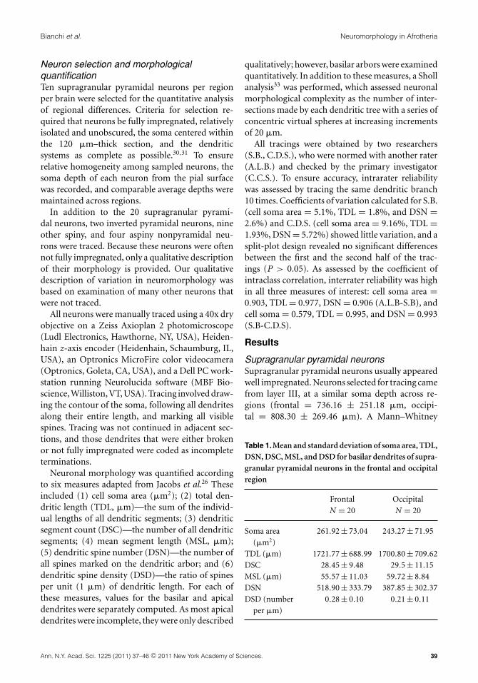
Neuron selection and morphologicalquantificationTen supragranular pyramidal neurons per regionper brain were selected for the quantitative analysisof regional differences. Criteria for selection re-quired that neurons be fully impregnated, relativelyisolated and unobscured, the soma centered withinthe 120 �m–thick section, and the dendriticsystems as complete as possible.30,31 To ensurerelative homogeneity among sampled neurons, thesoma depth of each neuron from the pial surfacewas recorded, and comparable average depths weremaintained across regions.
In addition to the 20 supragranular pyrami-dal neurons, two inverted pyramidal neurons, nineother spiny, and four aspiny nonpyramidal neu-rons were traced. Because these neurons were oftennot fully impregnated, only a qualitative descriptionof their morphology is provided. Our qualitativedescription of variation in neuromorphology wasbased on examination of many other neurons thatwere not traced.
All neurons were manually traced using a 40x dryobjective on a Zeiss Axioplan 2 photomicroscope(Ludl Electronics, Hawthorne, NY, USA), Heiden-hain z-axis encoder (Heidenhain, Schaumburg, IL,USA), an Optronics MicroFire color videocamera(Optronics, Goleta, CA, USA), and a Dell PC work-station running Neurolucida software (MBF Bio-science, Williston, VT, USA). Tracing involved draw-ing the contour of the soma, following all dendritesalong their entire length, and marking all visiblespines. Tracing was not continued in adjacent sec-tions, and those dendrites that were either brokenor not fully impregnated were coded as incompleteterminations.
Neuronal morphology was quantified accordingto six measures adapted from Jacobs et al.26 Theseincluded (1) cell soma area (�m2); (2) total den-dritic length (TDL, �m)—the sum of the individ-ual lengths of all dendritic segments; (3) dendriticsegment count (DSC)—the number of all dendriticsegments; (4) mean segment length (MSL, �m);(5) dendritic spine number (DSN)—the number ofall spines marked on the dendritic arbor; and (6)dendritic spine density (DSD)—the ratio of spinesper unit (1 �m) of dendritic length. For each ofthese measures, values for the basilar and apicaldendrites were separately computed. As most apicaldendrites were incomplete, they were only described
qualitatively; however, basilar arbors were examinedquantitatively. In addition to these measures, a Shollanalysis33 was performed, which assessed neuronalmorphological complexity as the number of inter-sections made by each dendritic tree with a series ofconcentric virtual spheres at increasing incrementsof 20 �m.
All tracings were obtained by two researchers(S.B., C.D.S.), who were normed with another rater(A.L.B.) and checked by the primary investigator(C.C.S.). To ensure accuracy, intrarater reliabilitywas assessed by tracing the same dendritic branch10 times. Coefficients of variation calculated for S.B.(cell soma area = 5.1%, TDL = 1.8%, and DSN =2.6%) and C.D.S. (cell soma area = 9.16%, TDL =1.93%, DSN = 5.72%) showed little variation, and asplit-plot design revealed no significant differencesbetween the first and the second half of the trac-ings (P > 0.05). As assessed by the coefficient ofintraclass correlation, interrater reliability was highin all three measures of interest: cell soma area =0.903, TDL = 0.977, DSN = 0.906 (A.L.B-S.B), andcell soma = 0.579, TDL = 0.995, and DSN = 0.993(S.B-C.D.S).
Results
Supragranular pyramidal neuronsSupragranular pyramidal neurons usually appearedwell impregnated. Neurons selected for tracing camefrom layer III, at a similar soma depth across re-gions (frontal = 736.16 ± 251.18 �m, occipi-tal = 808.30 ± 269.46 �m). A Mann–Whitney
Table 1.Mean and standard deviation of soma area, TDL,DSN, DSC, MSL, and DSD for basilar dendrites of supra-granular pyramidal neurons in the frontal and occipitalregion
Frontal Occipital
N = 20 N = 20
Soma area
(�m2)
261.92 ± 73.04 243.27 ± 71.95
TDL (�m) 1721.77 ± 688.99 1700.80 ± 709.62
DSC 28.45 ± 9.48 29.5 ± 11.15
MSL (�m) 55.57 ± 11.03 59.72 ± 8.84
DSN 518.90 ± 333.79 387.85 ± 302.37
DSD (number
per �m)
0.28 ± 0.10 0.21 ± 0.11
Ann. N.Y. Acad. Sci. 1225 (2011) 37–46 c© 2011 New York Academy of Sciences. 39

Neuromorphology in Afrotheria Bianchi et al.
U-test showed no significant differences in the somadepth between regions (U = 152, P > 0.05).
Morphology. Supragranular pyramidal neuronstypically exhibited spiny basilar dendrites extend-ing radially from the cell body and a single apicaldendrite traveling toward the pial surface. Quan-titative data are provided in Table 1. Visual in-spection of 15 relatively complete apical dendritesrevealed one thick dendrite that ascended perpen-dicularly to the pial surface while forming thinner,but spiny, oblique branches in most cases. In someinstances, however, apical dendrites originated fromthe soma as a single shaft, split after approximately30 �m into two thick branches that either trav-eled obliquely toward the pial surface, forming a
V-shaped pattern, or, more frequently, ascended inparallel.
Regional differences. We analyzed regional differ-ences in pyramidal neuron morphology using one-way ANOVAs with MSL and DSD as the dependentvariables and region as the independent variable.These analyses showed that DSD (F1,38 = 5.167,P = 0.029) was significantly greater in the frontalregion. No significant differences were found inMSL (F1,38 = 0.252, P > 0.05), which was simi-lar across regions. Because the other variables ofinterest, TDL, DSN, DSC, and cell body area werenot normally distributed, we used Mann–WhitneyU-tests to examine regional differences. Results re-vealed no significant differences in either cell body
Figure 1. A–D illustrate regional differences in the soma area, TDL, DSN, and DSD for basilar dendritic arbors of supragranularpyramidal neurons. E shows different Sholl profiles for frontal and occipital supragranular pyramidal neurons, calculated as thetotal number of intersections on each dendritic system per 20 �m of dendritic length.
40 Ann. N.Y. Acad. Sci. 1225 (2011) 37–46 c© 2011 New York Academy of Sciences.

Bianchi et al. Neuromorphology in Afrotheria
area (U = 155.00, P > 0.05), TDL (U = 199.00,P > 0.05) or DSC (U = 193.00, P > 0.05). GreaterDSN was found in neurons in the frontal region, butit fell short of conventional significance levels (U =136.500, P = 0.086, Fig. 1).
In a Sholl analysis, similar profiles of basilar den-dritic branching characterized frontal and occipitalneurons in both the average of the total numberof intersections (frontal = 4.59 ± 4.09, occipital =5.07 ± 4.53) and the distance from the soma, whichpeaked at 60 �m in both regions. In the frontal re-gion, however, the number of intersections tendedto decrease less steeply as the distance from the somaincreased, possibly indicating a relatively larger re-ceptive field (Fig. 1).
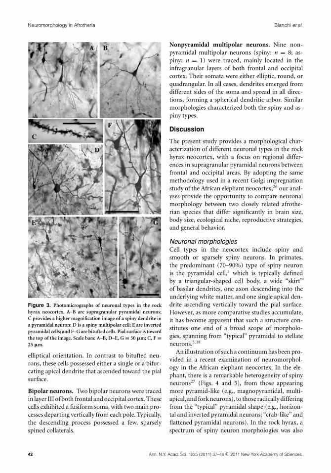
Other neuronal morphologiesIn addition to the supragranular pyramidal neu-rons, a variety of other spiny and aspiny neurontypes were identified, including inverted pyrami-dal, fork-shaped, bitufted, bipolar, and multipolarcells (Figs. 2 and 3). For these neuronal types, aqualitative description of morphology and laminardistribution is provided here.
Inverted pyramidal neurons. Two inverted pyra-midal neurons were traced in the infragranular lay-ers of the occipital region. These neurons exhibiteda spiny basilar dendritic skirt that ascended towardthe pial surface, and a thick apical dendrite with sev-eral side branches that descended toward the whitematter.
Fork neurons. One fork neuron was traced in layerIII of the frontal region. Fork neurons have beenpreviously described in the human insular cortex.34
They have a single large tapered basal dendrite, withtwo thick apical dendrites that bifurcate close to thesoma and project toward layer I in a narrow Y-likefashion. However, only the cell body and proximalapical dendritic segments were fully impregnated inthis neuron.
Bitufted neurons. Three bitufted neurons (spiny:n = 2; aspiny: n = 1) were traced in layer IIIand V of the occipital cortex. Both spiny and as-piny types showed similar vertical morphologies,with two dendritic processes emerging from eachpole of an elongated soma. A possible variant ofthese neurons showed an elongated soma, fromwhich a spiny basilar skirt developed vertically ina horse tail–like fashion, giving the neuron an
Figure 2. Tracings of pyramidal and nonpyramidal morpholo-gies in the rock hyrax neocortex. Among pyramidal morpholo-gies, A–G illustrate frontal pyramidal neurons, H–L illustrateoccipital pyramidal neurons, and M and N illustrate invertedpyramidal neurons. Among nonpyramidal morphologies, O,R–T, and V–Y are multipolar cells; P and Q are bipolar cells; andU and T are bitufted cells. O, P, R, and S are aspiny or sparselyspined. Q, T, U, W, and Y are spiny. Pial surface is toward thetop of the figure. Scale bar = 100 �m.
Ann. N.Y. Acad. Sci. 1225 (2011) 37–46 c© 2011 New York Academy of Sciences. 41
Neuromorphology in Afrotheria Bianchi et al.
Figure 3. Photomicrographs of neuronal types in the rockhyrax neocortex. A–B are supragranular pyramidal neurons;C provides a higher magnification image of a spiny dendrite ina pyramidal neuron; D is a spiny multipolar cell; E are invertedpyramidal cells; and F–G are bitufted cells. Pial surface is towardthe top of the image. Scale bars: A–B, D–E, G = 50 �m; C, F =25 �m.
elliptical orientation. In contrast to bitufted neu-rons, these cells possessed either a single or a bifur-cating apical dendrite that ascended toward the pialsurface.
Bipolar neurons. Two bipolar neurons were tracedin layer III of both frontal and occipital cortex. Thesecells exhibited a fusiform soma, with two main pro-cesses departing vertically from each pole. Typically,the descending process possessed a few, sparselyspined collaterals.
Nonpyramidal multipolar neurons. Nine non-pyramidal multipolar neurons (spiny: n = 8; as-piny: n = 1) were traced, mainly located in theinfragranular layers of both frontal and occipitalcortex. Their somata were either elliptic, round, orquadrangular. In all cases, dendrites emerged fromdifferent sides of the soma and spread in all direc-tions, forming a spherical dendritic arbor. Similarmorphologies characterized both the spiny and as-piny types.
Discussion
The present study provides a morphological char-acterization of different neuronal types in the rockhyrax neocortex, with a focus on regional differ-ences in supragranular pyramidal neurons betweenfrontal and occipital areas. By adopting the samemethodology used in a recent Golgi impregnationstudy of the African elephant neocortex,26 our anal-yses provide the opportunity to compare neuronalmorphology between two closely related afrothe-rian species that differ significantly in brain size,body size, ecological niche, reproductive strategies,and general behavior.
Neuronal morphologiesCell types in the neocortex include spiny andsmooth or sparsely spiny neurons. In primates,the predominant (70–90%) type of spiny neuronis the pyramidal cell,5 which is typically definedby a triangular-shaped cell body, a wide “skirt”of basilar dendrites, one axon descending into theunderlying white matter, and one single apical den-drite ascending vertically toward the pial surface.However, as more comparative studies accumulate,it has become apparent that such a structure con-stitutes one end of a broad scope of morpholo-gies, spanning from “typical” pyramidal to stellateneurons.5,18
An illustration of such a continuum has been pro-vided in a recent examination of neuromorphol-ogy in the African elephant neocortex. In the ele-phant, there is a remarkable heterogeneity of spinyneurons27 (Figs. 4 and 5), from those appearingmore pyramid-like (e.g., magnopyramidal, multi-apical, and fork neurons), to those radically differingfrom the “typical” pyramidal shape (e.g., horizon-tal and inverted pyramidal neurons; “crab-like” andflattened pyramidal neurons). In the rock hyrax, aspectrum of spiny neuron morphologies was also
42 Ann. N.Y. Acad. Sci. 1225 (2011) 37–46 c© 2011 New York Academy of Sciences.

Bianchi et al. Neuromorphology in Afrotheria
Figure 4. Photomicrographs of two supragranular pyramidal neurons from the African elephant frontal cortex (A), a layer III/Vinverted pyramidal neuron (arrowhead) from frontal cortex (B), and a high magnification view of a spine-rich apical dendrite fromoccipital cortex (C). Pial surface is toward the top of the image. For A, scale bar = 100 �m. For B, scale bar = 200 �m. For C, scalebar = 50 �m.
observed; however, fewer separate neuronal typeswere identified, including only pyramidal, forkedshaped, inverted pyramidal, and bitufted neurons.
Of the pyramidal-like neurons, some presented“atypical” features (e.g., bifurcating apical den-drites, multiapical and inverted soma) that havebeen similarly observed in other Atlantogenata (i.e.,the clade formed by Afrotheria and Xenarthra), suchas elephant shrews, anteaters, and sloths,18 as wellas a monotreme, the short-beaked echidna.10 Therock hyrax, nevertheless, was characterized by a pre-dominance of “typical” pyramidal cells. Indeed, onemain respect in which the rock hyrax differed fromthe elephant was the high frequency of pyramidalcells with “canonical” apical dendrites that ascendedvertically toward the pial surface. In contrast, in theelephant, most spiny neurons had apical dendritesthat bifurcated at or near the soma, resulting in twoobliquely ascending secondary branches that joinedwith others to form V-shaped apical bundles (Fig. 5).
While being consistent with descriptions in thehorse and the cow,35 as well as the two-toed slothand anteater,18 the widely bifurcating structure ob-served in the elephant represents a striking depar-ture from the vertically orientated apical dendritesthat typify the rodent and primate neocortex.36
Together with previous observations, this suggeststhat the notion of a neocortical architecture com-mon to all mammals, defined by pyramidal-shapedprojection neurons with apical dendrites bundledtogether at the core of minicolumns,36 may not cap-ture the actual variation that is present among dif-ferent mammalian lineages. In fact, the observa-tion of a greater frequency of “atypical” features intaxa close to the origin of the mammalian radiationhas led to the suggestion that, while variation inprojection neuron morphology characterizes mostmammalian species, strong selection for verticallyoriented apical dendrites might have begun toemerge with the evolution of Boreoeutheria (i.e.,Euarchontoglires and Laurasiatheria).18
As shown by a recent reevaluation of the conceptof the cortical column as elementary unit of the neo-cortex, however, the functional and evolutionarysignificance of a vertical neocortical organizationremains unclear.37 Thus, while it is interesting thatthe afrotherian rock hyrax exhibits high frequencyof “typical” pyramidal features, further studies onspecies such as shrews, flying lemurs, lagomorphs,and megachiropterans will be needed to deter-mine whether these features universally characterizeBoreoeutheria as a phylogenetic group.
Ann. N.Y. Acad. Sci. 1225 (2011) 37–46 c© 2011 New York Academy of Sciences. 43

Neuromorphology in Afrotheria Bianchi et al.
Figure 5. Photomicrograph of neuronal morphologies in the African elephant. In A, a magnopyramidal-taproot or matriarchneuron, a large layer V neuron with a long descending taproot dendrite. An inverted pyramidal neuron (a) and a large, multiapicalpyramidal neuron (b) from frontal cortex are represented in B. Two multiapical pyramidal neurons (arrowheads) from occipitalcortex can be seen in C. In D, several supragranular pyramidal neurons from the frontal cortex are present, each with widelybifurcating apical dendrites projecting toward the pial surface. For A, B, C, scale bar = 200 �m. For D, scale bar = 100 �m.
Greater similarity with other eutherian mammalswas observed in the morphology of aspiny andsparsely spiny neurons, including those withmultipolar, bitufted, and bipolar shapes. In terms ofmorphology and laminar distribution, these neu-rons resembled those described in monotremes,10
humans, carnivores, artiodactyls, lagomorphs,38
rodents,39 dolphins,40 and elephants.26 Becauseaspiny cortical neurons generally correspond toinhibitory GABAergic subtypes, this suggests thatthere may be relatively more evolutionary conser-vation in regard to the morphology of cell typesthat comprise the intrinsic microcircuitry of themammalian cerebral cortex41 (however, see Ref. 5).Considerable phylogenetic diversity in the densityand biochemical phenotype of these inhibitoryinterneurons, however, is apparent.3,7,8,18
Regional differences in supragranularpyramidal morphologyA comparison of regional differences in the morph-ology of supragranular pyramidal neurons in therock hyrax revealed spinier dendrites in frontal com-pared to occipital cortex. This result is consistentwith previous findings in the elephant26 as well asseveral primate species,5,29 including humans.6 Incontrast with these species, however, the rock hyraxshowed no differences in the extent and complexityof dendritic arbors.
Spine density and dendritic branching patternsare essential in determining the receptive fieldand integrative capacity of the neuron.42 In pri-mates, regional variation in neuronal morphologyis thought to reflect area-specific functional spe-cializations.29,30 Specifically, neurons with greater
44 Ann. N.Y. Acad. Sci. 1225 (2011) 37–46 c© 2011 New York Academy of Sciences.
Bianchi et al. Neuromorphology in Afrotheria
dendritic complexity and higher spine density mightsubserve the synthesis of a more diverse array ofinputs.29 Thus, the higher spine density found inthe frontal cortex of the rock hyrax provides indi-rect evidence that this region is involved in func-tions that entail greater computational demand.However, given the scarcity of data on the physi-ology and connectivity of the rock hyrax cerebralcortex, it is uncertain whether the functional sig-nificance of regional differences in spine densityis comparable to that of primates. Moreover, ourfailure to find significant differences in other im-portant aspects of the cell morphology indicatesmore similarity in neuronal structure across regionsthan has previously been reported in primates. Therelatively small sample size used this study, how-ever, may have prevented the observation of greatermorphological variation. As such, our interpreta-tion of structural and functional regional differencesremains tentative. The present study contributes,however, to the growing body of research delineat-ing neuromorphological variation in the neocor-tex of afrotherian species, and adds to our under-standing of the evolution of neuronal diversity inmammals.
Acknowledgments
We thank Drs. Mary Ann Raghanti and AlbertLewandowski for assistance related to this research.Brain materials used in this study were loanedby the Cleveland Metroparks Zoo. This work wassupported by the National Science Foundation(BCS-0515484, BCS-0549117, BCS-0827531, DGE-0801634) and the James S. McDonnell Foundation(22002078).
Conflicts of interest
The authors declare no conflicts of interest.
Supporting Information
Additional supporting information may be foundin the online version of this article:
Supplementary Figure 1. Low magnification pho-tomicrograph of Golgi-impregnated neurons inthe elephant occipital cortex. Note the wide lat-eral spread of the bifurcating apical dendrites inthe large layer V neurons (arrowheads). Pial sur-face is toward the top of the image. Scale bar =200 �m.
Supplementary Figure 2. Low magnification pho-tomicrograph of Golgi-impregnated neurons in ele-phant frontal cortex illustrating several neuronaltypes: (A) supragranular pyramidal neurons withwidely bifurcating apical branches, (BB) horizontalpyramidal neurons with sectioned apical dendrites,and (C) several inverted pyramidal neurons. Pialsurface is toward the top of the image. Scale bar =200 �m.
Supplementary Figure 3. Low magnification pho-tomicrograph of Golgi-impregnated neurons in ele-phant frontal cortex illustrating several neuronaltypes: (A) a supragranular pyramidal neuron, (B) amagnopyramidal-taproot or matriarch neuron, and(C) a magnopyramidal neuron with a sectioned api-cal dendrite. Pial surface is toward the top of theimage. Scale bar = 200 �m.
Supplementary Figure 4. Photomicrograph of amultipolar, Golgi-impregnated aspiny interneuronin elephant frontal cortex. Pial surface is toward thetop of the image. Axon is highlighted by the arrow-head. Scale bar = 100 �m.
Please note:Wiley-Blackwell is not responsible forthe content or functionality of any supporting ma-terials supplied by the authors. Any queries (otherthan missing material) should be directed to thecorresponding author for the article.
References
1. Nieuwenhuys, R. 1994. The neocortex. An overview ofits evolutionary development, structural organization andsynaptology. Anat. Embryol. 190: 307–337.
2. De Felipe, L., L. Alonso-Nanclarens & J.I. Arellano. 2002.Microstructure of the neocortex: comparative aspects.J. Neurocytol. 31: 299–316.
3. Hof, P.R. & C.C. Sherwood. 2007. The evolution of neuronclasses in the neocortex of mammals. In Evolution of NervousSystems in Mammals. Evolution of Nervous System, vol. 3.L.A. Krubitzer & J.H. Kaas, Eds.: 113–124. Academic Press.Oxford.
4. Somogji, P., G. Tamas, R. Lujan, et al. 1998. Salient featuresof synaptic organization in the cerebral cortex. Brain Res.Rev. 26: 113–135.
5. Elston, G.N. 2007. Specializations in pyramidal cell struc-ture during primate evolution. In: Evolution of Nervous Sys-tems. J.H. Kaas & T.M. Preuss, Eds.: 191–242. AcademicPress. Oxford.
6. Jacobs, B. & A.B. Scheibel. 2002. Regional dendritic varia-tion in primate cortical pyramidal cells. In: Cortical Areas:Unity And Diversity. A. Schuz & R. Miller, Eds.: pp 111–131.Taylor and Francis. London.
Ann. N.Y. Acad. Sci. 1225 (2011) 37–46 c© 2011 New York Academy of Sciences. 45

Neuromorphology in Afrotheria Bianchi et al.
7. Hof, P.R., I.I. Glezer, F. Conde, et al. 1999. Cellular dis-tribution of the calcium-binding proteins parvalbumin,calbindin, and calretinin in the neocortex of mammals: phy-logenetic and developmental patterns. J. Chem. Neuroanat.16: 77–116.
8. Hof, P.R. & C.C. Sherwood. 2005. Morphomolecular neu-ronal phenotypes in the neocortex reflect phylogenetic re-lationships among certain mammalian orders. Anat. Rec. A287: 1153–1163.
9. Hassiotis, M., G. Paxinos & K.W.S. Ashwell. 2003. Theanatomy of the cerebral cortex of the echidna (Tachyglossusaculeatus). Comp. Biochem. Physiol. A 136: 827–850.7.
10. Hassiotis, M. & K.W.S. Ashwell. 2003. Neuronal classesin the isocortex of a monetreme, the Australian echidna(Tachyglossus aculeatus). Brain Behav. Evol. 61: 6–27.
11. Ashwell, K.W.S., L.-L. Zhang & L.R. Marotte. 2005. Cyto-and chemoarchitecture of the cortex of the Tammar wallaby(Macropus eugenii). Area organization. Brain Behav. Evol.66: 114–136.
12. Waite, P.M.E., L.R. Marotte & R.F. Mark. 1991. Developmentof whiskers representation in the cortex of the Tammarwallaby (Macropus eugenii). Dev. Brain Res. 58: 35–41.
13. Waite, P.M.E., L.R. Marotte, C.A. Leamey, et al. 1998. De-velopment of whisker-related patterns in marsupials: factorscontrolling timing. Trends Neurosci. 21: 265–269.
14. Weller, W.L. 1993. SmI cortical barrels in an Australian mar-supial, Trichosurus vulpecula (brush-tailed possum): struc-tural organization, patterned distribution, and somatotopicrelationships. J. Comp. Neurol. 337: 471–492.
15. Tyler, C.J., S.A. Dunlop, R.D. Lund, et al. 1998. Anatomicalcomparison of the macaque and marsupial visual cortex:common features that may reflect retention of essential cor-tical elements. J. Comp. Neurol. 400: 449–468.
16. Dell, L-A., J-L. Kruger, A. Bhagwandin, et al. 2010. Nuclearorganization of cholinergic, putative catecholaminergic andserotonergic systems in the brains of two megachiropteranspecies. J. Chem. Neuroanat. 40: 177–195.
17. Murphy, W.J., T.H. Pringle, T.A. Crider, et al. 2007. Usinggenomic data to unravel the root of the placental mammalphylogeny. Genome Res. 17: 413–421.
18. Sherwood, C.C., C. D. Stimpson, C. Butti, et al. 2009. Neo-cortical neuron types in Xenarthra and Afrotheria: implica-tions for brain evolution in mammals. Brain Struct. Funct.213: 301–328.
19. Pieters, R.P., N.Gravett, K. Fuxe, et al. 2010. Nuclear or-ganization of cholinergic, putative catecholaminergic andserotonergic nuclei in the brain of the eastern rock elephantshrew, Elephantulus myurus. J. Chem. Neuroanat . 39: 175–88.
20. Manger, P.R., P. Pillay, B.C. Maseko, et al. 2009. Acquisitionof brains from the African elephant (Loxodonta africana):perfusion-fixation and dissection. J. Neurosci. Methods 179:16–21.
21. Manger, P.R, J. Corti, N. Ebrahim, et al. 2008. Is 21st centuryneuroscience too focussed on the rat/mouse model of brainfunction and dysfunction? Front. Neuroanat. 2: 1–7.
22. Dengler-Crish, C.M., S.D. Crish, M.J. O’Riain, et al. 2006.Organization of the somatosensory cortex in elephant shrew(E. edwardii). Anat. Rec. A 288: 859–866.
23. Krubitzer, L., H. Kunzle & J. Kaas. 1997. Organization of
the sensory cortex in a Madagascan insectivore, the tenrec(Echinops telfairi). J. Comp. Neurol. 379: 399–414.
24. Reep, R.L., J.I. Johnson, R.C. Switzer, et al. 1989. Manateecerebral cortex: cytoarchitecture of the frontal region inTrichechus manatus latirostris. Brain Behav. Evol. 34: 365–386.
25. Hakeem, A.Y., C.C. Sherwood, C.J. Bonar, et al. 2009. VonEconomo neurons in the elephant brain. Anat. Rec. 292:242–248.
26. Jacobs, B., J. Lubs, M. Hannan, et al. 2011. Neuronalmorphology in the African elephant (Loxodonta africana)neocortex. Brain Struct. Funct. 215: 273–298. doi:10.1007/s00429-010-0288-3.
27. Welker, W.I. & M. Carlson. 1976. Somatic sensory cortex ofhyrax (Procavia). Brain Behav. Evolut. 13: 294–301.
28. Bush, E.C. & J.M Allman. 2003. The scaling of white matterto gray matter in the cerebellum and neocortex. Brain BehavEvol. 65: 1–5.
29. Elston, G.N., R. Benavides-Piccione & J. DeFelipe. 2001: Thepyramidal cell in cognition: a comparative study in humansand monkeys. J. Neurosci. 21: 1–5.
30. Jacobs, B., L. Driscoll & M. Schall. 1997. Life-span dendriticand spine changes in areas 10 and 18 of human cortex: aquantitative Golgi study. J. Comp. Neurol. 386: 661–680.
31. Jacobs, B., M. Schall, M. Prather, et al. 2001. Regional den-dritic and spine variation in human cerebral cortex: a quan-titative Golgi study. Cereb. Cortex 11: 558–571.
32. Scheibel, M.E. & A.B. Scheibel. 1978. The meth-ods of Golgi. In Neuroanatomical Research Techniques.R.T. Robertson, Ed.: 89–114. Academic Press. New York.
33. Sholl, D.A. 1953. Dendritic organization of the neurons ofthe visual and motor cortices of the cat. J. Anat. 87: 387–406.
34. Ngowyang, G. 1932. Beschreibung einer Art vonSpezialzellen in der Inselrinde. J. Psychol. Neurol. 44: 671–674.
35. Barasa A. 1960. Forma, grandezza e densita dei neuroni dellacorteccia cerebrale in mammiferi di grandezza corporeadifferente. Z. Zellforsch. 53: 69–89.
36. Mountcastle, V.B. 1997. The columnar organization of theneocortex. Brain 120: 701–722.
37. Horton, J.C. & D.L. Adam. 2005. The cortical column: astructure without a function. Phil. Trans. R. Soc. B. 370:837–862.
38. Ballesteros, Y.I., A. Munos & J. Contreras. 2005. Doublebouquet cell in the human cerebral cortex and a comparisonwith other mammals. J. Comp. Neurol. 486: 344–360.
39. Feldman, M.L. & A. Peters. 1978 The forms of non-pyramidal neurons in the visual cortex of the rat. J. Comp.Neurol. 179: 761–794.
40. Ferrer, I., M. Perrera.1988. Structure and nerve cell or-ganization in the cerebral cortex of the dolphin Stenellacoeruleoalba: a Golgi study with a particular attention tothe primary auditory area. Anat. Embryol. 178: 161–173.
41. Elston, G.N. 2003. Cortex, cognition, and the cell: insightinto the pyramidal neuron and the prefrontal function.Cereb. Cortex 13: 1124–1138.
42. Raghanti, M.A., M.A. Spocter, C. Butti, et al. 2010. Acomparative perspective on minicolumns and inhibitoryGABAergic interneurons in the neocortex. Front. Neu-roanat. 4: 1–10.
46 Ann. N.Y. Acad. Sci. 1225 (2011) 37–46 c© 2011 New York Academy of Sciences.