Synthesis and conformational analysis of phosphorylated β-(1→2) linked mannosides
Upload
independentCategory
view
1download
0
Published Ahead of Print 26 February 2007. 10.1128/MCB.00086-06.
2007, 27(9):3530. DOI:Mol. Cell. Biol. HoughtonLimin Xiao, Eilyn R. Lacy, Richard W. Kriwacki and Peter J. Jun Zhan, John B. Easton, Shile Huang, Ashutosh Mishra, In Vivo Serine 98 That Is Phosphorylated by ASK1Involves a Small Domain That Includes
Cip1Negative Regulation of ASK1 by p21
http://mcb.asm.org/content/27/9/3530Updated information and services can be found at:
These include:
SUPPLEMENTAL MATERIAL Supplemental material
REFERENCEShttp://mcb.asm.org/content/27/9/3530#ref-list-1at:
This article cites 65 articles, 34 of which can be accessed free
CONTENT ALERTS more»articles cite this article),
Receive: RSS Feeds, eTOCs, free email alerts (when new
http://journals.asm.org/site/misc/reprints.xhtmlInformation about commercial reprint orders: http://journals.asm.org/site/subscriptions/To subscribe to to another ASM Journal go to:
on Septem
ber 2, 2014 by guesthttp://m
cb.asm.org/
Dow
nloaded from
on Septem
ber 2, 2014 by guesthttp://m
cb.asm.org/
Dow
nloaded from

MOLECULAR AND CELLULAR BIOLOGY, May 2007, p. 3530–3541 Vol. 27, No. 90270-7306/07/$08.00�0 doi:10.1128/MCB.00086-06Copyright © 2007, American Society for Microbiology. All Rights Reserved.
Negative Regulation of ASK1 by p21Cip1 Involves a Small Domain ThatIncludes Serine 98 That Is Phosphorylated by ASK1 In Vivo�†
Jun Zhan,1 John B. Easton,1 Shile Huang,2 Ashutosh Mishra,3 Limin Xiao,4 Eilyn R. Lacy,4‡Richard W. Kriwacki,4 and Peter J. Houghton1*
Department of Molecular Pharmacology,1 Hartwell Center,3 and Department of Structural Biology,4
St. Jude Children’s Research Hospital, 332 North Lauderdale St., Memphis, Tennessee 38105-2794,and Department of Biochemistry & Molecular Biology, Louisiana State University Health
Sciences Center, 1501 Kings Highway, Shreveport, Louisiana 71130-39322
Received 13 January 2007/Returned for modification 13 February 2007/Accepted 18 February 2007
The cyclin-dependent kinase inhibitor p21Cip1 regulates multiple cellular functions and protects cells fromgenotoxic and other cellular stresses. Activation of apoptosis signal-regulating kinase 1 (ASK1) induced byinhibition of mTOR signaling leads to sustained phospho-c-Jun that is suppressed in cells with functional p53or by forced expression of p21Cip1. Here we show that small deletions of p21Cip1 around S98 abrogate itsassociation with ASK1 but do not affect binding to Cdk1, hence distinguishing between the cell cycle-regulatingfunctions of p21Cip1 and its ability to suppress activation of the ASK1/Jun N-terminal protein kinase (JNK)pathway. p21Cip1 is phosphorylated in vitro by both ASK1 and JNK1 at S98. In vivo phosphorylation of p21Cip1,predominantly carried out by ASK1, is associated with binding to ASK1 and inactivation of ASK1 kinasefunction. Binding of p21Cip1 to ASK1 requires ASK1 kinase function and may involve phosphorylation of S98.
The cyclin-dependent kinase (CDK) inhibitor p21Cip1 isknown to control the cell cycle and DNA replication. In addi-tion, it has been shown to be involved in many biologicalprocesses, including differentiation, stress response, apoptosis,development, and tumorigenesis (reviewed in references 7 and12). Consistent with its numerous cellular functions, p21Cip1
has been found to participate in a number of specific protein-protein interactions. In addition to associating with cyclinCDKs and PCNA (22, 58, 63), p21Cip1 has also been reportedto bind to transcription factors (c-Myc [33], E2F [11], C/EBP-�[54], and STAT3 [10]), proteins involved in apoptosis (pro-caspase 3 [48], stress-associated protein kinase [47], and apop-tosis signal-regulating kinase 1 [ASK1] [4]), and other proteins(SET [14], calmodulin [52], HPV-16 E7 [16], GADD45 [30],TOK-1 [41], and protein kinase CK 2 [20]).
p21Cip1 plays a dual role in cell cycle progression, regulatingboth DNA synthesis and CDK activity. p21Cip1 binds to cyclinE-CDK complexes via an amino-terminal binding site, and thisinteraction inhibits the activity of these complexes, resulting ina block in cell cycle progression (46). p21Cip1 also blocks cellcycle progression in S phase due to its ability to bind andinhibit PCNA, a processivity factor for replication polymerases(31), through a carboxyl-terminal interaction (21) and leads toinhibition of DNA synthesis (58). However, more recent stud-ies have shown that p21Cip1 also has positive effects on G1
progression (46). p21Cip1 was shown to stabilize interactions
between Cdk4 and cyclin D and promote the formation ofactive complexes in a concentration-dependent manner (60).Several studies (15, 62) have demonstrated that p21Cip1 medi-ates the assembly and activation of cyclin D1-CDK4/6 com-plexes (36), which function as sensors of growth factors at G1-Sphase.
p21Cip1 has also been shown to play a role in enhancing cellsurvival. For example, terminally differentiated cells, such asmuscle cells (37), hematopoietic stem cells (9), and macro-phages (2, 21), were found to contain elevated levels ofp21Cip1, and the inhibition of the up-regulation of this proteinresulted in apoptotic cell death. Furthermore, p21Cip1 can pro-tect various types of cells from death following exposure tocytotoxic agents and ionizing radiation. The level of p21Cip1 incells determines the sensitivity to cisplatin and ionizing radia-tion. p21Cip1 overexpressed in inducible expression systems oradenovirus gene transfer increases cell survival against prosta-glandin A2 or p53 overproduction (17, 18). The reduction orabsence of p21Cip1 expression, however, sensitizes cells todoxorubicin hydrochloride, camptothecin etoposide, ionizingradiation, or prostaglandin A2 (42, 53, 59, 60). Because thep21Cip1 protein level is elevated in a diversity of tumors, par-ticularly in late-stage, aggressive tumors such as glioblastomamultiforme, the complex functions of p21Cip1 suggest that ad-vanced tumors take advantage of the positive effects of p21Cip1
on cell cycle progression and cell survival (3, 13, 15).The mechanisms by which p21Cip1 can prevent cells from
undergoing apoptosis are not well understood. One mecha-nism is assumed to involve p21Cip1-dependent cell cycle arrest(often at G2/M) that would permit repair of DNA damage(43). Another distinct mechanism is linked to the ability ofp21Cip1 to bind and inactivate cyclin A/Cdk2 complexes. It hasbeen shown that caspase 3-mediated cleavage of p21Cip1 is animportant mechanism of cyclin A/Cdk2 activation associatedwith death in different cell types (1, 29, 39). Apoptosis induced
* Corresponding author. Mailing address: Department of MolecularPharmacology, St. Jude Children’s Research Hospital, 332 North Lau-derdale St., Memphis, TN 38105-2794. Phone: (901) 495-3440. Fax:(901) 495-4290. E-mail: [email protected].
‡ Present address: Centocor, Inc., 145 King of Prussia Road, Rad-nor, PA 19087.
† Supplemental material for this article may be found at http://mcb.asm.org/.
� Published ahead of print on 26 February 2007.
3530
on Septem
ber 2, 2014 by guesthttp://m
cb.asm.org/
Dow
nloaded from

by various stimuli appears to be mediated by caspase 3 cleav-age of p21Cip1 and subsequent up-regulation of cyclin A/Cdk2activity. Caspase-dependent Cdk2 activity is a requisite effectorof apoptotic death, and it may be necessary for death-associ-ated chromatin condensation, cell shrinking, and loss of adhe-sion to substrate (23).
Recent data also point to more direct mechanisms by whichp21Cip1 may inhibit cell death. p21Cip1 interacts with pro-caspase 3 through the amino terminus and suppresses its acti-vation by masking the serine proteinase cleavage site (29, 40,48, 49). Procaspase 3-p21Cip1 complex formation occurred inmitochondria and required phosphorylation of p21Cip1 by pro-tein kinase A (50). Importantly, phosphorylation of p21Cip1 atT145 by Akt may cause relocalization of p21Cip1 to the cyto-plasm (65), where it may form a complex with ASK1 andinhibit the stress-activated mitogen-activated protein kinase(MAPK) cascade and cell death (25).
ASK1, a mammalian MAPK kinase kinase (MAPKKK), ac-tivates the Jun N-terminal protein kinase (JNK) and p38 path-ways and is activated in response to various cytotoxic stresses,including hydrogen peroxide (H2O2), Fas ligation, tumor ne-crosis factor (TNF), serum withdrawal, and cancer chemother-apeutic agents (8, 19, 27, 55). Overexpression of ASK1 inepithelial cells in low serum conditions induced apoptosis (27),and ASK1-deficient cells were resistant to H2O2- and TNF-induced apoptosis (56), indicating that ASK1 plays a pivotalrole in stress-induced apoptosis. The kinase activity of ASK1 istightly regulated within cells; under nonstressed conditionsASK1 is inhibited by association with its physiological inhibi-tor, thioredoxin (Trx). When cells are exposed to H2O2 orTNF, reactive oxygen species-dependent oxidation of Trx oc-curs, resulting in dissociation of Trx from ASK1 and therebyactivation of ASK1 (40). Oligomerization-dependent auto-phosphorylation appears to be the next step required for fullactivation of ASK1 after the release from Trx (19). On theother hand, the mechanism(s) of how the activated ASK1returns to an inactive form has not been elucidated.
Recently we found that the association of p21Cip1 with ASK1can block rapamycin-induced apoptosis in human and murinecells (25). However, the mechanism by which p21Cip1 associ-ates with ASK1 is not understood. Here we report thatp21Cip1 association with ASK1 involves a small region aroundS98. ASK1 can phosphorylate p21Cip1 at S98 both in vivo andin vitro, and this phosphorylation appears to be important forthe association of p21Cip1 with ASK1.
MATERIALS AND METHODS
Chemicals and antibodies. The JNK inhibitor SP600125, the p38 inhibitorSB203580, and anisomycin (AN), an activator of JNK and p38, were purchasedfrom A. G. Scientific (San Diego, CA). ASK C-terminal antibodies (Sc-5294),p21Cip1 rabbit polyclonal antibody (Sc-756), and mouse antihemagglutinin (anti-HA) monoclonal antibody (Sc-7392) were from Santa Cruz Biotechnology(Santa Cruz, CA). p21Cip1 mouse monoclonal antibody was from BD Pharmin-gen (Franklin Lakes, NJ). Anti-HA rabbit polyclonal antibody was from BDClontech (Mountain View, CA). Purified JNK1 and ASK1 kinases were obtainedfrom Upstate Biotechnology (Lake Placid, NY).
Cell lines and growth conditions. The Rh30 human rhabdomyosarcoma cellline expresses both wild-type (WT) and mutant (R273C) p53 alleles and wasgrown in antibiotic-free RPMI 1640 under conditions described previously (25).HEK293T cells were maintained in antibiotic-free Dulbecco’s minimal essentialmedium (DMEM) supplemented with 10% fetal bovine serum (FBS) at 37°Cand 5% CO2.
Cell transfection. 293T cells were transfected by using FuGene 6 (Roche).Briefly, HEK293T cells were seeded at 2 � 106 cells/10-cm-diameter dish. Trans-fection was performed 48 h after seeding the cells. Nine microliters of FuGene6 was added directly to serum-free DMEM, to a total volume of 100 �l, withgentle mixing. Two micrograms of p21Cip1 plasmid DNA (or plasmids encodingmutants) and 1 �g of ASK1 plasmid DNA were added to prediluted FuGene 6in 100 �l of DMEM. The mixture was gently tapped to ensure adequate mixingand set at room temperature for 30 to 45 min. The DNA-FuGene 6 transfectionmixture was added to the dish dropwise, and at the same time the plate contentswere swirled to ensure even dispersal.
Adenoviral infection. Stable lines expressing Rh30/p21 (WT) and Rh30/�NLSp21 (truncated at residue 140 and lacking the nuclear localization signal)under control of the metallothionein promoter have been described previously(25). Both cell lines were infected with replication-deficient adenovirus thatexpresses WT or N-terminally or C-terminally deleted human ASK1 constructs.First, all infections were performed in 2% FBS-DMEM for 2 h, and then cellswere incubated in 10% FBS-DMEM for 22 h, followed by culture in serum-freemedium. The expression of p21Cip1 or NLSp21Cip1 was induced with Zn (60 �M)for 24 h, at which time cell lysates were prepared for immunoprecipitation andWestern blot analysis.
Immunoprecipitation and Western blot analysis. Cells were briefly washedwith ice-cold phosphate-buffered saline. On ice, cells were lysed in 3-[(3-chol-amidopropyl)-dimethylammonio]-1-propanesulfonate (CHAPS) buffer (40 mMHEPES [N-2-hydroxyethylpiperazine-NN-2-ethanesulfonic acid], pH 7.4, 120mM NaCl, 1 mM EDTA, 10 mM pyrophosphate, 10 mM glycophosphate, 50 mMNaF, 1.5 mM Na3VO4, 0.3% CHAPS, protease inhibitor mixture [1:1,000;Sigma]). For immunoprecipitation, lysates were sonicated for 10 s and centri-fuged at 14,000 rpm for 15 min at 4°C. The protein concentration was determinedby the Bradford method (Bio-Rad, Hercules, CA). Immune immunoprecipita-tion and Western blot analyses were performed as previously described (25), withminor modifications.
Protein purification. p21Cip1 WT and p21Cip1-S98E were expressed in Esche-richia coli with a C-terminal His epitope tag using the expression vector pET24d.The harvested E. coli cells were disrupted by ultrasonication and centrifuged.The pellet was dissolved in 6 M urea and purified from the soluble cellularfraction by Ni2� affinity chromatography in the presence of 6 M urea. p21Cip1-containing fractions were further purified by reverse-phase C4 high-performanceliquid chromatography using a 0.1% trifluoroacetic acid/acetonitrile buffer sys-tem. The protein sequence was confirmed by matrix-assisted laser desorptionionization–time of flight (MALDI-TOF) mass spectrometry (MS). The purifiedp21Cip1 proteins were used for in vitro kinase assays.
In vitro kinase assay. Cells were briefly washed with ice-cold phosphate-buffered saline and disrupted in CHAPS buffer and then immunoprecipitatedwith rabbit polyclonal antibodies to ASK1 (8, 25) and protein A/G-agarose. Theactivity of ASK1 was measured according to the phosphorylation of Mal-E-MKK6 in the presence of [�-P32]ATP, and radiolabeled material was quantitatedusing a Storm 860 phosphorimager (Amersham Biosciences, Sunnyvale, CA).After detection of phosphorylation of MKK6, polyvinylidene difluoride mem-branes were immunoblotted with ASK1 antibody. For the phosphorylation ofp21Cip1 by JNK or ASK1 in vitro, full-length p21Cip1 and mutant p21-S98E werepurified from bacteria. One microgram of purified p21Cip1 or its mutant, p21-S98E, was incubated with purified active ASK1 or JNK1 kinase (Upstate Bio-technology) in the presence of [�-P32]ATP in kinase buffer (10 mM Tris, pH 7.4,10 mM MgCl2) (25). Radiolabeling material incorporated into p21Cip1 was quan-titated as described above. The same membrane was blotted using anti-p21Cip1
antibody.Mass spectral analysis of the phosphorylation of p21Cip1 at serine 98. Kinase
reactions using recombinant p21Cip1 and ASK1 proteins were carried out asdescribed above. Reaction mixtures were aliquoted into separate tubes. Onealiquot of each reaction mixture was treated with 10 U of calf intestinal alkalinephosphatase and incubated at 37°C for 2 h to dephosphorylate the protein. Thecontrol aliquots and alkaline phosphatase-treated aliquots were run on a 10 to20% sodium dodecyl sulfate gel and stained with SYPRO Ruby protein stain.Bands of interest were excised, reduced, and alkylated with iodoacetamide, andchymotrypsin-trypsin double digests were prepared. The digests were desaltedusing C18 reversed phase zip tips (Millipore Corporation, Bedford, MA), spottedon MALDI-TOF plates, and subjected to peptide mass fingerprinting-MS.
MS was performed using the model 4700 proteomics analyzer from AppliedBiosystems (Foster City, CA). It employs MALDI in conjunction with tandemTOF mass analyzers. The digest was introduced into the instrument in a crys-talline matrix of �-cyano-4-hydroxycinnamic acid with diammonium citrate as theadditive (final concentration, 2 mM). Database searches were performed withApplied Biosystems GPS explorer software, which uses the Mascot search en-
VOL. 27, 2007 INHIBITION OF ASK1 BY BINDING OF p21Cip1 3531
on Septem
ber 2, 2014 by guesthttp://m
cb.asm.org/
Dow
nloaded from

gine. Protein assignments were made on the basis of MS and MS-MS spectra.NCBInr (November 2004) was used for protein identification.
In vivo kinase assay. p21Cip1 or its mutants were coexpressed with ASK1 orJNK1 in 293T cells. Twenty-four hours after transfection, the cells were washedin phosphate-free DMEM medium and labeled overnight with [32P]orthophos-phate in the phosphate-free DMEM medium. AN (10 �M) was added 3 h beforeharvest of the cells. The JNK inhibitor SP600125 (10 �M) was added 10 minbefore harvesting the cells. Cells were lysed in CHAPS buffer and immunopre-cipitated with p21Cip1 antibody. Phosphorylated p21Cip1 was quantitated as de-scribed above. The same membrane was probed using anti-p21Cip1 antibody.
Plasmid construction. p21Cip1 expression vector pMT/6CB�/p21 has beendescribed previously (25). pcDNA/ASK1-WT-HA and pcDNA/ASK1-KM-HAexpress WT and kinase-dead ASK1, respectively. All the new p21Cip1 plasmidswere constructed in the Flag-tagged vector pCMV-Tag4A. Full-length p21Cip1
and �NLS p21Cip1 were generated by inserting the fragment into pCMV-Tag4Aat BamHI and EcoRI sites, yielding pCMV-Tag4A164 and pCMV-Tag4A140,respectively. All the point mutations and small deletions were made using theQuikChange mutagenesis kit (Stratagene). The ASK1 cDNA was cloned intopMH, a mammalian expression vector (SacII/NotI site), and expressed with anHA epitope tag. The point mutations of ASK1 were constructed based onpcDNA/ASK1-WT-HA, and all mutations were confirmed by restriction enzymedigestion and DNA sequencing. The primers used in the constructions are listedin Table S1 in the supplemental material.
RESULTS
The kinase domain of ASK1 is essential for its associationwith p21Cip1. Previously, we demonstrated that rapamycin, aspecific inhibitor of mTOR, induced apoptosis under condi-tions of serum-free growth only in cells lacking functional p53.In contrast, rapamycin induced cytostasis in fibroblasts withfunctional p53, or in p53�/� embryo fibroblasts where p53 hadbeen reexpressed (24). Rapamycin-induced apoptosis was as-sociated with a prolonged stress response resulting from acti-vation of ASK1 and leading to increased phospho-c-Jun (p-c-Jun). In contrast to the apoptosis induced by rapamycin in cellslacking p53, inhibition of mTOR resulted in a transient eleva-tion of p-c-Jun in cells with functional p53 or in which p21Cip1
was overexpressed in the absence of p53. In these cells, p21Cip1
was found to associate with ASK1 and inhibit its kinase activity(25). However, the mechanism by which p21Cip1 interacts withASK1 and the signaling pathway(s) involved remain to beelucidated.
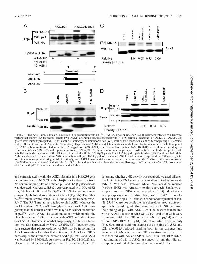
Our first objective was to determine which domain(s) ofASK1 plays a role in association with p21Cip1. Rh30/p21 orRh30/�NLSp21 cells, engineered to stably express full-lengthp21 or �NLSp21 (amino acids [aa] 1 to 140) lacking the nu-clear localization sequence under a metallothionein promoter(25) were infected with replication-deficient adenovirus ex-pressing WT, HA-tagged ASK1, or mutants with deletions inthe N or C terminus. Adenovirus-infected cells were inducedwith Zn for 24 h to allow the expression of p21Cip1 and �NLSp21Cip1. The results (Fig. 1A) show that both full-lengthp21Cip1 and �NLS p21Cip1 immunoprecipitated with ASK1 andASK1 peptides lacking either the N or C terminus, as deter-mined by anti-HA or by using a monoclonal antibody against aC-terminal epitope (aa 1076 to 1375) of ASK1. These resultsindicate that neither the N terminus nor the C terminus wasinvolved in p21Cip1 association but suggest that the overlappingregion, the ASK1 kinase domain (aa 678 to 936), may beinvolved in association of ASK1 with p21Cip1. To further definethe region of ASK1 that interacts with p21Cip1, we generatedplasmids that would encode small regions of the N- or C-terminal regions of ASK1 as well as point mutants in the kinase
domain. Most of these ASK1 deletion constructs failed to beexpressed at a detectable level in Rh30 or HEK293T cells;however, as shown in Fig. 1B, the N-terminal fragment ofASK1 (aa 1 to 672), which is readily detected, fails to bindp21Cip1. In addition, the K709R mutation that abrogates ASK1kinase activity substantially abolished their association (Fig.1B). This ASK1-K709R mutant lacks kinase activity and hasdominant-negative activity (26). We wondered, therefore,whether binding of p21Cip1 to ASK1 was disrupted by thismutation or whether ASK1 kinase activity was required forassociation.
To distinguish between these possibilities we generated sev-eral ASK1 kinase domain point or double point mutations andexpressed these in 293T cells. In vitro kinase assays for ASK1activity showed that the GT689/690AA mutant had markedlydecreased kinase activity, whereas no activity was detected forthe R802T mutant (Fig. 1C). Mutation at the ATP binding site(GT689/690AA) or at the catalytic site (R802T) of ASK1 es-sentially abolished its association with p21Cip1 (Fig. 1D). Incontrast, the Y792F mutant retained some kinase activity(�35% relative to ASK1-WT), consistent with decreasedp21Cip1 binding activity (�24% binding relative to ASK1-WT).Thus, ASK1 mutants with diminished kinase activity appear tobind less readily to p21Cip1, suggesting that ASK1 activity isimportant in its association with p21Cip1.
The JNK inhibitor SP600125 disrupts the association ofASK1 and p21Cip1. ASK1 is a MAPKKK and phosphorylatesand activates a subgroup of MAPKK, such as MKK3/6, andMKK4/7 kinases upstream of p38MAPK and JNK, respectively(8, 19, 27, 55). We hypothesized that either ASK1 or a kinasedownstream modified p21Cip1 and that this modification me-diated the interaction of p21Cip1 with ASK1. To test whetherJNK or p38 plays any role in the association, we used specifickinase inhibitors in the association experiments. HEK293Tcells were transfected with expression plasmids encoding HA-ASK1 and �NLS p21Cip1. Cells were exposed to SP600125(JNK inhibitor) or SB203580 (p38 inhibitor) for 10 min priorto cell harvesting. WT ASK1, but not the K709R mutant,associated with �NLS p21Cip1. The JNK inhibitor (Fig. 2A)and not the p38 inhibitor (Fig. 2B) disrupted the association ofASK1 with p21Cip1. As a control we used a CDK4/6 inhibitor(PD332991) which did not affect the ASK1/p21Cip1 interaction(Fig. 2A). As shown in Fig. 2C, SP600125 (10 �M, 10 min)suppressed p-c-Jun. Taken at face value, the results indicatethat JNK activation is essential for the possible modification ofp21Cip1 and subsequent association with ASK1. However, thisassumes the specificity of SP600125 for inhibition of JNK.
Mutations in p21Cip1 around S98 modulate ASK1 associa-tion. To investigate the possible role of JNK in the p21Cip1/ASK1 association, we generated p21Cip1 mutations (T57A andS130A) at reported JNK phosphorylation sites in the �NLSp21construct (32). However, we did not see any effect of these twomutations on association of these proteins (data not shown). Inlight of this result, we considered two possibilities: that a kinaseother than JNK modulates p21/ASK1 association (e.g., ASK1)or that there is an unknown JNK phosphorylation site(s) inp21Cip1, in addition to T57 and S130, that may be important forp21/ASK1 interactions.
The sequence of p21Cip1 indicates a putative JNK phosphory-lation site at S98. Consequently, we generated �NLSp21S98A
3532 ZHAN ET AL. MOL. CELL. BIOL.
on Septem
ber 2, 2014 by guesthttp://m
cb.asm.org/
Dow
nloaded from
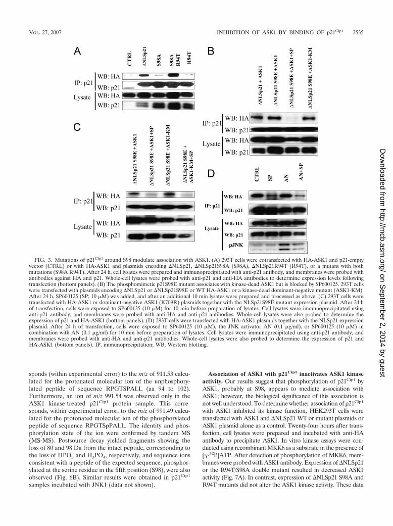
and cotransfected it with HA-ASK1 plasmids into HEK293 cellsor cotransfected �NLSp21 with HA--galactosidase (control).No coimmunoprecipitation between p21 and HA--galactosidasewas detected, whereas �NLSp21 coprecipitated with HA-ASK1(Fig. 3A, lanes CTRL and �NLSp21). The S98A mutation almostcompletely abolished association with ASK1 (Fig. 3A). Two otherp21Cip1 mutants were tested, R94T and a double mutant, S98A/R94T. The R94T mutant also failed to bind ASK1, whereas thedouble mutant (S98A/R94T) strongly associated with ASK1, sug-gesting that the domain around S98 may be critical for associationof p21Cip1 with ASK1. The S98E mutation, which mimics thephosphorylation of S98, associates with ASK1 and also kinase-dead ASK1. However, somewhat surprisingly, this latter interac-tion was also abrogated by SP600125 (Fig. 3B). Together thesedata suggest that phosphorylation of S98 may be important forASK1 association but also that activation of ASK1 or JNK isnecessary, as the interaction between �NLS p21S98E and ASK1was blocked by SP600125. As shown in Fig. 3C, SP600125 alsoblocked the interaction of p2198E with kinase-dead ASK1. To
determine whether JNK activity was required, we used differentsmall interfering RNA constructs in an attempt to down-regulateJNK in 293T cells. However, while JNK2 could be reduced(�60%), JNK1 was refractory to this approach. Similarly, at-tempts to use the JNK-interacting peptide (6, 38) did not atten-uate phosphorylation of c-Jun. Also, jnk1�/� jnk2�/� double-knockout cells or jnk1�/� cells with conditional regulation of jnk2(28, 35, 44) were not available. We therefore used a differentapproach, by asking whether stimulation of JNK increasedthe binding of p21 with ASK1. 293T cells were transfectedwith HA-Ask1 together with �NLS p21 and after 24 h werestimulated with the JNK activator AN (0.1 �g/ml) with orwithout SP600125 (10 �M). AN stimulated JNK activity(Fig. 3D), but this did not increase the binding of ASK1 andp21. SP600125 reduced binding both in the absence andpresence of AN, even when JNK activation was greater incells treated with AN and SP600125. Thus, SP600125 inhib-ited binding of p21 to ASK1 at concentrations that did notcompletely inhibit AN-induced activation of JNKs.
FIG. 1. The ASK1 kinase domain is involved in its association with p21Cip1. (A) Rh30/p21 or Rh30/�NLSp21 cells were infected by adenoviralvectors that express HA-tagged full-length (WT-ASK1) or epitope-tagged constructs with N- or C-terminal deletions (�N-ASK1, �C-ASK1). Celllysates were immunoprecipated (IP) with anti-p21 antibody and immunoblotted (WB) with either a monoclonal antibody recognizing a C-terminalepitope (C-ASK1) or anti-HA or anti-p21 antibody. Expression of ASK1 and deletion mutants in whole-cell lysates is shown in the bottom panel.(B) 293T cells were transfected with the HA-tagged WT (ASK1-WT), the kinase-dead mutant (ASK1K709R), or a plasmid encoding theN-terminal 672 aa (ASK672) and a plasmid encoding �NLSp21. Cell lysates were immunoprecipated with anti-p21 antibody and probed withanti-HA antibody. Control cells (CTRL) were tranfected with the �NLSp21 plasmid and HA-tagged -galactosidase. (C) Mutations that inhibitASK1 catalytic activity also reduce ASK1 association with p21. HA-tagged WT or mutant ASK1 constructs were expressed in 293T cells. Lysateswere immunoprecipitated using anti-HA antibody, and ASK1 kinase activity was determined in vitro using the MKK6 peptide as a substrate.(D) 293T cells were cotransfected with the �NLSp21 plasmid together with plasmids encoding HA-tagged WT or mutant ASK1. The associationof ASK1 with p21Cip1 was determined as described above.
VOL. 27, 2007 INHIBITION OF ASK1 BY BINDING OF p21Cip1 3533
on Septem
ber 2, 2014 by guesthttp://m
cb.asm.org/
Dow
nloaded from

ASK1 phosphorylates p21Cip1 in vitro. While ASK1 has notbeen reported to phosphorylate p21Cip1, we found that p21Cip1
can be phosphorylated in vitro by recombinant ASK1 kinase(Fig. 4A). Interestingly, this phosphorylation was inhibited bythe JNK-specific inhibitor SP600125 but was not significantlyinhibited by the p38 inhibitor SB203580. ASK1 immunopre-cipitated from HEK293T cell lysates also phosphorylated re-combinant p21Cip1 in vitro, whereas lysates from cells trans-fected with ASK-KM, a dominant-negative kinase-inactivemutant, had far less activity. As a control we used recombinantp27Kip1, which is not phosphorylated by ASK1 (Fig. 4B). Invitro SP600125 potently inhibited phosphorylation of bothMKK6 substrate and p21Cip1 by recombinant ASK1 at concen-trations as low as 0.1 �M (Fig. 4C). Thus, the effect ofSP600125 inhibiting ASK1/p21Cip1 binding in vivo could bethrough inhibition of either ASK1 or JNK.
ASK1 phosphorylates �NLS p21 in vivo. To determinewhether in vivo S98 of �NLSp21 can be phosphorylated,HEK293T cells were transiently transfected with ASK1 orJNK1 plasmids and coexpressed with �NLS p21Cip1 WT or thep21S98E mutant. Twenty-four hours after transfection, cellswere washed with phosphate-free medium and incubated with[32P]orthophosphate in phosphate-free medium overnight.Cell lysates were immunoprecipitated with p21Cip1 polyclonalantibody, and incorporated radiolabeled material in p21Cip1
was quantified (density of the 32P image/p21 Western blotimage). As shown in Fig. 4D, p21Cip1 is phosphorylated byASK1 in vivo, although the signal is quite low. There is somereduction in phosphorylation of the p21S98E mutant by ASK1,indicating that S98 may be a predominant but not exclusivephosphorylation site for ASK1 in vivo. Very little �NLSp21Cip1
was phosphorylated in cells overexpressing JNK1.To further test whether JNK phosphorylates p21Cip1 in vivo,
we expressed the WT ASK1 or dominant-negative ASK1-KMmutant (K709R) together with �NLS p21Cip1 in 293T cells.Thus, where the dominant-negative ASK-KM mutant is ex-pressed, only JNK and not ASK1 is activated when cells aretreated with AN. As shown in Fig. 4E, AN potently activatesJNK isoenzymes. However, there was no increase of phosphor-ylation on p21Cip1, indicating that JNK may not play a role in
phosphorylation of p21Cip1. This is also consistent with otherresults, where we did not find any detectable association ofJNK with p21Cip1 in coprecipitation assays (data not shown). Incontrast, p21Cip1 was phosphorylated, albeit at a low level, incells transfected with ASK1 irrespective of AN treatment.
Rapamycin induces endogenous ASK1-p21Cip1 interaction.Previously, we demonstrated that rapamycin activated ASK1 inrhabdomyosarcoma cells and that this resulted in associationwith p21Cip1 when overexpressed. To determine whether rapa-mycin induced endogenous ASK1 activation and associationwith endogenous p21Cip1, we treated 293T cells with rapamycinfor 1 h and immunoprecipitated endogenous p21Cip1. Al-though levels of p21Cip1 are very low in these cells, ASK1coprecipitated only in the presence of rapamycin. Consistentwith previous studies, rapamycin induced phosphorylation ofc-Jun (Fig. 5A). Rapamycin also activated ASK1, as deter-mined by in vitro kinase assay using recombinant p21Cip1 as asubstrate (Fig. 5B). Consistent with previously collected data(24), rapamycin (10 ng/ml) also induced prolonged activationof ASK1 in 293T cells which have low p21Cip1 but induced onlytransient activation in these cells when transfected with full-length p21Cip1 plasmid (Fig. 5C). The latter result is consistentwith our previous observation of suppression of the rapamycin-induced stress response (p-c-Jun) in cells with high p21ip1 ex-pression (24). Further, as shown in Fig. 5D, rapamycin inducesprolonged activation of ASK1 in p21�/� mouse embryo fibro-blasts (MEFs). p21�/� MEFs were treated with rapamycin forup to 2 h, and ASK1 was immunoprecipitated and used in anin vitro kinase reaction with recombinant p21. These resultsare consistent with previous studies showing that prolongedactivation of ASK1 induced by rapamycin occurred only in cellswith mutated p53 or deficient in p21cip1 (24). Consistent withASK1 activation, phosphorylation of c-Jun was increased byrapamycin treatment (Fig. 5E).
Determination of p21Cip1 phosphorylation at S98 by MSanalysis. Phosphorylation of p21Cip1 at S98 was investigated byMS characterization of chymotrypsin-trypsin peptides derivedfrom the kinase-treated recombinant p21Cip1 protein. Phos-phorylation at S98 was confirmed based on the following: anion signal of m/z 911.64 was observed (Fig. 6A) which corre-
FIG. 2. The JNK1 kinase inhibitor SP600125 disrupts the association of ASK1 and p21Cip1. (A) 293T cells were transfected with HA-ASK1 ordominant-negative ASK1 (ASK1K709R) plasmids together with the �NLSp21 expression plasmid, �NLSp21, or HA-tagged -galactosidase(CTRL). After 24 h, cells were exposed to SP600125 (10 �M) or a CDK4/6 inhibitor (PD332991 [PD]; 10 �M) for 10 min before preparation oflysates. Cell lysates were immunoprecipitated using anti-p21 antibody, and membranes were probed with anti-HA and anti-p21 antibodies.Whole-cell lysates were also probed to determine the expression of p21 and HA-ASK1 (bottom panels). (B) Cells were prepared as above butexposed to the p38 inhibitor SB203580 (SB; 10 �M) for 10 min prior to lysis. The results show that SB203580 does not block the association ofp21 with ASK1. (C) Cells were exposed to increasing concentrations of SP600125 (10 min), and the levels of phosphorylated c-Jun in cell lysateswere determined. -Tubulin was used to normalize protein loading. IP, immunoprecipitation; WB, Western blotting.
3534 ZHAN ET AL. MOL. CELL. BIOL.
on Septem
ber 2, 2014 by guesthttp://m
cb.asm.org/
Dow
nloaded from
sponds (within experimental error) to the m/z of 911.53 calcu-lated for the protonated molecular ion of the unphosphory-lated peptide of sequence RPGTSPALL (aa 94 to 102).Furthermore, an ion of m/z 991.54 was observed only in theASK1 kinase-treated p21Cip1 protein sample. This corre-sponds, within experimental error, to the m/z of 991.49 calcu-lated for the protonated molecular ion of the phosphorylatedpeptide of sequence RPGTSpPALL. The identity and phos-phorylation state of the ion were confirmed by tandem MS(MS-MS). Postsource decay yielded fragments showing theloss of 80 and 98 Da from the intact peptide, corresponding tothe loss of HPO3 and H3PO4, respectively, and sequence ionsconsistent with a peptide of the expected sequence, phosphor-ylated at the serine residue in the fifth position (S98), were alsoobserved (Fig. 6B). Similar results were obtained in p21Cip1
samples incubated with JNK1 (data not shown).
Association of ASK1 with p21Cip1 inactivates ASK1 kinaseactivity. Our results suggest that phosphorylation of p21Cip1 byASK1, probably at S98, appears to mediate association withASK1; however, the biological significance of this association isnot well understood. To determine whether association of p21Cip1
with ASK1 inhibited its kinase function, HEK293T cells weretransfected with ASK1 and �NLSp21 WT or mutant plasmids orASK1 plasmid alone as a control. Twenty-four hours after trans-fection, cell lysates were prepared and incubated with anti-HAantibody to precipitate ASK1. In vitro kinase assays were con-ducted using recombinant MKK6 as a substrate in the presence of[�-32P]ATP. After detection of phosphorylation of MKK6, mem-branes were probed with ASK1 antibody. Expression of �NLSp21or the R94T/S98A double mutant resulted in decreased ASK1activity (Fig. 7A). In contrast, expression of �NLSp21 S98A andR94T mutants did not alter the ASK1 kinase activity. These data
FIG. 3. Mutations of p21Cip1 around S98 modulate association with ASK1. (A) 293T cells were cotransfected with HA-ASK1 and p21-emptyvector (CTRL) or with HA-ASK1 and plasmids encoding �NLSp21, �NLSp21S98A (S98A), �NLSp21R94T (R94T), or a mutant with bothmutations (S98A R94T). After 24 h, cell lysates were prepared and immunoprecipitated with anti-p21 antibody, and membranes were probed withantibodies against HA and p21. Whole-cell lysates were probed with anti-p21 and anti-HA antibodies to determine expression levels followingtransfection (bottom panels). (B) The phosphomimetic p21S98E mutant associates with kinase-dead ASK1 but is blocked by SP600125. 293T cellswere transfected with plasmids encoding �NLSp21 or �NLSp21S98E or WT HA-ASK1 or a kinase-dead dominant-negative mutant (ASK1-KM).After 24 h, SP600125 (SP; 10 �M) was added, and after an additional 10 min lysates were prepared and processed as above. (C) 293T cells weretransfected with HA-ASK1 or dominant-negative ASK1 (K709R) plasmids together with the NLSp21S98E mutant expression plasmid. After 24 hof transfection, cells were exposed to SP600125 (10 �M) for 10 min before preparation of lysates. Cell lysates were immunoprecipitated usinganti-p21 antibody, and membranes were probed with anti-HA and anti-p21 antibodies. Whole-cell lysates were also probed to determine theexpression of p21 and HA-ASK1 (bottom panels). (D) 293T cells were transfected with HA-ASK1 plasmids together with the NLSp21 expressionplasmid. After 24 h of transfection, cells were exposed to SP600125 (10 �M), the JNK activator AN (0.1 �g/ml), or SP600125 (10 �M) incombination with AN (0.1 �g/ml) for 10 min before preparation of lysates. Cell lysates were immunoprecipitated using anti-p21 antibody, andmembranes were probed with anti-HA and anti-p21 antibodies. Whole-cell lysates were also probed to determine the expression of p21 andHA-ASK1 (bottom panels). IP, immunoprecipitation; WB, Western blotting.
VOL. 27, 2007 INHIBITION OF ASK1 BY BINDING OF p21Cip1 3535
on Septem
ber 2, 2014 by guesthttp://m
cb.asm.org/
Dow
nloaded from

are consistent with the coimmunoprecipitation results, in whichS98A and R94T mutants did not associate with ASK1 and did notinhibit ASK1 kinase activity, while the double mutant R94T/S98A, which restores the association with ASK1, also restores theability to inactivate ASK1 kinase activity.
A region of p21Cip1 (aa 94 to 98) modulates its interactionwith ASK1. p21Cip1 binds to several proteins, including CDKsand cyclins as well as PCNA (22, 58, 63), through well-defineddomains. We were interested, therefore, in identifying such adomain of p21Cip1 that mediates its association with ASK1.The kinase domain of ASK1 has significant homology to thekinase domains of cdk1 and cdk2. The domain of p21Cip1 thatinteracts with cdk’s maps to amino acid residues 49 to 80 (61).To determine whether this region also plays a role in associa-tion with ASK1, we generated a �NLSp21 mutant expressing apeptide with a small deletion at positions 53 to 58 (p21�53/8).As a control we expressed �NLSp21�120/5, a deletion mutantlacking residues 120 to 125. The results (Fig. 7B) show thatboth deletion mutants still interact with ASK1, while p21�53/8
does not associate with Cdk1. This indicates that differentregions of p21Cip1 are involved in interaction with cdk’s andASK1. We hypothesized that a region around residues 94 to 98may be involved in association of p21Cip1 with ASK1. To testthis idea we generated deletion mutants (�80-85, �94-98, and�108-112) around this region. The results show that the �94-98peptide did not coprecipitate with ASK1, whereas the othertwo deletions did not effect association with ASK1 (Fig. 7C).Therefore, a region (aa 94 to 98) of p21Cip1 appears to beessential for ASK1 association.
DISCUSSION
The multiple functions of p21Cip1 are determined by proteinpartners that form specific protein complexes and by its ownsubcellular localization. Nuclear p21Cip1 functions to inhibitcell cycle progression, while cytoplasmic p21Cip1 appears toprevent apoptosis. However, the mechanisms by which p21Cip1
can prevent cells from undergoing apoptosis and promote cell
FIG. 4. Ask1 phosphorylates p21Cip1 in vitro and in vivo. (A) Bacterially expressed purified p21Cip1 was incubated with recombinant ASK1 inthe presence of [�-32P]ATP with or without addition of SP600125 (10 �M) or SB203580 (10 �M). In vitro kinase assays were carried out asdescribed in Materials and Methods. (B) 293T cells were transfected with plasmids encoding HA-ASK1 or the epitope-tagged kinase-dead mutantASK-KM. After 24 h, cell lysates were prepared and immunoprecipitated using anti-ASK1 antibody. Immunoprecipitates were used in vitro tophosphorylate either purified p21 or purified p27Kip1. (C). The JNK inhibitor SP600125 potently inhibits ASK1-mediated in vitro phosphorylationof MKK6 or p21Cip1. Recombinant ASK1 was incubated with purified Mal-E-MKK6 peptide, a known substrate, or purified p21Cip1 in the presenceof [�-32P]ATP and increasing concentrations of SP600125 as described in Materials and Methods. (D) ASK1 can phosphorylate p21cip1 in vivo.293T cells were transfected with plasmids encoding �NLSp21 or its mutant S98E and cotransfected with ASK1 or JNK1 expression plasmids. TheJNK inhibitor SP600125 was added 10 min before harvesting of cells. Cell lysates were immunoprecipated using anti-p21Cip1 antibody, andphosphorylated p21Cip1 was detected using a PhosphorImager. The same membrane was blotted using antibodies against p21Cip1. Levels of ASK1and JNK1 were determined on whole-cell lysates (bottom panel). Relative activities for phosphorylation of �NLSp21 were calculated from the ratioof optical densities of the 32P-p21Cip1/p21Cip1 Western blot signals and normalized to that for �NLSp21. (E) ASK1 or ASK1-KM expressionplasmids were cotransfected together with the �NLSp21 plasmid into 293T cells. After 24 h, cells were either not stimulated or stimulated withAN (10 �g/ml), a known activator of JNK1 and -2, in the presence of [32P]orthophosphate. After 3 h, p21Cip1 was immunoprecipitated, andphosphorylated p21 was detected using a PhosphorImager. The membrane was reprobed using anti-p21 antibody. The lower panels show levelsof pJNK1, and the level of pJNK2 in whole-cell lysates was determined using anti-phospho-JNK antibody. Relative activities for phosphorylationof �NLSp21 were calculated from the ratio of optical densities of the 32P-p21Cip1/p21Cip1 Western blot signals.
3536 ZHAN ET AL. MOL. CELL. BIOL.
on Septem
ber 2, 2014 by guesthttp://m
cb.asm.org/
Dow
nloaded from

survival are not well understood. Here, we provide data todemonstrate that p21Cip1 interacts with ASK1 and down-reg-ulates its kinase activity, thereby attenuating the stress re-sponse induced by inhibition of mTOR signaling by rapamycin.Our results indicate that the interaction of p21Cip1 with ASK1is mediated by a small region of p21Cip1 (residues 94 to 98) thatincludes S98 and that is phosphorylated by both Ask1 and JNKin vitro. Phosphorylation of p21Cip1 at S98, which in vivo ap-pears to be regulated by ASK1, may therefore mediate nega-tive feedback in the ASK1 signaling pathway.
The C terminus of p21Cip1 (residues 141 to 164) has severalimportant functions (57, 58); this region contains the NLS, andhence the �NLS p21Cip1 (aa 1 to 140) is predominantly cyto-plasmic (25, 45). We found that �NLS p21Cip1 has increasedassociation with ASK1 compared to wild-type full-lengthp21Cip1. Since ASK1 is localized in the cytoplasm (2), it isreasonable that �NLS p21Cip1 has greater opportunity to in-teract with ASK1. Furthermore, the �NLS p21Cip1 lacks thesite that binds the C8 �-subunit of the 20S proteasome and
hence is more stable than the full-length p21Cip1. This may beanother reason why �NLS p21Cip1 shows increased associationwith ASK1.
As a cell cycle regulator, p21Cip1 has been shown to associatewith CDKs (22) (63) through their kinase domains. The highsimilarity between CDK1, CDK2, and ASK1 kinase domains(over 40% alignment) suggests that ASK1 may also use itskinase domain to interact with p21Cip1. Our data do demon-strate that the kinase domain of ASK1 is involved in its inter-action with p21Cip1. Since p21Cip1 has a very flexible structureand performs a variety of functions (34), it is possible thatp21Cip1 interacts with other kinases via their similar kinasedomains, affecting their biological function. For example, it hasbeen shown that p21Cip1 associates with JNK and affects itskinase activity (64). p21Cip1 also forms a complex with Rhokinase and inhibits its activity in vivo and in vitro (51).
We observed that not only the kinase domain per se but alsothe kinase activity of ASK1 is required for its association withp21Cip1. This suggests that ASK1 or a kinase(s) downstream
FIG. 5. Rapamycin treatment induces ASK1 activation and binding of p21Cip1. (A) 293T cells were treated with rapamycin (100 ng/ml) for 1 h,and lysates were prepared, immunoprecipitated (IP) (anti-p21 antibody), and processed as described in Materials and Methods. In the presenceof rapamycin, ASK1 coimmunoprecipitated with p21Cip1, and rapamycin also induced phosphorylation of c-Jun. WB, Western blotting. (B) 293Tcells were treated with rapamycin (100 ng/ml) for 1 h, and ASK1 was immunoprecipitated and incubated with recombinant p21Cip1 in the presenceof [�-32P]ATP. In vitro kinase assays were carried out as described in Materials and Methods. (C) 293T cells (left) or cells transfected with the p21plasmid (right) were exposed to rapamycin (10 ng/ml) for up to 2 h. ASK1 was immunoprecipitated, and the activity in phosphorylatingrecombinant p21Cip1 was assessed in vitro. ASK1 activity relative to the control is quantitated in the histogram. (D) Rapamycin (Rap) inducesprolonged ASK1 activation in the absence of p21. p21 null MEFs were treated with rapamycin (100 ng/ml) for various lengths of time, from 0 to2 h, in 10% fetal calf serum medium. Cell lysates were immunoprecipitated with ASK1 antibody. Bacterially expressed purified p21 as ASK1 kinasesubstrate was incubated with immunoprecipitated ASK1 in the presence of [32P]ATP. In vitro kinase assays were carried out as described inMaterials and Methods. (E) The activation of ASK1 was also confirmed by the increased phosphorylation of c-Jun by rapamycin treatment. Thesame cell lysate was subjected to Western blotting by p-c-Jun and c-Jun.
VOL. 27, 2007 INHIBITION OF ASK1 BY BINDING OF p21Cip1 3537
on Septem
ber 2, 2014 by guesthttp://m
cb.asm.org/
Dow
nloaded from

FIG. 6. Determination of phosphorylation of p21(S98) by MS. Phosphorylation of S98 in p21Cip1 protein was identified by MS characterizationof chymotrypsin-trypsin peptides derived from the ASK1-treated recombinant p21Cip1 protein. MS and MS-MS spectra were obtained on a model4700 MALDI-tandem TOF spectrometer. MS (A) and MS-MS (B) spectra derived from the singly charged unphosphorylated tryptic peptideRPGTSPALL (panel A, upper spectrum) and the phosphorylated tryptic peptide RPGTpSPALL (panel A, center spectrum) are shown. (B) Theamino acid sequence from the peptide could be identified from the b and y ion fragment peaks.
3538 ZHAN ET AL. MOL. CELL. BIOL.
on Septem
ber 2, 2014 by guesthttp://m
cb.asm.org/
Dow
nloaded from

modifies p21Cip1 and that the modification is necessary forp21Cip1-ASK1 association. It is known that ASK1 can activateJNK and p38 (8, 27, 55). To determine which kinase(s) isessential for association of p21Cip1 with ASK1, we examinedtheir association in the presence of JNK and p38 inhibitors. Wefound that the JNK inhibitor SP600125 almost completely in-hibited p21Cip1 binding to ASK1. The immediate suggestionfrom these data is that JNK modifies p21Cip1 and leads to itsassociation with ASK1. SP600125 is a reversible ATP compet-itive inhibitor with more than 20-fold greater selectivity forJNK than a range of kinases and other enzymes (4). However,our data show that SP600125 can also potently inhibit recom-binant ASK1 activity both in vitro with MKK6 or p21Cip1 as asubstrate and also in vivo. Consequently, phosphorylation ofp21Cip1 by ASK1 may also mediate the association of theseproteins.
ASK1 is a member of the MAPKKK family, and its normalsubstrate is a subgroup of MAPKKs. The present study repre-sents, to our knowledge, the first time that a MAPKKK/ASK1has been shown to phosphorylate p21Cip1. In contrast, we didnot observe phosphorylation of recombinant p27Kip1 in vitro.Using purified p21Cip1 as a substrate in the in vitro kinaseassay, we found several 32P-labeled bands, perhaps indicatingthat there is more than one ASK1 phosphorylation site besidesS98. Furthermore, rapamycin treatment activated ASK1,which phosphorylated p21Cip1 in vitro. Using MS we identifiedS98 phosphorylation but failed to identify additional sites afterin vitro kinase reactions with recombinant ASK1 or JNK1.Several kinases have been reported to phosphorylate p21Cip1
and mediate important biological processes. For example,phosphorylation of T145 by AKT leads to cytoplasmic local-ization and switches p21Cip1 function from cell cycle inhibitionto protection from apoptosis (65).
A mutant that mimics phosphorylation at S98, p21S98E,associates with ASK1 in coimmunoprecipitation assays; how-ever, it was unexpected that the JNK inhibitor SP600125 wouldalso disrupt this association. It is intriguing that a relativelyshort exposure to SP600125 results in dissociation of p21Cip
from ASK1 that may be a consequence of inhibitor-inducedconformational change. This may account for the observationthat SP600125 blocked the association of the p21R94T/S98Amutant with ASK1 and similarly blocked association of thep21S98E mutant with kinase-dead ASK1.
Even though in vitro JNK1 can phosphorylate p21Cip1 atS98, our data do not support JNKs as the predominant in vivokinases. Overexpression of JNK1, or activation by AN, failedto increase p21Cip1 phosphorylation in vivo. Furthermore,SP600125 inhibited p21-ASK1 association at concentrationsthat did not completely block AN-induced activation of JNK.To unambiguously define whether JNK played a role in p21S98phosphorylation in vivo, we attempted to down-regulateJNK1/2 using small interfering RNA or block JNK activationusing a peptide that prevents JNK from binding to JNK-inter-acting peptide (6, 38), but we were unable to modify JKNactivity in either 293T or 3T3 cells. jnk1�/� jnk2�/� double-knockout (35, 44) or jnk1�/� cells with conditional jnk2 (28)were not available for these experiments. Single mutations atother JNK consensus sites (T57, S130) did not decreasep21Cip1-ASK1 interactions (data not shown). However, a mu-tation that blocks phosphorylation of S98 (S98A) severely at-tenuates binding of p21Cip1 to ASK1. These data, together withthose for the S98E mutant, strongly implicate phosphorylationof p21S98 as being important for binding of WT p21 to ASK1.
Rather surprisingly, the p21R94T mutation restored theability of the S98A mutant to bind ASK1. This suggested thata region around residues 94 to 98 could be important in this
FIG. 7. The association of ASK1 with p21Cip1 inactivates ASK1 kinase activity and requires a domain (aa 94 to 98) for association. (A) 293Tcells were transfected with epitope-tagged ASK1 or ASK1-KM alone or ASK1 cotransfected with �NLSp21, �NLSp21S98A, �NLSp211R94T, orthe double mutant. Cell lysates were immunoprecipated using anti-HA antibody, and precipitated samples were used for in vitro kinase assays withpurified MKK6 as the substrate. The phosphorylation of MKK6 was detected using a PhosphorImager. The same membrane was probed usinganti-HA antibody. WB, Western blotting. (B) 293T cells were either mock transfected (CTRL) or cotransfected with HA-ASK1 and WT �NLSp21or �NLSp21 deletion mutants (�53-58, lacking the cdk interaction domain) or a control mutant (�120-125). After 24 h, cell lysates were prepared,and p21 was immunoprecipitated (IP). Membranes were probed with antibodies against p21, Cdk1, and HA. The lower panel shows levels ofHA-ASK1 and p21 in whole-cell lysates. (C) Identification of the region of p21 involved in association with ASK1. 293T cells were cotransfectedwith HA-ASK1 and �NLSp21 or �NLSp21 deletion mutants (�80-85, �94-98, or �108-112). After 24 h, cell lysates were prepared, and p21 wasimmunoprecipitated. Membranes were probed with anti-p21 and anti-HA antibodies. The lower panel shows levels of HA-ASK1 and p21 inwhole-cell lysates. The association of ASK1 with �NLSp21 or mutants was derived from the ratio of HA to p21 signal in the Western analyses andnormalized to the ratio of ASK1 to �NLSp21.
VOL. 27, 2007 INHIBITION OF ASK1 BY BINDING OF p21Cip1 3539
on Septem
ber 2, 2014 by guesthttp://m
cb.asm.org/
Dow
nloaded from

association. To narrow the domain of p21Cip1 involved in theinteraction with ASK1, we produced a series of p21Cip1 mu-tants with small deletions (Fig. 6). As a positive control weintroduced deletions into the cdk binding domain (�53-58) andshowed that it failed to associate with Cdk1 in coprecipitationassays. Deletion of a small sequence around S98 (�94-98)significantly (90%) decreased binding to ASK1, whereasother deletions (�80-85, �108-112) had no observable effect.
Our results suggest that in vivo ASK1 phosphorylatesp21Cip1 at S98 and that this phosphorylation is in part neces-sary for its association with ASK1. However, the finding thatthe p21R94T/S98A binds strongly to ASK1 suggests additionalcomplexities, and further experimentation will be required tofully elucidate the molecular detail of association of theseproteins. We next questioned the biological significance ofp21Cip1-ASK1 association. We found that binding of p21Cip1
inactivates ASK1 kinase activity. In contrast, ASK1 remainedactive in rapamycin-treated p21Cip1 null MEFs. These data areconsistent with our previous finding that suppression of rapa-mycin-induced activation of ASK1 correlated with p21Cip1
binding (25). The details of the mechanism by which p21Cip1
inactivates ASK1 kinase activity are not completely under-stood. Possibly, p21Cip1 may mask the kinase domain and pre-vent its activation of downstream kinases, such as MKK3/6 orMKK4/7. We observed that only a relatively small fraction ofASK1 associated with p21Cip1. We have shown previously (26)that rapamycin treatment leads to a rapid suppression of theactivity of protein phosphatase 5 (PP5) that is physically asso-ciated with ASK1 and dissociation of the PP2A� regulatorysubunit (PR72) from the ASK1 complex. One possibility is thatp21Cip1 stabilizes this complex, maintaining PP5 activity, andnegatively regulates phosphorylation of T838 and T845 ofASK1 and its kinase activity. Alternatively, p21Cip1 binding toASK1 may recruit another PP. This possibility is under inves-tigation.
The role of p21Cip1 in attenuating apoptosis induced byvarious stimuli is well established. Results from many studiessuggest that protection of cells is a consequence of cell cyclearrest, potentially allowing for damage repair in the case ofDNA-damaging stimuli. Interestingly, rapamycin, a specific in-hibitor of mTOR, in some cells blocks the translation ofp21Cip1 subsequent to DNA damage induced by agents such ascisplatin, selectively potentiating cytotoxicity in cells with func-tional p53/p21Cip1 (5, 64). Our previous studies using cell lineswith mutations of p53 showed that inhibition of mTOR re-sulted in a rapid and sustained activation of ASK1 and JNK,increased p-c-Jun, and apoptosis. In cells expressing WT p53this activation was rapidly suppressed, and suppression wasp21Cip1 dependent. Thus, p21Cip1 may protect cells from DNAdamage by inducing cell cycle arrest and by suppressing stressresponses caused by nutritional deprivation: for example,amino acid starvation that is mimicked by rapamycin treat-ment.
ACKNOWLEDGMENTS
Limin Xiao is acknowledged for assistance preparing p21-S98E forthese studies.
This work was supported by Public Health Service awards CA77776(P.J.H.), CA96996 (P.J.H.), and CA82491 (R.W.K.); grant CA21785(Cancer Center Support Grant) from the National Cancer Institute;and American, Lebanese, Syrian Associated Charities (ALSAC).
REFERENCES
1. Adachi, S., H. Ito, M. Tamamori-Adachi, Y. Ono, T. Nozato, S. Abe, M.Ikeda, F. Marumo, and M. Hiroe. 2001. Cyclin A/cdk2 activation is involvedin hypoxia-induced apoptosis in cardiomyocytes. Circ. Res. 88:408–414.
2. Asada, M., T. Yamada, H. Ichijo, D. Delia, K. Miyazono, K. Fukumuro, andS. Mizutani. 1999. Apoptosis inhibitory activity of cytoplasmic p21(Cip1/WAF1) in monocytic differentiation. EMBO J. 18:1223–1234.
3. Barboule, N., P. Mazars, V. Baldin, S. Vidal, S. Jozan, P. Martel, and A.Valette. 1995. Expression of p21WAF1/CIP1 is heterogeneous and unrelatedto proliferation index in human ovarian carcinoma. Int. J. Cancer 63:611–615.
4. Bennett, B. L., D. T. Sasaki, B. W. Murray, E. C. O’Leary, S. T. Sakata, W.Xu, J. C. Leisten, A. Motiwala, S. Pierce, Y. Satoh, S. S. Bhagwat, A. M.Manning, and D. W. Anderson. 2001. SP600125, an anthrapyrazolone inhib-itor of Jun N-terminal kinase. Proc. Natl. Acad. Sci. USA 98:13681–13686.
5. Beuvink, I., A. Boulay, S. Fumagalli, F. Zilbermann, S. Ruetz, T. O’Reilly, F.Natt, J. Hall, H. A. Lane, and G. Thomas. 2005. The mTOR inhibitorRAD001 sensitizes tumor cells to DNA-damaged induced apoptosis throughinhibition of p21 translation. Cell 120:747–759.
6. Bonny, C., A. Oberson, S. Negri, C. Sauser, and D. F. Schorderet. 2001.Cell-permeable peptide inhibitors of JNK: novel blockers of beta-cell death.Diabetes 50:77–82.
7. Boulaire, J., A. Fotedar, and R. Fotedar. 2000. The functions of the cdk-cyclin kinase inhibitor p21WAF1. Pathol. Biol. (Paris) 48:190–202.
8. Chang, H. Y., H. Nishitoh, X. Yang, H. Ichijo, and D. Baltimore. 1998.Activation of apoptosis signal-regulating kinase 1 (ASK1) by the adapterprotein Daxx. Science 281:1860–1863.
9. Cheng, T., N. Rodrigues, H. Shen, Y. Yang, D. Dombkowski, M. Sykes, andD. T. Scadden. 2000. Hematopoietic stem cell quiescence maintained byp21cip1/waf1. Science 287:1804–1808.
10. Coqueret, O., and H. Gascan. 2000. Functional interaction of STAT3 tran-scription factor with the cell cycle inhibitor p21WAF1/CIP1/SDI1. J. Biol.Chem. 275:18794–18800.
11. Delavaine, L., and N. B. La Thangue. 1999. Control of E2F activity byp21Waf1/Cip1. Oncogene 18:5381–5392.
12. Dotto, G. P. 2000. p21(WAF1/Cip1): more than a break to the cell cycle?Biochim. Biophys. Acta 1471:M43–M56.
13. Erber, R., W. Klein, T. Andl, C. Enders, A. I. Born, C. Conradt, J. Bartek,and F. X. Bosch. 1997. Aberrant p21(CIP1/WAF1) protein accumulation inhead-and-neck cancer. Int. J. Cancer 74:383–389.
14. Estanyol, J. M., M. Jaumot, O. Casanovas, A. Rodriguez-Vilarrupla, N.Agell, and O. Bachs. 1999. The protein SET regulates the inhibitory effect ofp21(Cip1) on cyclin E-cyclin-dependent kinase 2 activity. J. Biol. Chem.274:33161–33165.
15. Esteve, V., N. Canela, A. Rodriguez-Vilarrupla, R. Aligue, N. Agell, I. Min-garro, O. Bachs, and E. Perez-Paya. 2003. The structural plasticity of the Cterminus of p21Cip1 is a determinant for target protein recognition. Chem-biochem 4:863–869.
16. Funk, J. O., S. Waga, J. B. Harry, E. Espling, B. Stillman, and D. A.Galloway. 1997. Inhibition of CDK activity and PCNA-dependent DNAreplication by p21 is blocked by interaction with the HPV-16 E7 oncoprotein.Genes Dev. 11:2090–2100.
17. Gorospe, M., C. Cirielli, X. Wang, P. Seth, M. C. Capogrossi, and N. J.Holbrook. 1997. p21(Waf1/Cip1) protects against p53-mediated apoptosis ofhuman melanoma cells. Oncogene 14:929–935.
18. Gorospe, M., X. Wang, K. Z. Guyton, and N. J. Holbrook. 1996. Protectiverole of p21(Waf1/Cip1) against prostaglandin A2-mediated apoptosis ofhuman colorectal carcinoma cells. Mol. Cell. Biol. 16:6654–6660.
19. Gotoh, Y., and J. A. Cooper. 1998. Reactive oxygen species- and dimeriza-tion-induced activation of apoptosis signal-regulating kinase 1 in tumor ne-crosis factor-alpha signal transduction. J. Biol. Chem. 273:17477–17482.
20. Gotz, C., P. Wagner, O. G. Issinger, and M. Montenarh. 1996. p21WAF1/CIP1 interacts with protein kinase CK2. Oncogene 13:391–398.
21. Gulbis, J. M., Z. Kelman, J. Hurwitz, M. O’Donnell, and J. Kuriyan. 1996.Structure of the C-terminal region of p21(WAF1/CIP1) complexed withhuman PCNA. Cell 87:297–306.
22. Harper, J. W., G. R. Adami, N. Wei, K. Keyomarsi, and S. J. Elledge. 1993.The p21 Cdk-interacting protein Cip1 is a potent inhibitor of G1 cyclin-dependent kinases. Cell 75:805–816.
23. Harvey, K. J., D. Lukovic, and D. S. Ucker. 2000. Caspase-dependent Cdkactivity is a requisite effector of apoptotic death events. J. Cell Biol. 148:59–72.
24. Huang, S., L. N. Liu, H. Hosoi, M. B. Dilling, T. Shikata, and P. J. Houghton.2001. p53/p21(CIP1) cooperate in enforcing rapamycin-induced G(1) arrestand determine the cellular response to rapamycin. Cancer Res. 61:3373–3381.
25. Huang, S., L. Shu, M. B. Dilling, J. Easton, F. C. Harwood, H. Ichijo, and P. J.Houghton. 2003. Sustained activation of the JNK cascade and rapamycin-induced apoptosis are suppressed by p53/p21(Cip1). Mol. Cell 11:1491–1501.
26. Huang, S., L. Shu, J. Easton, F. C. Harwood, G. S. Germain, H. Ichijo, andP. J. Houghton. 2004. Inhibition of mammalian target of rapamycin activates
3540 ZHAN ET AL. MOL. CELL. BIOL.
on Septem
ber 2, 2014 by guesthttp://m
cb.asm.org/
Dow
nloaded from

apoptosis signal-regulating kinase 1 signaling by suppressing protein phos-phatase 5 activity. J. Biol. Chem. 279:36490–36496.
27. Ichijo, H., E. Nishida, K. Irie, P. ten Dijke, M. Saitoh, T. Moriguchi, M.Takagi, K. Matsumoto, K. Miyazono, and Y. Gotoh. 1997. Induction ofapoptosis by ASK1, a mammalian MAPKKK that activates SAPK/JNK andp38 signaling pathways. Science 275:90–94.
28. Jaeschke, A., M. Karasarides, J. J. Ventura, A. Ehrhardt, C. Zhang, R. A.Flavell, K. M. Shokat, and R. J. Davis. 2006. JNK2 is a positive regulator ofthe cJun transcription factor. Mol. Cell 23:899–911.
29. Jin, Y. H., K. J. Yoo, Y. H. Lee, and S. K. Lee. 2000. Caspase 3-mediatedcleavage of p21WAF1/CIP1 associated with the cyclin A-cyclin-dependentkinase 2 complex is a prerequisite for apoptosis in SK-HEP-1 cells. J. Biol.Chem. 275:30256–30263.
30. Kearsey, J. M., P. J. Coates, A. R. Prescott, E. Warbrick, and P. A. Hall.1995. Gadd45 is a nuclear cell cycle regulated protein which interacts withp21Cip1. Oncogene 11:1675–1683.
31. Kelman, Z., and M. O’Donnell. 1995. Structural and functional similarities ofprokaryotic and eukaryotic DNA polymerase sliding clamps. Nucleic AcidsRes. 23:3613–3620.
32. Kim, G. Y., S. E. Mercer, D. Z. Ewton, Z. Yan, K. Jin, and E. Friedman. 2002.The stress-activated protein kinases p38 alpha and JNK1 stabilize p21(Cip1)by phosphorylation. J. Biol. Chem. 277:29792–29802.
33. Kitaura, H., M. Shinshi, Y. Uchikoshi, T. Ono, S. M. Iguchi-Ariga, and H.Ariga. 2000. Reciprocal regulation via protein-protein interaction betweenc-Myc and p21(cip1/waf1/sdi1) in DNA replication and transcription. J. Biol.Chem. 275:10477–10483.
34. Kriwacki, R. W., L. Hengst, L. Tennant, S. I. Reed, and P. E. Wright. 1996.Structural studies of p21Waf1/Cip1/Sdi1 in the free and Cdk2-bound state:conformational disorder mediates binding diversity. Proc. Natl. Acad. Sci.USA 93:11504–11509.
35. Kuan, C. Y., D. D. Yang, D. R. Samanta Roy, R. J. Davis, P. Rakic, and R. A.Flavell. 1999. The Jnk1 and Jnk2 protein kinases are required for regionalspecific apoptosis during early brain development. Neuron 22:667–676.
36. LaBaer, J., M. D. Garrett, L. F. Stevenson, J. M. Slingerland, C. Sandhu,H. S. Chou, A. Fattaey, and E. Harlow. 1997. New functional activities for thep21 family of CDK inhibitors. Genes Dev. 11:847–862.
37. Lawlor, M. A., and P. Rotwein. 2000. Insulin-like growth factor-mediatedmuscle cell survival: central roles for Akt and cyclin-dependent kinase in-hibitor p21. Mol. Cell. Biol. 20:8983–8995.
38. Lee, Y. H., J. Giraud, R. J. Davis, and M. F. White. 2003. c-Jun N-terminalkinase (JNK) mediates feedback inhibition of the insulin signaling cascade.J. Biol. Chem. 278:2898–2902.
39. Levkau, B., H. Koyama, E. W. Raines, B. E. Clurman, B. Herren, K. Orth,J. M. Roberts, and R. Ross. 1998. Cleavage of p21Cip1/Waf1 and p27Kip1mediates apoptosis in endothelial cells through activation of Cdk2: role of acaspase cascade. Mol. Cell 1:553–563.
40. Liu, H., H. Nishitoh, H. Ichijo, and J. M. Kyriakis. 2000. Activation ofapoptosis signal-regulating kinase 1 (ASK1) by tumor necrosis factor recep-tor-associated factor 2 requires prior dissociation of the ASK1 inhibitorthioredoxin. Mol. Cell. Biol. 20:2198–2208.
41. Ono, T., H. Kitaura, H. Ugai, T. Murata, K. K. Yokoyama, S. M. Iguchi-Ariga, and H. Ariga. 2000. TOK-1, a novel p21Cip1-binding protein thatcooperatively enhances p21-dependent inhibitory activity toward CDK2 ki-nase. J. Biol. Chem. 275:31145–31154.
42. Polyak, K., T. Waldman, T. C. He, K. W. Kinzler, and B. Vogelstein. 1996.Genetic determinants of p53-induced apoptosis and growth arrest. GenesDev. 10:1945–1952.
43. Raj, K., P. Ogston, and P. Beard. 2001. Virus-mediated killing of cells thatlack p53 activity. Nature 412:914–917.
44. Sabapathy, K., W. Jochum, K. Hochedlinger, L. Chang, M. Karin, and E. F.Wagner. 1999. Defective neural tube morphogenesis and altered apoptosis inthe absence of both JNK1 and JNK2. Mech. Dev. 89:115–124.
45. Schepers, H., M. Geugien, B. J. Eggen, and E. Vellenga. 2003. Constitutivecytoplasmic localization of p21(Waf1/Cip1) affects the apoptotic process inmonocytic leukaemia. Leukemia 17:2113–2121.
46. Sherr, C. J., and J. M. Roberts. 1999. CDK inhibitors: positive and negativeregulators of G1-phase progression. Genes Dev. 13:1501–1512.
47. Shim, J., H. Lee, J. Park, H. Kim, and E. J. Choi. 1996. A non-enzymatic p21protein inhibitor of stress-activated protein kinases. Nature 381:804–806.
48. Suzuki, A., Y. Tsutomi, K. Akahane, T. Araki, and M. Miura. 1998. Resis-tance to Fas-mediated apoptosis: activation of caspase 3 is regulated by cellcycle regulator p21WAF1 and IAP gene family ILP. Oncogene 17:931–939.
49. Suzuki, A., Y. Tsutomi, M. Miura, and K. Akahane. 1999. Caspase 3 inac-tivation to suppress Fas-mediated apoptosis: identification of binding do-main with p21 and ILP and inactivation machinery by p21. Oncogene 18:1239–1244.
50. Suzuki, A., Y. Tsutomi, N. Yamamoto, T. Shibutani, and K. Akahane. 1999.Mitochondrial regulation of cell death: mitochondria are essential for pro-caspase 3-p21 complex formation to resist Fas-mediated cell death. Mol.Cell. Biol. 19:3842–3847.
51. Tanaka, H., T. Yamashita, M. Asada, S. Mizutani, H. Yoshikawa, and M.Tohyama. 2002. Cytoplasmic p21(Cip1/WAF1) regulates neurite remodelingby inhibiting Rho-kinase activity. J. Cell Biol. 158:321–329.
52. Taules, M., A. Rodriguez-Vilarrupla, E. Rius, J. M. Estanyol, O. Casanovas,D. B. Sacks, E. Perez-Paya, O. Bachs, and N. Agell. 1999. Calmodulin bindsto p21(Cip1) and is involved in the regulation of its nuclear localization.J. Biol. Chem. 274:24445–24448.
53. Tian, H., E. K. Wittmack, and T. J. Jorgensen. 2000. p21WAF1/CIP1 anti-sense therapy radiosensitizes human colon cancer by converting growtharrest to apoptosis. Cancer Res. 60:679–684.
54. Timchenko, N. A., T. E. Harris, M. Wilde, T. A. Bilyeu, B. L. Burgess-Beusse,M. J. Finegold, and G. J. Darlington. 1997. CCAAT/enhancer binding pro-tein alpha regulates p21 protein and hepatocyte proliferation in newbornmice. Mol. Cell. Biol. 17:7353–7361.
55. Tobiume, K., T. Inage, K. Takeda, S. Enomoto, K. Miyazono, and H. Ichijo.1997. Molecular cloning and characterization of the mouse apoptosis signal-regulating kinase 1. Biochem. Biophys. Res. Commun. 239:905–910.
56. Tobiume, K., A. Matsuzawa, T. Takahashi, H. Nishitoh, K. Morita, K.Takeda, O. Minowa, K. Miyazono, T. Noda, and H. Ichijo. 2001. ASK1 isrequired for sustained activations of JNK/p38 MAP kinases and apoptosis.EMBO Rep. 2:222–228.
57. Touitou, R., J. Richardson, S. Bose, M. Nakanishi, J. Rivett, and M. J.Allday. 2001. A degradation signal located in the C-terminus of p21WAF1/CIP1 is a binding site for the C8 alpha-subunit of the 20S proteasome.EMBO J. 20:2367–2375.
58. Waga, S., G. J. Hannon, D. Beach, and B. Stillman. 1994. The p21 inhibitorof cyclin-dependent kinases controls DNA replication by interaction withPCNA. Nature 369:574–578.
59. Waldman, T., C. Lengauer, K. W. Kinzler, and B. Vogelstein. 1996. Uncou-pling of S phase and mitosis induced by anticancer agents in cells lacking p21.Nature 381:713–716.
60. Waldman, T., Y. Zhang, L. Dillehay, J. Yu, K. Kinzler, B. Vogelstein, and J.Williams. 1997. Cell-cycle arrest versus cell death in cancer therapy. Nat.Med. 3:1034–1036.
61. Wang, Y., I. Filippov, C. Richter, R. Luo, and R. W. Kriwacki. 2005. SolutionNMR studies of an intrinsically unstructured protein within a dilute, 75 kDaeukaryotic protein assembly; probing the practical limits for efficiently as-signing polypeptide backbone resonances. ChemBioChem 6:2242–2246.
62. Weiss, R. H., A. Joo, and C. Randour. 2000. p21(Waf1/Cip1) is an assemblyfactor required for platelet-derived growth factor-induced vascular smoothmuscle cell proliferation. J. Biol. Chem. 275:10285–10290.
63. Xiong, Y., G. J. Hannon, H. Zhang, D. Casso, R. Kobayashi, and D. Beach.1993. p21 is a universal inhibitor of cyclin kinases. Nature 366:701–704.
64. Xue, Y., N. T. Ramaswamy, X. Hong, and J. C. Pelling. 2003. Association ofJNK1 with p21waf1 and p53: modulation of JNK1 activity. Mol. Carcinog.36:38–44.
65. Zhou, B. P., Y. Liao, W. Xia, B. Spohn, M. H. Lee, and M. C. Hung. 2001.Cytoplasmic localization of p21Cip1/WAF1 by Akt-induced phosphorylationin HER-2/neu-overexpressing cells. Nat. Cell Biol. 3:245–252.
VOL. 27, 2007 INHIBITION OF ASK1 BY BINDING OF p21Cip1 3541
on Septem
ber 2, 2014 by guesthttp://m
cb.asm.org/
Dow
nloaded from
Copyright © 2022 FDOKUMEN