Negative feedback loop in T cell activation through IB kinase-induced phosphorylation and...
6
Negative feedback loop in T cell activation through IB kinase-induced phosphorylation and degradation of Bcl10 Camille Lobry, Tatiana Lopez, Alain Israe ¨ l, and Robert Weil* Unite ´ de Signalisation Mole ´ culaire et Activation Cellulaire, Unite ´ de Recherche Associe ´ e 2582, Centre National de la Recherche Scientifique, Institut Pasteur, 25 Rue du Dr. Roux, 75724 Paris Cedex 15, France Edited by Tak Wah Mak, University of Toronto, Toronto, ON, Canada, and approved November 27, 2006 (received for review August 11, 2006) Activation of the transcription factor NF-B after stimulation through antigen receptors is important for lymphocyte differen- tiation, activation, proliferation, and protection against apoptosis. Much progress has been made in understanding the molecular events leading to NF-B activation, but how this activation is eventually down-regulated is less well understood. Recent studies have indicated that Bcl10 functions downstream of lymphocyte antigen receptors to promote the activation of the IB kinase complex leading to the phosphorylation and degradation of the IB inhibitors of NF-B. Bcl10 has also been implicated in the pathogenesis of mucosa-associated lymphoid tissue lymphoma, possibly in association with its nuclear localization. Here, we provide evidence that the IB kinase complex phosphorylates Bcl10 after T cell antigen receptor stimulation and causes its proteolysis via the -TrCP ubiquitin ligase/proteasome pathway. These find- ings document a negative regulatory activity of the IKK complex and suggest that Bcl10 degradation is part of the regulatory mechanisms that precisely control the response to antigens. Mu- tants of Bcl10 in the IKK phosphorylation site are resistant to degradation, accumulate in the nucleus, and lead to an increase in IL-2 production after T cell antigen receptor stimulation. NF-kB ubiquitination signal transduction T he NF-B signaling cascade is critically involved in the regulation of the immune, inflammatory, and antiapoptotic responses (1). The NF-B proteins are sequestered in the cytoplasm through physical interaction with inhibitors of the IB family. After a number of extracellular signals, IKK, a cytoplas- mic kinase complex, becomes activated and phosphorylates the IBs, leading to their degradation through the ubiquitin- proteasome pathway. NF-B dimers then translocate to the nucleus and activate their target genes. T cell activation by the antigen receptor leads to the activation of multiple signaling pathways, including NF-B. Research over the past decade has uncovered more than a dozen proteins that link the T cell antigen receptor (TCR) to IKK (1). Among those, Bcl10 plays a pivotal role in regulating T cell functions, together with two interacting partners: CARMA1 (CARD11/Bimp3), a member of the family of membrane-associated guanylate kinases (MAGUK), which was identified by several groups as a Bcl10 interacting protein (2–5), and MALT1, a member of the paracaspase family (6, 7). Thus, Bcl10, CARMA1, and MALT1 (also called the CBM complex) represent three essential players of NF-B activation in antigen-stimulated lymphocytes, and their deficiency results in profound defects in proliferation, cytokine production, and NF-B activation in response to antigen stimulation (8–10). Recently, it has been shown that two kinases, PDK1 and its downstream target PKC, are involved in the recruitment of the CBM and the IKK complexes to the lipid rafts of the plasma membrane (11). This in turn leads to the activation of the ubiquitin ligase activity of TRAF6 (12) and the polyubiquitina- tion of NEMO through Lys-63-linked chains (13). Autoubiqui- tination of TRAF6 allows recruitment and activation of a complex that includes the kinase TAK1, which acts as an IKK kinase (14). While the events that lead to NF-B activation are reasonably well understood, how this activation is terminated is less clear. Resynthesis of the IB inhibitors as well as deubiquitination of certain critical components of the cascade by the CylD and A20 deubiquitinases have been identified as negative regulators of the pathway. Here, we demonstrate that, in T cells, Bcl10 is phosphorylated on multiple sites by IKK in response to TCR activation and subsequently degraded in the nucleus through the ubiquitin/proteasome machinery. These results suggest that neg- ative regulation of Bcl10 by IKK represents a negative-feedback loop required to selectively terminate NF-B signaling in re- sponse to TCR activation. Consequently, expression of a non- degradable form of Bcl10 in a mouse T cell hybridoma gives rise to an enhancement of IL-2 production in response to TCR stimulation. Interestingly, expression of this mutant also results in its nuclear localization in response to TCR activation, and to its accumulation in speckles similar to the ones observed in certain types of MALT lymphoma. Results Bcl10 Degradation Depends on NEMO and CARMA1 and Is Blocked by Proteasome Inhibitors. It has been recently shown that Bcl10 undergoes degradation upon TCR stimulation of various T cell types (15). To test whether Bcl10 degradation depends on NEMO and CARMA1, we performed TCR ligation using antibodies against CD3 and CD28 in WT Jurkat cells and in NEMO- or CARMA1-deficient Jurkat cell lines (16, 17). As reported previously by Scharschmidt and collaborators (15), we observed that Bcl10 was degraded within 2 h in WT Jurkat cells after TCR ligation (Fig. 1A, lanes 1–6) or phorbol 12-myristate 13-acetate (PMA)/ionomycin treatment (Fig. 1B Left, lanes 1– 4). Interestingly, we observed that Bcl10 was not degraded in NEMO-deficient (Fig. 1 A, lanes 7–12, and B, lanes 5–8) or CARMA1-deficient cells (lanes 13–18). To test whether Bcl10 degradation is specific for TCR ligation, Jurkat cells were treated with TNF- (Fig. 1B Right). No Bcl10 degradation was observed, whereas IB was efficiently de- graded (data not shown). Taken together, these data demonstrate that Bcl10 is specif- ically degraded after TCR or PMA stimulation and that this degradation process requires NEMO and CARMA1. We also Author contributions: C.L. and R.W. designed research; C.L. and T.L. performed research; C.L. and R.W. analyzed data; and C.L., T.L., A.I., and R.W. wrote the paper. The authors declare no conflict of interest. This article is a PNAS direct submission. Abbreviations: PMA, phorbol 12-myristate 13-acetate; TCR, T cell antigen receptor. *To whom correspondence should be addressed. E-mail: [email protected]. This article contains supporting information online at www.pnas.org/cgi/content/full/ 0606982104/DC1. © 2007 by The National Academy of Sciences of the USA 908 –913 PNAS January 16, 2007 vol. 104 no. 3 www.pnas.orgcgidoi10.1073pnas.0606982104
-
Upload
independent -
Category
Documents
-
view
1 -
download
0
Transcript of Negative feedback loop in T cell activation through IB kinase-induced phosphorylation and...
Negative feedback loop in T cell activationthrough I�B kinase-induced phosphorylationand degradation of Bcl10Camille Lobry, Tatiana Lopez, Alain Israel, and Robert Weil*
Unite de Signalisation Moleculaire et Activation Cellulaire, Unite de Recherche Associee 2582, Centre National de la Recherche Scientifique,Institut Pasteur, 25 Rue du Dr. Roux, 75724 Paris Cedex 15, France
Edited by Tak Wah Mak, University of Toronto, Toronto, ON, Canada, and approved November 27, 2006 (received for review August 11, 2006)
Activation of the transcription factor NF-�B after stimulationthrough antigen receptors is important for lymphocyte differen-tiation, activation, proliferation, and protection against apoptosis.Much progress has been made in understanding the molecularevents leading to NF-�B activation, but how this activation iseventually down-regulated is less well understood. Recent studieshave indicated that Bcl10 functions downstream of lymphocyteantigen receptors to promote the activation of the I�B kinasecomplex leading to the phosphorylation and degradation of theI�B inhibitors of NF-�B. Bcl10 has also been implicated in thepathogenesis of mucosa-associated lymphoid tissue lymphoma,possibly in association with its nuclear localization. Here, weprovide evidence that the I�B kinase complex phosphorylates Bcl10after T cell antigen receptor stimulation and causes its proteolysisvia the �-TrCP ubiquitin ligase/proteasome pathway. These find-ings document a negative regulatory activity of the IKK complexand suggest that Bcl10 degradation is part of the regulatorymechanisms that precisely control the response to antigens. Mu-tants of Bcl10 in the IKK phosphorylation site are resistant todegradation, accumulate in the nucleus, and lead to an increase inIL-2 production after T cell antigen receptor stimulation.
NF-kB � ubiquitination � signal transduction
The NF-�B signaling cascade is critically involved in theregulation of the immune, inflammatory, and antiapoptotic
responses (1). The NF-�B proteins are sequestered in thecytoplasm through physical interaction with inhibitors of the I�Bfamily. After a number of extracellular signals, IKK, a cytoplas-mic kinase complex, becomes activated and phosphorylates theI�Bs, leading to their degradation through the ubiquitin-proteasome pathway. NF-�B dimers then translocate to thenucleus and activate their target genes. T cell activation by theantigen receptor leads to the activation of multiple signalingpathways, including NF-�B. Research over the past decade hasuncovered more than a dozen proteins that link the T cell antigenreceptor (TCR) to IKK (1). Among those, Bcl10 plays a pivotalrole in regulating T cell functions, together with two interactingpartners: CARMA1 (CARD11/Bimp3), a member of the familyof membrane-associated guanylate kinases (MAGUK), whichwas identified by several groups as a Bcl10 interacting protein(2–5), and MALT1, a member of the paracaspase family (6, 7).Thus, Bcl10, CARMA1, and MALT1 (also called the CBMcomplex) represent three essential players of NF-�B activationin antigen-stimulated lymphocytes, and their deficiency results inprofound defects in proliferation, cytokine production, andNF-�B activation in response to antigen stimulation (8–10).
Recently, it has been shown that two kinases, PDK1 and itsdownstream target PKC�, are involved in the recruitment of theCBM and the IKK complexes to the lipid rafts of the plasmamembrane (11). This in turn leads to the activation of theubiquitin ligase activity of TRAF6 (12) and the polyubiquitina-tion of NEMO through Lys-63-linked chains (13). Autoubiqui-tination of TRAF6 allows recruitment and activation of a
complex that includes the kinase TAK1, which acts as an IKKkinase (14).
While the events that lead to NF-�B activation are reasonablywell understood, how this activation is terminated is less clear.Resynthesis of the I�B inhibitors as well as deubiquitination ofcertain critical components of the cascade by the CylD and A20deubiquitinases have been identified as negative regulators ofthe pathway. Here, we demonstrate that, in T cells, Bcl10 isphosphorylated on multiple sites by IKK in response to TCRactivation and subsequently degraded in the nucleus through theubiquitin/proteasome machinery. These results suggest that neg-ative regulation of Bcl10 by IKK represents a negative-feedbackloop required to selectively terminate NF-�B signaling in re-sponse to TCR activation. Consequently, expression of a non-degradable form of Bcl10 in a mouse T cell hybridoma gives riseto an enhancement of IL-2 production in response to TCRstimulation. Interestingly, expression of this mutant also resultsin its nuclear localization in response to TCR activation, and toits accumulation in speckles similar to the ones observed incertain types of MALT lymphoma.
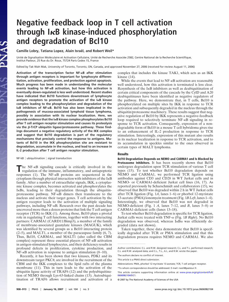
ResultsBcl10 Degradation Depends on NEMO and CARMA1 and Is Blocked byProteasome Inhibitors. It has been recently shown that Bcl10undergoes degradation upon TCR stimulation of various T celltypes (15). To test whether Bcl10 degradation depends onNEMO and CARMA1, we performed TCR ligation usingantibodies against CD3 and CD28 in WT Jurkat cells and inNEMO- or CARMA1-deficient Jurkat cell lines (16, 17). Asreported previously by Scharschmidt and collaborators (15), weobserved that Bcl10 was degraded within 2 h in WT Jurkat cellsafter TCR ligation (Fig. 1A, lanes 1–6) or phorbol 12-myristate13-acetate (PMA)/ionomycin treatment (Fig. 1B Left, lanes 1–4).Interestingly, we observed that Bcl10 was not degraded inNEMO-deficient (Fig. 1 A, lanes 7–12, and B, lanes 5–8) orCARMA1-deficient cells (lanes 13–18).
To test whether Bcl10 degradation is specific for TCR ligation,Jurkat cells were treated with TNF-� (Fig. 1B Right). No Bcl10degradation was observed, whereas I�B� was efficiently de-graded (data not shown).
Taken together, these data demonstrate that Bcl10 is specif-ically degraded after TCR or PMA stimulation and that thisdegradation process requires NEMO and CARMA1. We also
Author contributions: C.L. and R.W. designed research; C.L. and T.L. performed research;C.L. and R.W. analyzed data; and C.L., T.L., A.I., and R.W. wrote the paper.
The authors declare no conflict of interest.
This article is a PNAS direct submission.
Abbreviations: PMA, phorbol 12-myristate 13-acetate; TCR, T cell antigen receptor.
*To whom correspondence should be addressed. E-mail: [email protected].
This article contains supporting information online at www.pnas.org/cgi/content/full/0606982104/DC1.
© 2007 by The National Academy of Sciences of the USA
908–913 � PNAS � January 16, 2007 � vol. 104 � no. 3 www.pnas.org�cgi�doi�10.1073�pnas.0606982104
demonstrated that a proteasome inhibitor, ALLN, efficientlyblocked Bcl10 degradation (Fig. 1C) and induced the appear-ance of a slower migrating form of Bcl10 after 30 min ofCD3/CD28 costimulation (lanes 4–6), which likely represents ahyperphosphorylated isoform of Bcl10 (see below).
Inhibition of IKK Prevents Bcl10 Degradation. Because we observedthat TCR-induced Bcl10 degradation depends on NEMO andthe proteasome pathway, we hypothesized that Bcl10 could bephosphorylated by the NEMO/IKK complex. Analysis of theBcl10 protein sequence revealed a Threonine and a Serine(Thr-81 and Ser-85) in an environment similar to the consensusphosphorylation site/E3 ligase recognition sequence foundwithin I�Bs, p105, p100, and Foxo3A (Fig. 2A).
To determine whether Bcl10 degradation could be blocked bypharmacological inhibition of IKK, we tested the effect of theIKK inhibitor Bay 11-7085 on PMA/ionomycin-induced degra-dation of Bcl10 (Fig. 2B) and observed a complete inhibition ofBcl10 as well as I�B� phosphorylation (compare lanes 6–8 and2–4). Because Wegener et al. (18), using the same type ofexperiments, did not observe any effect on signal-induced Bcl10degradation, we decided to inactivate IKK by RNA interference(RNAi) (Fig. 2C). The results confirmed and extended the dataobtained in our BAY 11-7085 experiment: down-regulation ofIKK� (Fig. 2C, bottom panel, lanes 9–12) completely inhibitedPMA/ionomycin-induced Bcl10 degradation, whereas that ofIKK� was less efficient (lanes 5–8).
Bcl10 Is Phosphorylated by the NEMO/IKK Complex. To test whetherIKK� could induce Bcl10 phosphorylation, we transfected HEK-293T cells with Flag-tagged Bcl10 alone or together with in-
creasing amounts of VSV-tagged IKK� (Fig. 2D Left). Underthese conditions, we observed that IKK� overexpression led tothe accumulation of slower migrating forms of Bcl10 (lanes 4–6).Treatment with � phosphatase (Fig. 2D Right) indicates thatthese slowly migrating bands represent hyperphosphorylatedforms of Bcl10.
To evaluate whether IKK directly phosphorylates Bcl10 invitro, we generated a recombinant GST-Bcl10 fusion protein. Wethen performed in vitro kinase assays using GST-Bcl10 as asubstrate and immunoprecipitated IKK� or RIP2 derived fromtransfected HEK-293T cells as a source of kinase activity (Fig.2E). Under these conditions, GST-Bcl10 was phosphorylated byIKK� (Fig. 2E Left, lanes 5–8) but not by Rip2 (lanes 1–4). Thisresult demonstrates that IKK�, but not RIP2, directly phosphor-ylates Bcl10. RIP2 may, however, activate an intermediatekinase, because it has been shown to be involved in TCR-inducedBcl10 phosphorylation (19).
Further experiments were performed to determine whetherthe putative consensus sequence of Bcl10 is the actual target ofIKK phosphorylation. To this end, we produced several non-overlapping fragments of Bcl10 fused to GST and used them assubstrates in an IKK�-immunocomplex kinase assay. Three ofthese fragments were phosphorylated by IKK�, F1 (amino acids1–38), F3 (74–112), and F5 (155–196) (Fig. 3A, lanes 1, 5, and9), whereas the other fragments, F2 (39–73), F4 (113–154), andF6 (197–232) were not (Fig. 3A, lanes 3, 7, and 11). To narrowdown the potential phosphorylation site(s), we mutated severalindividual Ser or Thr residues in fragments F1, F3, and F5 andused them as substrates in the IKK� kinase assay. In F1,mutation of Ser-7 abolished phosphorylation, whereas mutationof Thr-4, -9, or -14 did not (Fig. 3B, compare lane 5 with lanes1, 3, 7, and 9). As mentioned above, F3 contained the consensusrecognition site for IKK phosphorylation/�-TrCP ubiquitina-tion. Therefore, we mutated Thr-81 and Ser-85 to Ala andobserved that the phosphorylation of the mutant peptide wastotally abrogated (Fig. 3C, compare lane 3 with lane 1), con-firming that the DTLVES sequence is a bona fide IKK�phosphorylation site. Finally, various mutations (Ser or Thrclusters) were introduced into F5 as indicated in Fig. 3D, and invitro phosphorylation experiments were performed. Althoughmutation of three clusters (amino acids 160–164, 170–171, and188–191) did not result in a decrease of F5 phosphorylation(compare lanes 3, 5, 9 to lane 1), phosphorylation was attenuatedby substitution of Ser-167 and Thr-168 to Ala (compare lane 7to lane 1). Incidentally, we observed that the appearance of themajor slowly migrating band observed in Fig. 2 was stronglyaffected by the substitution of these phosphorylation sites [seesupporting information (SI) Fig. 7].
We finally examined whether F1, F3, and F5 could be phos-phorylated by IKK� in vitro. Our results demonstrated that IKK�and IKK� phosphorylate Bcl10 in vitro on the same phosphor-ylation sites (data not shown).
Bcl10 Interacts with and Is Ubiquitinated by �-TrCP. Because thesequence surrounding Thr-81 and Ser-85 shows a strong homologyto the consensus recognition site for the E3 ubiquitin ligase �-TrCP,its phosphorylation by IKK is expected to recruit �-TrCP to Bcl10.To assess whether Bcl10/�-TrCP interaction can take place ex vivo,the two proteins were expressed in HEK-293T cells either alone,together, or in association with active or inactive forms of IKK�, asindicated in Fig. 4. Bcl10/�-TrCP association was demonstrated bycoimmunoprecipitation and was found to depend on the cotrans-fection of IKK� (Fig. 4, compare lane 7 with lane 6). Mutation ofthe two phosphoacceptor residues (T81A/S85A) abolished Bcl10–�-TrCP interaction (Fig. 4, lanes 9 and 10). In addition, we couldalso detect an interaction between Bcl10 and IKK� (Fig. 4, lane 7),which was reduced by the mutation of the two phosphoacceptorresidues (lane 10). In addition, Bcl10/�-TrCP association requires
Fig. 1. NEMO-, CARMA1-, and proteasome-dependent degradation of Bcl10in T cells in response to PMA and CD3/CD28 costimulation. (A) Kinetics of Bcl10degradation in response to CD3/CD28 costimulation. Jurkat parental cells(lanes 1–6) as well as NEMO-deficient (lanes 7–12) or CARMA1-deficient (lanes13–18) cells were treated with CD3/CD28 antibodies for the indicated period,and the amount of Bcl10 was measured by immunoblotting. The level ofexpression of IKK� is shown as a loading control. (B) PMA/ionomycin but notTNF-� induces Bcl10 degradation. Jurkat cells (Left, lanes 1–4, and Right) orNEMO-deficient cells (lanes 5–8) were stimulated for the indicated period(hours) with PMA or TNF-�. Levels of Bcl10 and �-tubulin as a loading controlwere determined by Western blotting. (C) Bcl10 is degraded by the protea-some. Jurkat cells were pretreated with ALLN for 30 min and treated withALLN and CD3/CD28 antibodies for the indicated period, and the levels ofBcl10, IKK�, and IKK� were determined by Western blotting.
Lobry et al. PNAS � January 16, 2007 � vol. 104 � no. 3 � 909
IMM
UN
OLO
GY

the presence of IKK� and of the two conserved phosphoacceptorresidues of Bcl10 and results in Bcl10 ubiquitination (SI Fig. 8).
Thr-81 and Ser-85 of Bcl10 Contribute to TCR-Induced Bcl10 Degrada-tion. Experiments in Figs. 2 and 4 were performed by overexpres-sion in nonlymphoid cells. To further our understanding of theregulation of Bcl10 in T cells, we tested the impact of TCRstimulation on the degradation of Bcl10 in E29.1 cells (an antigen-specific murine T cell hybridoma) stably expressing the WT formof human Bcl10 or the T81A/S85A mutant. After incubation withanti-TCR mAb H57 in the presence of cycloheximide (to avoidBcl10 resynthesis) for the indicated period, E29.1 cells were lysed,
Fig. 2. IKK�-dependent Bcl10 phosphorylation is required for its degra-dation. (A) Sequence alignment of proteins that are polyubiquitinated bythe �-TrCP E3 ligase. The ubiquitination of these proteins is caused byphosphorylation by IKK, except for VPU and �-catenin. The phosphoaccep-tor sites are the S and T indicated in bold. In the consensus sequence, �
represents an hydrophobic amino acid, and X represents any amino acid. (B)Pharmacological inhibition of IKK inhibits Bcl10 degradation. Jurkat cellswere pretreated with the IKK inhibitor Bay 11-7085 for 30 min. Time courseanalysis of Bcl10 expression (bottom gel) and I�B� phosphorylation [usinga phosphospecific anti-I�B� antibody (middle gel)] reveals that Bay 11-7085abrogates PMA/ionomycin-induced I�B� phosphorylation and Bcl10 deg-radation. IKK2 was used as a loading control. (C) siRNA-mediated depletionof IKK�, and to a lesser extent IKK�, inhibits Bcl10 degradation. Jurkat cellswere either left untransfected or transfected with IKK� siRNA, IKK� siRNA,or both, as mentioned in the figure. Cells were then treated with PMA/ionomycin for the indicated time. The level of expression of IKK�, IKK�,actin (loading control), and Bcl10 were determined by Western blotting.(D) (Left) Ex vivo phosphorylation of Bcl10 by IKK�. (Left) Flag-tagged Bcl10was expressed in HEK-293T cells in the absence (�) or presence (�) ofincreasing amounts of VSV-tagged IKK�. Bcl10 was analyzed by Westernblotting with anti-Flag antibody and reveals the IKK-dependent accumu-lation of higher-molecular-weight products (* and **). Expression of trans-fected IKK� was also monitored by immunoblotting with anti-VSV. (Right)Lysates of cells cotransfected with Flag-Bcl10 and VSV-IKK� (lane 2) weretreated with �-phosphatase alone (lane 4) or with �-phosphatase andphosphatase inhibitors (lane 3), and analyzed by Western blotting withanti-Bcl10 anti-serum. (E) In vitro phosphorylation of Bcl10 by IKK�. (Left)VSV-Rip2 and VSV-IKK� were transiently transfected in HEK293T cells, andkinase activity was assayed (KA) by immune complex reaction in thepresence of increasing amounts of GST-Bcl10 (1, 2.5, 5, and 10 �g). (Right)VSV-IKK� WT, Flag-IKK� DA (a constitutively active mutant), and VSV-IKK�
DN (a kinase-dead mutant) were transiently transfected into HEK-293T cellsand immunoprecipitated. In vitro kinase assays (KA) were performed by using2.5 �g of GST-Bcl10, GST-I�B� N-ter (as a positive control), or GST-I�B� C-ter (asa negative control).
Fig. 3. Mapping of IKK�-induced Bcl10 phosphorylation sites. (A) In vitrophosphorylation of Bcl10 fragments (F1–F6) by IKK�. VSV-tagged IKK�, eitherWT or dominant negative (DN), were expressed in HEK-293T cells, and immu-noprecipitates were used for in vitro kinase assays (KA) with fragments ofBcl10 fused to GST, as indicated above the lanes (the relevant bands areindicated by asterisks). (B–D) Mutation analysis of IKK-mediated Bcl10 phos-phorylation. GST fused to fragment F1 [amino acids 11–38 (B)], F3 [74–112 (C)],or F5 [155–196 (D)] of Bcl10 and mutants of the indicated Ser (S) or Thr (T)residues were subjected to in vitro kinase assays as described in A.
910 � www.pnas.org�cgi�doi�10.1073�pnas.0606982104 Lobry et al.
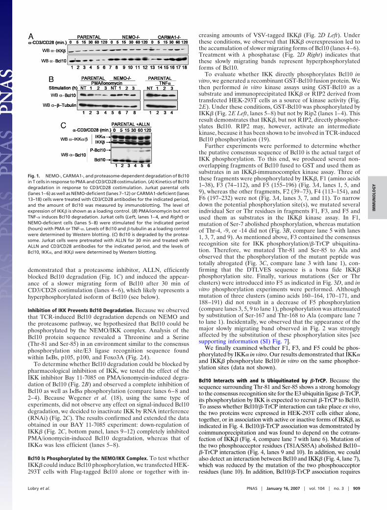
and Bcl10 degradation was measured by immunoblotting with anantibody that only recognizes its human form. This experimentshowed that TCR stimulation induced a strong reduction of WTBcl10 within 2 h after stimulation (Fig. 5A, lanes 1–4), whereas theT81A/S85A mutant was resistant to this degradation (lanes 5–8).
Enhancement of IL-2 Production in Bcl10 T81A/S85A-Expressing Cells.Because Bcl10 degradation results in down-regulation of NF-�Bactivity, it is expected that its stabilization will lead to anincreased production of IL-2 in activated T cells. To confirm thisprediction, individual E29.1 clones expressing either Bcl10 WTor Bcl10 T81A/S85A were isolated by limiting dilution. Theseclones were first assayed for expression of Bcl10 by Western blotanalysis. Three cell lines expressing comparable levels of eitherWT or mutant Bcl10 were selected for further studies (Fig. 5BLower). E29.1 cell lines were stimulated with PMA/ionomycin,and IL-2 production was assayed by ELISA (Fig. 5B Upper).When compared with E29.1 clones expressing the empty vectorMSCV or Bcl10 WT, cells expressing mutant Bcl10 demon-strated a marked enhancement of IL-2 production.
Mutation of the T81/S85 Bcl10 Phosphorylation Sites Results in ItsNuclear Localization upon TCR Stimulation. Because nuclear local-ization of Bcl10 has been reported in a number of cases of MALT
Fig. 6. Subcellular localization of WT and mutant (T81A/S85A and S7A/T81A/S85A/S167A/T168A) Bcl10 in untreated (A) or TCR-activated (B and C) E29.1cells. Mutation of the T81/S85 phosphorylation site is sufficient to inducenuclear localization of Bcl10 upon TCR stimulation. A similar localization canbe observed for the WT molecule in the presence of proteasome inhibitors[MG132 (C)]. In each panel, E29.1 cells were stained with Hoechst to revealDNA (Left) and with anti-Bcl10 antiserum (Center).
Fig. 4. The presence of active IKK� is necessary for the association betweenBcl10 and �-TrCP. HEK-293T cells were transfected with the indicated con-structs. Cell extracts were immunoprecipitated with anti-Flag and immuno-blotted with anti-VSV (Upper), and total lysates (TL) were blotted with theindicated antibodies (Lower).
Fig. 5. The Bcl10 T81A/S85A mutation prevents degradation and increasesIL-2 production. (A) Bcl10 degradation in response to TCR stimulation isprevented by mutation of the T81/S85 phosphorylation site. E29.1 murine Tcells transduced with WT human Bcl10 (lanes 1–4) or the T81A/S85A mutant(lanes 5–8) were treated with anti-TCR mAb for the indicated period in thepresence of cycloheximide, and the expression of Bcl10 was determined byWestern blotting. (B) Increased IL-2 production in cells expressing Bcl10 T81A/S85A. (B Lower) Levels of Bcl10 were measured in E29.1 cells expressing theempty vector MSCV (lane 1), in three individual clones expressing WT Bcl10(lanes 2–4) and in three individual clones expressing Bcl10 T81A/S85A (lanes5–7). (B Upper) IL-2 production after 14 h of PMA/ionomycin treatment wasassayed in the E29.1 clones.
Lobry et al. PNAS � January 16, 2007 � vol. 104 � no. 3 � 911
IMM
UN
OLO
GY
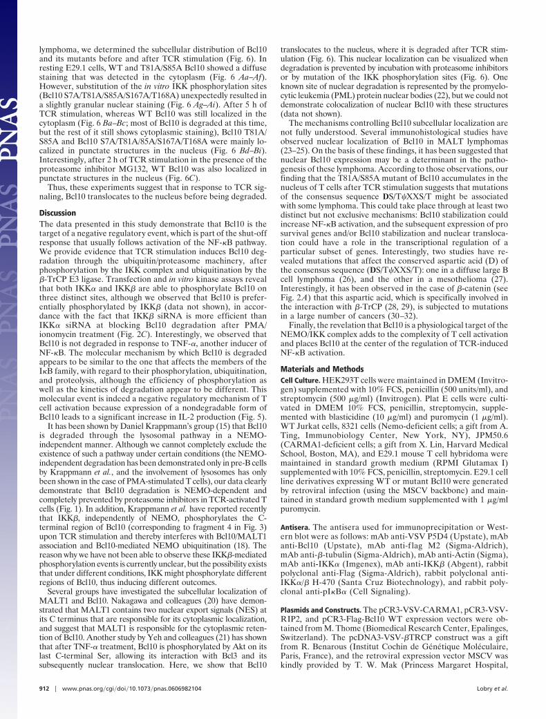
lymphoma, we determined the subcellular distribution of Bcl10and its mutants before and after TCR stimulation (Fig. 6). Inresting E29.1 cells, WT and T81A/S85A Bcl10 showed a diffusestaining that was detected in the cytoplasm (Fig. 6 Aa–Af).However, substitution of the in vitro IKK phosphorylation sites(Bcl10 S7A/T81A/S85A/S167A/T168A) unexpectedly resulted ina slightly granular nuclear staining (Fig. 6 Ag–Ai). After 5 h ofTCR stimulation, whereas WT Bcl10 was still localized in thecytoplasm (Fig. 6 Ba–Bc; most of Bcl10 is degraded at this time,but the rest of it still shows cytoplasmic staining), Bcl10 T81A/S85A and Bcl10 S7A/T81A/85A/S167A/T168A were mainly lo-calized in punctate structures in the nucleus (Fig. 6 Bd–Bi).Interestingly, after 2 h of TCR stimulation in the presence of theproteasome inhibitor MG132, WT Bcl10 was also localized inpunctate structures in the nucleus (Fig. 6C).
Thus, these experiments suggest that in response to TCR sig-naling, Bcl10 translocates to the nucleus before being degraded.
DiscussionThe data presented in this study demonstrate that Bcl10 is thetarget of a negative regulatory event, which is part of the shut-offresponse that usually follows activation of the NF-�B pathway.We provide evidence that TCR stimulation induces Bcl10 deg-radation through the ubiquitin/proteasome machinery, afterphosphorylation by the IKK complex and ubiquitination by the�-TrCP E3 ligase. Transfection and in vitro kinase assays revealthat both IKK� and IKK� are able to phosphorylate Bcl10 onthree distinct sites, although we observed that Bcl10 is prefer-entially phosphorylated by IKK� (data not shown), in accor-dance with the fact that IKK� siRNA is more efficient thanIKK� siRNA at blocking Bcl10 degradation after PMA/ionomycin treatment (Fig. 2C). Interestingly, we observed thatBcl10 is not degraded in response to TNF-�, another inducer ofNF-�B. The molecular mechanism by which Bcl10 is degradedappears to be similar to the one that affects the members of theI�B family, with regard to their phosphorylation, ubiquitination,and proteolysis, although the efficiency of phosphorylation aswell as the kinetics of degradation appear to be different. Thismolecular event is indeed a negative regulatory mechanism of Tcell activation because expression of a nondegradable form ofBcl10 leads to a significant increase in IL-2 production (Fig. 5).
It has been shown by Daniel Krappmann’s group (15) that Bcl10is degraded through the lysosomal pathway in a NEMO-independent manner. Although we cannot completely exclude theexistence of such a pathway under certain conditions (the NEMO-independent degradation has been demonstrated only in pre-B cellsby Krappmann et al., and the involvement of lysosomes has onlybeen shown in the case of PMA-stimulated T cells), our data clearlydemonstrate that Bcl10 degradation is NEMO-dependent andcompletely prevented by proteasome inhibitors in TCR-activated Tcells (Fig. 1). In addition, Krappmann et al. have reported recentlythat IKK�, independently of NEMO, phosphorylates the C-terminal region of Bcl10 (corresponding to fragment 4 in Fig. 3)upon TCR stimulation and thereby interferes with Bcl10/MALT1association and Bcl10-mediated NEMO ubiquitination (18). Thereason why we have not been able to observe these IKK�-mediatedphosphorylation events is currently unclear, but the possibility existsthat under different conditions, IKK might phosphorylate differentregions of Bcl10, thus inducing different outcomes.
Several groups have investigated the subcellular localization ofMALT1 and Bcl10. Nakagawa and colleagues (20) have demon-strated that MALT1 contains two nuclear export signals (NES) atits C terminus that are responsible for its cytoplasmic localization,and suggest that MALT1 is responsible for the cytoplasmic reten-tion of Bcl10. Another study by Yeh and colleagues (21) has shownthat after TNF-� treatment, Bcl10 is phosphorylated by Akt on itslast C-terminal Ser, allowing its interaction with Bcl3 and itssubsequently nuclear translocation. Here, we show that Bcl10
translocates to the nucleus, where it is degraded after TCR stim-ulation (Fig. 6). This nuclear localization can be visualized whendegradation is prevented by incubation with proteasome inhibitorsor by mutation of the IKK phosphorylation sites (Fig. 6). Oneknown site of nuclear degradation is represented by the promyelo-cytic leukemia (PML) protein nuclear bodies (22), but we could notdemonstrate colocalization of nuclear Bcl10 with these structures(data not shown).
The mechanisms controlling Bcl10 subcellular localization arenot fully understood. Several immunohistological studies haveobserved nuclear localization of Bcl10 in MALT lymphomas(23–25). On the basis of these findings, it has been suggested thatnuclear Bcl10 expression may be a determinant in the patho-genesis of these lymphoma. According to those observations, ourfinding that the T81A/S85A mutant of Bcl10 accumulates in thenucleus of T cells after TCR stimulation suggests that mutationsof the consensus sequence DS/T�XXS/T might be associatedwith some lymphoma. This could take place through at least twodistinct but not exclusive mechanisms: Bcl10 stabilization couldincrease NF-�B activation, and the subsequent expression of prosurvival genes and/or Bcl10 stabilization and nuclear transloca-tion could have a role in the transcriptional regulation of aparticular subset of genes. Interestingly, two studies have re-vealed mutations that affect the conserved aspartic acid (D) ofthe consensus sequence (DS/T�XXS/T): one in a diffuse large Bcell lymphoma (26), and the other in a mesothelioma (27).Interestingly, it has been observed in the case of �-catenin (seeFig. 2A) that this aspartic acid, which is specifically involved inthe interaction with �-TrCP (28, 29), is subjected to mutationsin a large number of cancers (30–32).
Finally, the revelation that Bcl10 is a physiological target of theNEMO/IKK complex adds to the complexity of T cell activationand places Bcl10 at the center of the regulation of TCR-inducedNF-�B activation.
Materials and MethodsCell Culture. HEK293T cells were maintained in DMEM (Invitro-gen) supplemented with 10% FCS, penicillin (500 units/ml), andstreptomycin (500 �g/ml) (Invitrogen). Plat E cells were culti-vated in DMEM 10% FCS, penicillin, streptomycin, supple-mented with blasticidine (10 �g/ml) and puromycin (1 �g/ml).WT Jurkat cells, 8321 cells (Nemo-deficient cells; a gift from A.Ting, Immunobiology Center, New York, NY), JPM50.6(CARMA1-deficient cells; a gift from X. Lin, Harvard MedicalSchool, Boston, MA), and E29.1 mouse T cell hybridoma weremaintained in standard growth medium (RPMI Glutamax I)supplemented with 10% FCS, penicillin, streptomycin. E29.1 cellline derivatives expressing WT or mutant Bcl10 were generatedby retroviral infection (using the MSCV backbone) and main-tained in standard growth medium supplemented with 1 �g/mlpuromycin.
Antisera. The antisera used for immunoprecipitation or West-ern blot were as follows: mAb anti-VSV P5D4 (Upstate), mAbanti-Bcl10 (Upstate), mAb anti-f lag M2 (Sigma-Aldrich),mAb anti-�-tubulin (Sigma-Aldrich), mAb anti-Actin (Sigma),mAb anti-IKK� (Imgenex), mAb anti-IKK� (Abgent), rabbitpolyclonal anti-Flag (Sigma-Aldrich), rabbit polyclonal anti-IKK�/� H-470 (Santa Cruz Biotechnology), and rabbit poly-clonal anti-pI�B� (Cell Signaling).
Plasmids and Constructs. The pCR3-VSV-CARMA1, pCR3-VSV-RIP2, and pCR3-Flag-Bcl10 WT expression vectors were ob-tained from M. Thome (Biomedical Research Center, Epalinges,Switzerland). The pcDNA3-VSV-�TRCP construct was a giftfrom R. Benarous (Institut Cochin de Genetique Moleculaire,Paris, France), and the retroviral expression vector MSCV waskindly provided by T. W. Mak (Princess Margaret Hospital,
912 � www.pnas.org�cgi�doi�10.1073�pnas.0606982104 Lobry et al.
Toronto, ON, Canada). The pcDNA3-VSV-IKK� was generatedin our laboratory. The WT ubiquitin expression vector encodingan octameric tandem fusion of HA-ubiquitin was a gift from M.Treier (European Molecular Biology Laboratory, Heidelberg,Germany).
All PCR-generated DNA fragments used to construct theGST fusions were cloned into the BamHI–EcoRI restriction siteof the pGEX-KT vector (Amersham Pharmacia). The GST-I�B� N-ter and the GST-I�B� C-ter has been described (33).The Flag-Bcl10 mutants were generated by PCR mutagenesis.
MSCV-Bcl10 WT or mutants were created by PCR and clonedinto HpaI–XhoI restriction sites of the MSCV retroviral vector(Clontech).
Activation, Immunoprecipitations, and Immunoblots. TCR activation.E29.1 cells were activated with H57 mAb (R & D Systems)coated overnight at 4°C on six-well plates. Human T cells wereactivated as described (34).Treatment with chemicals or siRNA. The following reagents werepurchased and used as indicated: 100 ng/ml PMA (Sigma-Aldrich) and 1 �M calcium ionophore (Sigma-Aldrich), Bay11-7085 (5 �g/ml; Calbiochem), 10 �g/ml human TNF-�(PeproTech) or mouse TNF-� (Apotech), 100 �M N-acetyl-Leu-Leu-norleucinal (calpain inhibitor I; Sigma-Aldrich), 25 �MMG 132 (Sigma-Aldrich) or 50 �g/ml cycloheximide (Sigma-Aldrich). For transient expression of siRNA, we used smart poolIKK� and smart pool IKK� (Dharmacon).
After treatment, cells were lysed in 100 �l of 1� TNE buffer(50 mM Tris, pH 8.0/0.5% Nonidet P-40/200 mM NaCl/0.1 mMEDTA, supplemented with protease and phosphatase inhibitors)and subjected either to direct immunoblotting or to immuno-precipitation followed by immunoblotting as described in thefigure legends (34).
Immunofluorescence Microscopy. E29.1 stable clones were acti-vated as described above and then harvested on circular glass
slides coated with polylysine (Sigma-Aldrich) in a 24-well dish.The cells were then fixed in 4% formaldehyde in PBS for 20 minat room temperature followed by treatment with 50 mM am-monium chloride. Cells were then permeabilized in 0.2% Tritonin PBS and washed in PBS supplemented with 1 mg/ml BSA. Theprimary antibody (anti-Bcl10) was applied for 1 h at roomtemperature. Cells were then washed and incubated for 45 minwith the anti-mouse CY3-labeled secondary antibody, while thenucleus was stained with DAPI. Cell preparations were mountedin Mowiol, and the specimens were examined by using anApoTome imaging system (Imager Z1; Zeiss) with a �60/1.4OIL DIC objective (Zeiss). Images were analyzed by usingAxioVision software (Zeiss).
Immune Complex Kinase Assays. HEK 293T cells were transfectedwith an IKK�-expressing vector, lysed with 1� TNE buffer after48 h, and immunoprecipitated. Production of fusion proteins andkinase assays were performed as described (34) by using the kinasebuffer 20 mM Hepes/10 mM MgCl2/50 mM NaCl (pH 7.5).
IL-2 Assays. To evaluate PMA/ionomycin-induced IL-2 produc-tion by the different E29.1 clones, wells of 24-well Falcon tissueculture plates (2 � 105 cells per well) were activated in duplicatewith 5 ng/ml PMA and 1 mg/ml ionomycin for 14 h at 37°C. Serialdilutions of supernatant were then tested in triplicate for IL-2content. These assays were performed by measuring IL-2 pro-duction with an ELISA kit (R & D Systems).
We thank Margot Thome for critical reading of the manuscript and forthe gift of Bcl10 and CARMA1 expression vectors, Adrian Ting for thegift of the 8321 NEMO-deficient cell line, and Xin Lin for the gift of theCARMA1-deficient cell line. This work was supported by Ligue Contrele Cancer Contract R05/75-22 (R.W.), Association pour la Recherche surle Cancer Contract 3119 (A.I.), and Canceropole Axe 2 (A.I.). C.L. is therecipient of a Ministere de la Recherche et Technologie fellowship.
1. Weil R, Israel A (2006) Cell Death Differ 13:826–833.2. Bertin J, Wang L, Guo Y, Jacobson MD, Poyet JL, Srinivasula SM, Merriam
S, DiStefano PS, Alnemri ES (2001) J Biol Chem 276:11877–11882.3. Gaide O, Martinon F, Micheau O, Bonnet D, Thome M, Tschopp J (2001)
FEBS Lett 496:121–127.4. McAllister-Lucas LM, Inohara N, Lucas PC, Ruland J, Benito A, Li Q, Chen
S, Chen FF, Yamaoka S, Verma IM, et al. (2001) J Biol Chem 276:30589–30597.
5. Wang L, Guo Y, Huang WJ, Ke X, Poyet JL, Manji GA, Merriam S,Glucksmann MA, DiStefano PS, Alnemri ES, Bertin J (2001) J Biol Chem276:21405–21409.
6. Lucas PC, Yonezumi M, Inohara N, McAllister-Lucas LM, Abazeed ME, ChenFF, Yamaoka S, Seto M, Nunez G (2001) J Biol Chem 276:19012–19019.
7. Uren AG, O’Rourke K, Aravind LA, Pisabarro MT, Seshagiri S, Koonin EV,Dixit VM (2000) Mol Cell 6:961–967.
8. Egawa T, Albrecht B, Favier B, Sunshine MJ, Mirchandani K, O’Brien W,Thome M, Littman DR (2003) Curr Biol 13:1252–1258.
9. Ruefli-Brasse AA, French DM, Dixit VM (2003) Science 302:1581–1584.10. Ruland J, Duncan GS, Elia A, del Barco Barrantes I, Nguyen L, Plyte S, Millar
DG, Bouchard D, Wakeham A, Ohashi PS, Mak TW (2001) Cell 104:33–42.11. Lee KY, D’Acquisto F, Hayden MS, Shim JH, Ghosh S (2005) Science
308:114–118.12. Sun L, Deng L, Ea CK, Xia ZP, Chen ZJ (2004) Mol Cell 14:289–301.13. Zhou H, Wertz I, O’Rourke K, Ultsch M, Xiao W, Dixit VM (2004) Nature
427:167–171.14. Chen ZJ (2005) Nat Cell Biol 7:758–765.15. Scharschmidt E, Wegener E, Heissmeyer V, Rao A, Krappmann D (2004) Mol
Cell Biol 24:3860–3873.16. He KL, Ting AT (2002) Mol Cell Biol 22:6034–6045.17. Wang D, Matsumoto R, You Y, Che T, Xue-Yan L, Gaffen SL, Lin X (2004)
Mol Cell Biol 24:164–171.
18. Wegener E, Oeckinghaus A, Papadopoulou N, Lavitas L, Schmidt-Supprian M,Ferch U, Mak TW, Ruland J, Heissmeyer V, Krappmann D (2006) Mol CellBiol 23:13–23.
19. Ruefli-Brasse AA, Lee WP, Hurst S, Dixit VM (2003) J Biol Chem 279:1570–1574.20. Nakagawa M, Hosakawa Y, Yonezumi M, Izumiyama K, Susuki R, Tsuzuki S,
Asaka M, Seto M (2005) Blood 106:4210–4216.21. Yeh PY, Kuo SH, Yeh KH, Chuang SE, Hsu CH, Chang WC, Lin HI, Gao M,
Cheng AL (2005) J Biol Chem 281:167–175.22. Bailey D, O’Hare P (2005) Biochem J 329:271–281.23. Liu H, Ye H, Dogan A, Ranaldi R, Hamoudi RA, Bearzi I, Isaacson PG, Du
MQ (2001) Blood 98:1182–1187.24. Maes B, Demunter A, Peeters B, De Wolf-Peeters C (2002) Blood 99 1398–1404.25. Ye H, Dogan A, Karran L, Willis TG, Chen L, Wlodarska I, Dyer MJ, Isaacson
PG, Du MQ (2000) Am J Pathol 157:1147–1154.26. Lee SH, Shin MS, Kim HS, Park WS, Kim SY, Lee HK, Park JY, Oh RR, Jang
JJ, Park KM, et al. (1999) Cancer Res 59:5674–5677.27. Willis TG, Jadayel DM, Du MQ, Peng H, Perry AR, Abdul-Rauf M, Price H,
Karran L, Majekodunmi O, Wlodarska I, et al. (1999) Cell 96:35–45.28. Megy S, Bertho G, Gharbi-Benarous J, Evrard-Todeschi N, Coadou G, Segeral E,
Iehle C, Quemeneur E, Benarous R, Girault J-P (2005) J Biol Chem 280:29107–29116.
29. Wu G, Xu G, Schulman BA, Jeffrey PD, Harper JW, Pavletich N (2003) MolCell 11:1445–1456.
30. Chan EF, Gat U, McNiff JM, Fuchs E (1999) Nat Genet 21:410–413.31. Legoix P, Bluteau O, Bayer J, Perret C, Balabaud C, Belghiti J, Franco D,
Thomas G, Laurent-Puig P, Zucman-Rossi J (1999) Oncogene 18:4044–4046.32. Machin P, Catasus L, Pons C, Munos J, Matias-Guiu X, Prat J (2002) Hum
Pathol 33:206–212.33. Weil R, Laurent-Winter C, Israel A (1997) J Biol Chem 272:9942–9949.34. Weil R, Schwamborn K, Alcover A, Bessia C, Di Bartolo V, Israel A (2003)
Immunity 18:13–26.
Lobry et al. PNAS � January 16, 2007 � vol. 104 � no. 3 � 913
IMM
UN
OLO
GY


![Topic 11 Revision [129 marks] - IB Questionbank](https://static.fdokumen.com/doc/165x107/6315a7f3b1e0e0053b0f274d/topic-11-revision-129-marks-ib-questionbank.jpg)