Nasal cardiac myosin peptide treatment and OX40 blockade protect mice from acute and chronic...
11
Nasal cardiac myosin peptide treatment and OX40 blockade protect mice from acute and chronic virally-induced myocarditis Georgia Fousteri 1 , Amy Dave, Bret Morin, Shaida Omid, Michael Croft, Matthias G. von Herrath * Diabetes Center, La Jolla Institute for Allergy and Immunology, La Jolla, CA 92037, USA article info Article history: Received 15 November 2010 Received in revised form 17 January 2011 Accepted 18 January 2011 Keywords: Autoimmune myocarditis IL-10 Treg Cardiac myosin Peptide therapy Coxsackie virus B3 OX40 abstract Myocarditis poses a severe health problem, can lead to dilated cardiomyopathy (DCM) and death, and is thought to be triggered by infections. Enteroviruses such as Coxsackie virus B 3 (CVB3) have been implicated as a culprit, since they can cause acute and chronic heart disease in susceptible mice. CVB was detected in human cardiac myocytes in some cases, whereas acute CVB infection was thought to have caused death. Here we studied, whether nasal administration of cardiac myosin (CM) major histocom- patibility class (MHC) II peptides CM 947e960 and CM 735e747 and OX40 blockade would be able to ameliorate immunopathology and heart disease in BALB/C mice infected with CVB3. We found that nasal CM-peptide prophylactic treatment significantly reduced myocarditis and mortality by enhancing Treg and IL-10 induction and that blockade of OX40 signaling could reduce heart inflammation when administered late during pathogenesis. Altogether, these results chart the way for novel prevention and intervention strategies for viral myocarditis. Ó 2011 Elsevier Ltd. All rights reserved. 1. Introduction Myocarditis is an inflammatory disease of the heart, which may be the precursor of heart failure due to dilated cardiomyopathy (DCM) [1e4]. In this study, we selected two coxsackie virus group B type 3 (CVB3) virally-induced myocarditis (VIM) mouse models, in which disease occurs after infection with CVB3. In the first model, acute VIM with significant damage to cardiomyocytes and sudden death within the first 10 days after infection is induced [5e7]. In the second model, following infection with heart-passaged CVB3 (hpCVB3), the initial, non-lethal signs of acute myocarditis become chronic and autoimmune in nature. In both cases, the myocardium becomes infiltrated by macrophages, B cells, and other lympho- cytes, and heart damage with fibrosis is often observed [8,9]. The chronic inflammatory disease in the second model is believed to be due to an autoimmune response driven by effector T cells responding to cardiac antigens [10e12]. Interestingly, among the most common pathogens related to myocarditis development in humans are enteroviruses and CVB3 in particular [5]. Most mouse strains including the C57BL/6 strain are resistant to acute myocarditis and moreover do not progress in developing the chronic features of the disease, while a certain proportion of BALB/C males but not females are susceptible. However, resistance in C57BL/6 mice can be overcome by treatment with LPS [13]. Here we studied the effect of nasal treatment simultaneously with cardiac myosin (CM) derived major histocompatibility class (MHC) II epitopes CM 947e960 and CM 735e747 (CM combo) [14] in male BALB/C mice in acute and chronic CVB3-induced myocarditis. The rationale for this study was that CM-specific regulatory T cells (Tregs) would be induced after nasal treatment with CM epitopes, a scenario that has been proven to be beneficial in other autoimmune settings [15e17] and in experimental autoimmune myocarditis (EAM) in particular [18,19]. Similar to other auto- antigen-induced autoimmune pathologies, EAM results in suscep- tible mice after subcutaneous injection of CM in complete Freund’s adjuvant (CFA) and pertussis toxin. In both EAM and VIM models, there is evidence that Tregs are involved in disease penetrance and/or precipitation. For example, induction of acute VIM in females during the estrus phase of the ovarian cycle seems to invigorate the function of Tregs, protecting from heart disease and sudden death [20]. The role of Th1 and Th2 effector cells in the pathogenesis of both EAM and VIM is still unclear, although tumor necrosis factor (TNF) [21,22] and more recently IL-6 have been implicated as important mediators of the disease [23]. Here, we report that intranasal CM combination peptide treatment protected male BALB/C mice from acute myocarditis, sudden death, and * Corresponding author. Tel.: þ1 858 205 0646; fax: þ1 858 752 6993. E-mail address: [email protected] (M.G. von Herrath). 1 Present address: San Raffaele Scientific Institute, Via Olgettina 58, Milan 20132, Italy. Contents lists available at ScienceDirect Journal of Autoimmunity journal homepage: www.elsevier.com/locate/jautimm 0896-8411/$ e see front matter Ó 2011 Elsevier Ltd. All rights reserved. doi:10.1016/j.jaut.2011.01.006 Journal of Autoimmunity 36 (2011) 210e220
Transcript of Nasal cardiac myosin peptide treatment and OX40 blockade protect mice from acute and chronic...

lable at ScienceDirect
Journal of Autoimmunity 36 (2011) 210e220
Contents lists avai
Journal of Autoimmunity
journal homepage: www.elsevier .com/locate/ jaut imm
Nasal cardiac myosin peptide treatment and OX40 blockade protectmice from acute and chronic virally-induced myocarditis
Georgia Fousteri 1, Amy Dave, Bret Morin, Shaida Omid, Michael Croft, Matthias G. von Herrath*
Diabetes Center, La Jolla Institute for Allergy and Immunology, La Jolla, CA 92037, USA
a r t i c l e i n f o
Article history:Received 15 November 2010Received in revised form17 January 2011Accepted 18 January 2011
Keywords:Autoimmune myocarditisIL-10TregCardiac myosinPeptide therapyCoxsackie virus B3OX40
* Corresponding author. Tel.: þ1 858 205 0646; faxE-mail address: [email protected] (M.G. von Herra
1 Present address: San Raffaele Scientific Institute, VItaly.
0896-8411/$ e see front matter � 2011 Elsevier Ltd.doi:10.1016/j.jaut.2011.01.006
a b s t r a c t
Myocarditis poses a severe health problem, can lead to dilated cardiomyopathy (DCM) and death, and isthought to be triggered by infections. Enteroviruses such as Coxsackie virus B 3 (CVB3) have beenimplicated as a culprit, since they can cause acute and chronic heart disease in susceptible mice. CVB wasdetected in human cardiac myocytes in some cases, whereas acute CVB infection was thought to havecaused death. Here we studied, whether nasal administration of cardiac myosin (CM) major histocom-patibility class (MHC) II peptides CM947e960 and CM735e747 and OX40 blockade would be able toameliorate immunopathology and heart disease in BALB/C mice infected with CVB3. We found that nasalCM-peptide prophylactic treatment significantly reduced myocarditis and mortality by enhancing Tregand IL-10 induction and that blockade of OX40 signaling could reduce heart inflammation whenadministered late during pathogenesis. Altogether, these results chart the way for novel prevention andintervention strategies for viral myocarditis.
� 2011 Elsevier Ltd. All rights reserved.
1. Introduction
Myocarditis is an inflammatory disease of the heart, which maybe the precursor of heart failure due to dilated cardiomyopathy(DCM) [1e4]. In this study, we selected two coxsackie virus group Btype 3 (CVB3) virally-induced myocarditis (VIM) mouse models, inwhich disease occurs after infection with CVB3. In the first model,acute VIM with significant damage to cardiomyocytes and suddendeathwithin the first 10 days after infection is induced [5e7]. In thesecond model, following infection with heart-passaged CVB3(hpCVB3), the initial, non-lethal signs of acute myocarditis becomechronic and autoimmune in nature. In both cases, the myocardiumbecomes infiltrated by macrophages, B cells, and other lympho-cytes, and heart damage with fibrosis is often observed [8,9]. Thechronic inflammatory disease in the second model is believed to bedue to an autoimmune response driven by effector T cellsresponding to cardiac antigens [10e12]. Interestingly, among themost common pathogens related to myocarditis development inhumans are enteroviruses and CVB3 in particular [5]. Most mousestrains including the C57BL/6 strain are resistant to acute
: þ1 858 752 6993.th).ia Olgettina 58, Milan 20132,
All rights reserved.
myocarditis and moreover do not progress in developing thechronic features of the disease, while a certain proportion of BALB/Cmales but not females are susceptible. However, resistance inC57BL/6 mice can be overcome by treatment with LPS [13]. Herewestudied the effect of nasal treatment simultaneously with cardiacmyosin (CM) derived major histocompatibility class (MHC) IIepitopes CM947e960 and CM735e747 (CM combo) [14] in male BALB/Cmice in acute and chronic CVB3-induced myocarditis.
The rationale for this study was that CM-specific regulatory Tcells (Tregs) would be induced after nasal treatment with CMepitopes, a scenario that has been proven to be beneficial in otherautoimmune settings [15e17] and in experimental autoimmunemyocarditis (EAM) in particular [18,19]. Similar to other auto-antigen-induced autoimmune pathologies, EAM results in suscep-tible mice after subcutaneous injection of CM in complete Freund’sadjuvant (CFA) and pertussis toxin. In both EAM and VIM models,there is evidence that Tregs are involved in disease penetranceand/or precipitation. For example, induction of acute VIM infemales during the estrus phase of the ovarian cycle seems toinvigorate the function of Tregs, protecting from heart disease andsudden death [20]. The role of Th1 and Th2 effector cells in thepathogenesis of both EAM and VIM is still unclear, although tumornecrosis factor (TNF) [21,22] and more recently IL-6 have beenimplicated as important mediators of the disease [23]. Here, wereport that intranasal CM combination peptide treatment protectedmale BALB/C mice from acute myocarditis, sudden death, and

G. Fousteri et al. / Journal of Autoimmunity 36 (2011) 210e220 211
progression to the chronic stage of the disease, through inductionof IL-10 and Tregs, and some downregulation of IFNg production.
In a second, complimentary approach, the pathogenic effector Tcell population was targeted. Of potential significance to myocar-ditis, OX40 (CD134), a costimulatory receptor of the TNFR super-family, has been detected on pathogenic effector T cells, coincidingwith increased levels of OX40-ligand (OX40L) expressed ondiseased cardiomyocytes [24e26]. Therefore we reasoned to testthe efficiency of targeting this interaction to protect fromprogression to the chronic stage of the disease. Interestingly,a beneficial effect of targeting OX40eOX40L signaling through anti-OX40L blockade administration was revealed in halting diseaseprogression.
2. Materials and methods
2.1. Mice
Male BALB/C mice 6e8 weeks of age were obtained from theJackson Laboratories (Bar Harbor, ME). The animal work describedwas carried out in accordance with the IACUC of the La Jolla Insti-tute for Allergy and Immunology.
2.2. Acute and chronic virally-induced myocarditis models
Acute myocarditis model: 103 plaque forming units (PFU) CVB3(Nancy strain), a sublethal dose, was used for the induction of acuteheart inflammation followed by sudden death and was prepared aspreviously described [27].
Chronic myocarditis model: For inducing chronic disease, 103 PFUheart-passaged viral stocks of CVB3 (hpCB3) were prepared andused according to [5].
In all cases, viral stocks were stored at �80 �C in Dulbecco’sModified Eagle Medium (DMEM). BALB/C mice were infectedintraperitoneally (i.p.).
2.3. Peptide treatments
Nasal administration-acute myocarditis model: Mice were treatedfor 3 consecutive days prior to infection with CVB3 (days �3, �2,�1) with the CM-derived peptides CM947e960 and CM735e747 (CMcombo) after diluting in sterile 1� DPBS and mixing. 40 mg of eachpeptide was given in the mouse nostril while anesthetized withisoflurane. Peptides were purchased from Abgent with more than90% purity (San Diego, CA).
Nasal administration-chronic myocarditis model: Mice weretreated with the aforementioned CM-peptide mix for 3 consecutivedays: a) prior to infectionwith hpCVB3 (days�3,�2,�1), b) duringthe acute phase of the immune response (days 7, 8, 9), and/or c)during the transition to the chronic phase of the immune response(days 20, 21, 22).
2.4. Flow cytometry
Flow cytometry antibodies were purchased from BD-Pharmin-gen, e-Bioscience and Biolegend (all San Diego, CA). Single-cellsuspensions, after a 2.4G2 blocking step, were stained with thefollowing directly conjugated antibodies: CD4-Pacific Blue orPECy7, CD69-FITC, CD62L-APC, CD45.2-PerCPCy5.5, CD8-APCCy7 orFITC, CD44-Pacific Blue, CTLA-4-PE, CD25-APCCy7, CD127-PeCy7and OX40-PE (isotype controls for all antibodies were included). Forintracellular Foxp3 detection, cells were fixed with Fix/Perm bufferand stained with anti-Foxp3-APC (e-Bioscience). Cells wereacquired on an LSRII flow cytometer (BD Biosciences) and analyzedusing FlowJo software (Treestar, Ashland, OR).
2.5. Histopathology and scoring
At the indicated time points, hearts were removed and cutlongitudinally into half with a scalpel; one half fixed in 4% PFAovernight and kept in 70% ethanol until further processing. Tissueswere paraffin embedded with the cut surface down and sections of5 mm thickness were cut for hematoxylin and eosin (H & E) staining.Paraffin blocks and sections were prepared and stained by theHistology and Immunohistochemistry Shared Resource (Universityof California, San Diego, Moores Cancer Center, CA). Inflamed heartswere characterized by the presence of interstitial infiltrate ofusually mononuclear cells associated with focal myocyte necrosisand in acute cases fibrosis. In some cases, extensive tissue necrosiswas seen in the pericardium (pericarditis). Heart sections werescored blindly according to the following scoring criteria: grade 1:0e10% of the total heart tissue shows immune inflammation(presence of lymphocytic infiltration) or any kind of tissuepathology i.e. pericarditis, grade 2: 11e25%; grade 3: 26e50%;grade 4: greater than 50%.
2.6. ELISPOT
IL-10, IL-17 and IFNg (BD-Pharmingen and Biolegend) producingcytokines were detected by ELISPOTas previously described [28]. Inbrief, splenocytes from CM peptide-immunized and control,hpCVB3-infected mice were diluted by factor of two starting at106 cells/well down to 0.25�106. Cells were cultured for three daysin the presence of CM-combo peptide and recombinant IL-2 (50 U/ml) (Roche, Pleasanton, CA). Color was developed with 3-amino-9-ethylcarbazole (AEC) (SigmaeAldrich, St Louis, MO) and H2O2.Lastly, plates were rinsed in water, left air dried, and spots repre-senting cytokine-producing cells were photographed and countedusing a computer-assisted image analysis (Zeiss KS ELISPOT reader).
2.7. Antibody treatments
Mice were treated at the indicated time points with150 mg/mouse anti-OX40L (clone RM134L, Biolegend) or anti-CD25(clone PC61, BD-Pharmingen) or anti-IL-10 (clone JES5-2A5) andisotype antibody controls.
2.8. Adoptive transfer experiments
Splenocytes from CM combo treated, hpCVB3-infected micewere pooled together. Splenocytes were depleted of CD8 T cells byusing anti-CD8 antibody incubation (BioLegend, San Diego, CA,USA), followed by incubation with sheep anti-Rat IgG coatedDynabeads (Invitrogen, San Diego, CA, USA). Following the CD8-depletion step, cells were resuspended at 10 � 106/ml in 10%vol/volRPMI containing 50 U/ml rhIL-2 and 20 mg/ml combo peptide mix,and cultured for 3 days. Next cells were further purified intoCD4þCD25þ or CD4þCD25� fractions using the CD4þCD25þ Regu-latory T cell isolation kit (Miltenyi Biotech, Auburn, CA). At least1 �106 total purified lymphocytes were stained with CFSE [29] andadoptively transferred intravenously (i.v.) into new BALB/C mice,which were subsequently infected with hpCVB3.
2.9. Heart infiltrate isolation
Heart infiltrate was prepared as previously described [23]. Inbrief, mice were killed with CO2 and immediately perfused with1� DPBS. One half of the each mouse heart was kept for histology(described above), whereas the other half was minced anddigested with 0.2% collagenase type II, 0.25% pancreatin (bothfrom SigmaeAldrich, USA) and 0.1% DNase I (Roche Applied

G. Fousteri et al. / Journal of Autoimmunity 36 (2011) 210e220212
Science, Basel, Switzerland) for 7 min at 37 �C. Digestion wasstopped with the addition of 0.1 M EDTA. Following digestion,single-cell suspensions were prepared by filtering through stain-less steel mesh 200 gauge.
2.10. Statistics
Unpaired Student’s t-test was implemented for all statisticalanalysis apart from differences in disease severity, which werecalculated with the Mann Whitney U test. p values lower than 0.05(*) were considered significant.
3. Results
3.1. Intranasal CM-combo peptide (CM947e960 and CM735e747)administration protects male BALB/C mice from sublethal CVB3infection
Acute myocarditis can be induced in BALB/C male mice afterinfection with 103 plaque forming units (PFU) of CVB3 (Nancystrain). Acute myocarditis is an inflammation of the heart muscle,which within 10 days after infection typically proceeds into heart
Fig. 1. Prevention from death caused by sublethal CVB3 infection after nasal CM-peptide treold male BALB/C mice were infected with 103 PFU CVB3. Mice were immunized nasally for 3�3, �2, �1) or left untreated. (A) Survival was followed over time post-infection (p.i.) withfifteenth day after infectionwere subsequently divided in two groups: one that was treated wthat remained untreated (n ¼ 7). Of these, two more mice died, one belonging into the anti-first antibody infection and one more belonging to the untreated group, which died at 19e245 days after infection shown in (BeC). CVB3-infected BALB/C mice treated with anti-OX40Lstained sections from hearts of CVB3-infected and naïve mice treated with CM peptides or
failure and sudden death. Disease is more severe in males thanfemales and some studies suggest a role of estrogens on the activityof immunosuppressive Treg populations [20]. Here, we attemptedto induce cardiac-specific Tregs by nasal immunization with theCM-derived peptides CM947e960 and CM735e747, which were previ-ously described to induce EAM in BALB/Cmice [14,18].We sought tocombine both epitopes in order to enhance our protective effect(combination therapy). In order to understand the effect of nasalCM-combo peptide vaccination in CVB3-induced acute myocarditis,male BALB/C mice were treated for 3 consecutive days prior toinfection with CVB3. Mice were separated in 2 experimentalgroups: 1) nasal CM-peptide treatment (n ¼ 12), and 2) no treat-ment (n ¼ 16). As shown in Fig. 1A, a significant reduction in thepercentage of mice dying from the sublethal CVB3 infection wasseen. The majority of nasal peptide treated mice survived (11 outof 12), while 5 out of 16 (31.25%) died in the group not treated withthe CM peptides by the twentieth day after infection. While most ofthe untreated mice died in the first 10 days after the infection, fewperished later, one at 17 and one at 20 days after infection(next section in more detail). However, in both groups of mice,significant heart inflammation was detectable in those mice thatsurvived the infection (Fig. 1BeC).
atment, and reduction in cardiac inflammation after anti-OX40L blockade. Seven-weekconsecutive days with CM947e960 and CM735e747 (40 mg each) before viral infection (daysCVB3. *p < 0.05. Thirteen mice surviving the infection in the untreated group by theith anti-OX40L, 150 mg per mouse on days 15, 17, 19 from infection (n ¼ 6), and a secondOX40L group that was already sick at the time of the treatment and died soon after the0 days after infection. In the remaining mice, myocarditis severity score was evaluateddisplayed the highest reduction in heart inflammation. Hematoxylin and eosin (H & E)anti-OX40L.

G. Fousteri et al. / Journal of Autoimmunity 36 (2011) 210e220 213
3.2. Anti-OX40L blockade inhibits myocarditis progression in micesurviving the lethal CVB3 infection
Given the results described above, we sought to investigate theeffect of anti-OX40 blockade (clone RM134L) treatment in haltingmyocarditis severity. To this end, 13 mice in the untreated groupthat remained alive by the fifteenth day after infectionwere dividedinto two sub-groups: one, which remained untreated (n ¼ 7), anda second group that received three anti-OX40L blocking antibodyinjections (150 mg/mouse) on days 15, 17 and 19 after infection(n ¼ 6). Of those, one mouse in the anti-OX40L group and alreadysick at the time of treatment died after the first antibody injectionon day 17, while one more mouse in the group that remaineduntreated died 2e3 days later. We cannot conclude whether anti-OX40L therapy protected the mice from further lethality to CVB3infection, however, histological examination of the heart tissue atday 45 revealed a significant reduction in inflammation after OX40Lblockade treatment, whereas nasal CM-peptide treated and controlmice displayed similar degrees of inflammation (Fig. 1BeC). Alto-gether, nasal CM-peptide treatment can protect male BALB/C micefrom acute myocarditis acute myocarditis-associated mortality,while persistent CVB3-driven heart inflammation can be reducedby anti-OX40L blockade treatment.
3.3. Nasal CM-combo peptide treatment increases Treg frequency inblood and spleen and protects from autoimmune myocarditis
Since autoimmune myocarditis is more relevant to the humandisease, the majority of our follow-up studies were conducted withthe chronic hpCVB3-induced model. Similarly, we attempted toinduce Tregs by nasal immunizationwith the CM-derived peptides,following several nasal immunization protocols, since we were notsure which would deliver the most beneficial effect. In male BALB/Cmice, infection with hpCVB3 virus leads to a peak in myocarditisaround day 7 post-infection (p.i.), which resolves after w21 daysand is followed by a second T cell-driven chronic phase of thedisease, which occurs around day 28e35 p.i. [8]. To this end, threeconsecutive daily treatments from the day of infection (d ¼ 0) wereconducted: a) before viral infection (�3, �2, �1), b) during theacute phase of the immune response [7e9], 3), and/or c) during thetransition to the chronic phase of the immune response [20e22].We also conducted a combination of a), b) and c). Interestingly, thebest outcome was obtained when immunizations had started priorto hpCVB3 infection (data not shown). Moreover, the best protec-tionwas seenwhen immunizations were initiated prior to infection
Fig. 2. Intranasal CM-peptide immunization protocol. A limited number of mouse strains, instage after infection with a cardiotropic strain of CVB3. The peak of inflammation of this earlthe figure. The mice usually do not die from the viral myocarditis in this model but rather prby the production of antibodies to cardiac myosin (indicated by the second wave, autoimmuPFU heart-passaged CVB3 (hpCVB3, Nancy strain). Mice were immunized nasally for 3 conselines) time points.
and repeated at the peak of the response and again at the transitionto the chronic phase (Fig. 2).
Upon nasal treatment with CM peptides following the schemepresented in Fig. 2, no effect on heart inflammation (mostly seen aspericarditis) was seen 11 days after infection with hpCVB3 (Fig. 3).This time point though coincides with the contraction phase of theinitial response, where minimal residual inflammation can usuallybe detected. Histological examination of the heart tissues 35 daysafter infection, when the chronic nature of the disease starts topeak, showed significantly reduced inflammation in the hearts ofthe mice treated with the CM peptides (mean score 1.5 � 0.5)compared to non treated (mean score: 2.5 � 1.5) suggesting thatinduction of tolerance toward the targeted autoantigens e CMpeptides e was achieved (Fig. 3). Interestingly, viral titerspredominantly in the heart evaluated on day 11 after infectionwithplaque assay showed no differences, suggesting that the treatmentdid not significantly alter the antiviral immune response (data notshown).
As disease is thought to be driven by T cells and antagonized byTreg, CD4þ and CD8þ T cell populations were analyzed in thespleen, heart and blood by flow cytometry on days 5, 11, and 35after infectionwith hpCVB3. Since no significant disease-associatedalterations in the markers expressed on CD8þ T cells were observed(data not shown), the results we obtained in the CD4þ populationare documented. Nasal treatment with CM peptides showed nodifference in the CD4þ effector memory-like population identifiedas CD44hiCD62Llow cells (data not shown). Interestingly, uponexamining Treg fluctuation over time, we observed that after CM-combo peptide immunization, there was a transient and signifi-cantly higher induction in the blood compared to the control group.Two days after the last nasal immunization prior to infection withhpCVB3, Treg frequency in the blood was w7% for the controlgroup, while the frequency in immunized mice was 9% (Fig. 4A).However, this difference was lost after CVB3 infection at three timepoints analyzed (5, 11 and 35 days after infection). In the spleens ofinfected mice that received CM-peptide treatments on the otherhand, the Treg frequency slightly peaked at 35 days after infection,somewhat correlating with protection from the disease (Fig. 4AeB).No differences were observed in the Treg frequency within theheart infiltrate (Fig. 4C).
In order to further demonstrate the involvement of Treg inmediating tolerance after nasal CM-peptide treatment, CD4þ T cellsfrom treated and uninfected mice were purified two days after thelast treatment and separated into CD25þ and CD25� fractions. Bothfractions were CFSE labeled and subsequently adoptively trans-ferred by i.v. injection into recently infected mice (day 6 after
cluding male BALB/C, seem to develop an acute viral myocarditis followed by a chronicy viral myocarditis is around nine days after the infection indicated by the first wave inogress to a chronic myocarditis with generalized mononuclear infiltration accompaniedne myocarditis). Here, six to eight-week old male BALB/C mice were infected with 103
cutive days with CM947e960 and CM735e747 (40 mg each) at the indicated (vertical small

Fig. 3. Reduced cardiac inflammation after nasal CM-combo peptide vaccination. Seven-week old BALB/C mice were infected with 103 PFU hpCVB3 (Nancy strain). Mice weretreated according to the protocol shown in Fig. 2. (A) Severity scores of individual diseased and control (naïve) BALB/C mice at 11 days (upper panels) or 35 days (lower panels) post-infection (p.i.) (B) H & E histological heart sections from uninfected, CM-peptide combo immunized and untreated CVB3-infected mice for the same time points after infection.
G. Fousteri et al. / Journal of Autoimmunity 36 (2011) 210e220214
infection). We monitored the engraftment of the cells by weeklyblood withdraws and we saw no signs of proliferation, while Foxp3levels became reduced over time in mice that received the CD25þ
fraction (data not shown). Next, we evaluated for the presence ofheart inflammation in mice receiving these T cells, assessed 35 daysafter virus infection. Interestingly, as shown in Fig. 4DeE,CD4þCD25þ cells from the nasal CM-peptide treated group wereable to halt autoimmune myocarditis progression. While additionalexperimentation is necessary to fully reveal their in vivo mecha-nism of action, the data discussed further below indirectly impli-cate IL-10 production in tolerance induction.
3.4. Nasal CM-combo peptide treatment increases IL-10 productionin the spleen
In this study, we measured IL-17, IFNg and IL-10 levels withELISPOT immunoassay in spleens derived from mice immunizedwith CM-combo peptide at 11 and 35 days post-infection (p.i.).Interestingly, we could not identify clear alterations in theexpression levels of IL-17. Nevertheless, as it is clearly shown inFig. 5AeB (and data not shown), an increased expression IL-10 wasseen in mice treated nasally, while IFNg was somewhat but notstatistically significantly dampened 11 days after disease induction.In order to examine the importance of the elevated IL-10 levels,anti-IL-10 blocking approaches were conducted simultaneouslywith nasal CM-peptide treatments. More specifically, mice thatwere nasally immunized following the regimen described in Fig. 2,were treated intraperitoneally (i.p.) twice with 150 mg anti-IL-10(clone JES-A25) at days 2 and 22 after infection. As controls, miceinfected with CVB3 but not treated with CM peptides were treatedwith anti-IL-10. As shown in Fig. 5CeD, upon in vivo IL-10neutralization, the majority of CM-peptide nasally treated micedisplayed an enhanced degree of autoimmune myocarditis. Theseresults provide significant evidence for IL-10 in mediating protec-tion from the disease after nasal CM-peptide treatment.
Interestingly, upon closer look at bystander cytokine production(without stimulation) ELISPOT results, an interesting observationwas made: CVB3 viral infection-induced increased levels of IFNg
and IL-10 in splenocytes from mice 11 days after infection (Fig. 5).While the elevation of IFNg was somewhat expected, we werestartled to observe the elevated of IL-10 levels caused by CVB3.Interestingly, IL-10 neutralization seemed to somewhat exacerbatedisease progression suggesting that among the mechanismscontrolling excessive immunopathology following CVB3 infectionis through IL-10 production. Following similar cytokine productionanalysis at a later time point, 35 days after infection, no suchbystander IL-10 cytokine production was seen (data not shown).
3.5. Anti-CD25 treatment increases bystander IL-10 production andprotects from autoimmune myocarditis
Our results in Fig. 4 implicated a role for CD4þCD25þFoxp3þ
Tregs in tolerance induction after nasal CM-peptide treatment. Inorder to further address how this population might be involved intolerance induction, we followed an additional approach: Tregswere depleted concomitantly with CM-peptide treatments. Moreprecisely, mice vaccinated nasally with CM peptides following theregimen described in Fig. 2, were concomitantly treated four timesi.p. with 150 mg anti-CD25 (clone PC61), at days �8, �2, 18 and 22from infection. In the control group, mice infected with CVB3 nottreatedwith CM peptides were included. Interestingly, upon in vivoCD25 depletion significantly reduced autoimmunemyocarditis wasseen in all of the groups (data not shown). We next examined forthe presence of CD4þCD25þFoxp3þ cells on days 11 and 35 afterinfection and for the presence of cytokines by ELISPOT immuno-assay. Anti-CD25 in vivo administration was effective in depletingCD4þCD25þFoxp3þ cells in CM-peptide treated and control mice.Phenotypic analysis conducted in spleen, blood and heart infiltrateshowed great reduction in the frequency of CD4þCD25þFoxp3þ
cells (Fig. 6A and data not shown). Since effectors are alsoexpressing CD25 upon CVB3 infection, we assume that theprotective effect observed after anti-CD25 depletion was due toelimination of the auto-reactive effectors and not the Tregs.
Taking our model further, we analyzed the cytokine productionprofile inmice after anti-CD25 treatment by ELISPOT analysis. To ourgreat surprise, an unexpected outcome was revealed: anti-CD25

Fig. 4. Treg frequency increases after nasal CM-peptide immunization. (A) Spleen and or blood derived lymphocytes from CM-peptide-immunized mice were stained and analyzedwith flow cytometry to determine the frequency of Treg (CD25þFoxp3þ). The frequency was determined over time after gating on total CD4þ/CD127low T cells, n ¼ 4 mice per group.The data are significantly different among CM-peptide treated group and no treatment only for the indicated time points. These can be identified by the asterisk “*”, whichdetermines a p value �0.05. (B) Representative flow cytometry plots indicating the frequency of Treg in the spleen after gating on CD4þ/CD127low cells, 35 days post-infection (p.i.)with hpCVB3. (C) Treg frequency in the heart infiltrate at the indicated time points p.i. (D) Histological scoring and representative microphotographs from hearts of mice afterreceiving 1 � 106 CFSE-labeled CD4þCD25þ or CD4þCD25� cells from CM-combo peptide-immunized mice.
G. Fousteri et al. / Journal of Autoimmunity 36 (2011) 210e220 215
blockade increased the bystander IL-10 cytokine production in thespleens of the anti-CD25-treated mice (Fig. 6B). Based on thesefindings, we assume that anti-CD25 treatment abolished the gener-ation of CD25þ Tregs and but also effectors, while it enhanced theoverall bystander IL-10 production already set in place by the CVB3infection. These results provide a novel concept regarding the regu-lation of the antiviral CVB3 response and consequently autoimmunemyocarditis in infected mice. Progression to CVB3-induced myocar-ditis appears to be controlled to a great extent by an IL-10 circuit thatrequires further investigation in order to be fully elucidated.
3.6. OX40 is primarily expressed on CD8þ and Tregs; blockade ofOX40eOX40L signaling reduces CVB3-induced autoimmunemyocarditis
Here, we investigated the effect of blocking OX40eOX40Linteraction in affecting autoimmune myocarditis progression. Atfirst, we monitored the OX40 levels within the CD4þ and CD8þ
populations in spleen and blood. Within the CD4þ, Treg cellsconstitutively high expressed OX40 (Mean Fluorescence Intensity
[MFI]: 180 � 10), which increased over time after CVB3 infection(Fig. 7AeC and data not shown). While non-Treg cells did notexpress neither significantly upregulated OX40, we observed anincrease in CD8þ cells. More precisely, CD8 cells constitutivelyexpressed low levels of OX40 (MFI: 28 � 2), which increased at thetransition to the chronic stage of myocarditis (MFI: 32 � 2)(Fig. 7AeC and data not shown). The elevated expression of OX40 onCD8þ and moreover Tregs further perplexes our second hypothesis,where anti-OX40L blocking treatments were initially considered.
Mouse Treg constitutively express OX40, and some studies havesuggested that OX40 might regulate the expansion or survival ofTreg. However, whether this is a natural physiological activity ofOX40 for Treg is unclear as other studies have shown that OX40signaling can prevent the induction of adaptive Foxp3þ (aTreg) or IL-10þ Treg, and can also downregulate suppressive activity in naturalFoxp3þ Treg. Ultimately, the effect of blocking OX40eOX40L inter-actions will depend on the balance of OX40 signaling to either aneffector T cell or aTreg. As most studies of OX40L neutralization inautoimmune and inflammatorymodels have shown reduced diseaseand inflammation, this implies that targeting OX40L predominantly
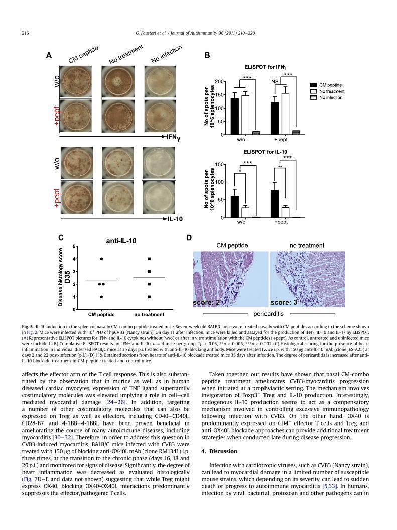
Fig. 5. IL-10 induction in the spleen of nasally CM-combo peptide treated mice. Seven-week old BALB/C mice were treated nasally with CM peptides according to the scheme shownin Fig. 2. Mice were infected with 103 PFU of hpCVB3 (Nancy strain). On day 11 after infection, mice were killed and assayed for the production of IFNg, IL-10 and IL-17 by ELISPOT.(A) Representative ELISPOT pictures for IFNg and IL-10 cytokines without (w/o) or after in vitro stimulation with the CM peptides (þpept). As control, untreated and uninfected micewere included. (B) Cumulative ELISPOT results for IFNg and IL-10, n ¼ 4 mice per group. *p < 0.05, **p < 0.005, ***p < 0.001. (C) Histological scoring for the presence of heartinflammation in individual diseased BALB/C mice at 35 days p.i. treated with anti-IL-10 blocking antibody. Mice were treated twice i.p. with 150 mg anti-IL-10 mAb (clone JES-A25) atdays 2 and 22 post-infection (p.i.). (D) H & E stained sections from hearts of anti-IL-10 blockade treated mice 35 days after infection. The degree of pericarditis is increased after anti-IL-10 blockade treatment in CM-peptide treated and control mice.
G. Fousteri et al. / Journal of Autoimmunity 36 (2011) 210e220216
affects the effector arm of the T cell response. This is also substan-tiated by the observation that in murine as well as in humandiseased cardiac myocytes, expression of TNF ligand superfamilycostimulatory molecules was elevated implying a role in cellecellmediated myocardial damage [24e26]. In addition, targetinga number of other costimulatory molecules that can also beexpressed on Treg as well as effectors, including CD40eCD40L,CD28-B7, and 4-1BBe4-1BBL have been proven beneficial inameliorating the course of many autoimmune diseases, includingmyocarditis [30e32]. Therefore, in order to address this question inCVB3-induced myocarditis, BALB/C mice infected with CVB3 weretreated with 150 mg of blocking anti-OX40L mAb (clone RM134L) i.p.three times, at the transition to the chronic phase (days 16, 18 and20 p.i.) andmonitored for signs of disease. Significantly, the degree ofheart inflammation was decreased as evaluated histologically(Fig. 7DeE and data not shown) suggesting that while Treg mightexpress OX40, blocking OX40-OX40L interactions predominantlysuppresses the effector/pathogenic T cells.
Taken together, our results have shown that nasal CM-combopeptide treatment ameliorates CVB3-myocarditis progressionwhen initiated at a prophylactic setting. The mechanism involvesinvigoration of Foxp3þ Treg and IL-10 production. Interestingly,endogenous IL-10 production seems to act as a compensatorymechanism involved in controlling excessive immunopathologyfollowing infection with CVB3. On the other hand, OX40 ispredominantly expressed on CD4þ effector T cells and Treg andanti-OX40L blockade approaches can provide additional treatmentstrategies when conducted late during disease progression.
4. Discussion
Infection with cardiotropic viruses, such as CVB3 (Nancy strain),can lead to myocardial damage in a limited number of susceptiblemouse strains, which depending on its severity, can lead to suddendeath or progress to autoimmune myocarditis [5,33]. In humans,infection by viral, bacterial, protozoan and other pathogens can in

Fig. 6. Anti-CD25 treatment depletes Treg and effector cells while it upregulates bystander IL-10 production in the spleen. Mice were treated with anti-CD25 (clone PC61) depletingantibody four times i.p. with 150 mg at days �8, �2, 18 and 22 p.i., n ¼ 3e4 mice per group. (A) Spleen cells from CM-peptide-immunized treated mice were stained and analyzed forflow cytometry to determine the frequency CD4þCD25þFoxp3þ (Treg) on days 11 and 35 after infection. The frequency was determined after gating on total CD4þ T cells. (B)Representative ELISPOT results for IL-10 without or after stimulation with the CM peptides in mice treated or not treated with the anti-CD25 depleting antibody.
G. Fousteri et al. / Journal of Autoimmunity 36 (2011) 210e220 217
some severe cases cause heart infarction followed by heart failureand in some rare cases death [34,35]. The most common virusassociated with persisting, chronic inflammation of the myocar-dium in humans is CVB3. Interestingly, in the majority of thesecases, persistent viral genome can be found, suggesting that theremaining inflammatory response is due to an ongoing antiviralimmunity [36]. Despite the lack of strong direct evidence inhumans [37], results in mice suggest that autoimmunity as a resultof cardiotropic CVB3 infection can occur [8]. In such cases, cardiacmyosin specific antibodies and T cell clones that persist after viralclearance can be found that can cause de novo myocarditis uponadoptive transfer into unmanipulated hosts [10e12]. Here, we tookadvantage of a virally-induced myocarditis model, where acutedisease followed by sudden death and progression to autoimmunemyocarditis can be recapitulated in BALB/C male mice followinginfection with CVB3. Given the difficulties to fully translate ourfindings to humans, we were able to show that nasal CM-peptidetreatment initiated at a prophylactic setting can greatly reduceacute infection-induced death and inhibit transition to the chronicphase. Moreover, further targeting the OX40eOX40L costimulatorypathway during the transition to the chronic stage, strongly haltedmyocarditis progression.
Amelioration of disease progression following autoantigenadministration in a tolerogenic manner is known to occur in severalautoimmune settings [38], including diseases induced after path-ogen infection [39]. Of importance to myocarditis, whole CM nasaladministration or oral CM-peptide (614e629) treatment was ableprotect susceptible mice from EAM [18,19]. EAM studies showedthat IL-10 induction and Treg invigorationwas involved, which shutdown the effector Th1 and Th17 response. Recent findings attributea detrimental role for IL-23 and IL-17 in EAM. Mice lacking T-betexpression and thus IFNg responses develop severe EAM, due toincreased IL-17 production in the inflamed heart [40]. In anotherstudy however, IL-17 was found to be important in postmyocarditiscardiac remodeling and the progression to DCM [33]. However, therole of IL-17 in CVB3-induced myocarditis is less clear and here wecould not attribute any specific effect on CVB3-induced diseaseprogression after nasal CM-peptide treatment.
On the other hand, we were able to show that IL-10 and Treginduction were set in place to achieve tolerance after CM-peptidecombo immunization, where CVB3 myocarditis was induced. Moreprecisely, Treg identified as CD4þCD25þFoxp3þ cells, were slightlyincreased in the blood and spleen of CM-combo immunized mice,but not in controls. In addition, adoptive transfer of CD4þCD25þ
cells from immunized but uninfected mice into new hosts, was able
to confer protection from CVB3-induced myocarditis. Interestingly,anti-CD25 depletion following CM-peptide treatment did notrestore progression to autoimmune myocarditis. To our surprise,disease progression was greatly halted. Interestingly, in anotherstudy, oral administration of antigens coupled to the B subunit ofthe cholera toxin (CTB) into mice depleted of CD25þ cells still gaverise to systemic tolerance [41]. While depletion of effector CD25þ
cells could account for this and our observation, the increased IL-10levels in the spleens of anti-CD25-treated mice suggest that inCVB3-infected animals IL-10 plays a significant role in controllingdisease severity per se.
IL-10 seemed to play a role in mediating tolerance inductionafter CM-combo peptide treatment; while IL-10 levels increased inspleens of CM-treated mice, IFNg levels were reduced. The spleenseems to be a vital organ in long-term tolerance maintenanceinduced after mucosal antigen administration in mice involvingTreg and IL-10 (Tr1) induction [42]. In the present study, bystanderIL-10 productionwas increased in CVB3 infected compared to naïvemice. No direct evidence regarding the source of IL-10 was seen,however our CD25-depletion studies suggest that IL-10 wasderived from a non-CD25þ T cell source, perhaps Tr1 cells [43]. Therole of IL-10 in CM peptide induced tolerance and CVB3-inducedmyocarditis was revealed through an additional approach. Whenanti-IL-10 neutralizing antibodies were concomitantly adminis-teredwith CM-combo peptide treatments, diseasewas significantlyexacerbated. The severity was worse than in the control, non anti-IL-10-treated mice, suggesting that IL-10 production is among themechanisms taking place that control excessive immunopathologydue to CVB3 infection. Interestingly, in studies of lymphocyticchoriomeningitis virus (LCMV) in mice, IL-10 was associated withviral persistence [44,45]. Moreover, in the acute LCMV infectionmodel followed by viral clearance, IL-10 production directlyinhibited effector and memory CD4 T cell responses [46]. These andour observations here suggest that IL-10 production following viralinfection is involved in controlling excessive CD4 T cell immunity.
In addition to enhancing Treg generation, we reveal anotherapproach to therapy of myocarditis. Costimulatory receptorsignaling to T cells has been found to be important for the fulldifferentiation of CD4þ cells into Th1, Th2 and more recently Th17phenotypes [47e49]. Of importance to myocarditis, OX40 (CD134),a costimulatory receptor of the TNFR superfamily, was detected onpathogenic effector T cells, coinciding with increased levels ofOX40-ligand (OX40L) expression on diseased cardiomyocytes[24e26]. Whether increased expression of TNF-superfamily cos-timulatory molecules on cardiac myocytes represents the cause or

Fig. 7. OX40 is expressed predominantly on Treg and CD8þ cells; Reduced cardiac inflammation after i.p. anti-OX40L blockade treatment. Eight-week old BALB/C mice were infectedwith 103 PFU of hpCVB3 (Nancy strain). (A) Representative flow cytometry plot depicting Treg versus non-Treg gating based on which OX40 mean fluorescent intensity (MFI) levelswere analyzed. (B) Histogram overlay showing the levels of OX40 on Treg (blue line), non-Treg (red line) and CD8þ cells (black line) versus isotype control (filled gray line). (C)Splenocytes from CVB3-infected mice were stained and analyzed with flow cytometry to determine the MFI of OX40 on Treg, non-Treg and total CD8þ T cells for the indicated timepoints post-infection (p.i.) with CVB3. n ¼ 3e4 mice per group. (DeE) Mice were treated i.p. with 150 mg of blocking anti-OX40L mAb (clone RM134L) three times, at days 16, 18 and20 p.i. Severity scores of individual diseased BALB/C mice at 35 days p.i. are shown in (D), while in (E) H & E stained sections from their hearts. (For interpretation of the references tocolor in this figure legend, the reader is referred to the web version of this article.)
G. Fousteri et al. / Journal of Autoimmunity 36 (2011) 210e220218
the result of the disease is not clear. Infiltrating cells producing Th1cytokines such as IFNg, could induce the expression of cos-timulatory molecules, while the virus might directly induce theirexpression which in turn leads to activation of T cells [26]. Perhapsfitting with the latter scenario, signaling through OX40-ligand wasshown to induce vascular endothelial cells to produce RANTES/CCL5 [50] which might selectively facilitate the diapedesis of Th1pathogenic T cells at the site of inflammation [51]. Interestingly, inEAM, targeting CD28/CTLA-4/B7, CD40/CD40L, ICOS/ICOSL and 4-1BB/4-1BBL costimulatory pathways attenuated disease develop-ment [30,32,52e54]. Therefore we reasoned that targeting theOX40eOX40L signaling cascade through anti-OX40L blockadewould result in further halting disease progression.
Apart from the potentially indirect role in recruiting effector Tcells at the site of inflammation, OX40 has been shown to directlymediate the binding of T cells to vascular endothelial cells [47,55].On the other hand, OX40 costimulation in other systems has been
shown to inhibit the generation of Foxp3þ aTreg from naïveprecursors, suggesting that OX40 can both promote effector T cellgeneration while at the same time antagonizing the differentiationof aTreg [56e58]. Alternatively, some studies have highlighted thatOX40 might also positively aid natural Treg survival or theirresponse to IL-2 in some situations [59,60]. Given the potentialcomplexity of blocking OX40L in a situation where both effector Tcells and Treg were responding, we initially monitored theexpression levels of OX40 during the course of CVB3 infection.While no significant expressionwas detected on CD8þ T cells, CD4þ
T cells and Treg expressed OX40 particularly at the peak of theantiviral response. Based on these findings, we reasoned to blockthe OX40 signaling axes at a window, where the Treg compartmentand the anti-CVB3 responsemight be less affected. To this end, anti-OX40L blocking antibody was administered in CVB3-infected miceat the transition phase toward autoimmune myocarditis. Quitesignificantly, we found no exacerbation in disease progression as

G. Fousteri et al. / Journal of Autoimmunity 36 (2011) 210e220 219
might be expected if OX40were supporting Treg activity, but ratherwe observed a strong reduction in disease severity implying anaction in blocking the effector/pathogenic response.
Altogether, in the present study wewere able to show that nasaladministration of CM peptides, initiated before CVB3 infection, cansignificantly reduce acute and chronic myocarditis in BALB/C malemice. The key finding was that therapeutic activity is more efficientwhen treatment is initiated before the onset of the autoimmunedisease and this activity involves Treg induction and IL-10increases. How this approach might impact the physiologicalantiviral response is not clear, however no significant alterations inendogenous CVB3 titers were seen. Translating our findings toclinical setting faces many obstacles posed by the difficulty inattributing the cause of heart immune pathology to a viral orautoimmune cause. Interestingly though, in our acute CVB3-induced myocarditis studies, where immune pathology is mostlyvirally-driven, a significant reduction in mortality was seen, sug-gesting that such an approachwould be beneficial in treating CVB3-induced myocarditis or other infection associated autoimmunedisorders. In line with these observations, female BALB/C mice areless susceptible to acute myocarditis, and some studies implicateestrogens in the activity of Treg [20]. Altogether, our results alongwith previous studies suggest that CM-combo peptide treatmentmight be proven beneficial in viral-induced myocarditis, since Treginvigoration could result in reduced damage to the host and controlof excessive immunopathology.
5. Conclusions
Our findings highlight the importance of Treg and IL-10 incontrolling excessive immunopathology following acute CVB3infection as well as progression to autoimmune myocarditis.
Acknowledgments
This work was funded by an NIAID P01 grant to MGvH. GeorgiaFousteri was an American Heart Association Western States Affil-iate Awardee for the academic period 2008e2010. Additionalsupport fromNIH grants AI67341 and CA91837 toMC. The sponsorsfor this study had no role in study design; in the collection, analysis,and interpretation of data; in the writing of the report; and in thedecision to submit the paper for publication.
References
[1] Afanasyeva M, Georgakopoulos D, Rose NR. Autoimmune myocarditis: cellularmediators of cardiac dysfunction. Autoimmun Rev 2004;3:476e86.
[2] Leuschner F, Katus HA, Kaya Z. Autoimmune myocarditis: past, present andfuture. J Autoimmun 2009;33:282e9.
[3] Rose NR, Herskowitz A, Neumann DA. Autoimmunity in myocarditis: modelsand mechanisms. Clin Immunol Immunopathol 1993;68:95e9.
[4] Taqueti VR, Mitchell RN, Lichtman AH. Protecting the pump: controllingmyocardial inflammatory responses. Annu Rev Physiol 2006;68:67e95.
[5] Cihakova D, Rose NR. Pathogenesis of myocarditis and dilated cardiomyop-athy. Adv Immunol 2008;99:95e114.
[6] Henke A, Huber S, Stelzner A, Whitton JL. The role of CD8þ T lymphocytes incoxsackievirus B3-induced myocarditis. J Virol 1995;69:6720e8.
[7] Huber SA, Gauntt CJ, Sakkinen P. Enteroviruses and myocarditis: viral path-ogenesis through replication, cytokine induction, and immunopathogenicity.Adv Virus Res 1998;51:35e80.
[8] Fairweather D, Rose NR. Coxsackievirus-induced myocarditis in mice: a modelof autoimmune disease for studying immunotoxicity. Methods2007;41:118e22.
[9] Cihakova D, Sharma RB, Fairweather D, Afanasyeva M, Rose NR. Animalmodels for autoimmune myocarditis and autoimmune thyroiditis. MethodsMol Med 2004;102:175e93.
[10] Neu N, Beisel KW, Traystman MD, Rose NR, Craig SW. Autoantibodies specificfor the cardiac myosin isoform are found in mice susceptible to Coxsack-ievirus B3-induced myocarditis. J Immunol 1987;138:2488e92.
[11] Neumann DA, Lane JR, LaFond-Walker A, Allen GS, Wulff SM, Herskowitz A,et al. Heart-specific autoantibodies can be eluted from the hearts of Cox-sackievirus B3-infected mice. Clin Exp Immunol 1991;86:405e12.
[12] Neumann DA, Rose NR, Ansari AA, Herskowitz A. Induction of multiple heartautoantibodies in mice with coxsackievirus B3- and cardiac myosin-inducedautoimmune myocarditis. J Immunol 1994;152:343e50.
[13] Lane JR, Neumann DA, Lafond-Walker A, Herskowitz A, Rose NR. LPS promotesCB3-inducedmyocarditis in resistantB10.Amice.Cell Immunol1991;136:219e33.
[14] Pummerer CL, Luze K, Grassl G, Bachmaier K, Offner F, Burrell SK, et al.Identification of cardiac myosin peptides capable of inducing autoimmunemyocarditis in BALB/c mice. J Clin Invest 1996;97:2057e62.
[15] Nagler-Anderson C, Terhoust C, Bhan AK, Podolsky DK. Mucosal antigenpresentation and the control of tolerance and immunity. Trends Immunol2001;22:120e2.
[16] Mestecky J, Russell MW, Elson CO. Perspectives on mucosal vaccines: ismucosal tolerance a barrier? J Immunol 2007;179:5633e8.
[17] von Herrath MG, Harrison LC. Antigen-induced regulatory T cells in autoim-munity. Nat Rev Immunol 2003;3:223e32.
[18] Gonnella PA, Del Nido PJ, McGowan FX. Oral tolerization with cardiac myosinpeptide (614e629) ameliorates experimental autoimmune myocarditis: roleof STAT 6 genes in BALB/CJ mice. J Clin Immunol 2009;29:434e43.
[19] Kaya Z, Dohmen KM, Wang Y, Schlichting J, Afanasyeva M, Leuschner F, et al.Cutting edge: a critical role for IL-10 in induction of nasal tolerance inexperimental autoimmune myocarditis. J Immunol 2002;168:1552e6.
[20] Huber SA. Coxsackievirus B3-induced myocarditis: infection of females duringthe estrus phase of the ovarian cycle leads to activation of T regulatory cells.Virology 2008;378:292e8.
[21] Smith SC, Allen PM. Neutralization of endogenous tumor necrosis factor amelio-rates the severity of myosin-induced myocarditis. Circ Res 1992;70:856e63.
[22] Bachmaier K, Pummerer C, Kozieradzki I, Pfeffer K, Mak TW, Neu N, et al. Low-molecular-weight tumor necrosis factor receptor p55 controls induction ofautoimmune heart disease. Circulation 1997;95:655e61.
[23] PoffenbergerMC, Straka N, ElWarry N, Fang D, Shanina I, HorwitzMS. Lack of IL-6during coxsackievirus infection heightens the early immune response resulting inincreased severity of chronic autoimmune myocarditis. PLoS One 2009;4:e6207.
[24] Seko Y, Ishiyama S, Nishikawa T, Kasajima T, Hiroe M, Suzuki S, et al.Expression of tumor necrosis factor ligand superfamily costimulatory mole-cules CD27L, CD30L, OX40L and 4-1BBL in the heart of patients with acutemyocarditis and dilated cardiomyopathy. Cardiovasc Pathol 2002;11:166e70.
[25] Seko Y, Takahashi N, Oshima H, Shimozato O, Akiba H, Kobata T, et al.Expression of tumour necrosis factor (TNF) receptor/ligand superfamilyco-stimulatory molecules CD40, CD30L, CD27L, and OX40L in murine heartswith chronic ongoing myocarditis caused by coxsackie virus B3. J Pathol 1999;188:423e30.
[26] Seko Y, Takahashi N, Oshima H, Shimozato O, Akiba H, Takeda K, et al.Expression of tumour necrosis factor (TNF) ligand superfamily co-stimulatorymolecules CD30L, CD27L, OX40L, and 4-1BBL in murine hearts with acutemyocarditis caused by Coxsackievirus B3. J Pathol 2001;195:593e603.
[27] Filippi CM, Estes EA, Oldham JE, von Herrath MG. Immunoregulatory mech-anisms triggered by viral infections protect from type 1 diabetes in mice. J ClinInvest 2009;119:1515e23.
[28] Fousteri G, Dave A, Bot A, Juntti T, Omid S, von HerrathM. Subcutaneous insulinB:9-23/IFA immunisation induces Tregs that control late-stage prediabetes inNOD mice through IL-10 and IFNgamma. Diabetologia 2010; 53:1958e1970.
[29] Chatzidakis I, Fousteri G, Tsoukatou D, Kollias G, Mamalaki C. An essential rolefor TNF in modulating thresholds for survival, activation, and tolerance ofCD8þ T cells. J Immunol 2007;178:6735e45.
[30] Seko Y, Takahashi N, Azuma M, Yagita H, Okumura K, Yazaki Y. Expression ofcostimulatory molecule CD40 in murine heart with acute myocarditis andreduction of inflammation by treatment with anti-CD40L/B7-1 monoclonalantibodies. Circ Res 1998;83:463e9.
[31] Seko Y, Takahashi N, Azuma M, Yagita H, Okumura K, Yazaki Y. Effects of invivo administration of anti-B7-1/B7-2 monoclonal antibodies on murine acutemyocarditis caused by coxsackievirus B3. Circ Res 1998;82:613e8.
[32] Haga T, Suzuki J, Kosuge H, Ogawa M, Saiki H, Haraguchi G, et al. Attenuationof experimental autoimmune myocarditis by blocking T cell activationthrough 4-1BB pathway. J Mol Cell Cardiol 2009;46:719e27.
[33] Baldeviano GC, Barin JG, Talor MV, Srinivasan S, Bedja D, Zheng D, et al.Interleukin-17A is dispensable for myocarditis but essential for the progres-sion to dilated cardiomyopathy. Circ Res 2010; 106:1646e1655.
[34] Feldman AM, McNamara D. Myocarditis. N Engl J Med 2000;343:1388e98.[35] Guilherme L, Kalil J. Rheumatic fever and rheumatic heart disease: cellular
mechanisms leading autoimmune reactivity and disease. J Clin Immunol2010; 30:17e23.
[36] Kuhl U, Pauschinger M, Seeberg B, Lassner D, Noutsias M, Poller W, et al. Viralpersistence in the myocardium is associated with progressive cardiacdysfunction. Circulation 2005;112:1965e70.
[37] Neumann DA, Burek CL, Baughman KL, Rose NR, Herskowitz A. Circulatingheart-reactive antibodies in patients with myocarditis or cardiomyopathy.J Am Coll Cardiol 1990;16:839e46.
[38] Fousteri G, von Herrath M, Bresson D. Mucosal exposure to antigen: cause orcure of type 1 diabetes? Curr Diab Rep 2007;7:91e8.
[39] Collins LV, Eriksson K, Ulrich RG, Tarkowski A. Mucosal tolerance to a bacterialsuperantigen indicates a novel pathway to prevent toxic shock. Infect Immun2002;70:2282e7.

G. Fousteri et al. / Journal of Autoimmunity 36 (2011) 210e220220
[40] Rangachari M, Mauermann N, Marty RR, Dirnhofer S, Kurrer MO,Komnenovic V, et al. T-bet negatively regulates autoimmune myocarditis bysuppressing local production of interleukin 17. J Exp Med2006;203:2009e19.
[41] George Chandy A, Hultkrantz S, Raghavan S, Czerkinsky C, Lebens M,Telemo E, Holmgren J. Oral tolerance induction by mucosal administration ofcholera toxin B-coupled antigen involves T-cell proliferation in vivo and is notaffected by depletion of CD25þ T cells. Immunology 2006;118:311e20.
[42] Alvarez D, Swirski FK, Yang TC, Fattouh R, Croitoru K, Bramson JL, et al.Inhalation tolerance is induced selectively in thoracic lymph nodes butexecuted pervasively at distant mucosal and nonmucosal tissues. J Immunol2006;176:2568e80.
[43] Battaglia M, Gianfrani C, Gregori S, Roncarolo MG. IL-10-producing T regula-tory type 1 cells and oral tolerance. Ann N Y Acad Sci 2004;1029:142e53.
[44] Ejrnaes M, Filippi CM, Martinic MM, Ling EM, Togher LM, Crotty S, et al.Resolution of a chronic viral infection after interleukin-10 receptor blockade.J Exp Med 2006;203:2461e72.
[45] Brooks DG, Trifilo MJ, Edelmann KH, Teyton L, McGavern DB, Oldstone MB.Interleukin-10 determines viral clearance or persistence in vivo. Nat Med2006;12:1301e9.
[46] Brooks DG, Walsh KB, Elsaesser H, Oldstone MB. IL-10 directly suppresses CD4but not CD8 T cell effector and memory responses following acute viralinfection. Proc Natl Acad Sci U S A 2010;107:3018e23.
[47] Nohara C, Akiba H, Nakajima A, Inoue A, Koh CS, Ohshima H, et al. Amelio-ration of experimental autoimmune encephalomyelitis with anti-OX40 ligandmonoclonal antibody: a critical role for OX40 ligand in migration, but notdevelopment, of pathogenic T cells. J Immunol 2001;166:2108e15.
[48] Zhang Z, Rosenbaum JT, Zhong W, Lim C, Hinrichs DJ. Costimulation of Th17cells: adding fuel or putting out the fire in the inflamed gut? Semin Immu-nopathol 2010; 32:55e70.
[49] Croft M. Co-stimulatory members of the TNFR family: keys to effective T-cellimmunity? Nat Rev Immunol 2003;3:609e20.
[50] Kotani A, Hori T, Matsumura Y, Uchiyama T. Signaling of gp34 (OX40 ligand)induces vascular endothelial cells to produce a CC chemokine RANTES/CCL5.Immunol Lett 2002;84:1e7.
[51] Kawai T, Seki M, Hiromatsu K, Eastcott JW, Watts GF, Sugai M, et al. Selectivediapedesis of Th1 cells induced by endothelial cell RANTES. J Immunol1999;163:3269e78.
[52] Ford ML, Larsen CP. Translating costimulation blockade to the clinic: lessonslearned from three pathways. Immunol Rev 2009;229:294e306.
[53] FutamatsuH, Suzuki J, KosugeH, Yokoseki O, KamadaM, ItoH, et al. Attenuationof experimental autoimmunemyocarditis by blocking activated T cells throughinducible costimulatory molecule pathway. Cardiovasc Res 2003;59:95e104.
[54] Seko Y, Takahashi N, Ishiyama S, Nishikawa T, Kasajima T, Hiroe M, et al.Expression of costimulatory molecules B7-1, B7-2, and CD40 in the heart ofpatients with acute myocarditis and dilated cardiomyopathy. Circulation1998;97:637e9.
[55] Imura A, Hori T, Imada K, Ishikawa T, Tanaka Y, Maeda M, et al. The humanOX40/gp34 system directly mediates adhesion of activated T cells to vascularendothelial cells. J Exp Med 1996;183:2185e95.
[56] Duan W, So T, Croft M. Antagonism of airway tolerance by endotoxin/lipo-polysaccharide through promoting OX40L and suppressing antigen-specificFoxp3þ T regulatory cells. J Immunol 2008;181:8650e9.
[57] Gramaglia I, Weinberg AD, Lemon M, Croft M. Ox-40 ligand: a potent cos-timulatory molecule for sustaining primary CD4 T cell responses. J Immunol1998;161:6510e7.
[58] So T, Croft M. Cutting edge: OX40 inhibits TGF-beta- and antigen-drivenconversion of naive CD4 T cells into CD25þFoxp3þ T cells. J Immunol2007;179:1427e30.
[59] Griseri T, Asquith M, Thompson C, Powrie F. OX40 is required for regulatory Tcell-mediated control of colitis. J Exp Med 2010;207:699e709.
[60] Piconese S, Pittoni P, Burocchi A, Gorzanelli A, Care A, Tripodo C, et al. A non-redundant role for OX40 in the competitive fitness of Treg in response to IL-2.Eur J Immunol 2010;40:2902e13.