Narrow structure in the coherent population trapping resonance in rubidium
11
1 Narrow structure in the coherent population trapping resonances in rubidium and Rayleigh scattering S Gateva, L Gurdev, E Alipieva, E Taskova and G Todorov Institute of Electronics, Bulgarian Academy of Sciences, 72 Tzarigradsko Chaussee, 1784 Sofia, Bulgaria e-mail: [email protected] Abstract. The measurement of the coherent-population-trapping (CPT) resonances in uncoated Rb vacuum cells has shown that the shape of the resonances is different in different cells. In some cells the resonance has a complex shape - a narrow Lorentzian structure, which is not power broadened, superimposed on the power broadened CPT resonance. The results of the performed investigations on the fluorescence angular distribution are in agreement with the assumption that the narrow structure is a result of atom interaction with Rayleigh scattering light. The results are interesting for indication of the vacuum cleanness of the cells and building of magnetooptical sensors. PACS numbers: 42.50.Gy, 33.20.Fb, 42.65.Es 1. Introduction The development of the applications of the Coherent Population Trapping (CPT) resonances in high- resolution spectroscopy, quantum information storage and processing, metrology (atomic clocks), magnetometry, lasing without inversion, laser cooling, ultraslow group velocity propagation of light, etc. [1-6] increases the interest in the processes which determine the shape of the resonances. For many applications narrow resonance signals and high signal-to-noise ratios are important. Narrow
-
Upload
independent -
Category
Documents
-
view
6 -
download
0
Transcript of Narrow structure in the coherent population trapping resonance in rubidium
1
Narrow structure in the coherent population trapping resonances in rubidium and Rayleigh scattering
S Gateva, L Gurdev, E Alipieva, E Taskova and G Todorov Institute of Electronics, Bulgarian Academy of Sciences, 72 Tzarigradsko Chaussee, 1784 Sofia, Bulgaria e-mail: [email protected]
Abstract. The measurement of the coherent-population-trapping (CPT) resonances in
uncoated Rb vacuum cells has shown that the shape of the resonances is different in different
cells. In some cells the resonance has a complex shape - a narrow Lorentzian structure, which
is not power broadened, superimposed on the power broadened CPT resonance. The results of
the performed investigations on the fluorescence angular distribution are in agreement with the
assumption that the narrow structure is a result of atom interaction with Rayleigh scattering
light. The results are interesting for indication of the vacuum cleanness of the cells and
building of magnetooptical sensors.
PACS numbers: 42.50.Gy, 33.20.Fb, 42.65.Es
1. Introduction
The development of the applications of the Coherent Population Trapping (CPT) resonances in high-
resolution spectroscopy, quantum information storage and processing, metrology (atomic clocks),
magnetometry, lasing without inversion, laser cooling, ultraslow group velocity propagation of light,
etc. [1-6] increases the interest in the processes which determine the shape of the resonances. For
many applications narrow resonance signals and high signal-to-noise ratios are important. Narrow

2
structures in the CPT resonance shape were measured at different experimental configurations, in
vacuum, buffer gas and coated cells. The effect was studied for spatially and temporary separated laser
fields.
Optical Ramsey fringes induced by Zeeman coherence were investigated in Rb by Zibrov et
al. [7,8]. Experimental data were presented showing Ramsey fringes in frequency and time domain for
vacuum cell, as well as for buffer gas cell. In a series of later works [9-12], the observed sharp central
peak on a broad pedestal in the Electromagnetically Induced Transparency (EIT) resonance shapes
(the Diffusion-Induced Ramsey Narrowing [10,11]) was studied. The broad pedestal is associated with
the single pass interaction time and is power broadened. The sharp central peak is the central Ramsey
fringe, which adds coherently for all Ramsey sequences. Its width changes with the laser beam
diameter. At low laser power, small beam diameter and low buffer gas pressure the sharp central peak
is not Lorentzian in shape and is insensitive to power broadening. At high laser intensity the central
peak loses its contrast and is Lorentzian in shape and power broadened.
Another narrow structure in the CPT resonance was registered in Rb by Alipieva et al. [13]. In
an uncoated room temperature vacuum cell, the CPT resonance was prepared in the so called Hanle
effect configuration. The observed in fluorescence narrow resonance is with Lorentzian shape, it is not
radiation broadened and its amplitude increases with the laser power. Its width (FWHM) ∆ L does not
change with the laser beam diameter and is in agreement with the assumption that the broadening is
affected mainly by relaxation that is due to atomic collisions with the cell walls. Details about the
shape of the CPT resonances and the narrow structure are given in [13-17]. The resonance shapes,
measured at different geometries of excitation and registration, show that the narrow structure at the
center of the resonance can be considered as a result of a weak field – atom interaction [17].
Narrow structures in the CPT resonance have also been measured in Na [18,19]. In [18] the
non-Lorentzian structure is explained, supposing the existence of scattered light in the volume of the

3
Figure 1. Experimental geometry.
cell. In [19] the narrow structure in Na was attributed to multiple interactions of the atoms with the
main beam. In the latter case, the comparison of the experimental and theoretical shapes shows that in
the range of +1 mG there is a good coincidence between them, but in the range of +10 mG the
experimental shape is narrower than the theoretical one.
One of the possible scattering processes influencing the CPT resonance shape is Rayleigh
scattering [20] because in this case the scattered light maintains coherence with the incident beam and
in this way a diffusion of the coherent light is created. For example, Rayleigh scattering has been used
for studying the properties of cold atoms [21] and optical lattices ([22] and references therein).
The purpose of the present work is to investigate the hypothesis of the influence of the
Rayleigh scattering light on the formation of the narrow structure in the CPT resonance described in
[13].
2. Experimental setup
All the investigations were performed under the experimental conditions of [13] in the so called Hanle
effect configuration. The experimental geometry is shown schematically in figure 1. The resonances
were examined in uncoated vacuum cells containing a natural mixture of Rb isotopes at room
temperature (300 K). A single-frequency linearly polarized diode laser beam (2 mm in diameter,
22 mW in power) was propagating along the cell’s axis x. A magnetic field Bscan, created by a
solenoid, was applied collinearly to the laser beam. The gas cell and the solenoid were placed in a 3

4
layer µ-metal magnetic shield. The fluorescence was detected at 90o to the laser beam direction by a
photodiode. The signals from the photodiode were amplified and stored in a PC, which also controlled
the magnetic field scan. All measurements were performed on the Fg =2→Fe =1 transition of the 87Rb
D1 line.
3. Shape of the CPT resonances
3.1. Experimental
The investigations of the CPT resonance in different uncoated Rb vacuum cells show that the shape of
the resonances is different in different cells. In figure 2a the CPT signals measured in two cells with
similar dimensions (cell a: length la=4.6 cm, diameter da=2.4 cm and cell b: length lb=4.8 cm, diameter
db=3.2 cm) are given. In cell a the resonance has the typical (for high power laser beam with Gaussian
distribution of the beam intensity) triangular shape. In cell b the resonance contains a narrow structure
superimposed on a broad pedestal.
3.2. Theoretical
For explanation of this difference a numerical solution of the full system of the density matrix
equations for 87Rb D1 line transition was performed following the procedure described in [17]. The
irreducible tensor operator formalism was used and the influence of the velocity distribution of the
atoms, the Gaussian distribution of the laser beam intensity and the experimental geometry was taken
into account. The theoretical shape obtained coincides with the shape of the fluorescence resonance in
cell a (figure 2b) and the pedestal of the resonance in cell b (figure 2c). The difference between the
experimental and theoretical shapes in cell b (figure 2c) is a Lorentzian which width is of the order of
1/(2 )L πτ∆ = , defined by the mean time τ between two atom collisions with the cell’s walls. This is
the narrow structure from [13].

5
-40 -20 0 20 40
0.0
0.2
0.4
0.6
0.8
1.0
Am
plit
ude (
arb
. units
)
Magnetic field (mG)
a
-40 -20 0 20 40
0.0
0.2
0.4
0.6
0.8
1.0
Am
plit
ud
e (
arb
. un
its)
Magnetic field (mG)
b
-40 -20 0 20 40
0.0
0.2
0.4
0.6
0.8
1.0
Am
plit
ud
e (
arb
. u
nits)
Magnetic field (mG)
c
Figure 2. CPT resonance normalized shapes: a/ experimental in cell a (scattered circles curve) and cell b (dash line curve); b/ experimental in cell a (scattered circles curve) and theoretical (solid curve); c/ experimental in cell b (dash line curve), theoretical (solid curve) and the difference between them (dash-dot line curve).
6
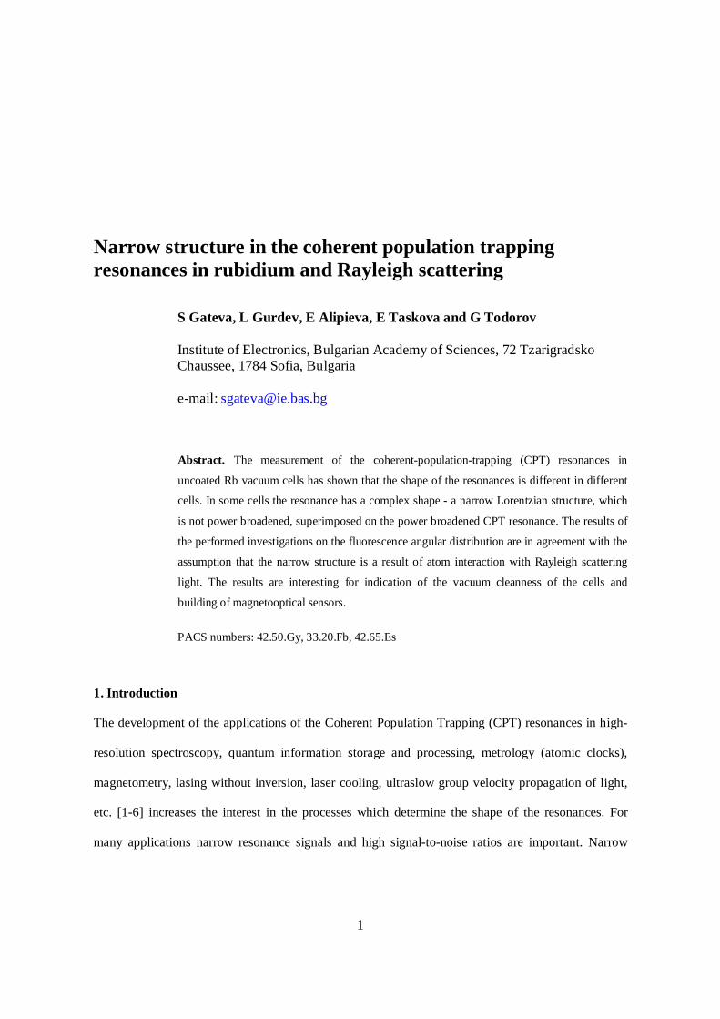
4. Rayleigh scattering light angular distribution
To check the supposition that this narrow structure is a result of Rayleigh scattered light we have
performed a series of experiments and evaluations related to the light field angular distribution. For
linear polarization of the incident light, the power of the Rayleigh-scattering light PR registered at 90o
to the laser beam direction is [20,23]:
20( , ) ( ) sinRP P Nλ ϕ α σ λ ϕ= , (1)
where P0 is the incident light power, α is the registration efficiency coefficient, N is the scattering
particles density, ( , )σ λ ϕ = ( )σ λ sin2 ϕ is the differential cross-section of Rayleigh scattering of
polarized light with wavelength λ in the plane perpendicular to the beam direction and ϕ is the angle
between the laser light polarization and the direction of registration. According to equation (1) the
Rayleigh scattering light has a maximum at ϕ =90o and is zero at ϕ =0o.
In Rb vacuum cell at room temperature the Rb pressure is of the order of 4.10-5 Pa and at this
pressure the Rayleigh scattered light from Rb atoms can not be registered in our experiment [20,23] –
this is the case of cell a. However, if the vacuum cell is not pumped very well, there will be some
residual air, water and oil vapor, as well as rare but relatively strongly scattering submicron particles,
which will scatter the light in the whole cell – this is the case of cell b. In this case we are able to
register the Rayleigh scattered light. The measurement of the power of the scattered light when the
laser is tuned out of line has the typical 2sin ϕ dependence from equation (1) with maximum at
ϕ =90o and minimum at ϕ =0o (figure 3).
7
-90 -45 0 45 90 135
50
100
150
200
250
Sca
tte
red
lig
ht p
ow
er
(arb
. u
nits
)
ϕ (o)
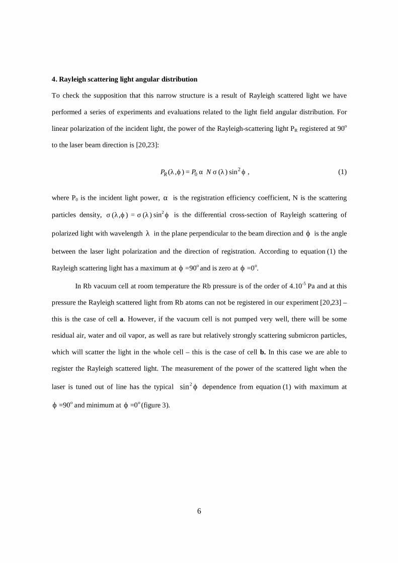
Figure 3. The measured scattered light power at 90o to the laser beam at different angle ϕ of orientation of the
polarization when the laser is out of line.
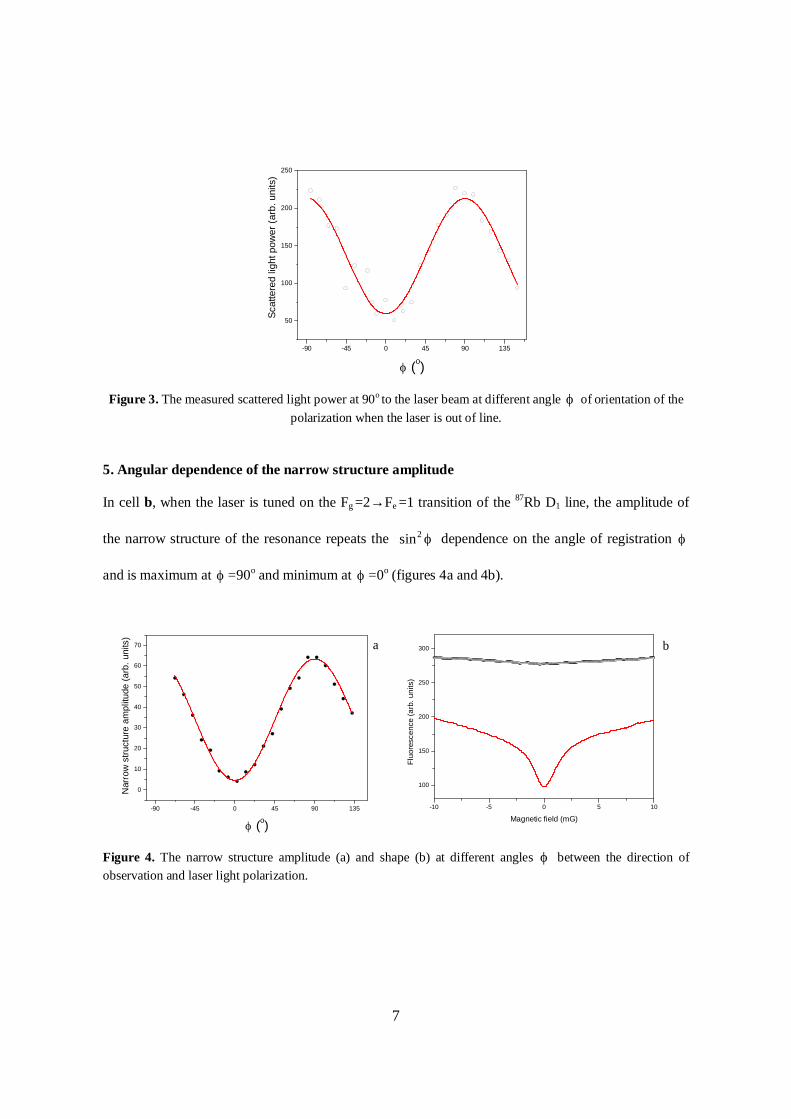
5. Angular dependence of the narrow structure amplitude
In cell b, when the laser is tuned on the Fg =2→Fe =1 transition of the 87Rb D1 line, the amplitude of
the narrow structure of the resonance repeats the 2sin ϕ dependence on the angle of registration ϕ
and is maximum at ϕ =90o and minimum at ϕ =0o (figures 4a and 4b).
Figure 4. The narrow structure amplitude (a) and shape (b) at different angles ϕ between the direction of
observation and laser light polarization.
-10 -5 0 5 10
100
150
200
250
300 b
Flu
ore
sce
nce (
arb
. u
nits)
Magnetic field (mG)
-90 -45 0 45 90 135
0
10
20
30
40
50
60
70
Narr
ow
str
uctu
re a
mp
litu
de
(a
rb.
un
its)
ϕ (o)
a

8
Theoretically, the main part of the unpolarized fluorescence intensity is determined by the
population of the upper level. The corresponding observation tensor doesn’t depend on the observation
direction. Therefore the observed amplitude dependence could be explained as reflection of the
Rayleigh scattering effect on the excitation. The resonances are with Lorentzian shape, because the
scattered light is very low in power - when the Rabi frequency of the light field Ω is small enough, so
that 2 / e gγ γΩ < ( eγ is the population decay rate from the excited state into the ground states, and
gγ is the ground state coherence relaxation rate), there is no power broadening [1]. If the mean free
path of the atom is longer than the cell dimensions, gγ is defined by the mean time between two
successive collisions with the cell walls. Since the weak field is due to Rayleigh scattering, the
amplitude of the Lorentzian will depend on the density of the scattering particles and can be used as
indicator of the level of the vacuum cleanness of the cell.
6. CPT resonance dependence on the geometry of registration
The comparison of the resonances at two different geometries of observation in Rb vacuum
cell c (length lc=2 cm, diameter dc=2 cm) shows that the broad pedestal doesn’t change, while
the shape and the width of the narrow structure change. In the first case, the photodiode is on
the cell wall (figure 5, solid curve), the angle of view is large comprising almost the whole
cell and the fluorescence mostly of atoms out of the laser beam is registered. In this case the
narrow structure is Lorentzian in shape and its width ∆ L is defined by the relaxation on the
cell walls. In the second case, a lens is used in front of the photodiode in order to restrict the
observation field of view just to the laser beam volume. In this case the measured narrow
structure is narrower than ∆ L (figure 5, scattered circles curve). For explanation of such

9
Figure 5. Narrow structure of the CPT resonance at different geometries of registration in cell c (length lc=2 cm, diameter dc=2 cm) a/ the photodiode is on the cell wall (solid curve) and b/ the light from the laser beam is projected by a lens on the photodiode (scattered circles curve).
narrowing the influence of the diffusion-induced Ramsey narrowing in presence of low
pressure buffer gas of residual air, water and other submicron particles has to be taken into
account. This result explains the difference in the resonances in [10] and [13]. In [13] the
registered fluorescence is mainly from atoms out of the laser beam volume, which interact
with the Rayleigh scattered light. In this case the narrow component is Lorentzian. In [10] the
resonance is registered in EIT and only atoms from the laser beam volume contribute to the
signal. In this case the diffusion-induced Ramsey narrowing is responsible for the narrow
structure.
7. Conclusion
The measurement of the CPT resonances in the fluorescence of uncoated Rb vacuum cells in the so
called Hanle effect configuration has shown that the shape of the resonances is different in different
cells. In some not very well pumped cells, where residual gaseous and particulate matter exists, the
resonance has a complex shape - a narrow structure which is not power broadened, superimposed on
-20 -10 0 10 20
0.0
0.2
0.4
0.6
0.8
1.0
Magnetic field (mG)
Am
plit
ud
e (
arb
. u
nits
)

10
the power broadened CPT resonance [13]. The results of the performed investigations on the
fluorescence angular distribution are in agreement with the assumption that this structure is due to
atom interaction with the Rayleigh scattering light. The shape of the narrow structure is Lorentzian
and so far as the mean free path of the atom is longer than the cell dimensions, its width is defined by
the mean time between two successive collisions with the cell walls. The investigation is interesting
for indication of the vacuum cleanness of the cells and building of magnetooptical sensors.
Acknowledgements
The authors would like to acknowledge the Bulgarian NSF for the financial support (grant DO-02-
108/2009).
References
[1] Arimondo E 1996 Coherent population trapping in laser spectroscopy Prog Opt 35 257-354
[2] Moruzzi G and Strumia F 1991 The Hanle Effect and Level-Crossing Spectroscopy (New York:
Plenum)
[3] Wynands R and Nagel A 1999 Precision spectroscopy with coherent dark states Appl Phys B 68
1-25
[4] Budker D and Romalis M 2007 Optical Magnetometry Nature Physics 3 227-234
[5] Alexandrov E B, Auzinsh M, Budker D, Kimball D F, Rochester S M and Yaschuk V V 2005
Dynamic effects in nonlinear magneto-optics of atoms and molecules JOSA B 22 7-20
[6] Budker D, Gawlik W, Kimball D F, Rochester S M, Yashchuk V V and Weis A 2002 Resonant
nonlinear magneto-optical effects in atoms Rev Mod Phys 74 1153-1201
[7] Zibrov A S and Matsko A B 2001 Optical Ramsey fringes induced by Zeeman coherence Phys
Rev A 65 013814
[8] Zibrov A S, Novikova I and Matsko A B 2001 Observation of Ramsey fringes in an atomic cell
with buffer gas Opt Lett 26 1311-1313
[9] Novikova I, Xiao Y, Phillips D F and Walsworth R L 2005 EIT and diffusion of atomic
coherence J Mod Optics 52 2381-2390
[10] Xiao Y, Novikova I, Phillips D F and Walsworth R L 2006 Diffusion-Induced Ramsey
Narrowing Phys Rev Lett 96 043601

11
[11] Xiao Y, Novikova I, Phillips D F and Walsworth R L 2008 Repeated interaction model for
diffusion-induced Ramsey narrowing Opt Express 16 14128-14141
[12] Xiao Y 2009 Spectral line narrowing in electromagnetically induced transparency Modern
Physics Letters B 23 661-680
[13] Alipieva E, Gateva S, Taskova E and Cartaleva S 2003 Narrow structure in the CPT resonance
in Rb Opt Lett 28, 1817-1819
[14] Gateva S, Alipieva E and Taskova E 2005 Power dependence of the coherent-population-
trapping resonances registered in fluorescence and transmission: Resonance-width narrowing
effects Phys Rev A 72 025805
[15] Gateva S, Petrov L, Alipieva E, Todorov G, Domelunksen V and Polischuk V 2007 Shape of
the coherent-population-trapping resonances and high-rank polarization moments Phys Rev A
76 025401
[16] Gateva S, Alipieva E, Petrov L, Taskova E and Todorov G 2007 Single frequency coherent-
population-trapping resonances for magnetic field measurement J Optoelectron Adv Mater 10
98-103
[17] Gateva S, Alipieva E, Domelunksen V, Polischuk V, Taskova E, Slavov D and Todorov G 2008
Shape of the coherent-population-trapping resonances registered in fluorescence at different
experimental geometries Proc SPIE 7027 70270I
[18] Alzetta G, Cartaleva S, Gozzini S, Karaulanov T, Lucchesini A, Marinelli C, Moi L, Nasyrov K
and Sarova V 2004 Lossless formation of Electromagnetically Induced Transparency in Sodium
atoms Proc SPIE 5449 280-291
[19] Alzetta G, Cartaleva S, Gozzini S, Karaulanov T, Lucchesini A, Marinelli C, Moi L, Nasyrov
K, Sarova V and Vaseva K 2005 Magnetic Coherence Resonance Profiles in Na and K Proc
SPIE 5830 181-185
[20] Boyd R W 1992 Nonlinear Optics (New York: Academic Press)
[21] Datsyuk V M, Sokolov I M, Kupriyanov D V and Havey M D 2006 Diffuse light scattering
dynamics under conditions of electromagnetically induced transparency Phys Rev A 74 043812
[22] Carminati F-R, Sanchez-Palencia L, Schiavoni M, Renzoni F and Grynberg G 2003 Rayleigh
Scattering and Atomic Dynamics in Dissipative Optical Lattices Phys Rev Lett 90 043901
[23] Measures R M 1984 Laser Remote Sensing: Fundamentals and Applications (New York:
Wiley)