Multifocal adenomatous oncocytic hyperplasia of the parotid gland

Na+ Modulates Anion Permeation and Blockade of P2X7 Receptorsfrom Mouse Parotid Glands
Juan Pablo Reyes2, Patricia Pérez-Cornejo1, Carmen Y. Hernández-Carballo2, AlakaSrivastava3, Victor G. Romanenko3, Mireya Gonzalez3, James E. Melvin3, and JorgeArreola2,41Facultad de Medicina, Universidad Autónoma de San Luis Potosí, San Luis Potosí, SLP 78290,México2Instituto de Física, Universidad Autónoma de San Luis Potosí, San Luis Potosí, SLP 78290, México3Center for Oral Biology, University of Rochester Medical Center, Rochester, NY 146424Pharmacology and Physiology Department, University of Rochester Medical Center, Rochester,NY 14642
AbstractWe previously reported that mouse parotid acinar cells display an anion conductance (IATPCl) whenstimulated by external ATP in Na+-free extracellular solutions. It has been suggested that the P2X7receptor channel (P2X7R) might underlie IATPCl. In this work we show that IATPCl can be activatedby ATP, ADP, AMP-PNP, ATPγS and CTP. This is consistent with the nucleotide sensitivity ofP2X7R. Accordingly, acinar cells isolated from P2X7R−/− mice lacked IATPCl. Experiments withP2X7R heterologously expressed resulted in ATP-activated currents (IATP-P2X7) partially carried byanions. In Na+-free solutions, IATP-P2X7 had an apparent anion permeability sequence of SCN− >I− ≅ NO3
− > Br− > Cl− > acetate, comparable to that reported for IATPCl under the same conditions.However, in the presence of physiologically relevant concentrations of external Na+, the Cl−permeability of IATP-P2X7 was negligible, albeit permeation of Br− or SCN− was clearly resolved.Relative anion permeabilities were not modified by addition of 1 mM Carbenoxolone—a blocker ofPannexin-1. Moreover, Cibacron Blue 3GA, which blocks the Na+ current activated by ATP in acinarcells but not IATPCl, blocked IATP-P2X7 in a dose-dependent manner when Na+ was present, but failedto do so in TEA+-containing solutions. Thus, our data indicate that P2X7R is fundamental forIATPCl generation in acinar cells and that external Na+ modulates ion permeability and conductivityas well as drug affinity in P2X7R.
KeywordsSalivary Glands; P2X7 Receptors; Anion Permeability; Fluid Secretion; ATP and Na+
IntroductionAdenosine 5′- triphosphate (ATP) acts as an external signaling molecule in a variety of tissues(for reviews see North, 2002 and Burnstock, 2007). Many of the cellular responses to externalATP are mediated by activation of either G-protein coupled metabotropic P2Y and/orionotropic P2X purinoceptors present in the plasma membrane of cells in numerous tissues
Corresponding Author Jorge Arreola, Ph.D. Instituto de Física-UASLP Dr. Manuel Nava #6 San Luis Potosí, SLP 78290 México Tel.52(444)8262363 Fax 52(444)8133874 [email protected].
NIH Public AccessAuthor ManuscriptJ Membr Biol. Author manuscript; available in PMC 2009 December 10.
Published in final edited form as:J Membr Biol. 2008 May ; 223(2): 73–85. doi:10.1007/s00232-008-9115-7.
NIH
-PA Author Manuscript
NIH
-PA Author Manuscript
NIH
-PA Author Manuscript

(Burnstock, 2007; North and Barnard, 1997; Ralevic and Burnstock, 1998). Epithelial cellsexpress both P2Y and P2X receptors (Tenneti et al., 1998; Fukushi, 1999; Schwiebert andZsembery, 2003; Li et al., 2003, Hayashi et al., 2005; Ma et al., 2006). While the detailedphysiological role of these receptors remains unclear, it has been shown that external ATPinduces multiple cellular events important to the functions of epithelia (Roman and Fitz,1999; Leipziger, 2003). For example, external ATP increases both cationic and anionicconductances in the plasma membrane of epithelial cells (Leipziger, 2003; Li et al., 2005;Arreola and Melvin, 2003). Specifically, activation of P2Y receptors generates a G protein-coupled increase in InsP3 that leads to Ca2+ release from intracellular stores whilst activationof P2X receptor channels directly mediates Na+ and Ca2+ entry (for a complete review seeBurnstock, 2007). The resulting increase in [Ca2+]i associated with activation of either classof P2 receptors is likely to be important for epithelial fluid secretion since Ca2+ induces openingof Ca2+-dependent Cl− and K+ channels which are key to initiate and to sustain fluid andelectrolyte secretion (Melvin et al., 2005).
Salivary acinar cells express at least four different types of purinoceptors, P2Y1 and P2Y2 aswell as P2X4 and P2X7 (Turner et al., 1999; Li et al., 2003) whose stimulation results in anincrease in intracellular Ca2+ (McMillan et al., 1993). Although the physiological role ofP2X4 and P2X7 ionotropic receptors expressed in these cells still remains unknown, it has beenshown that stimulation of exocrine gland acinar cells with external ATP activates Ca2+-dependent ion currents (Li et al., 2003) including those associated with Ca2+-dependent Cl−channels. Additionally, in mouse parotid acinar cells bathed in Na+-free medium, external ATPactivates a Ca2+-independent anion conductance named IATPCl (Li et al., 2005; Arreola andMelvin 2003). We found that the kinetics and channel pharmacology of IATPCl are differentfrom other anion channels previously described in salivary gland acinar cells, including theCa2+-dependent, ClC-2, CFTR and volume-sensitive Cl channels (Arreola and Melvin 2003;Zeng et al., 1997). Moreover, the P2X receptor antagonist Cibacron Blue 3GA (CB) did notblock IATPCl when recorded in solutions containing TEACl and 0 Na+. In contrast, CB blocksthe ATP-activated Na+ current presumably flowing through P2X receptors (Arreola andMelvin, 2003). Thus, we suggested that IATPCl has an anion conductance component with novelcharacteristics, including more sensitivity to Bz-ATP than ATP and resistance to the P2Xreceptor antagonist CB. Recently, Li et al. (2005), using acinar and duct cells from parotidglands of wild type and P2X7R−/− mice, suggested that IATPCl results from regulation ofP2X7R by external Cl− and Na+. They also concluded that P2X7R are not permeable to Cl−.This study prompted us to further investigate the role of P2X7R in the generation of IATPClusing a pharmacological approach and P2X7R−/− mice. In addition, recombinant mouseP2X7R cloned from parotid acinar cells was used to determine if the resulting ATP-activatedcurrents (IATP-P2X7) displayed anion permeability sensitive to CB and extracellular Na+ similarto IATPCl. Our data shows that P2X7R is essential for IATPCl generation, that IATP-P2X7 hasindeed anion permeability similar to that of IATPCl and a conductivity which is modulated byextracellular Na+. Furthermore, CB blocks P2X7R when Na+ is present but in the absence ofexternal Na+ CB potentiates the current through the receptor. Thus, our data indicate thatP2X7R is fundamental for IATPCl generation and that the functional structure of P2X7R maybe critically dependent on external Na+.
MethodsP2X7R−/− mice
P2X7R−/− mice were kindly provided by Dr. Christopher A. Gabel (Solle et al., 2001) and bredat the University of Rochester’s vivarium. Wild type (WT) C57BL/6J (Jackson Laboratories,ME) and P2X7R null mice age 2 to 6 months were used following protocols approved by theAnimal Resources Committee of the University of Rochester.
Reyes et al. Page 2
J Membr Biol. Author manuscript; available in PMC 2009 December 10.
NIH
-PA Author Manuscript
NIH
-PA Author Manuscript
NIH
-PA Author Manuscript
Cloning of P2X7R and P2X4R from the mouse parotid glandTotal RNA was isolated from mouse parotid gland (RNEASY Mini Kit, Qiagen, Valencia,CA) followed by DNAse treatment. First-strand cDNA was synthesized from total RNA usingthe ISCRIPT cDNA synthesis kit (BIO-RAD, Hercules, CA). Full-length cDNAs wereamplified by PCR using the following gene specific primers:
P2X7R (accession no. NM_011027): 5′-GAA TTC GGC TTA TGC CGG CTT GCTGC-3′; 5′-CGG CTT TCA GTA GGG ATA CTT GAA GCC-3′
P2X4R (accession no. AF089751): 5′-CAT TAG AAT TCA CGA CGC GGA GCA GC-3′;5′-CGA GAG AAT TCG GTT AGG CAT CAC TGG TC-3′
PCR products were isolated (PCR cleaning kit, Qiagen, Valencia, CA) and their sequencesverified. The 5′ and 3′ untranslated regions of P2X7R and P2X4R contained EcoRI sites tofacilitate insertion of each clone into a bicistronic expression vector (pIRES2-EGFP, Clontech),generating pIRES2-EGFP-P2X7R and pIRES2-EGFP-P2X4R vectors, respectively. Inaddition, the mouse P2X7R clone was inserted in an inverted fashion (pIRES2-EGFP-P2X7Rinv) into the pIRES2-EGFP vector as a negative control. The cDNA sequences obtainedfor mouse parotid P2X7R and P2X4R were identical to those previously published in the NCBIdata base).
Biotinylation of Cell Surface ProteinsBiotinylation of cell surface proteins was performed according to the manufacturer’sinstructions (Pierce, Rockford, IL). Parotid acinar cells were prepared as previously described(Evans et al., 1999). Briefly, C57Bl/6J +/+ and P2X7−/− mice were rendered unconscious byexposure to CO2 and killed by exsanguinations prior to isolation of cells by collagenasedigestion. Dispersed cells were washed twice with ice-cold PBS (pH 8.0) prior to the additionof 80 μl of 10 mM Sulfo NHS-SS-Biotin stock solution per ml of PBS. After 30 minutes atroom temperature (22 °C), the reaction was stopped by addition of ice-cold 50 mM Tris (pH8.0) for 10 minutes, and then the cells were washed twice with ice-cold PBS.
Preparation of Crude and Affinity-enriched Plasma Membrane ProteinsBiotinylated cells were collected by centrifugation at 1,000g for 45 sec and the pelletresuspended in 5 ml of ice-cold homogenizing buffer containing: 250 mM sucrose (JT Baker;Philipsburg, NJ), 10 mM triethanolamine, leupeptin (1 μg/ml) and phenylmethanesulfonylfluoride (0.1 mg/ml). Cells were homogenized with ULTRA TURRAX T8 Homogenizer (IKA,Werke, Germany). Unbroken cells and nuclei were pelleted at 4,000g for 10 minutes at 4°C.The supernatant was saved and the pellet resuspended and centrifuged in the same volume ofhomogenization buffer as before. The collected supernatants were pooled and centrifuged at22,000g for 20 min at 4°C. The pellet was suspended in the same buffer and centrifuged at46,000g (Beckman SS28 rotor) for 30 minutes at 4°C. The resultant crude pellet wasresuspended in 1 ml of hypotonic buffer containing 10 mM HEPES (pH 7.5), 1.5 mMMgCl2, 10 mM KCl, Complete Protease Inhibitor Cocktail (1 tablet/50 ml; Roche AppliedScience, Indianapolis, IN), 1mM Na3VO4 and 1 mM NaF, and then incubated with 200 μl ofDYNABEADS M-280 streptavidin at 4°C overnight. Beads were collected with a magneticplate and washed with hypotonic buffer to obtain the membrane associated fractions. Thestreptavidin beads carrying the enriched plasma membrane fractions were suspended in 100mM DTT for 2 hr according to the manufacturer’s instructions (Dynal Biotech ASA, Oslo,Norway) and then centrifuged at 10,600g for 3 minutes. The supernatants were collected andused for electrophoretic analysis.
Reyes et al. Page 3
J Membr Biol. Author manuscript; available in PMC 2009 December 10.
NIH
-PA Author Manuscript
NIH
-PA Author Manuscript
NIH
-PA Author Manuscript
Electrophoresis and Western Blot AnalysisProtein (30 μg) was heated at 95°C for 5 minutes prior to separation in a 7.5% or 10% SDS-PAGE Tris-glycine mini-gel (Bio-Rad; Hercules, CA). Protein was transferred ontopolyvinylidene (PVDF) membranes (Invitrogen, Carlsbad, CA) overnight at 4°C using atransfer buffer containing 10 mM CAPS (3-[Cyclohexylamino]-1-propanesulphonic acid), pH11 in 10% methanol. Membranes were blocked overnight at 4°C with 5% non-fat dry milk in25 mM Tris pH 7.5, 150 mM NaCl (TBS) and then incubated with primary antibody forP2X7R (Millipore-Chemicon International Inc.; Temecula, CA) at a dilution of 1:300 in 2.5%non-fat dry milk solution at 4°C overnight. After washing with TBS containing 0.05%Tween-20 (TBS-T), the membranes were incubated with horseradish peroxidase-conjugatedgoat-anti rabbit IgG secondary antibody (Pierce, Rockford, IL) at a dilution of 1:2,500 in TBS-T/2.5% non-fat dry milk for 1 h at room temperature. Labeled proteins were visualized usingenhanced chemiluminescence (ECL detection kit, GE-Amersham Biosciences; Piscataway,NJ).
Single parotid acinar cell isolationSingle acinar cells were isolated from wild type or P2X7R−/− mice parotid glands as previouslydescribed (Arreola et al., 1995). Briefly, glands were dissected from exsanguinated mice underCO2 anesthesia and minced in Ca2+-free minimum essential medium (MEM; Gibco BRL,Gaithersburg, MD) supplemented with 1% bovine serum albumin (BSA; Fraction V, SigmaChemical, St. Louis, MO). The tissue was treated for 20 min (37°C) in MEM Ca2+-free solutioncontaining 0.02% trypsin + 1 mM EDTA (ethylenediaminetetraacetic acid) + 2 mM glutamine+ 1% BSA. Digestion was stopped with 2 mg/ml of soybean trypsin inhibitor (Sigma Chemical,Saint Louis, MO) and the tissue further dispersed by two sequential treatments of 60 min eachwith collagenase (0.04 mg/ml; Boehringer Mannheim, Indianapolis, IN) in MEM-Ca2+ free +2 mM glutamine + 1% BSA. Dispersed cells were centrifuged and washed with basal mediumEagle (BME; Gibco BRL, Gaithersburg, MD). The final pellet was resuspended in BME + 2mM glutamine and cells plated onto poly-L-lysine-coated glass coverslips forelectrophysiological recordings.
Culture and transient transfection of HEK-293 cellsHEK-293 cells (InVitrogen; Carlsbad, California, USA) were maintained in DMEM medium(Gibco, BRL, Gaithersburg, MD) at 37°C in a 95% O2/ 5% CO2 atmosphere. Cells were grownon 30 mm Petri dishes to 50-60% confluence and then transfected with either pIRES2-EGFP-P2X7R, pIRES2-EGFP-P2X4R or pIRES2-EGFP-P2X7RInv DNA (2 μg per dish of a 1 μg/μlstock) according to the manufacturer instructions using POLYFECT transfection reagents(Qiagen; Valencia, CA). Transfected cells were detached from culture dishes using trypsin,plated onto 5 mm glass coverslips and allowed to re-attach for at least 5 h before use.
Electrophysiological RecordingsA single coverslip containing either freshly isolated mouse parotid acinar or transfectedHEK-293 cells was placed in a recording chamber mounted on the stage of a Nikon invertedfluorescence microscope equipped with UV illumination. EGFP fluorescence observed underblue light (~488 nm) illumination was used to identify transfected HEK-293 cells.
Currents were recorded at room temperature (20-22°C) using the conventional whole-cellpatch-clamp configuration (Hamill at al., 1981) and an Axopatch 200B amplifier (AxonInstruments; Foster City, CA). Pipettes made with Corning 8161 glass (Warner InstrumentsInc.; Hamden, CT) have a resistance between 3-4 MΩ when filled with pipette solutions. Thestandard pipette (internal) solution contained (in mM): TEA+(or Na+) Cl 140, EGTA 20,HEPES 20 (pH 7.3; tonicity was ~335 mosm/kg). Cells were bathed in a standard external
Reyes et al. Page 4
J Membr Biol. Author manuscript; available in PMC 2009 December 10.
NIH
-PA Author Manuscript
NIH
-PA Author Manuscript
NIH
-PA Author Manuscript

solution containing (in mM): TEA+(or Na+) Cl 140, CaCl2 0.5, D-mannitol 100, HEPES 20(pH 7.3; tonicity was ~375 mosm/kg). A 3 M KCl agar-bridge was used to ground a 300 μlrecording chamber. The internal solution was Ca2+-free while the external was hypertonic topreclude activation of Ca2+-dependent and volume-sensitive chloride currents, respectively,which are endogenous to mouse parotid acinar and HEK-293 cells (Perez-Cornejo and Arreola,2004; Hernandez-Carballo, et al., 2003). Tonicity of the solutions was determined using a vaporpressure osmometer (Wescor; Logan, UT).
Voltage ramps (from −100 to +100 mV in 1 s; every 2.5 seconds) were used to generate I-Vcurves and to estimate reversal potentials. To determine PTEA/PCl or PNa/PCl the externalsolutions were prepared as above with absolute TEACl or NaCl concentrations equal to (inmM): 35, 50, 70, 100 and 140 or 27, 53, 73, 93, 113, 133 and 153, respectively (internal[TEACl] or [NaCl] were fixed at 141 mM using the standard internal solutions). To assay theeffects of external [Na+] on IATP-P2X7 Br− or SCN− permeation, a mole fraction experimentwas performed in cells dialyzed with the standard TEA+ internal solution whilst keeping thetotal external cation concentration (Na+ + TEA+) = 140 mM. In these experiments, the pH ofthe external solution was adjusted to 7.3 with 20 mM HEPES, 0.5 mM CaCl2 was present andtonicity was adjusted to ~375 mosm/kg with D-mannitol. To determine the anion permeationof IATP-P2X7, Cl− was substituted with equimolar concentrations of SCN−, I−, NO3
−, Br− orAcetate (supplied as TEA salts) in the standard external solution.
Adenosine 5′-triphosphate (ATP), cytosine 5′-triphosphate (CTP), guanosine 5′-triphosphate(GTP), uridine 5′-triphosphate (UTP), adenosine 5′-diphosphate (ADP), adenosine 5′-monophosphate (AMP), adenosine (A), ATPγS, AMP-PNP, and TNP-ATP were added to theexternal solution at a final concentration of 5 mM, whereas NPE-caged ATP was added to afinal concentration of 250 μM. After nucleotide addition the solution pH was readjusted to 7.3with TEAOH or NaOH. Block of IATP-P2X7 by Cibacron Blue 3GA (CB) in the presence orabsence of Na+ was evaluated constructing dose-response curves at +80 and −80 mV. Solutionswere gravity-perfused at a flow rate of about 4 ml/min. Uncaging ATP was achieved bycontrolled photolysis using a pulsed Xenon arc lamp (T.I.L.L. Photonics, Eugene, OR)equipped with a fiber optic guide. The fiber optic was fed into an epifluorescence condenserattached to an inverted microscope. High intensity (80 J) 1-ms flashes of UV light (~360 nm)were applied to release the ATP.
Voltage commands were delivered from a holding potential of 0 mV. Data were acquired usingthe software Clampex 9.0 (Axon Instruments, Foster City, CA). Unless otherwise indicateddata were filtered at 5 kHz and sampled at 10 kHz.
AnalysisThe anion permeability sequence of P2X7R was determined performing standard protocols ofion substitution to determine reversal potential (Er) shifts. These Er values were plugged intothe Goldman, Hodgkin and Katz (GHK) equation to estimate the apparent permeability of eachanion relative to Cl−. This technique has been extensively applied to determine selectivity ofion channels (Hille, 2001). Since the purinoceptor channel is mainly permeable to cations(North, 2002; Coutinho-Silva and Persechini, 1997; Duan et al., 2003), we first determined therelative permeability of the major cations present in our external recording solutions (TEA+
and Na+) relative to Cl− and subsequently used these values to determine an apparentpermeability ratio to different anions.
PTEA/PCl or PNA/PCl ratios were separately determined by analyzing the effects of differentexternal [TEACl] (with internal [TEA] = 141 mM) or different external [NaCl] (with internal[NaCl] = 141 mM), on the raw reversal potentials using the GHK equation:
Reyes et al. Page 5
J Membr Biol. Author manuscript; available in PMC 2009 December 10.
NIH
-PA Author Manuscript
NIH
-PA Author Manuscript
NIH
-PA Author Manuscript
(1)
where [C+]e or [C+]i represent the external or internal concentration of either [TEA+] or[Na+], PC and PCl represent TEA or Na and Cl permeabilities, respectively, and R, T and Fhave their usual thermodynamic meanings.
Next, using the reversal potential shifts (ΔEr) induced by replacing Cl− in the standard externalsolution with anion X and the PTEA/PCl determined from equation 1, an apparent permeabilityratio for each anion relative to Cl− (PX/PCl) was then estimated using another form of GHKequation:
(2)
where [TEA+]i is the intracellular [TEA+] = 141 mM, [X−]e is the external SCN−, I−, NO−3,
Br−, or acetate concentration (140 mM), and [Cl−]e = 140 mM. Thus, these apparentpermeability ratios would be valid only for 141 mM internal TEACl conditions. Measurementsof Er were also carried out in the presence of 1 mM carbenoxolone (CBX). CBX was dissolvedin water and then added to the extracellular solution. In these experiments the cells were firstpre-incubated for 5 minutes with CBX and CBX was present throughout the experiment.
Liquid junction potentials (LJP) were measured for the standard external TEACl solution andfor TEA solutions where Cl− was substituted by SCN−, I−, NO3
−, Br− or Acetate (Table 1).Membrane potential values were not corrected by LJP and the resulting selectivity sequenceestimated as above described is not altered by LJP.
Dose-response curves for ATP, ADP and CB blockade in the presence of Na+ were fitted to aHill equation:
(3)
where Rmax and Rmin are the maximum or minimum percentage of response or block, [A] iseither external ATP or ADP concentration, [CB] is the CB concentration, EC50 and IC50 are[A] or [CB] to get 50% of maximum response or block, and n is the Hill coefficient.
Experimental data are presented as the mean ± SEM. EC50, IC50 and n values were obtainedfrom fitting average data, and no errors are given. The statistical significance between groupswas evaluated applying one-way ANOVA and the post-hoc Scheffé test (p<0.05).
MaterialsMost of the chemicals were obtained from Sigma Chemical (Saint Louis, MO) including(Tris)2 or Na2 salts of CTP, UTP, GTP, ATP, ADP, AMP, Adenosine, ATPγS, AMP-PNP andTNP-ATP, NPE-caged ATP, Carbenoloxone, unless as indicated in the text. EZ-Link Sulfo-NHS-SS-Biotin [Sulfosuccinimidyl-2-(biotinamido) ethyl-1,3-dithiopropionate] was from
Reyes et al. Page 6
J Membr Biol. Author manuscript; available in PMC 2009 December 10.
NIH
-PA Author Manuscript
NIH
-PA Author Manuscript
NIH
-PA Author Manuscript
Pierce (Rockford, IL) and M-280 streptavidin beads were from Dynal Biotech ASA (Oslo,Norway).
ResultsPharmacology of IATPCl and IATP-P2X7
We have previously shown that ATP or Bz-ATP stimulation of voltage clamped single parotidacinar cells (dialyzed and bathed in TEA+ containing solutions with 0 Na+) induces theappearance of a Ca2+-independent ion current (referred to as IATPCl) that is partially carriedby anions (Arreola and Melvin, 2003). We suggested that IATPCl resulted from activation of anovel Cl− conductance in parotid acinar cells, although recent data indicate that the ATP-activated currents, including IATPCl, are due to activation of cation-selective P2X7 receptors(Li et al., 2005). This prompted us to further evaluate the role of P2X7R in the generation ofIATPCl. We reasoned that if P2X7R are responsible for IATPCl then both currents, IATPCl andIATP-P2X7 (current through P2X7R expressed in HEK-293 cells and activated by ATP) shouldhave similar pharmacological sensitivity and ion permeability characteristics.
The ability of a wide variety of nucleotides and ATP derivatives to activate IATPCl was testedby application of a single concentration (5 mM). A summary of these responses is presentedin Figures 1A and B, which show representative raw traces obtained at +80 and −80 mV froma single acinar cell bathed in a standard external solution containing either 0 or 5 mM of theindicated nucleotides or ATP derivatives. IATPCl was readily activated by ATP and partiallyby CTP (panel A). Other nucleotides like GTP and UTP did not significantly activate thecurrent. Raw traces in panel B show that ADP was the only ATP derivative which activatedIATPCl, while AMP and adenosine failed to activate the current. Since ADP was able to activateIATPCl, we investigated whether metabolization of ATP to ADP was necessary for IATPClactivation. Figure 1C shows that AMP-PNP and ATPγS, two non-hydrolysable ATP analogswere able to activate IATPCl. Hence, ATP hydrolysis is not required for IATPCl activation. Asexpected, the antagonist TNP-ATP failed to activate the current. To further support the ideathat ATP per se activates IATPCl, the external [ATP] was rapidly increased by applying UVlight flashes to an extracellular solution containing 250 μM NPE-caged ATP. Figure 1C(rightmost traces) shows that a single UV flash (double head arrow) applied in the absence ofcaged-ATP did not activate IATPCl (flat trace). However, addition of 250 μM NPE-caged ATP(arrowheads) to the same cell resulted in current activation (stair-like trace) after applicationof UV flashes. Collectively, these results suggest that ATP per se is sufficient to activateIATPCl.
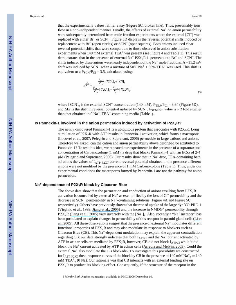
Our previous dose-response experiments done using 0 Na+, TEA+-containing solutions showedthat ATP activates IATPCl in acinar cells with an EC50 of 158±10 μM and a Hill coefficient of3.4±0.4 at +80 mV (Arreola and Melvin, 2003). Data in Figure 1A show that ADP was ableto activate IATPCl. Consequently, we carried out whole-cell recordings under the sameexperimental conditions with P2X7R cloned from the mouse parotid gland and expressed inHEK-293 cells to test the ability of external ATP and ADP to activate IATP-P2X7 and todetermine whether or not P2X7R alone is sufficient to account for the dose-response curveobserved in acinar cells. The resulting IATP-P2X7 dose-response curve to ATP obtained inNa+-free, TEA-containing external solution is depicted in Figure 2 (open squares). The EC50value estimated from fit with equation 3 was 59 μM with a Hill coefficient of 4 at +80 mV(n=7). Similarly, the IATP-P2X7 dose-response curve to ADP performed using 0 Na+, TEA+-containing external solutions (Fig. 2, closed symbols) shows that ADP activates IATPCl inacinar cells (closed circles; n=5) as well as IATP-P2X7 in HEK-293 cells (closed squares; n=8)with EC50 values of 469 and 472 μM, and Hill coefficients of 1.7 and 1.8, respectively. Thus,IATP-P2X7 and IATPCl are equally sensitive to ADP (472 vs. 469 μM), but IATP-P2X7 isapproximately 2.7 fold more sensitive to ATP than IATPCl (59 vs. 158 μM). We don’t know
Reyes et al. Page 7
J Membr Biol. Author manuscript; available in PMC 2009 December 10.
NIH
-PA Author Manuscript
NIH
-PA Author Manuscript
NIH
-PA Author Manuscript
the reason for this discrepancy but acinar cells express both P2X4 and P2X7 receptors in anunknown ratio and this could contribute to changes in ATP sensitivity. In addition, a higherecto-nucleotidase activity in acinar cells compared to HEK-293 cells could also contribute tochange the apparent ATP sensitivity of endogenous channels (Henz et al., 2006). Controlexperiments show that HEK-293 cells transfected with either P2X4 or an inverted sequence ofP2X7 did not exhibit the ATP-activated current (inset in Figure 4).
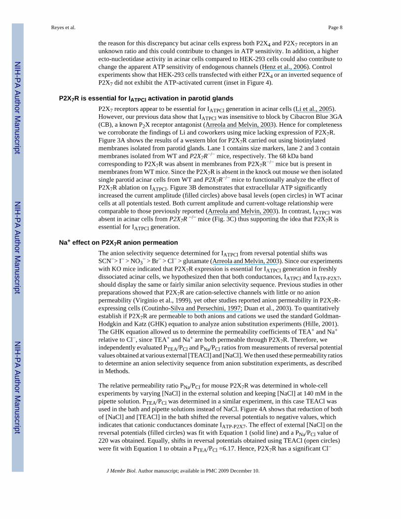
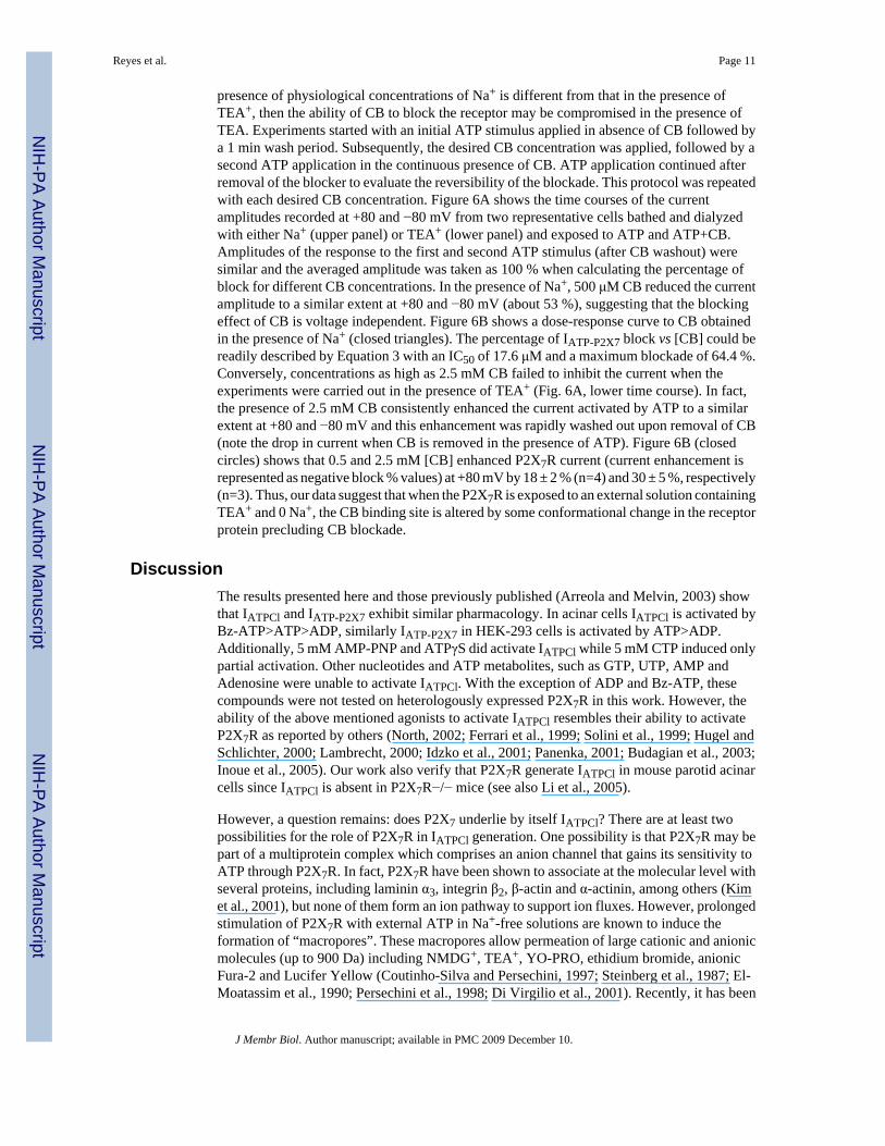
P2X7R is essential for IATPCl activation in parotid glandsP2X7 receptors appear to be essential for IATPCl generation in acinar cells (Li et al., 2005).However, our previous data show that IATPCl was insensitive to block by Cibacron Blue 3GA(CB), a known P2X receptor antagonist (Arreola and Melvin, 2003). Hence for completenesswe corroborate the findings of Li and coworkers using mice lacking expression of P2X7R.Figure 3A shows the results of a western blot for P2X7R carried out using biotinylatedmembranes isolated from parotid glands. Lane 1 contains size markers, lane 2 and 3 containmembranes isolated from WT and P2X7R−/− mice, respectively. The 68 kDa bandcorresponding to P2X7R was absent in membranes from P2X7R−/− mice but is present inmembranes from WT mice. Since the P2X7R is absent in the knock out mouse we then isolatedsingle parotid acinar cells from WT and P2X7R−/− mice to functionally analyze the effect ofP2X7R ablation on IATPCl. Figure 3B demonstrates that extracellular ATP significantlyincreased the current amplitude (filled circles) above basal levels (open circles) in WT acinarcells at all potentials tested. Both current amplitude and current-voltage relationship werecomparable to those previously reported (Arreola and Melvin, 2003). In contrast, IATPCl wasabsent in acinar cells from P2X7R −/− mice (Fig. 3C) thus supporting the idea that P2X7R isessential for IATPCl generation.
Na+ effect on P2X7R anion permeationThe anion selectivity sequence determined for IATPCl from reversal potential shifts wasSCN−> I− > NO3
− > Br− > Cl− > glutamate (Arreola and Melvin, 2003). Since our experimentswith KO mice indicated that P2X7R expression is essential for IATPCl generation in freshlydissociated acinar cells, we hypothesized then that both conductances, IATPCl and IATP-P2X7,should display the same or fairly similar anion selectivity sequence. Previous studies in otherpreparations showed that P2X7R are cation-selective channels with little or no anionpermeability (Virginio et al., 1999), yet other studies reported anion permeability in P2X7R-expressing cells (Coutinho-Silva and Persechini, 1997; Duan et al., 2003). To quantitativelyestablish if P2X7R are permeable to both anions and cations we used the standard Goldman-Hodgkin and Katz (GHK) equation to analyze anion substitution experiments (Hille, 2001).The GHK equation allowed us to determine the permeability coefficients of TEA+ and Na+
relative to Cl−, since TEA+ and Na+ are both permeable through P2X7R. Therefore, weindependently evaluated PTEA/PCl and PNa/PCl ratios from measurements of reversal potentialvalues obtained at various external [TEACl] and [NaCl]. We then used these permeability ratiosto determine an anion selectivity sequence from anion substitution experiments, as describedin Methods.
The relative permeability ratio PNa/PCl for mouse P2X7R was determined in whole-cellexperiments by varying [NaCl] in the external solution and keeping [NaCl] at 140 mM in thepipette solution. PTEA/PCl was determined in a similar experiment, in this case TEACl wasused in the bath and pipette solutions instead of NaCl. Figure 4A shows that reduction of bothof [NaCl] and [TEACl] in the bath shifted the reversal potentials to negative values, whichindicates that cationic conductances dominate IATP-P2X7. The effect of external [NaCl] on thereversal potentials (filled circles) was fit with Equation 1 (solid line) and a PNa/PCl value of220 was obtained. Equally, shifts in reversal potentials obtained using TEACl (open circles)were fit with Equation 1 to obtain a PTEA/PCl =6.17. Hence, P2X7R has a significant Cl−
Reyes et al. Page 8
J Membr Biol. Author manuscript; available in PMC 2009 December 10.
NIH
-PA Author Manuscript
NIH
-PA Author Manuscript
NIH
-PA Author Manuscript

permeability in the presence of TEA+ but little in the presence of physiologically relevantNa+ concentrations.
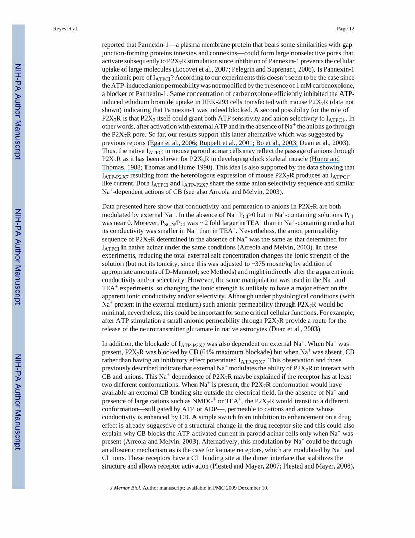
Once PTEA/PCl was estimated as required by Equation 2, we proceeded to determine the anionselectivity sequence of P2X7R in cells bathed and dialyzed with solutions containing TEA+.In these experiments external Cl− was replaced with equimolar concentrations of SCN−, I−,NO3
−, Br− or acetate under continuous stimulation with 5 mM ATP(Tris)2. If P2X7R channelsare indeed anion permeable, then the reversal potential of the current activated by external ATPwill shift according to the permeability of the anion being tested (Hille, 2001). Figure 4 showsa representative time course where both current amplitude at +80 mV (panel B) and thecorresponding reversal potential (panel C) of the current activated by ATP were continuouslymonitored in the same cell. Substitution of Cl− with SCN−, NO3
−, I− and Br− increased thecurrent amplitude while acetate decreased it. Cl− substitution by other anions resulted in asudden shift in Er, which was fully reversibly upon returning to Cl− containing media. Thisindicates that P2X7R display significant anion permeation. Anions that induced larger shiftsin the reversal potential induced larger outward current amplitude variations, indicating thatthe anions with the higher permeabilities also have higher conductivities. These results are incomplete agreement with the anion permeability of IATPCl in parotid acinar cells (Arreola andMelvin, 2003). Shifts in reversal potential induced by anion substitutions together with thePTEA/PCl ratio already determined (PTEA/PCl=6.17, Figure 4A) and Equation 2 were then usedto estimate apparent permeability ratios for these anions with respect to Cl−. Table 1summarizes the apparent anion permeability sequence for P2X7R: SCN− > I− ≅ NO3
− > Br−> Cl− > acetate. This sequence is identical to that determined in native mouse parotid acinarcells (Arreola and Melvin, 2003).
The above data show that P2X7R supports significant anion currents in Na+-free, TEA+-containing media. However, as shown in Figure 4A the P2X7R conductance in Na+-basedsolutions can be totally attributed to Na+ with negligible Cl− contribution. The Na+-dependentmodulation of permeation observed in the case of Cl− could be a phenomenon applicable toother anions too. To test this idea the external Cl− was replaced by SCN− and both the currentand reversal potential were determined. Figure 5A and B show the time course of this responserecorded from a representative cell. Time courses at −80 and +80 mV show that SCN−
unexpectedly reduces current amplitude and produces a big change in reversal potential (about−25 mV for this cell), which indicates that SCN− is highly permeable even in the presence ofexternal Na+. Despite this, SCN− has a smaller conductivity in the presence of Na+ than in thepresence of TEA+. This modulation by Na+ of anion permeation posed the next question: isthe ion permeability of P2X7 a function of Na+ concentration? Thus, we performed molefraction experiments where the external Na+ was changed while the total monovalent cationconcentration (Na+ + TEA+) was kept at 140 mM. Figure 5C displays the measured reversalpotentials recorded with the mole fraction solutions containing 100% Cl−. The data can be fitwith equation 4:
(4)
where [TEA]e represents the external [TEA+] and [Na]e represents the external [Na+]. Fromthis fit (continuous line) a PTEA/PCl ratio of 3.64 and a PNa/PCl ratio of 7.7 were estimated,these values are smaller than those obtained using either TEA+ or Na+ alone. If we assume thatthe ion fluxes are independent the PNa/PTEA value expected should be around 36 but insteadit falls around 2.1. Furthermore, when we plotted the expected reversal potential valuesassuming independent fluxes (PTEA/PCl = 6.17 and PNa/PCl = 220) against the [Na]e, we saw
Reyes et al. Page 9
J Membr Biol. Author manuscript; available in PMC 2009 December 10.
NIH
-PA Author Manuscript
NIH
-PA Author Manuscript
NIH
-PA Author Manuscript
that the experimentally values fall far away (Figure 5C, broken line). Thus, presumably ionsflow in a non-independent manner. Finally, the effects of external Na+ on anion permeabilitywere subsequently determined from mole fraction experiments where the external [Cl−] wasreplaced with either Br− or SCN−. Figure 5D displays the reversal potential shifts induced byreplacement with Br− (open circles) or SCN− (open squares). Both anions induced clearreversal potential shifts that were comparable to those observed in anion substitutionexperiments when 140 mM external TEA+ was present (see Figure 4 and Table 1). This resultdemonstrates that in the presence of external Na+ P2X7R is permeable to Br− and SCN−. Theshifts induced by these anions were nearly independent of the Na+ mole fractions. A −11.2 mVshift was induced by SCN− when a mixture of 50% Na+ + 50% TEA+ was used. This shift isequivalent to a PSCN/PCl = 3.5, calculated using:
(5)
where [SCN]e is the external SCN− concentration (140 mM), PTEA/PCl = 3.64 (Figure 5D),and ΔEr is the shift in reversal potential induced by SCN−. PSCN/PCl value is ~ 2 fold smallerthan that obtained in 0 Na+, TEA+-containing media (Table1).
Is Pannexin-1 involved in the anion permeation induced by activation of P2X7R?The newly discovered Pannexin-1 is a ubiquitous protein that associates with P2X7R. Longstimulation of P2X7R with ATP results in Pannexin-1 activation, which forms a macropore(Locovei et al., 2007; Pelegrin and Suprenant, 2006) permeable to large cations and anions.Therefore we asked: can the cation and anion permeability above described be attributed toPannexin-1? To test this idea, we repeated our experiments in the presence of a supramaximalconcentration of Carbenoxolone (1 mM), a drug that blocks Pannexin-1 with an EC50 of 2-4μM (Pelegrin and Suprenant, 2006). Our results show that in Na+-free, TEA-containing bathsolutions the values of IATP-P2X7 current reversal potential obtained in the presence differentanions were not modified by the presence of 1 mM Carbenoxolone (Table 1). Thus, under ourexperimental conditions the macropores formed by Pannexin-1 are not the pathway for anionpermeation.
Na+-dependence of P2X7R block by Cibacron BlueThe above data show that the permeation and conduction of anions resulting from P2X7Ractivation is controlled by external Na+ as exemplified by the loss of Cl− permeability and thedecrease in SCN− permeability in Na+-containing solutions (Figure 4A and Figure 5C,respectively). Others have previously shown that the rate of uptake of the large dye YO-PRO-1(Virginio et al., 1999; Jiang et al., 2005) and the increase in NMDG+ permeability throughP2X7R (Jiang et al., 2005) vary inversely with the [Na+]e. Also, recently a “Na+ memory” hasbeen postulated to explain changes in permeability of this receptor in parotid gland cells (Li etal., 2005). All these observations suggest that the presence of external Na+ modulates differentfunctional properties of P2X7R and may also modulate its response to blockers such asCibacron Blue (CB). This Na+-dependent modulation may explain the apparent contradictionregarding CB: our data strongly indicates that both IATPCl and the Na+ current activated byATP in acinar cells are mediated by P2X7R, however, CB did not block IATPCl while it didblock the Na+ current activated by ATP in acinar cells (Arreola and Melvin, 2003). Could theexternal Na+ also modulate the CB blockade? To investigate this possibility we constructedfor IATP-P2X7 dose-response curves of the block by CB in the presence of 140 mM Na+
e or 140mM TEA+
e (0 Na). Our rationale was that CB interacts with an external binding site onP2X7R to produce its blocking effect. Consequently, if the structure of the receptor in the
Reyes et al. Page 10
J Membr Biol. Author manuscript; available in PMC 2009 December 10.
NIH
-PA Author Manuscript
NIH
-PA Author Manuscript
NIH
-PA Author Manuscript
presence of physiological concentrations of Na+ is different from that in the presence ofTEA+, then the ability of CB to block the receptor may be compromised in the presence ofTEA. Experiments started with an initial ATP stimulus applied in absence of CB followed bya 1 min wash period. Subsequently, the desired CB concentration was applied, followed by asecond ATP application in the continuous presence of CB. ATP application continued afterremoval of the blocker to evaluate the reversibility of the blockade. This protocol was repeatedwith each desired CB concentration. Figure 6A shows the time courses of the currentamplitudes recorded at +80 and −80 mV from two representative cells bathed and dialyzedwith either Na+ (upper panel) or TEA+ (lower panel) and exposed to ATP and ATP+CB.Amplitudes of the response to the first and second ATP stimulus (after CB washout) weresimilar and the averaged amplitude was taken as 100 % when calculating the percentage ofblock for different CB concentrations. In the presence of Na+, 500 μM CB reduced the currentamplitude to a similar extent at +80 and −80 mV (about 53 %), suggesting that the blockingeffect of CB is voltage independent. Figure 6B shows a dose-response curve to CB obtainedin the presence of Na+ (closed triangles). The percentage of IATP-P2X7 block vs [CB] could bereadily described by Equation 3 with an IC50 of 17.6 μM and a maximum blockade of 64.4 %.Conversely, concentrations as high as 2.5 mM CB failed to inhibit the current when theexperiments were carried out in the presence of TEA+ (Fig. 6A, lower time course). In fact,the presence of 2.5 mM CB consistently enhanced the current activated by ATP to a similarextent at +80 and −80 mV and this enhancement was rapidly washed out upon removal of CB(note the drop in current when CB is removed in the presence of ATP). Figure 6B (closedcircles) shows that 0.5 and 2.5 mM [CB] enhanced P2X7R current (current enhancement isrepresented as negative block % values) at +80 mV by 18 ± 2 % (n=4) and 30 ± 5 %, respectively(n=3). Thus, our data suggest that when the P2X7R is exposed to an external solution containingTEA+ and 0 Na+, the CB binding site is altered by some conformational change in the receptorprotein precluding CB blockade.
DiscussionThe results presented here and those previously published (Arreola and Melvin, 2003) showthat IATPCl and IATP-P2X7 exhibit similar pharmacology. In acinar cells IATPCl is activated byBz-ATP>ATP>ADP, similarly IATP-P2X7 in HEK-293 cells is activated by ATP>ADP.Additionally, 5 mM AMP-PNP and ATPγS did activate IATPCl while 5 mM CTP induced onlypartial activation. Other nucleotides and ATP metabolites, such as GTP, UTP, AMP andAdenosine were unable to activate IATPCl. With the exception of ADP and Bz-ATP, thesecompounds were not tested on heterologously expressed P2X7R in this work. However, theability of the above mentioned agonists to activate IATPCl resembles their ability to activateP2X7R as reported by others (North, 2002; Ferrari et al., 1999; Solini et al., 1999; Hugel andSchlichter, 2000; Lambrecht, 2000; Idzko et al., 2001; Panenka, 2001; Budagian et al., 2003;Inoue et al., 2005). Our work also verify that P2X7R generate IATPCl in mouse parotid acinarcells since IATPCl is absent in P2X7R−/− mice (see also Li et al., 2005).
However, a question remains: does P2X7 underlie by itself IATPCl? There are at least twopossibilities for the role of P2X7R in IATPCl generation. One possibility is that P2X7R may bepart of a multiprotein complex which comprises an anion channel that gains its sensitivity toATP through P2X7R. In fact, P2X7R have been shown to associate at the molecular level withseveral proteins, including laminin α3, integrin β2, β-actin and α-actinin, among others (Kimet al., 2001), but none of them form an ion pathway to support ion fluxes. However, prolongedstimulation of P2X7R with external ATP in Na+-free solutions are known to induce theformation of “macropores”. These macropores allow permeation of large cationic and anionicmolecules (up to 900 Da) including NMDG+, TEA+, YO-PRO, ethidium bromide, anionicFura-2 and Lucifer Yellow (Coutinho-Silva and Persechini, 1997; Steinberg et al., 1987; El-Moatassim et al., 1990; Persechini et al., 1998; Di Virgilio et al., 2001). Recently, it has been
Reyes et al. Page 11
J Membr Biol. Author manuscript; available in PMC 2009 December 10.
NIH
-PA Author Manuscript
NIH
-PA Author Manuscript
NIH
-PA Author Manuscript
reported that Pannexin-1—a plasma membrane protein that bears some similarities with gapjunction-forming proteins innexins and connexins—could form large nonselective pores thatactivate subsequently to P2X7R stimulation since inhibition of Pannexin-1 prevents the cellularuptake of large molecules (Locovei et al., 2007; Pelegrin and Suprenant, 2006). Is Pannexin-1the anionic pore of IATPCl? According to our experiments this doesn’t seem to be the case sincethe ATP-induced anion permeability was not modified by the presence of 1 mM carbenoxolone,a blocker of Pannexin-1. Same concentration of carbenoxolone efficiently inhibited the ATP-induced ethidium bromide uptake in HEK-293 cells transfected with mouse P2X7R (data notshown) indicating that Pannexin-1 was indeed blocked. A second possibility for the role ofP2X7R is that P2X7 itself could grant both ATP sensitivity and anion selectivity to IATPCl.. Inother words, after activation with external ATP and in the absence of Na+ the anions go throughthe P2X7R pore. So far, our results support this latter alternative which was suggested byprevious reports (Egan et al., 2006; Ruppelt et al., 2001; Bo et al., 2003; Duan et al., 2003).Thus, the native IATPCl in mouse parotid acinar cells may reflect the passage of anions throughP2X7R as it has been shown for P2X5R in developing chick skeletal muscle (Hume andThomas, 1988; Thomas and Hume 1990). This idea is also supported by the data showing thatIATP-P2X7 resulting from the heterologous expression of mouse P2X7R produces an IATPCl-like current. Both IATPCl and IATP-P2X7 share the same anion selectivity sequence and similarNa+-dependent actions of CB (see also Arreola and Melvin, 2003).
Data presented here show that conductivity and permeation to anions in P2X7R are bothmodulated by external Na+. In the absence of Na+ PCl>0 but in Na+-containing solutions PClwas near 0. Morever, PSCN/PCl was ~ 2 fold larger in TEA+ than in Na+-containing media butits conductivity was smaller in Na+ than in TEA+. Nevertheless, the anion permeabilitysequence of P2X7R determined in the absence of Na+ was the same as that determined forIATPCl in native acinar under the same conditions (Arreola and Melvin, 2003). In theseexperiments, reducing the total external salt concentration changes the ionic strength of thesolution (but not its tonicity, since this was adjusted to ~375 mosm/kg by addition ofappropriate amounts of D-Mannitol; see Methods) and might indirectly alter the apparent ionicconductivity and/or selectivity. However, the same manipulation was used in the Na+ andTEA+ experiments, so changing the ionic strength is unlikely to have a major effect on theapparent ionic conductivity and/or selectivity. Although under physiological conditions (withNa+ present in the external medium) such anionic permeability through P2X7R would beminimal, nevertheless, this could be important for some critical cellular functions. For example,after ATP stimulation a small anionic permeability through P2X7R provide a route for therelease of the neurotransmitter glutamate in native astrocytes (Duan et al., 2003).
In addition, the blockade of IATP-P2X7 was also dependent on external Na+. When Na+ waspresent, P2X7R was blocked by CB (64% maximum blockade) but when Na+ was absent, CBrather than having an inhibitory effect potentiated IATP-P2X7. This observation and thosepreviously described indicate that external Na+ modulates the ability of P2X7R to interact withCB and anions. This Na+ dependence of P2X7R maybe explained if the receptor has at leasttwo different conformations. When Na+ is present, the P2X7R conformation would haveavailable an external CB binding site outside the electrical field. In the absence of Na+ andpresence of large cations such as NMDG+ or TEA+, the P2X7R would transit to a differentconformation—still gated by ATP or ADP—, permeable to cations and anions whoseconductivity is enhanced by CB. A simple switch from inhibition to enhancement on a drugeffect is already suggestive of a structural change in the drug receptor site and this could alsoexplain why CB blocks the ATP-activated current in parotid acinar cells only when Na+ waspresent (Arreola and Melvin, 2003). Alternatively, this modulation by Na+ could be throughan allosteric mechanism as is the case for kainate receptors, which are modulated by Na+ andCl− ions. These receptors have a Cl− binding site at the dimer interface that stabilizes thestructure and allows receptor activation (Plested and Mayer, 2007; Plested and Mayer, 2008).
Reyes et al. Page 12
J Membr Biol. Author manuscript; available in PMC 2009 December 10.
NIH
-PA Author Manuscript
NIH
-PA Author Manuscript
NIH
-PA Author Manuscript
Since our data does not provide a mechanistic explanation for P2X7R modulation by Na+ thisremains as attractive avenue to explore in future work.
In conclusion, this work shows that: 1) the P2X7R is essential for IATPCl activation in mouseparotid acinar cells; 2) Na+ regulates anion conductivity and permeation in P2X7R, i.e. in theabsence of Na+ and after stimulation with ATP the P2X7R become more permeable to anions;3) Na+ regulates the effect of CB on P2X7R, i.e. CB inhibits or enhances P2X7R current in thepresence or absence of Na+, respectively; and, 4) in the presence of physiologically relevantNa+ concentrations the P2X7R displays negligible Cl− permeability.
AcknowledgmentsThe authors thank Laurie Koek, Jennifer Scantlin, Mark Wagner, Carmen Y. Hernandez-Carballo and Monica Reynafor technical assistance. We also thank Dr. Ted Begenisich for his critical reading and comments on this work. Thiswork was supported in part by grants from NIH, (DE09692 and DE13539 to JEM), and from CONACyT, Mexico(42561 to JA).
References1. Arreola J, Melvin JE. A novel chloride conductance activated by extracellular ATP in mouse parotid
acinar cells. J. Physiol 2003;547:197–208. [PubMed: 12562938]2. Arreola J, Melvin JE, Begenisich T. Volume-activated chloride channels in rat parotid acinar cells. J.
Physiol 1995;484:677–687. [PubMed: 7623284]3. Bo X, Jiang LH, Wilson HL, Kim M, Burnstock G, Surprenant A, North RA. Pharmacological and
biophysical properties of the human P2X5 receptor. Mol. Pharmacol 2003;63:1407–1416. [PubMed:12761352]
4. Budagian V, Bulanova E, Brovko L, Orinska Z, Fayad R, Paus R, Bulfone-Paus S. Signaling throughP2X7 receptor in human T cells involves p56lck, MAP kinases, and transcription factors AP-1 andNF-kappa B. J. Biol. Chem 2003;278:1549–1560. [PubMed: 12424250]
5. Burnstock G. Physiology and pathophysiology of purinergic neurotransmission. Physiol. Rev2007;87:659–797. [PubMed: 17429044]
6. Coutinho-Silva R, Persechini PM. P2Z purinoceptor-associated pores induced by extracellular ATPin macrophages and J774 cells. Am. J. Physiol 1997;273:C1793–C1800. [PubMed: 9435482]
7. Di Virgilio F, Chiozzi P, Ferrari D, Falzoni S, Sanz JM, Morelli A, Torboli M, Bolognesi G, BaricordiOR. Nucleotide receptors: an emerging family of regulatory molecules in blood cells. Blood2001;97:587–600. [PubMed: 11157473]
8. Duan S, Anderson CM, Keung EC, Chen Y, Swanson RA. P2X7 receptor-mediated release ofexcitatory amino acids from astrocytes. J. Neurosci 2003;23:1320–1328. [PubMed: 12598620]
9. Egan TM, Samways DSK, Li Z. Biophysics of P2X receptors. Pflugers Arch. – Eur. J. Physiol2006;452:501–512. [PubMed: 16708237]
10. El-Moatassim C, Mani J, Dornand J. Extracellular ATP−4 permeabilizes thymocytes not only tocations but also to low-molecular-weight solutes. Eur. J. Pharmacol 1990;181:111–118. [PubMed:2387318]
11. Evans RL, Bell SM, Schultheis PJ, Shull GE, Melvin JE. Targeted Disruption of the Nhe1 GenePrevents Muscarinic Agonist-induced Up-regulation of Na+/H+ Exchange in Mouse Parotid AcinarCells. J. Biol. Chem 1999;274(41):29025–29030. [PubMed: 10506152]
12. Ferrari D, Stroh C, Schulze-Osthoff K. P2X7/P2Z purinoreceptor-mediated activation of transcriptionfactor NFAT in microglial cells. J. Biol. Chem 1999;274:13205–13210. [PubMed: 10224077]
13. Fukushi Y. Heterologous desensitization of muscarinic receptors by P2Z purinoceptors in rat parotidacinar cells. Eur. J. Pharmacol 1999;364:55–64. [PubMed: 9920185]
14. Hamill OP, Marty A, Neher E, Sakman B, Sigworth F. Improved patch-clamp techniques for high-resolution current recording from cells and cell-free membrane patches. Pflugers Archiv1981;391:85–100. [PubMed: 6270629]
Reyes et al. Page 13
J Membr Biol. Author manuscript; available in PMC 2009 December 10.
NIH
-PA Author Manuscript
NIH
-PA Author Manuscript
NIH
-PA Author Manuscript
15. Hayashi T, Kawakami M, Sasaki S, Katsumata T, Mori H, Yoshida H, Nakahari T. ATP regulationof ciliary beat frequency in rat tracheal and distal airway epithelium. Exp. Physiol 2005;90(4):535–544. [PubMed: 15769883]
16. Henz SL, Ribeiro CG, Rosa A, Chiarelli RA, Casali EA, Sarkis JJ. Kinetic characterization of ATPdiphosphohydrolase and 5′-nucleotidase activities in cells cultured from submandibular salivaryglands of rats. Cell Biol. Int 2006;30:214–220. [PubMed: 16480902]
17. Hernandez-Carballo CY, Perez-Cornejo P, Arreola J. Regulation of volume-sensitive chloridechannels by H+ and Cl− ions in HEK 293 cells. J. Gen. Physiol 2003;122:31a.
18. Hille, B. Ion channels of excitable membranes. Sinauer Associates; Sunderland, MA: 2001.19. Hugel S, Schlichter R. Presynaptic P2X receptors facilitate inhibitory GABAergic transmission
between cultured rat spinal cord dorsal horn neurons. J. Neurosci 2000;20:2121–2130. [PubMed:10704486]
20. Hume RI, Thomas SA. Multiple actions of adenosine 5′-triphosphate on chick skeletal muscle. J.Physiol 1988;406:503–524. [PubMed: 2474071]
21. Idzko M, Dichmann S, Panther E, Ferrari D, Herouy Y, Virchow C Jr. Luttmann W, Di Virgilio F,Norgauer J. Functional characterization of P2Y and P2X receptors in human eosinophils. J. CellPhysiol 2001;188:329–336. [PubMed: 11473359]
22. Inoue K, Denda M, Tozaki H, Fujishita K, Koizumi S, Inoue K. Characterization of multiple P2Xreceptors in cultured normal human epidermal keratinocytes. J. Invest. Dermatol 2005;124:756–763.[PubMed: 15816834]
23. Jiang LH, Rassendren F, Mackenzie A, Zhang YH, Surprenant A, North RA. N-methyl-D-glucamineand propidium dyes utilize different permeation pathways at rat P2X7 receptors. Am. J. Physiol2005;289:C1295–C1302.
24. Kim M, Jiang LH, Wilson HL, North RA, Surprenant A. Proteomic and functional evidence for aP2X7 receptor signaling complex. EMBO J 2001;20:6347–6358. [PubMed: 11707406]
25. Lambrecht G. Agonists and antagonists acting at P2X receptors: selectivity profiles and functionalimplications. Naunyn-Schmiedeberg’s Arch. Pharmacol 2000;362:340–350.
26. Leipziger J. Control of epithelial transport via luminal P2 receptors. Am. J. Physiol 2003;284:F419–F432.
27. Li Q, Luo X, Muallem S. Regulation of the P2X7 receptor permeability to large molecules byextracellular Cl− and Na+ J. Biol. Chem 2005;280:26922–26927. [PubMed: 15923180]
28. Li Q, Luo X, Zeng W, Muallem S. Cell-specific behavior of P2X7 receptors in mouse parotid acinarand duct cells. J. Biol. Chem 2003;278:47554–47561. [PubMed: 12968021]
29. Ma W, Korngreen A, Weil S, Cohen EB, Priel A, Kuzin L, Silberberg SD. Pore properties andpharmacological features of the P2X receptor channel in airway ciliated cells. J. Physiol 2006;571(Pt 3):503–517. [PubMed: 16423852]
30. McMillan MK, Soltoff SP, Cantley LC, Rudel RA, Talamo BR. Two distinct cytosolic calciumresponses to extracellular ATP in rat parotid acinar cells. Brit. J. Phamacol 1993;108:453–461.
31. Melvin JE, Yule D, Shuttleworth TJ, Begenisich T. Regulation of fluid and electrolyte secretion insalivary gland cells. Ann. Rev. Physiol 2005;67:445–469. [PubMed: 15709965]
32. North RA. Molecular physiology of P2X receptors. Physiol. Rev 2002;82:1013–1067. [PubMed:12270951]
33. North RA, Barnard EA. Nucleotide receptors. Curr. Opin. Neurobiol 1997;7:346–357. [PubMed:9232809]
34. Panenka W, Jijon H, Herx LM, Armstrong JN, Feighan D, Wei T, Yong VW, Ransohoff RM,MacVicar BA. P2X7-like receptor activation in astrocytes increases chemokine monocytechemoattractant protein-1 expression via mitogen-activated protein kinase. J. Neurosci2001;21:7135–7142. [PubMed: 11549724]
35. Pelegrin P, Surprenant A. Pannexin-1 couples to Maitotoxin- and Nigericin-induced Interleukin-1βrelease through a dye uptake-independent pathway. J. Biol. Chem 2007;282(4):2386–2394.[PubMed: 17121814]
36. Pérez-Cornejo P, Arreola J. Regulation of Ca2+-activated chloride channels by cAMP and CFTR inparotid acinar cells. Biochem. Biophys. Res. Commun 2004;316:612–617. [PubMed: 15033444]
Reyes et al. Page 14
J Membr Biol. Author manuscript; available in PMC 2009 December 10.
NIH
-PA Author Manuscript
NIH
-PA Author Manuscript
NIH
-PA Author Manuscript
37. Persechini PM, Bisaggio RC, Alves-Neto JL, Coutinho-Silva R. Extracellular ATP in thelymphohematopoietic system: P2Z purinoceptors and membrane permeabilization. Braz. J. Med.Biol. Res 1998;31:25–34. [PubMed: 9686176]
38. Plested AJR, Mayer ML. Structure and mechanism of kainate receptors modulation by anions. Neuron2007;53:829–841. [PubMed: 17359918]
39. Plested AJR, Mayer ML. Allosteric ion binding sites in kainate receptors. Biophys. J. Abstracts2008:362a–363a.
40. Ralevic V, Burnstock G. Receptors for purines and pyrimidines. Pharmacol. Rev 1998;50:413–492.[PubMed: 9755289]
41. Roman RM, Fitz JG. Emerging roles of purinergic signaling in gastrointestinal epithelial secretionand hepatobiliary function. Gastroenterology 1999;116:964–979. [PubMed: 10092320]
42. Ruppelt A, Ma W, Borchardt K, Silberberg SD, Soto F. Genomic structure, developmental distributionand functional properties of the chicken P2X5 receptor. J. Neurochem 2001;77:1256–1265.[PubMed: 11389176]
43. Schwiebert EM, Zsembery A. Extracellular ATP as a signaling molecule for epithelial cells. Biochim.Biophys. Acta 2003;1615:7–32. [PubMed: 12948585]
44. Solini A, Chiozzi P, Morelli A, Fellin R, Di Virgilio F. Human primary fibroblasts in vitro express apurinergic P2X7 receptor coupled to ion fluxes, microvesicle formation and IL-6 release. J. Cell Sci1999;112:297–305. [PubMed: 9885283]
45. Solle M, Labasi J, Perregaux DG, Stam E, Petrushova N, Koller BH, Griffiths RJ, Gabel CA. Alteredcytokine production in mice lacking P2X7 receptors. J. Biol. Chem 2001;276:125–132. [PubMed:11016935]
46. Steinberg TH, Newman AS, Swanson JA, Silverstein SC. ATP4− permeabilizes the plasma membraneof mouse macrophages to fluorescent dyes. J. Biol. Chem 1987;262:8884–8888. [PubMed: 3597398]
47. Tenneti L, Gibbons SJ, Talamo BR. Expression and trans-synaptic regulation of P2X4 and P2zreceptors for extracellular ATP in parotid acinar cells. J. Biol. Chem 1998;273:26799–26808.[PubMed: 9756924]
48. Thomas SA, Hume RI. Permeation of both cations and anions through a single class of ATP-activatedion channels in developing chick skeletal muscle. J. Gen. Physiol 1990;95:569–590. [PubMed:1692581]
49. Turner JT, London LA, Gibbons SJ, Talamo BR. Salivary gland P2 nucleotide receptors. Crit. Rev.Oral Biol. Med 1999;10:210–224. [PubMed: 10759423]
50. Virginio C, MacKenzie A, North RA, Surprenant A. Kinetics of cell lysis, dye uptake and permeabilitychanges in cells expressing the rat P2X7 receptor. J. Physiol 1999;519:335–346. [PubMed:10457053]
51. Zeng W, Lee MG, Yan M, Diaz J, Benjamin I, Marino CR, Kopito R, Freedman S, Cotton C, MuallemS, Thomas P. Immuno and functional characterization of CFTR in submandibular and pancreaticacinar and duct cells. Am. J. Physiol. Cell Physiol 1997;273:C442–C455.
Reyes et al. Page 15
J Membr Biol. Author manuscript; available in PMC 2009 December 10.
NIH
-PA Author Manuscript
NIH
-PA Author Manuscript
NIH
-PA Author Manuscript
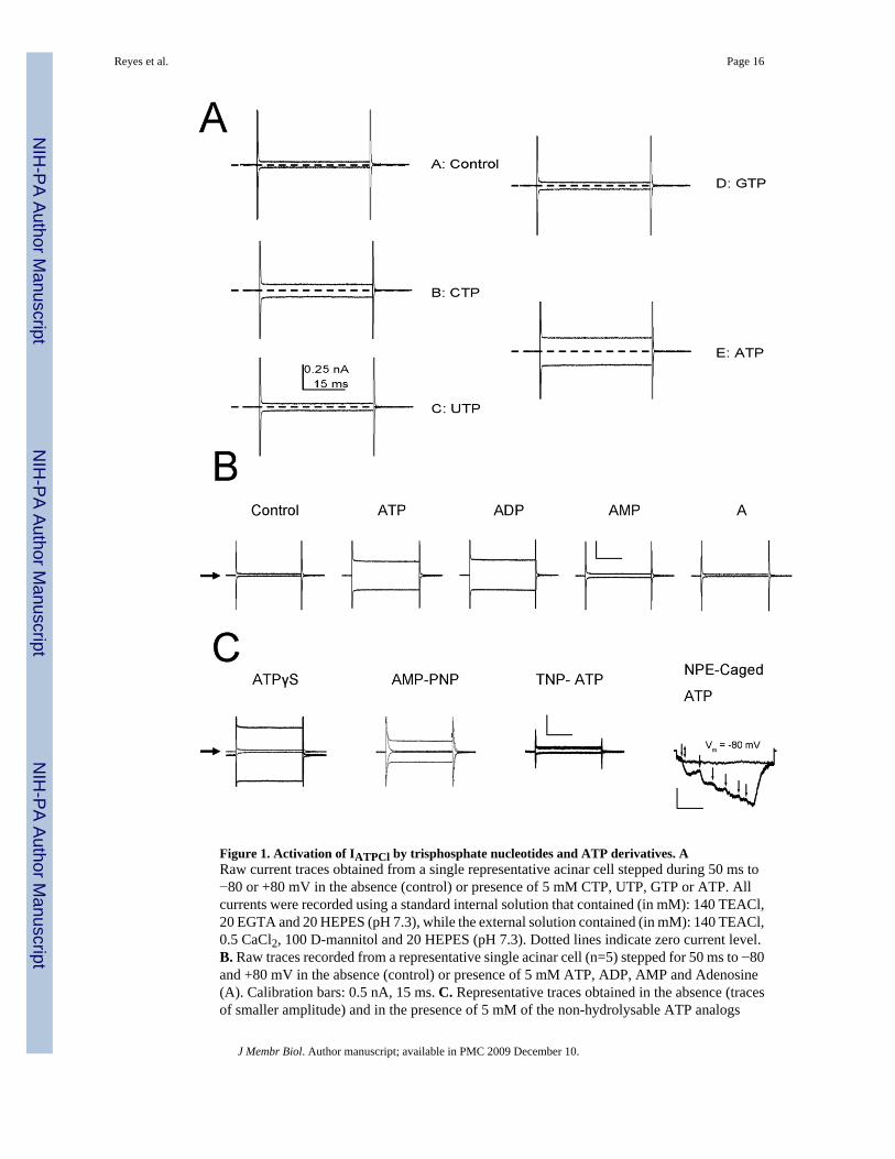
Figure 1. Activation of IATPCl by trisphosphate nucleotides and ATP derivatives. ARaw current traces obtained from a single representative acinar cell stepped during 50 ms to−80 or +80 mV in the absence (control) or presence of 5 mM CTP, UTP, GTP or ATP. Allcurrents were recorded using a standard internal solution that contained (in mM): 140 TEACl,20 EGTA and 20 HEPES (pH 7.3), while the external solution contained (in mM): 140 TEACl,0.5 CaCl2, 100 D-mannitol and 20 HEPES (pH 7.3). Dotted lines indicate zero current level.B. Raw traces recorded from a representative single acinar cell (n=5) stepped for 50 ms to −80and +80 mV in the absence (control) or presence of 5 mM ATP, ADP, AMP and Adenosine(A). Calibration bars: 0.5 nA, 15 ms. C. Representative traces obtained in the absence (tracesof smaller amplitude) and in the presence of 5 mM of the non-hydrolysable ATP analogs
Reyes et al. Page 16
J Membr Biol. Author manuscript; available in PMC 2009 December 10.
NIH
-PA Author Manuscript
NIH
-PA Author Manuscript
NIH
-PA Author Manuscript

ATPγS (n=3) or AMP-PNP (n=3) and of 5 mM of the P2 receptor blocker TNP-ATP (n=3).The slight shift from 0 current level in ATPγS was the result of using (Na+)2-ATP salt to activatethe current. Calibration bars: 0.5 nA, 15 ms.NPE-Caged ATP: representative traces of a singlecell stepped to −80 mV (n=4). Flat trace: The cell was initially exposed to short UV flash(double head arrow) in the absence of NPE-caged ATP (no response), then exposed to 250μM NPE-caged ATP (no response). Stair-like trace: UV flashes (arrows) were applied in thepresence NPE-caged ATP. Current returned to control level by perfusing the bath with thestandard external solution. Calibration bars: 0.1 nA, 10 ms. Black arrows on the left sideindicate zero current levels in all cases.
Reyes et al. Page 17
J Membr Biol. Author manuscript; available in PMC 2009 December 10.
NIH
-PA Author Manuscript
NIH
-PA Author Manuscript
NIH
-PA Author Manuscript
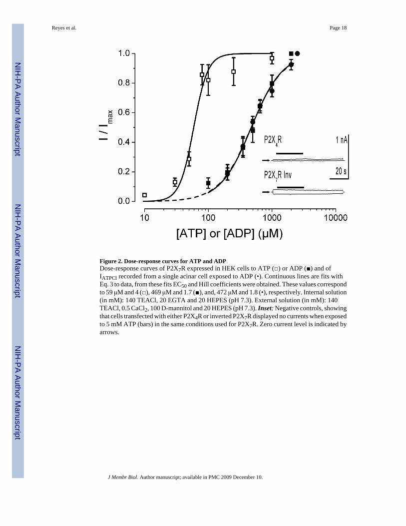
Figure 2. Dose-response curves for ATP and ADPDose-response curves of P2X7R expressed in HEK cells to ATP (□) or ADP (■) and ofIATPCl recorded from a single acinar cell exposed to ADP (•). Continuous lines are fits withEq. 3 to data, from these fits EC50 and Hill coefficients were obtained. These values correspondto 59 μM and 4 (□), 469 μM and 1.7 (■), and, 472 μM and 1.8 (•), respectively. Internal solution(in mM): 140 TEACl, 20 EGTA and 20 HEPES (pH 7.3). External solution (in mM): 140TEACl, 0.5 CaCl2, 100 D-mannitol and 20 HEPES (pH 7.3). Inset: Negative controls, showingthat cells transfected with either P2X4R or inverted P2X7R displayed no currents when exposedto 5 mM ATP (bars) in the same conditions used for P2X7R. Zero current level is indicated byarrows.
Reyes et al. Page 18
J Membr Biol. Author manuscript; available in PMC 2009 December 10.
NIH
-PA Author Manuscript
NIH
-PA Author Manuscript
NIH
-PA Author Manuscript

Figure 3. P2X7R are necessary for IATPCl generationPresence of P2X7R was detected in biotinylated membranes isolated from parotid glands fromWT and P2X7R−/− mice (A). Samples were run in a 7.5 % SDS-PAGE gel. Lane 1 containsSeeBlue Plus2 Pre-Stained Standard, lane 2 contains membranes from WT mice (50 μg ofprotein) and lane 3 contains membranes from knock-out mice (50 μg of protein). Current-voltage (I-V) relationships were constructed with currents recorded from acinar cells isolatedfrom WT (B; n=5) or P2X7R −/− mice (C; n=8) in the absence (open circles) or presence (closedcircles) of 5 mM (Tris)2-ATP. Currents were generated by application of 50 ms voltage stepsfrom −80 to +100 mV in 20 mV increments and then measured near the end of each step.Currents were recorded using an internal solution that contained (in mM): 140 TEACl, 20EGTA and 20 HEPES (pH 7.3); the external solution contained (in mM): 140 TEACl, 0.5CaCl2, 100 D-mannitol and 20 HEPES (pH 7.3).
Reyes et al. Page 19
J Membr Biol. Author manuscript; available in PMC 2009 December 10.
NIH
-PA Author Manuscript
NIH
-PA Author Manuscript
NIH
-PA Author Manuscript
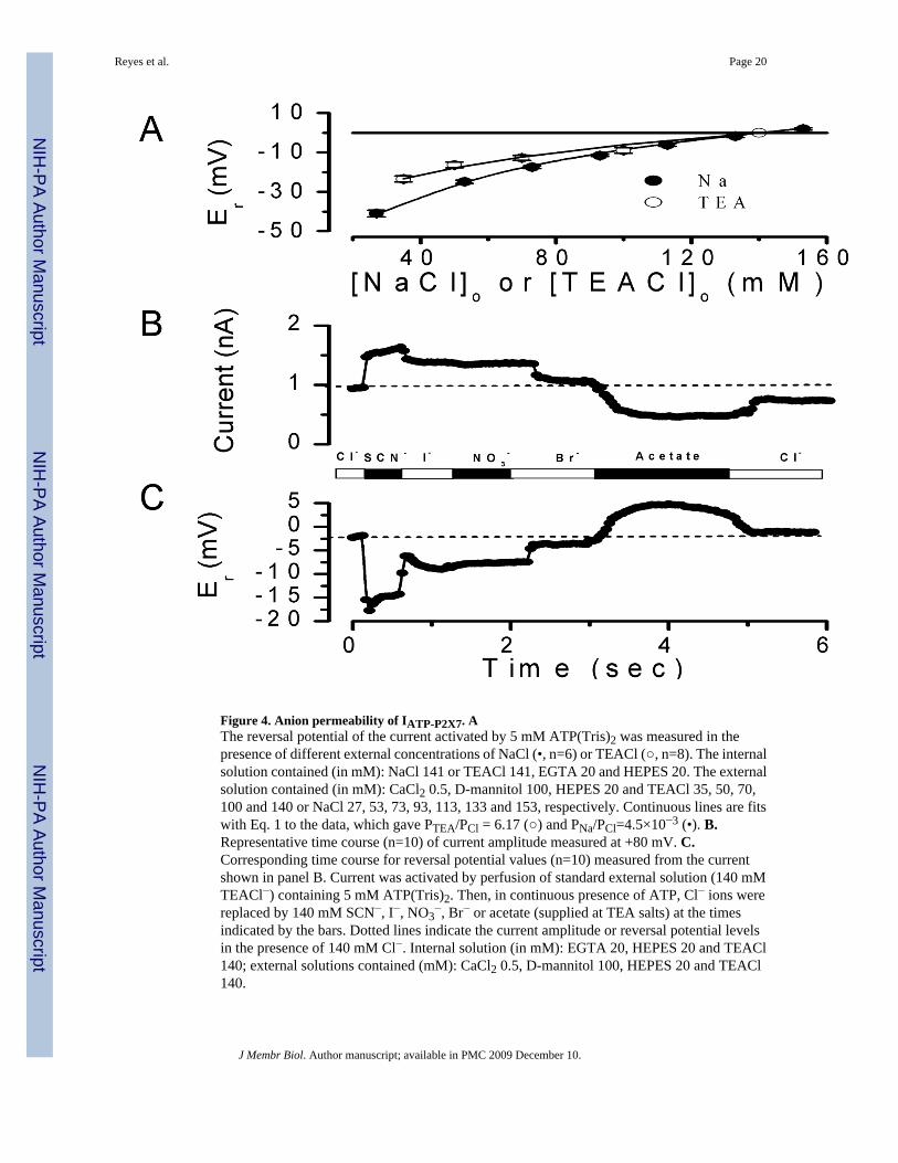
Figure 4. Anion permeability of IATP-P2X7. AThe reversal potential of the current activated by 5 mM ATP(Tris)2 was measured in thepresence of different external concentrations of NaCl (•, n=6) or TEACl (○, n=8). The internalsolution contained (in mM): NaCl 141 or TEACl 141, EGTA 20 and HEPES 20. The externalsolution contained (in mM): CaCl2 0.5, D-mannitol 100, HEPES 20 and TEACl 35, 50, 70,100 and 140 or NaCl 27, 53, 73, 93, 113, 133 and 153, respectively. Continuous lines are fitswith Eq. 1 to the data, which gave PTEA/PCl = 6.17 (○) and PNa/PCl=4.5×10−3 (•). B.Representative time course (n=10) of current amplitude measured at +80 mV. C.Corresponding time course for reversal potential values (n=10) measured from the currentshown in panel B. Current was activated by perfusion of standard external solution (140 mMTEACl−) containing 5 mM ATP(Tris)2. Then, in continuous presence of ATP, Cl− ions werereplaced by 140 mM SCN−, I−, NO3
−, Br− or acetate (supplied at TEA salts) at the timesindicated by the bars. Dotted lines indicate the current amplitude or reversal potential levelsin the presence of 140 mM Cl−. Internal solution (in mM): EGTA 20, HEPES 20 and TEACl140; external solutions contained (mM): CaCl2 0.5, D-mannitol 100, HEPES 20 and TEACl140.
Reyes et al. Page 20
J Membr Biol. Author manuscript; available in PMC 2009 December 10.
NIH
-PA Author Manuscript
NIH
-PA Author Manuscript
NIH
-PA Author Manuscript
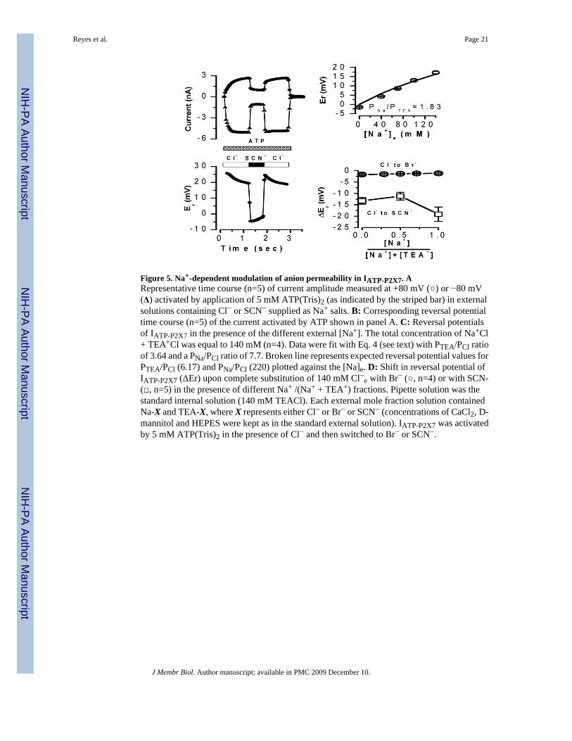
Figure 5. Na+-dependent modulation of anion permeability in IATP-P2X7. ARepresentative time course (n=5) of current amplitude measured at +80 mV (○) or −80 mV(Δ) activated by application of 5 mM ATP(Tris)2 (as indicated by the striped bar) in externalsolutions containing Cl− or SCN− supplied as Na+ salts. B: Corresponding reversal potentialtime course (n=5) of the current activated by ATP shown in panel A. C: Reversal potentialsof IATP-P2X7 in the presence of the different external [Na+]. The total concentration of Na+Cl+ TEA+Cl was equal to 140 mM (n=4). Data were fit with Eq. 4 (see text) with PTEA/PCl ratioof 3.64 and a PNa/PCl ratio of 7.7. Broken line represents expected reversal potential values forPTEA/PCl (6.17) and PNa/PCl (220) plotted against the [Na]e. D: Shift in reversal potential ofIATP-P2X7 (ΔEr) upon complete substitution of 140 mM Cl−e with Br− (○, n=4) or with SCN-(□, n=5) in the presence of different Na+ /(Na+ + TEA+) fractions. Pipette solution was thestandard internal solution (140 mM TEACl). Each external mole fraction solution containedNa-X and TEA-X, where X represents either Cl− or Br− or SCN− (concentrations of CaCl2, D-mannitol and HEPES were kept as in the standard external solution). IATP-P2X7 was activatedby 5 mM ATP(Tris)2 in the presence of Cl− and then switched to Br− or SCN−.
Reyes et al. Page 21
J Membr Biol. Author manuscript; available in PMC 2009 December 10.
NIH
-PA Author Manuscript
NIH
-PA Author Manuscript
NIH
-PA Author Manuscript

Figure 6. Block of IATP-P2X7 by Cibacron Blue is Na+-dependent. ATime course of P2X7R current amplitudes recorded at +80 (open symbols) and −80 mV (filledsymbols) in cells dialyzed and bathed with solutions containing 140 mM NaCl (triangles, upperpanel) or 140 mM TEACl (circles, lower panel). Black lines indicate application of 5 mM ATP(Tris)2 and double arrowhead lines indicate application of 0.5 mM or 2.5 mM CB (in thepresence of Na+ or TEA+, respectively). B: Dose-response curve of CB blockade obtained inNaCl (triangles) or TEACl conditions (circles). The number of cells tested were 4, 4, 5, 4, 3,5, 2, 7, 3 for 0.1, 1, 10, 50, 100, 250, 500, 1000 and 2500 μM CB, respectively. For theexperiments in TEA+ only two high-concentration data points are plotted: 1000 (n=4) and 2500(n=3). Continuous line is the fit with Eq. 3 and yielded an EC50=3.9 μM with maximum blockof 64 % and a Hill coefficient= 0.5. Note that CB stimulates P2X7R currents in cells dialyzedand bathed in Na+-free solutions containing 140 mM TEA+ (filled circles).
Reyes et al. Page 22
J Membr Biol. Author manuscript; available in PMC 2009 December 10.
NIH
-PA Author Manuscript
NIH
-PA Author Manuscript
NIH
-PA Author Manuscript

NIH
-PA Author Manuscript
NIH
-PA Author Manuscript
NIH
-PA Author Manuscript
Reyes et al. Page 23
Table 1Apparent anion permeability of IATP-P2X7
Shifts in reversal potential induced by anion substitution in Na+-free, TEA-containing solutions were used withequation 2 to determine PX/PCl. The term PTEA/PCl, which is present in equation 2, was determined from thedata in Figure 4 as described in the text. Er= reversal potential of the current; n= number of cells tested. Liquidjunction potentials are listed for different anion substitution solutions. The −0.6 mV listed in the Cl− row is thepotential measured upon returning to the standard external solution after the anion substitution measurement.ND = Not determined.
Anion Er (mV)Without CBX
Er (mV)With 1 mM
CBX
PX/PClWithout
CBX
LiquidJunctionPotential
(mV)SCN− −17.69±0.49
n=11−19.01±1.52
n=67.31±0.14 −1
I− −8.69±0.46n=11
−8.17±0.79n=6
3.28±0.16 −1.3
NO3− −7.46±0.40
n=11ND 2.83±0.16 +1.5
Br− −4.77±0.47n=10
ND 1.97±0.15 −0.3
Cl− −1.75±0.22n=12
−3.05±0.86n=6
1 −0.6
Acetate +5.18±0.86n=10
+12.22±3.57n=6
~0.0±0.00 +2.9
J Membr Biol. Author manuscript; available in PMC 2009 December 10.
Copyright © 2022 FDOKUMEN