An investigation into increased cases of idiopathic infantile ...
Upload
independentCategory
view
0download
0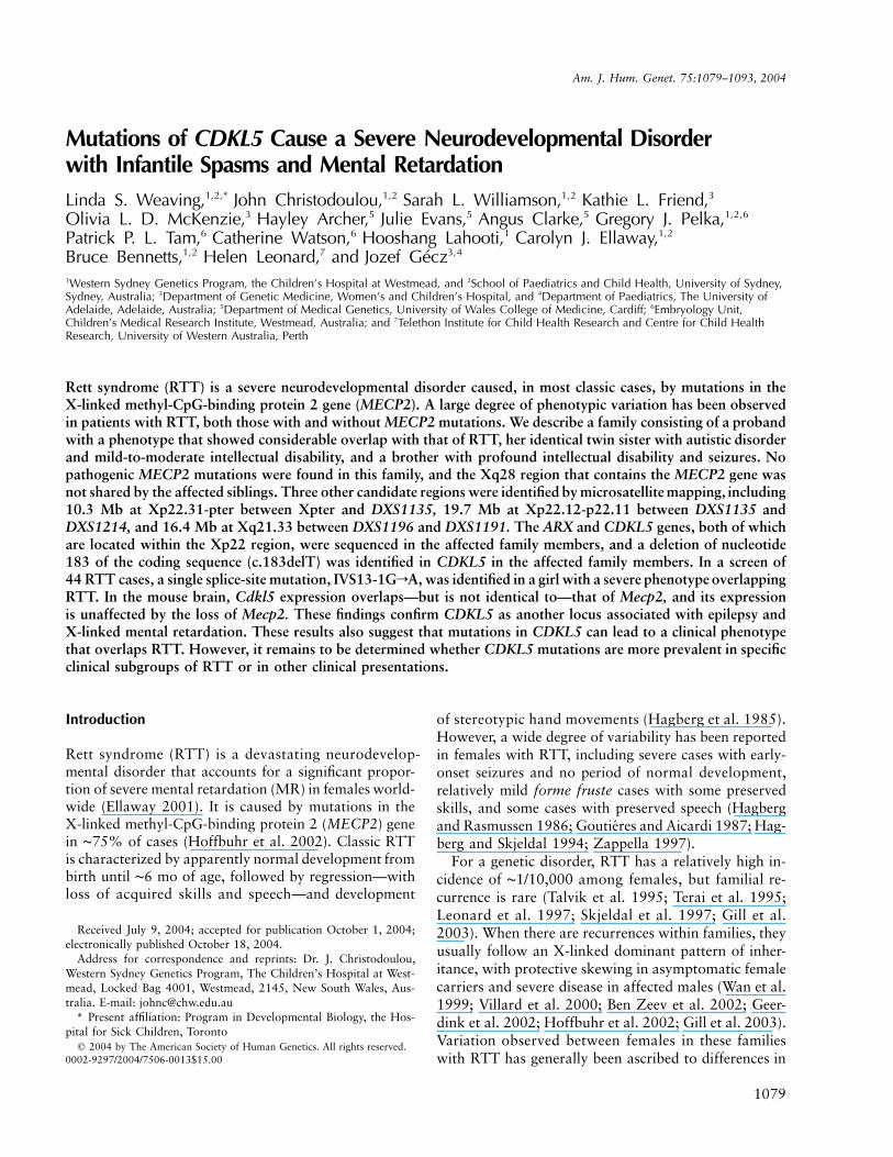
Am. J. Hum. Genet. 75:1079–1093, 2004
1079
Mutations of CDKL5 Cause a Severe Neurodevelopmental Disorderwith Infantile Spasms and Mental RetardationLinda S. Weaving,1,2,* John Christodoulou,1,2 Sarah L. Williamson,1,2 Kathie L. Friend,3Olivia L. D. McKenzie,3 Hayley Archer,5 Julie Evans,5 Angus Clarke,5 Gregory J. Pelka,1,2,6
Patrick P. L. Tam,6 Catherine Watson,6 Hooshang Lahooti,1 Carolyn J. Ellaway,1,2
Bruce Bennetts,1,2 Helen Leonard,7 and Jozef Gecz3,4
1Western Sydney Genetics Program, the Children’s Hospital at Westmead, and 2School of Paediatrics and Child Health, University of Sydney,Sydney, Australia; 3Department of Genetic Medicine, Women’s and Children’s Hospital, and 4Department of Paediatrics, The University ofAdelaide, Adelaide, Australia; 5Department of Medical Genetics, University of Wales College of Medicine, Cardiff; 6Embryology Unit,Children’s Medical Research Institute, Westmead, Australia; and 7Telethon Institute for Child Health Research and Centre for Child HealthResearch, University of Western Australia, Perth
Rett syndrome (RTT) is a severe neurodevelopmental disorder caused, in most classic cases, by mutations in theX-linked methyl-CpG-binding protein 2 gene (MECP2). A large degree of phenotypic variation has been observedin patients with RTT, both those with and without MECP2 mutations. We describe a family consisting of a probandwith a phenotype that showed considerable overlap with that of RTT, her identical twin sister with autistic disorderand mild-to-moderate intellectual disability, and a brother with profound intellectual disability and seizures. Nopathogenic MECP2 mutations were found in this family, and the Xq28 region that contains the MECP2 gene wasnot shared by the affected siblings. Three other candidate regions were identified by microsatellite mapping, including10.3 Mb at Xp22.31-pter between Xpter and DXS1135, 19.7 Mb at Xp22.12-p22.11 between DXS1135 andDXS1214, and 16.4 Mb at Xq21.33 between DXS1196 and DXS1191. The ARX and CDKL5 genes, both of whichare located within the Xp22 region, were sequenced in the affected family members, and a deletion of nucleotide183 of the coding sequence (c.183delT) was identified in CDKL5 in the affected family members. In a screen of44 RTT cases, a single splice-site mutation, IVS13-1GrA, was identified in a girl with a severe phenotype overlappingRTT. In the mouse brain, Cdkl5 expression overlaps—but is not identical to—that of Mecp2, and its expressionis unaffected by the loss of Mecp2. These findings confirm CDKL5 as another locus associated with epilepsy andX-linked mental retardation. These results also suggest that mutations in CDKL5 can lead to a clinical phenotypethat overlaps RTT. However, it remains to be determined whether CDKL5 mutations are more prevalent in specificclinical subgroups of RTT or in other clinical presentations.
Introduction
Rett syndrome (RTT) is a devastating neurodevelop-mental disorder that accounts for a significant propor-tion of severe mental retardation (MR) in females world-wide (Ellaway 2001). It is caused by mutations in theX-linked methyl-CpG-binding protein 2 (MECP2) genein ∼75% of cases (Hoffbuhr et al. 2002). Classic RTTis characterized by apparently normal development frombirth until ∼6 mo of age, followed by regression—withloss of acquired skills and speech—and development
Received July 9, 2004; accepted for publication October 1, 2004;electronically published October 18, 2004.
Address for correspondence and reprints: Dr. J. Christodoulou,Western Sydney Genetics Program, The Children’s Hospital at West-mead, Locked Bag 4001, Westmead, 2145, New South Wales, Aus-tralia. E-mail: [email protected]
* Present affiliation: Program in Developmental Biology, the Hos-pital for Sick Children, Toronto
� 2004 by The American Society of Human Genetics. All rights reserved.0002-9297/2004/7506-0013$15.00
of stereotypic hand movements (Hagberg et al. 1985).However, a wide degree of variability has been reportedin females with RTT, including severe cases with early-onset seizures and no period of normal development,relatively mild forme fruste cases with some preservedskills, and some cases with preserved speech (Hagbergand Rasmussen 1986; Goutieres and Aicardi 1987; Hag-berg and Skjeldal 1994; Zappella 1997).
For a genetic disorder, RTT has a relatively high in-cidence of ∼1/10,000 among females, but familial re-currence is rare (Talvik et al. 1995; Terai et al. 1995;Leonard et al. 1997; Skjeldal et al. 1997; Gill et al.2003). When there are recurrences within families, theyusually follow an X-linked dominant pattern of inher-itance, with protective skewing in asymptomatic femalecarriers and severe disease in affected males (Wan et al.1999; Villard et al. 2000; Ben Zeev et al. 2002; Geer-dink et al. 2002; Hoffbuhr et al. 2002; Gill et al. 2003).Variation observed between females in these familieswith RTT has generally been ascribed to differences in

1080 Am. J. Hum. Genet. 75:1079–1093, 2004
X-inactivation patterns (Miyamoto et al. 1997; Amir etal. 2000; Cheadle et al. 2000; Van den Veyver andZoghbi 2000; Ishii et al. 2001; Webb and Latif 2001;Hoffbuhr et al. 2002). In addition, an increased prev-alence of skewing of X inactivation has been reportedin patients with RTT, including those with no identifiedMECP2 mutation (Zoghbi et al. 1990b; Krepischi et al.1998; Ishii et al. 2001; Weaving et al. 2003). Skewinghas frequently been reported in association with X-linked disorders—and with X-linked MR in particular(Puck and Willard 1998; Plenge et al. 2002)—so itsoccurrence may be an indication that other X-linkedgenes cause RTT in a proportion of cases.
Mutations in MECP2 cause most cases of classic RTT,but a proportion of sporadic cases, and the majority offamilial cases, do not appear to have MECP2 mutations(Cheadle et al. 2000; Huppke et al. 2000; Hoffbuhr etal. 2001; Gill et al. 2003; Weaving et al. 2003), althoughsystematic screenings for large deletions (Laccone et al.2004) or mutations involving the recently describedMECP2 isoform (Kriaucionis and Bird 2004; Mnatza-kanian et al. 2004) have yet to be undertaken. This hasraised the possibility that RTT is a genetically hetero-geneous disorder, especially among atypical RTT cases,in which the prevalence of MECP2 mutations may beas low as 44% (Weaving et al. 2003).
Here, we report patients from two families whoseconditions show considerable clinical overlap with RTTand report mutations in CDKL5 in the first family andin one affected individual from the second family. Theseresults suggest that the phenotypic variability seen inRTT is, in part, due to genetic heterogeneity and thata clinical picture that overlaps RTT may be caused bymutations in CDKL5.
Material and Methods
MECP2 Mutation Screening, X-Inactivation Analyses,and X-Chromosome Exclusion and Haplotype MappingStudies
MECP2 screening was performed as described else-where (Leonard et al. 2001; Weaving et al. 2003). Inaddition, we performed multiplex-ligation–dependentprobe amplification [MLPA] analysis (MRC-Holland),in accordance with the manufacturer’s instructions. Wealso performed PCR and direct sequencing, to screenexon 1 of MECP2 for potentially pathogenic sequencevariations. Primers were designed to cover the whole ofexon 1, including 112 bp downstream of the intron 1splice junction and 490 bp upstream of exon 1 (forwardprimer: 5′-GCACTCGGTGCATCTGTGGACAGAG-3′;reverse primer: 5′-CATCCGCCAGCCGTGTCGTCC-GAC-3′). PCR was performed using 100 ng of genomicDNA as a template in a 20-ml reaction with a concen-
tration of 0.166 mM of both primers, and 0.75mM ofdNTPs, 1.5 U of Platinum Taq polymerase (Invitrogen),67 mM Tris-HCl (pH 8.3), 3.7 mM MgCl2, 0.5 M be-taine, 16.6 mM (NH4)2SO4, and 0.05% dimethyl sulf-oxide. PCR amplification conditions were 94�C for 7min; followed by 35 cycles of denaturation at 95�C for60 s, annealing at 65�C for 60 s, and extension at 72�Cfor 60 s; and a final extension step for 5 min at 72�C.PCR products were sequenced in forward and reversedirections and were analyzed as reported elsewhere(Weaving et al. 2003), with the forward primer for se-quencing being 5′-CAATTGACGGCATCGCCGCTGA-GAC-3′ and the reverse sequencing primer being the oneused for PCR amplification of this region.
X-chromosome inactivation status was analyzed usingDNA extracted from peripheral white blood cells or buc-cal samples. Skewing of X inactivation was measured inaccordance with the method of Allen et al. (1992). Forfamily 2, which was not informative at the androgenreceptor locus, X-chromosome inactivation analysiswas performed using an FMR1 CGG-repeat PCR-basedassay, essentially as described by Carrel and Willard(1996). Digestion conditions, primer sequences, andPCR conditions are available on request.
Preliminary X-chromosome mapping was performedusing the ABI Prism set, version 2, panel 28 (X chro-mosome), obtained from Applied Biosystems. This setcomprised primers for 18 fluorescently labeled micro-satellite markers dispersed at intervals of ∼10 cM overthe entire X chromosome. Further primer sequences foramplifying microsatellites within regions identified aspotential candidates were obtained from the GenomeDatabase (see Genome Database Web site). Genetic dis-tances and locations in centimorgans and megabaseswere obtained from the Cedar Genetics X-chromosomemap, the Ensembl Human Genome Browser, and theIntegrated X Chromosome Database (see Cedar GeneticsMap, Ensembl Human Genome Browser, and IntegratedX Chromosome Database Web sites). PCR conditionswere optimized for individual primers and are availableon request. Exclusion mapping was based on the hy-pothesis of an X-linked dominant disorder shared by thetwins and the brother and inherited from the mother infamily 1. Alleles that were not shared by the affectedindividuals were excluded. Alleles shared with the un-affected sister were also considered as highly unlikelycandidate regions and were excluded. In family 2, mi-crosatellite markers flanking the CDKL5 (DXS987 andDXS1683) and the ARX (DXS1684, DXS1177, andDXS1108) loci were used for haplotype analysis.
Candidate-Gene Analyses
After defining candidate regions through exclusionmapping, the Aristaless-like homeobox gene (ARX) and

Weaving et al.: STK9/CDKL5 Mutations and a Severe Neurodevelopmental Disorder 1081
the serine/threonine kinase 9 gene (STK9)—also knownas “cyclin-dependent kinase–like 5” (CDKL5 [MIM300203])—were chosen as candidates for screening.ARX screening was performed by sequencing of the cod-ing region by use of the method of Strømme et al.(2002b). CDKL5 was screened by sequencing of the cod-ing region by use of the method of Kalscheuer et al.(2003).
cDNA Analysis of the IVS13-1GrA Mutation
To amplify across CDKL5 exons 12–15, two primerswere designed: E12F, 5′-AGAGCCAACAGCCTGCAA-CTC-3′; and E15R, 5′-CATGGATCGAATGCTGGTT-GTC-3′ (Tm p 60�C; amplicon size 365 bp). Peripheralblood RNA of patients II:1 and II:2 from family 2 wasisolated using standard techniques. Random hexamer-primed cDNA was used for PCR with primers E12F andE15R. The 365-bp product was directly sequenced andwas cloned into pGEM-T (pGEM-T Vector System I[Promega]), in accordance with the manufacturer’s in-structions. XL10G cells (Stratagene) were used fortransformations.
To screen clones for the 1-bp deletion (c.2047delG),we designed a new mutant cDNA-specific primer, E14Rm(5 ′-TGTGGGTCATGATACACTCCACCT-3 ′). TheE14Rm primer was used in conjunction with the exon12 primer (E12F) to screen the clones. Despite the spec-ificity of the E14Rm primer for the c.2047delG allele,the normal CDKL5 alleles (C:A mismatch) were alsoamplified. However, as a result of the mismatch, theE14Rm primer introduced a base change, comparedwith the normal CDKL5 PCR product, which removesa DdeI restriction site. After PCR, the products weredigested with DdeI, to identify clones carrying thec.2047delG mutation. Sequencing (of normal and PCR/DdeI-positive clones) was used to confirm the results ofthe DdeI restriction-enzyme digest.
Ethical approval for this study was obtained from theChildren’s Hospital at Westmead Ethics Committee, andinformed consent was obtained from subjects and fromthe parents or guardians of patients.
In Situ Hybridization of Cdkl5 Expression in MouseBrains
The expression of Cdkl5 in the mouse brain at post-natal week 8 was examined by whole-mount in situhybridization. Mouse total RNA was prepared usingTrizol, and cDNA was synthesized using Superscript 2(Invitrogen) with oligodT primers, in accordance withthe manufacturer’s protocols. Riboprobes that hybridizeto a 467-bp sequence of exons 3–10 of the Cdkl5 genewere prepared using the cDNA as templates (mouseCdkl5 sequence obtained from Mouse Genome Infor-matics; see Mouse Genome Informatics Web site). The
forward primer was 5′-AAATGCAGACACAAGGAAA-CAC-3′, the reverse primer was 5′-AGTAGCAGTTCT-GGGGATCG-3′, and PCR was performed with 1 ml ofcDNA as template in a 20-ml reaction volume with aconcentration of 0.25 mM of both primers, and 0.24mM of dNTPs, 1.35 U of AmpliTaq Gold polymerase(Applied Biosystems), and 2.0 mM Mg2�. Touchdownamplification conditions were 94�C for 12 min; followedby touchdown with two cycles for each 2�C reductionin the annealing temperature, with denaturation at 94�Cfor 30 s, annealing at 65�C–55�C for 30 s, and extensionat 72�C for 30 s; then 30 cycles with an annealing tem-perature of 55�C for 30 s and an extension temperatureof 72�C for 30 s; and a final extension step at 72�C for10 min.
Amplified products were cloned into pGEM-T (Pro-mega) plasmids, and clones containing inserts in bothorientations were identified and sequenced. Antisenseand sense riboprobes were generated from the NotI lin-earized vector from the T7 RNA polymerase site, by useof the Ampliscribe RNA Polymerase Kit (Epicentre) withDigoxigenin-11-UTP (Roche). Brain samples for in situhybridization were fixed in 4% paraformaldehyde andwere dissected into ∼1-mm sagittal slices. Specimenswere dehydrated and stored in 100% methanol at�20�C until used for in situ hybridization by establishedprotocols (Davidson et al. 1999) with the followingmodifications: hybridization and high-stringency washeswere performed at 65�C, followed by RNase digestion,and BM Purple AP substrate (Roche) was used for colordevelopment. Photography was performed under anMZFLIII (Leica) dissecting microscope by use of a Spotdigital camera and Spot software, version 3.5 (Diag-nostic Instruments).
Mecp2 Mutant Mouse
Mecp2-deficient mice were generated by replacing thecoding sequence for the methyl-binding domain with afloxed Pgk-neo cassette. A nonfunctional splicing sitewas introduced at the 5′ end of the coding sequencedownstream of the selection cassette, to prevent correctsplicing of the transcript sequence coding the transcrip-tion repression domain, the C-terminal domain, and the3′ UTR. A 10.8-kb region containing exon 3 and thefirst 3 kb of exon 4 was excised with BamHI from theBAC clone B22804 containing the Mecp2 gene and wascloned into a pBC (Stratagene) vector. An HpaI and NotIdigest removed 2 kb of sequence containing exon 3 andthe first ∼170 bp of exon 4 that encode the methyl-binding domain. The floxed Pgk-neo cassette was in-serted into the HpaI/NotI ends of the targeting vector.The correctly targeted embryonic stem cells were usedto produce mutant mice by germline transmission on anR1-129 background. Mating of the Mecp2 mutant mice

1082 Am. J. Hum. Genet. 75:1079–1093, 2004
with Tnap-Cre mice resulted in the recombinase-medi-ated deletion of the floxed Pgk-neo.
MeCP2 Antibody Generation
An N-terminal peptide consisting of human MeCP2amino acids 9–27 (REEKSEDQDLQGLKDKPLK) wassynthesized (Auspep). A homology search, conducted us-ing the BLAST program (see National Center for Bio-technology Information [NCBI] Web site), revealed sig-nificant identity to only the MeCP2 protein. This peptidesequence was used to generate antisera in rabbits con-taining rabbit polyclonal IgG, subsequently referred toas “LR3” (Institute of Medical and Veterinary Science,Adelaide, Australia).
Western Analysis of MeCP2 Protein Expression in theMouse
Whole brain, liver, and lung tissues were collectedfrom mice after cervical dislocation. Nuclear extractswere prepared using established protocols (Kuramoto etal. 2003). In brief, tissues were homogenized (PolytronOmni 5000) in a low-salt buffer (10 mM HEPES-NaOHbuffer [pH 7.9], 10 mM KCl, 1 mM EDTA, 1 mMEGTA, 5 mM dithiothreitol [DTT], and 0.1% NonidetP-40) containing protease inhibitors and were centri-fuged (800 # g at 4�C for 30 min). The pellet wassubsequently mixed with high-salt buffer (500 mM ofKCl, 50 mM Tris (pH 8.0), 1.5 mM EDTA, 1 mM DTT,20% glycerol, and 0.1% Nonidet P-40) containing pro-tease inhibitors and was ultracentrifuged (180,000 # gat 4�C for 30 min), and the nuclear fraction in the su-pernatant was then used for western analyses.
Twenty micrograms of total protein was applied toeach lane of a 10% SDS-polyacrylamide gel and wasseparated by electrophoresis, then transferred to a ni-trocellulose membrane (Schleicher and Schuell). Westernblotting was performed using established procedures(Sambrook et al. 1989). In brief, the LR3 antibody wasdiluted 1:1,000 in PBS-T with 5% skim-milk powder,and the secondary horseradish peroxidase-conjugatedgoat anti-rabbit antibody (Santa Cruz) was diluted 1:2,000. Antibody binding was detected with enhancedchemiluminescence (Amersham Life Science).
Results
Clinical Description of Family 1
The first family in this study consisted of a probandwith a clinical phenotype that overlaps RTT (III:1) (fig.1), her identical twin sister with autistic disorder (III:2),and an elder brother with profound intellectual disabilityand seizures who died at the age of 16 years (III:3).Monozygosity of the twins was confirmed by X-chro-mosome microsatellite mapping and by human leuko-
cyte antigen (HLA) and red-cell antigen typing. Thefamily also included an unaffected older sister and anunaffected older brother. The unaffected brother (III:4)(fig. 1) was born with the remains of a dead twin at-tached to the placenta. The mother also had two first-trimester miscarriages prior to the birth of III:3. Theparents were nonconsanguineous and of white descent.
The proband (III:1) (fig. 1) was a female born by cae-sarean section at 35 wk of gestation, after a normalpregnancy. Her head circumference at birth was 34.6cm, and her birth weight was 2,340 g; both measureswere between the 50th and 98th percentiles. There wereno problems in the neonatal period. At 9 wk of age, shedeveloped infantile spasms. Despite this, her general de-velopment was normal until 10 mo of age. She sat un-supported at 6 mo and could be pulled to stand at 12mo of age. She learned to crawl but never walked with-out assistance. She developed babble, but this was lostbetween 10 and 15 mo, during which time she becametotally withdrawn, showing little interest in others. Shehad poor eye contact, preferring to look at bright objectsor lights. At that time, she also lost her pincer grasp.She subsequently developed hand-wringing and hand-mouthing stereotypies as well as breathing dysfunc-tion with hyperventilation and breath-holding episodes.When seen at 19 years of age, she had a thoracolumbarscoliosis corrected by surgical fusion and was wheelchairdependent. Her head circumference was between the 2ndand 50th percentiles, whereas her weight and heightwere well below the 3rd percentile. Her hands and feetwere small (below the 3rd percentile). She had severeMR and a mixed seizure disorder with generalized tonicclonic, myoclonic, and absence seizures. She had severeconstipation and generalized spasticity, hyperreflexia,and peripheral vasomotor disturbance. On the basis ofa recent consensus panel review, this patient would fitthe criteria for atypical RTT (Hagberg et al. 2002).
III:2 (fig. 1) was the identical twin sister of III:1. Herbirth weight was 1,820 g (below the 10th percentile),and her head circumference 30.5 cm (10th percentile).In contrast to III:1, III:2 was described as a “stroppy,”or hard-to-please, baby. She was difficult to feed and didnot like to be cuddled. She sat unsupported at the ageof 9 mo, started to walk at 17 mo, and spoke her firstwords at 18 mo. Although she was slow to learn newwords, she was able to speak in four-five–word sentencesat 4 years. At 5 years of age, her speech was clearlyabnormal, with echolalia, lack of spontaneity, and aninsistence on sameness in replying to others. She avoidedeye gaze and interaction with peers. She has never hadseizures and has no other recognizable features of RTT.She received a diagnosis of autism at the age of 5 years.When seen at 19 years of age, she was very active anddemanding and had reasonable verbal skills but mildMR (IQ assessed at ∼70). Her weight was between the
Weaving et al.: STK9/CDKL5 Mutations and a Severe Neurodevelopmental Disorder 1083
Figure 1 Pedigree of family 1, showing results of microsatellite-exclusion mapping. Haplotypes were derived using Cyrillic v2 software(Cyrillic Software) and are shown as differently shaded bars. The black bars represent grandpaternal alleles, the gray bars represent grandmaternalalleles, and the striped bars represent paternal alleles. Candidate regions are indicated by boxes. X-inactivation patterns are shown as ratiosbelow the haplotypes. Microsatellites used in this study and their chromosomal band and map positions are given at the far left of the pedigree.Map distances were derived from the Cedar Genetics Map of the X Chromosome, the Ensembl Human Genome Browser, and the IntegratedX Chromosome Database. The approximate positions of each of the candidate genes screened in this family are also indicated.
10th and 25th percentiles, her height was between the3rd and 10th percentiles, and her head circumferencewas between the 50th and 98th percentiles.
III:3 (fig. 1) had a mixed seizure disorder of the “Len-nox-Gastaut” type, profound intellectual impairment,severe global developmental delay, spastic quadriparesis,cortical blindness, and a marked kyphoscoliosis. He wasborn at term after a normal pregnancy, with a birthweight and head circumference in the 50th percentile.He developed infantile spasms in the neonatal period,with a sudden increase in their frequency and severityat 9 wk of age. An electroencephalogram performed at12 mo of age showed “hypsarrhythmia.” His develop-ment was reported by his parents to be normal for thefirst 4 mo of life, but, between 4 and 6 mo, he developedpoor head control and adopted a fisting posture ofboth hands. By 1 year of age, he had severe psycho-
motor retardation, with no evidence of visual or socialresponses, although auditory responses were present. At2 years of age, he showed further developmental re-gression, with a poor state of consciousness and frequentseizures. His seizures remained very difficult to control,despite numerous anticonvulsants, a trial of adrenocrti-cotropic hormone, and a ketogenic diet. At 14 years ofage, he was unresponsive and had frequent myoclonicjerks, roving eye movements, marked kyphoscoliosisand joint contractures, cortical blindness, and recurrentbouts of pneumonia due to gastroesophageal reflux. Hehad marked constipation. He was not dysmorphic, hishead circumference was in the 2nd percentile, and hisweight was below the 3rd percentile. He had no organ-omegaly and was markedly hypertonic and hyperre-flexic. Extensive investigations failed to establish a causeof his problems. These included a cerebral CT scan, ly-

1084 Am. J. Hum. Genet. 75:1079–1093, 2004
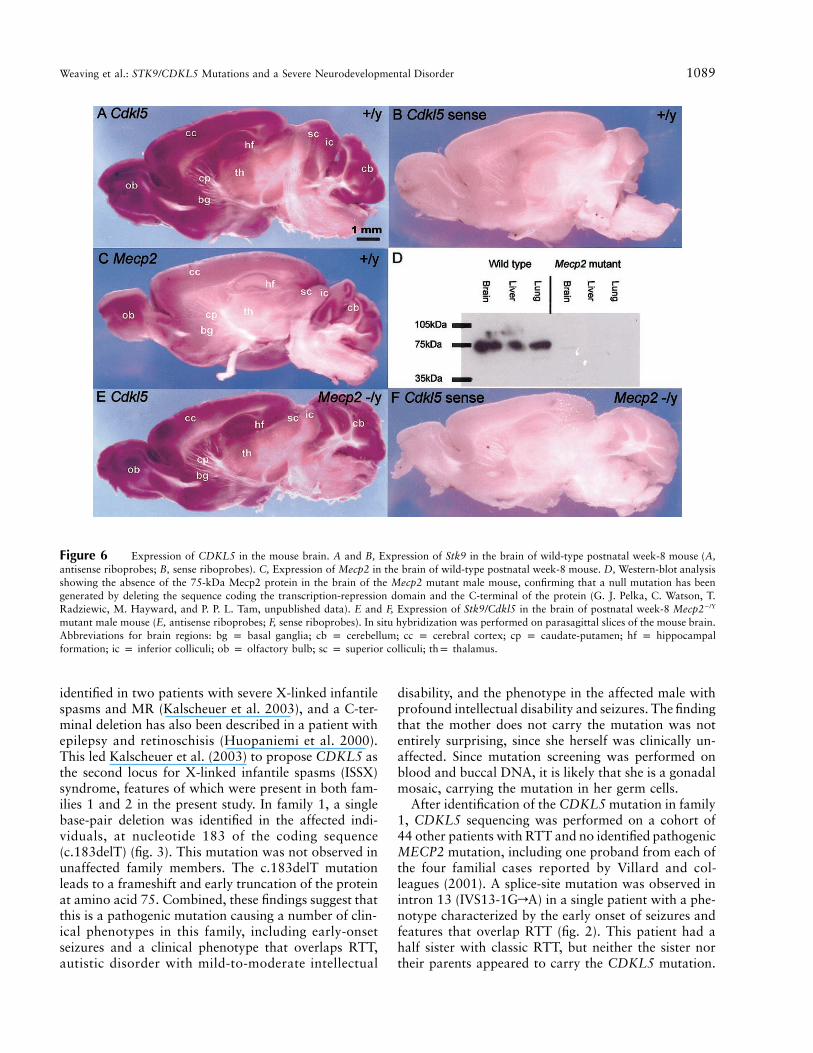
Figure 2 IVS13-1GrA mutation of CDKL5 in family 2. Chromatograms of the CDKL5 sequence that is mutated in family 2 are shown.A G-to-A transversion involving the intron 13 splice-acceptor site was identified in blood DNA from the subject with an atypical RTT–likephenotype (II:1) but not in her half sister (II:2) with classical RTT or either of her parents.
sosomal enzymes, karyotype, urinary amino- and or-ganic-acid analyses, and urinary succinylpurines. Plasmaelectrolytes, copper, acid-base status, urea, calcium,magnesium, uric acid, blood glucose, creatinine, andvery-long-chain fatty acids were also normal. His plasmalactate was mildly elevated, but the cerebrospinal fluid(CSF) lactate was normal. CSF neurotransmitters, glu-cose, and protein and viral studies for herpes simplex,measles, and varicella were normal. He had a normalmale karyotype, 46XY. Results of a skin biopsy showedno evidence of a storage disorder or neuroaxonal dys-trophy. He did not have the common mtDNA mutationsfor mitochondrial encephalopathy, lactic acidosis, andstroke-like episodes; myoclonic epilepsy ragged red fi-bers; Leigh syndrome; or neurogenic ataxia retinitis pig-mentosa in a peripheral blood sample. Rectal-biopsyelectron microscopy was normal. This boy died of re-spiratory failure at the age of 16 years, secondary toaspiration pneumonia. In retrospect, his parents reportepisodes of hyperventilation that were unrelated to feedsand/or aspiration.
Clinical Description of Family 2
This family has been reported elsewhere (family 9 inGill et al. 2003). The proband (II:1) (fig. 2) was bornat term after a normal pregnancy. Her birth weight wasin the 50th percentile. She smiled at 6 wk but shortlyafterward developed severe infantile spasms. Calcifica-tion on her brain CT scan was suggestive of intrauterinecytomegalovirus infection, but viral serology did notsupport this diagnosis. She developed intractable sei-zures (generalized tonic clonic, myoclonic, and absenceseizures) during childhood, which worsened during ad-olescence and improved somewhat in adulthood, withdaily brief myoclonic seizures. Developmentally, shemade very slow progress up to 5 years of age, gainingfine and gross motor skills to a basic level but then be-coming clumsy and losing these skills. She was a hy-potonic child, who sat unsupported at 17 mo and stoodat 4 years of age. She spoke several single words, builta tower of four bricks, spoon fed with help, and turnedsingle pages of a book. She lost these skills between 2

Weaving et al.: STK9/CDKL5 Mutations and a Severe Neurodevelopmental Disorder 1085
years and 5 years of age. She walked independently at7 years, with an unsteady gait, and developed a finepincer grasp. When reviewed at age 28 years, her height,weight, and head circumference were all below the 3rdpercentile. She had no verbal communication skills,marked intellectual impairment, and a scoliosis that hasnot required surgery. Hand stereotypies predominatedon the left side, although, in the past, these were midline.She hyperventilated when upset and had peripheral au-tonomic disturbance. She had a disturbed sleep patternand laughed spontaneously at night. She was not dys-morphic but was hirsute. She had dystonia with normalreflexes and had small feet (below the 3rd percentile).She was considered to have atypical RTT because ofher very poor early progress, early onset of severeepilepsy, late and slight regression, and little, if any,breathing abnormality or stereotypy (A. M. Kerr, per-sonal communication).
II:2 (fig. 2) was the younger half sister of the proband,related through the mother. She was born at term aftera normal pregnancy. Her birth weight was between the50th and 75th percentiles, and her head circumferencewas in the 9th percentile. There were early maternalconcerns regarding placidity, hypotonia, and a slightlyvacant look. Her head circumference fell below the 3rdpercentile over the first 2 years. She achieved skills to abasic level: babbling at 9 mo, sitting with a roundedback at 19 mo, and standing at 4 years, and she wasable to stack four bricks and unscrew lids prior to re-gression. She regressed between 3 years and 5 years ofage, losing her hand skills and stagnating in her motorabilities. She developed typical hand stereotypies, as wellas hyperventilation and breath-holding episodes. Shemade subsequent slow progress, walking independentlyat 7 years with an unsteady gait and developing a palmargrasp. When seen at 13 years of age, she was a happychild, keen to interact socially. She could babble “mu-mum” and “dada” and understood several basic com-mands, but her verbal skills were not considered suffi-cient for a diagnosis of preserved speech variant. Shehad a moderate scoliosis and suffered from sleep dis-turbance, labile mood, peripheral vasomotor distur-bance, and mild constipation. She has never had seizures.She had an Angelman-like facial appearance (however,methylation and uniparental disomy studies for Angel-man syndrome were negative [results not shown]). Herhead circumference and weight were both below the 3rdpercentile. She had marked ligamentous laxity and nojoint contractures. She had short fourth metatarsals andsmall feet (below the 3rd percentile). The clinical diag-nosis of classical RTT in this patient was independentlyconfirmed by Dr. A. M. Kerr of the Department of Psy-chological Medicine, University of Glasgow, Scotland(personal communication).
MECP2 Mutation Screening
The entire coding region of the MECP2 gene (includ-ing exon 1) was sequenced in the twins from family 1and their affected brother. A c.426CrT sequence vari-ation was identified in the twins but not in the affectedbrother. This coded for a silent polymorphism (F142F),but such changes can affect splicing enhancers in someinstances (Cartegni et al. 2002). Therefore, the PCRproduct containing this nucleotide change was se-quenced in the other family members. It was not iden-tified in the affected brother or father but was seen inthe unaffected sister and mother. It thus seems unlikelythat this nucleotide change is pathogenic. No otherMECP2 changes, including large genomic rearrange-ments screened by MPLA, were identified in this family(results not shown). Similarly, sequencing of the twoaffected individuals in family 2 revealed no pathogenicsequence variations in any of the four MECP2 exons,and MLPA analysis did not show any large rearrange-ments involving this gene.
X-Chromosome Exclusion and Haplotype Mapping
Microsatellite mapping was performed on family 1,to confirm that the twins were identical and to determinewhether the Xq28 region containing the MECP2 genewas shared among the affected siblings. Results of themicrosatellite mapping are shown in the family tree infigure 1. The microsatellite mapping was compatiblewith the twins being identical. The Xq28 region was notshared with their affected brother but was shared be-tween the twins and their unaffected sister. MECP2 wasthus excluded as a disease candidate gene in this family,but the inheritance pattern and phenotypic variation stillsuggested that this was an X-linked dominant condition.
Three potential candidate regions were identifiedthrough exclusion mapping (fig. 1). The first was 10.3Mb at Xp22.31-pter between Xpter and DXS1135, thesecond was 19.7 Mb at Xp22.12-p22.11 betweenDXS1135 and DXS1214, and the third was 16.4 Mbat Xq21.33 between DXS1196 and DXS1191. Simonicet al. (1997) observed an increase in the incidence of abreakpoint at Xp22.1 in patients with RTT, which leadsus to favor this region. Further support for Xp22 as acandidate region came from the report of a patientwith RTT and an (X;3)(p22.11;q13.31) translocation(Zoghbi et al. 1990a). The candidate genes ARX andCDKL5 both reside within the Xp22.12-p22.1 region.
In family 2, only microsatellite markers flanking theCDKL5 and MECP2 genes were used. These analysesshowed that, for both loci, the sisters inherited differentmaternal and paternal X-chromosome haplotypes (re-sults not shown). These results strongly suggest that theirclinical phenotypes of RTT have resulted from different

1086 Am. J. Hum. Genet. 75:1079–1093, 2004
Figure 3 c.183delT mutation of CDKL5 in family 1. A chromatogram of the CDKL5 sequence that is mutated in family 1 is shown.Nucleotide numbers begin from the A of the translation start codon in exon 2. Individual ID numbers are derived from figure 1. A deletion ofnucleotide 183 was observed in the blood DNA of affected individuals (III:1, III:2, and III:3) but not in unaffected family members.
causes: the CDKL5 gene mutation in II:1 and an as-yetunknown cause in II:2.
Candidate-Gene Mutation Screening in Family 1
The ARX gene was chosen as a candidate for screeningbecause of the pleiotropy of its mutations (Kato et al.2004; Partington et al. 2004). However, no mutationswere detected in the coding region of this gene infamily 1.
The STK9 gene was chosen as a second candidate forscreening, because the phenotype of infantile spasmsoverlapped that described elsewhere in association withCDKL5 rearrangements (Kalscheuer et al. 2003). A de-letion of nucleotide 183 (c.183delT) in exon 5 was iden-tified in the affected siblings but not in any of the un-affected members of family 1, including their mother,for whom the mutation was screened in DNA extractedfrom blood and buccal cells (fig. 3). This deletion resultsin a frameshift and a premature truncation of the geneproduct at amino acid 75.
CDKL5 Screening of Other MECP2-Negative Subjectswith RTT
After the identification of the c.183delT mutation inCDKL5 in family 1, screening of the CDKL5 gene wasperformed in 44 patients with RTT who were MECP2
mutation negative. These included cases ascertainedfrom Australia and the United Kingdom and one of theprobands from each of four familial cases reported byVillard et al. (2000, 2001) who showed highly skewedX inactivation. A splice-site mutation (IVS13-1GrA)was identified in patient II:1 from family 2, who has theearly-onset–seizure RTT variant (see “Clinical Descrip-tion of Family 2” section and fig. 2). This patient hada sister with classic RTT, but neither the sister nor theparents carried the CDKL5 mutation (fig. 2). This mu-tation leads to the loss of a DdeI site. In unaffectedindividuals, a 346-bp fragment encompassing this regionis cut into 185-bp and 135-bp fragments, whereas themutant allele is cut into 320-bp and 26-bp fragments(results not shown).
Characterization of the CDKL5 IVS13-1GrA Mutation
The CDKL5 IVS13-1GrA mutation identified in II:1(family 2) was not found in any of the other 43 MECP2-mutation-negative patients with RTT who were inves-tigated or in 236 control chromosomes. To explore thepathogenicity of IVS13-1GrA further, we designedprimers that would amplify the cDNA from exons 12–15, inclusive. A fragment of 365 bp was identified in anRT-PCR of blood RNA from patient II:1. An apparentlynormal CDKL5 cDNA sequence was obtained from se-

Weaving et al.: STK9/CDKL5 Mutations and a Severe Neurodevelopmental Disorder 1087
Figure 4 cDNA and translated product of the CDKL5 IVS-1GrA mutation. A, Sequence chromatograms of representative cDNA clonesisolated from II:1 (family 2) blood RNA/cDNA. In 5 (16.6%) of 30 clones examined, a c.2047delG CDKL5 cDNA was observed. B, Illustrationof the creation of a novel intron 13 acceptor splice site as a result of the IVS13-1GrA mutation in CDKL5 of individual II:1 from family 2.The protein sequence is shown below the genomic sequence.
quencing. However, after a more careful inspection ofthe sequence chromatograms, a minor second sequencesignal could be detected in the background of the normalsequence (mutation shown in fig. 4A). Interestingly, thebackground cDNA sequence appeared to be shifted byonly 1 bp. On the basis of this, we hypothesized thatthe IVS13-1GrA mutation results in the activation of anovel 3′ acceptor splice site only 1 bp away from theoriginal one (fig. 4B). To test this hypothesis, peripheralblood RNA of patient II:1 was used to amplify this re-gion (exons 12–15) of the CDKL5 cDNA by RT-PCR.Using cloning, mismatch PCR, restriction digest, andsequencing (see “Material and Methods” section), weidentified 5 clones (of 30 analyzed) that carried the hy-pothesized 1-bp CDKL5 cDNA deletion (c.2047delG).The frequency of the c.2047delG clone (16.6%) amongthe cloned exon 12–15 RT-PCR products is in agreementwith the X-inactivation studies, indicating that the chro-mosome carrying the c.2047delG is active in only 15%of peripheral blood cells (data not shown). Translation
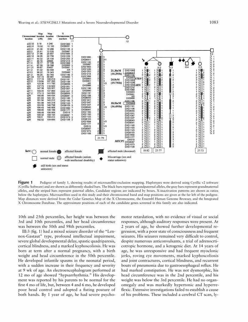
of the mutant spliced product would create a prematurestop codon (M783X), truncating the protein approxi-mately halfway through exon 16 (fig. 5).
X Inactivation
Results from the AR assay suggested that the mother,both of the twins, and their unaffected sister from family1 had skewed X inactivation (fig. 1). The twins had ahigh degree of skewing (82% and 77%) in their bloodDNA, but this appeared to favor the same X chromo-some. In the mother, the grandmaternal chromosomewas preferentially active in 79% of her lymphocytes,whereas, in DNA from a buccal sample, it was 96%.
In family 2, X inactivation was studied using an FMR1assay. The patient (II:1) had a skewed X-inactivationpattern with a ratio of 85:15. With the use of the avail-able family members, it was not possible to determinewhich X chromosome (normal or with the CDKL5 mu-tation) was preferentially inactivated. However, the anal-
1088 Am. J. Hum. Genet. 75:1079–1093, 2004
Figure 5 Schematic representation of the CDKL5 gene. CDKL5 exons are indicated by black boxes, and the last four exons of RS1, agene overlapping CDKL5, are indicated by white boxes. The alternative-splicing patterns are indicated below the diagram for the knownisoforms, I and II. Mutations and associated phenotypes that have been reported to date in association with CDKL5 mutations are given abovethe diagram, and those described in the present study are shown in bold.
ysis of the CDKL5 cDNA from this patient suggestedthat the chromosome carrying the CDKL5 IVS13-1GrAmutation was preferentially inactivated (active in ∼15%of peripheral blood cells).
Expression of Cdkl5 in Mouse Brains
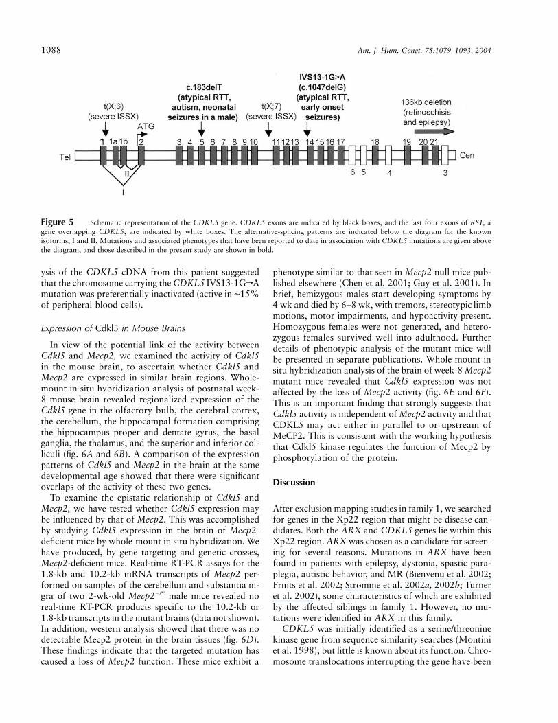
In view of the potential link of the activity betweenCdkl5 and Mecp2, we examined the activity of Cdkl5in the mouse brain, to ascertain whether Cdkl5 andMecp2 are expressed in similar brain regions. Whole-mount in situ hybridization analysis of postnatal week-8 mouse brain revealed regionalized expression of theCdkl5 gene in the olfactory bulb, the cerebral cortex,the cerebellum, the hippocampal formation comprisingthe hippocampus proper and dentate gyrus, the basalganglia, the thalamus, and the superior and inferior col-liculi (fig. 6A and 6B). A comparison of the expressionpatterns of Cdkl5 and Mecp2 in the brain at the samedevelopmental age showed that there were significantoverlaps of the activity of these two genes.
To examine the epistatic relationship of Cdkl5 andMecp2, we have tested whether Cdkl5 expression maybe influenced by that of Mecp2. This was accomplishedby studying Cdkl5 expression in the brain of Mecp2-deficient mice by whole-mount in situ hybridization. Wehave produced, by gene targeting and genetic crosses,Mecp2-deficient mice. Real-time RT-PCR assays for the1.8-kb and 10.2-kb mRNA transcripts of Mecp2 per-formed on samples of the cerebellum and substantia ni-gra of two 2-wk-old Mecp2�/Y male mice revealed noreal-time RT-PCR products specific to the 10.2-kb or1.8-kb transcripts in the mutant brains (data not shown).In addition, western analysis showed that there was nodetectable Mecp2 protein in the brain tissues (fig. 6D).These findings indicate that the targeted mutation hascaused a loss of Mecp2 function. These mice exhibit a
phenotype similar to that seen in Mecp2 null mice pub-lished elsewhere (Chen et al. 2001; Guy et al. 2001). Inbrief, hemizygous males start developing symptoms by4 wk and died by 6–8 wk, with tremors, stereotypic limbmotions, motor impairments, and hypoactivity present.Homozygous females were not generated, and hetero-zygous females survived well into adulthood. Furtherdetails of phenotypic analysis of the mutant mice willbe presented in separate publications. Whole-mount insitu hybridization analysis of the brain of week-8 Mecp2mutant mice revealed that Cdkl5 expression was notaffected by the loss of Mecp2 activity (fig. 6E and 6F).This is an important finding that strongly suggests thatCdkl5 activity is independent of Mecp2 activity and thatCDKL5 may act either in parallel to or upstream ofMeCP2. This is consistent with the working hypothesisthat Cdkl5 kinase regulates the function of Mecp2 byphosphorylation of the protein.
Discussion
After exclusion mapping studies in family 1, we searchedfor genes in the Xp22 region that might be disease can-didates. Both the ARX and CDKL5 genes lie within thisXp22 region. ARX was chosen as a candidate for screen-ing for several reasons. Mutations in ARX have beenfound in patients with epilepsy, dystonia, spastic para-plegia, autistic behavior, and MR (Bienvenu et al. 2002;Frints et al. 2002; Strømme et al. 2002a, 2002b; Turneret al. 2002), some characteristics of which are exhibitedby the affected siblings in family 1. However, no mu-tations were identified in ARX in this family.
CDKL5 was initially identified as a serine/threoninekinase gene from sequence similarity searches (Montiniet al. 1998), but little is known about its function. Chro-mosome translocations interrupting the gene have been
Weaving et al.: STK9/CDKL5 Mutations and a Severe Neurodevelopmental Disorder 1089
Figure 6 Expression of CDKL5 in the mouse brain. A and B, Expression of Stk9 in the brain of wild-type postnatal week-8 mouse (A,antisense riboprobes; B, sense riboprobes). C, Expression of Mecp2 in the brain of wild-type postnatal week-8 mouse. D, Western-blot analysisshowing the absence of the 75-kDa Mecp2 protein in the brain of the Mecp2 mutant male mouse, confirming that a null mutation has beengenerated by deleting the sequence coding the transcription-repression domain and the C-terminal of the protein (G. J. Pelka, C. Watson, T.Radziewic, M. Hayward, and P. P. L. Tam, unpublished data). E and F, Expression of Stk9/Cdkl5 in the brain of postnatal week-8 Mecp2�/Y
mutant male mouse (E, antisense riboprobes; F, sense riboprobes). In situ hybridization was performed on parasagittal slices of the mouse brain.Abbreviations for brain regions: bg p basal ganglia; cb p cerebellum; cc p cerebral cortex; cp p caudate-putamen; hf p hippocampalformation; ic p inferior colliculi; ob p olfactory bulb; sc p superior colliculi; thp thalamus.
identified in two patients with severe X-linked infantilespasms and MR (Kalscheuer et al. 2003), and a C-ter-minal deletion has also been described in a patient withepilepsy and retinoschisis (Huopaniemi et al. 2000).This led Kalscheuer et al. (2003) to propose CDKL5 asthe second locus for X-linked infantile spasms (ISSX)syndrome, features of which were present in both fam-ilies 1 and 2 in the present study. In family 1, a singlebase-pair deletion was identified in the affected indi-viduals, at nucleotide 183 of the coding sequence(c.183delT) (fig. 3). This mutation was not observed inunaffected family members. The c.183delT mutationleads to a frameshift and early truncation of the proteinat amino acid 75. Combined, these findings suggest thatthis is a pathogenic mutation causing a number of clin-ical phenotypes in this family, including early-onsetseizures and a clinical phenotype that overlaps RTT,autistic disorder with mild-to-moderate intellectual
disability, and the phenotype in the affected male withprofound intellectual disability and seizures. The findingthat the mother does not carry the mutation was notentirely surprising, since she herself was clinically un-affected. Since mutation screening was performed onblood and buccal DNA, it is likely that she is a gonadalmosaic, carrying the mutation in her germ cells.
After identification of the CDKL5 mutation in family1, CDKL5 sequencing was performed on a cohort of44 other patients with RTT and no identified pathogenicMECP2 mutation, including one proband from each ofthe four familial cases reported by Villard and col-leagues (2001). A splice-site mutation was observed inintron 13 (IVS13-1GrA) in a single patient with a phe-notype characterized by the early onset of seizures andfeatures that overlap RTT (fig. 2). This patient had ahalf sister with classic RTT, but neither the sister northeir parents appeared to carry the CDKL5 mutation.

1090 Am. J. Hum. Genet. 75:1079–1093, 2004
This phenomenon is not without precedent, since thepresence of a MECP2 mutation in one but not all af-fected members of a family unit (who may each showvariable clinical features) has been reported elsewhere(Gill et al. 2003). Our results from haplotype analysisof CDKL5 and MECP2 loci in the family further sup-port the hypothesis of different causes of the RTT phe-notype in the half sisters.
The IVS13-1GrA mutation changes the canonical 3′
acceptor splice site of intron 13 from AG to AA. Sucha 3′ acceptor splice site has not been detected in eukar-yotic genomes (Burset et al. 2000). It would, therefore,be highly unlikely for it to be recognized by the splicingmachinery and thus be likely to cause abnormal splicing.We demonstrated that the consequence of the IVS13-1GrA change is an activation of a cryptic splice site(81.34% consensus strength) just 1 bp downstream ofthe original site (96% consensus strength) (Shapiro andSenapathy 1987). We also showed that the IVS13-1GrA nucleotide change leads to the production ofCDKL5 mRNA with a deletion of the G at position2047 (c.2047delG; see fig. 4). This would lead to pro-duction of a truncated form of the STK9 protein andis the most likely cause of the clinical presentation ofearly-onset seizures and the phenotype that overlapsRTT observed in family 2 (patient II:1).
It is interesting to note that the seizures seen in theaffected individuals reported in this study and thosementioned in other published reports of CDKL5 genedefects (Huopaniemi et al. 2000; Kalscheuer et al. 2003)were early in their onset and were generally difficult tocontrol with anticonvulsant therapies. Early-onset sei-zures, usually more severe, have been described else-where in patients with RTT, and these seizures maysometimes precede the appearance of the more recog-nizable RTT features (Steffenburg et al. 2001). Steffen-burg et al. (2001) found an onset of seizures before apatient’s first birthday in 7 (18%) of 53 cases in theirseries, although none were actually classified as infantilespasms. In our Australian cohort with RTT (excludingfamily 1 in the present study), 18 (7%) of 258 patientswith verified RTT had seizures in the 1st year of life,and an MECP2 mutation was found in only 3 of 12individuals in whom mutation screening had been per-formed (H.L., unpublished data).
The twins from family 1 were confirmed as identicalby microsatellite mapping of the X chromosome andHLA typing but were discordant for their phenotype.Since the twins are genetically identical, their pheno-typic differences may only be attributable to epigeneticor environmental differences. Although rare, discor-dance between identical twins for the RTT phenotypehas been described elsewhere (Migeon et al. 1995; Ishiiet al. 2001). The most likely explanation for the dis-cordance in these instances was thought to be differ-
ential skewing of X inactivation, as was observed byHoffbuhr et al. (2001), although the development of amutation in one but not the other postzygotic embryoremains an untested possibility in these studies predat-ing the identification of MECP2 as the major causativegene in RTT. However, both of the twins in family 1had similar levels of skewing of the X chromosome (18:82 and 23:77), and this appeared to favor the samechromosome. As such, skewing of X inactivation doesnot explain their phenotypic differences, unless somediscordance between the brain, as the primarily affectedtissue, and blood is speculated. Such differences in X-inactivation patterns between different tissues have beendescribed elsewhere (Tan et al. 1993; Gale et al. 1994).Moreover, variability in X inactivation across differentbrain regions in individual heterozygous Mecp2308/X mu-tant mice has been reported (Young and Zoghbi 2004),and this, too, could contribute to differences in the se-verity of the clinical phenotype.
It should be noted that another group has recentlyidentified CDKL5 missense mutations in two patientswith diagnoses of RTT. These patients had early-onsetseizures and severe psychomotor retardation—charac-teristics of the early-onset–seizure RTT variant—as wellas a number of other typical clinical features associatedwith RTT, but they did not have the very noticeableintense eye gaze that is characteristic of patients withclassical RTT (Tao et al. 2004 [in this issue]). Patientswith atypical RTT, including those with the early-onset–seizure variant, are less likely to have MECP2 mutations(Hammer et al. 2002) and so should be considered asstrong candidates for CDKL5-mutation screening.
STK9 is a putative serine/threonine kinase of un-known function (Montini et al. 1998; Brunner et al.1999). The mechanism by which mutations in CDKL5produce phenotypes overlapping those seen in patientswith MECP2 mutations is, therefore, a puzzle, althoughthe most likely hypothesis is that they operate in thesame developmental pathway. Little work has beendone on CDKL5, but sequence comparisons have in-dicated that its protein product is most closely relatedto p56 KKIAMRE (the protein encoded by CDKL2)and p42 KKIALRE (the protein encoded by CDKL1),which share homology with the mitogen-activated pro-tein (MAP) kinase family of cyclin-dependent proteinkinases (Montini et al. 1998).
It has recently been reported that phosphorylation ofMeCP2 causes it to be released from a target promoter,resulting in transcriptional reactivation (Chen et al.2003). It is unknown at this stage whether MeCP2 andSTK9 interact directly with each other, with MeCP2possibly being a phosphorylation target for STK9;whether they operate via a common developmentalpathway; or whether they might exert their effectsthrough unrelated mechanisms. Future work to identify
Weaving et al.: STK9/CDKL5 Mutations and a Severe Neurodevelopmental Disorder 1091
the functional role of STK9, its relation to MeCP2, andthe mechanisms regulating both will be essential in un-derstanding the biological processes involved in the eti-ology of RTT and of related clinical phenotypes. Dis-section of this developmental pathway may also identifyfurther RTT candidate genes and potential targets fortherapy.
Together, our results suggest that CDKL5 mutationscan give rise to a phenotype that strongly resemblesRTT. However, given the variant phenotypes seen infamily 1 and in the three other reported cases withCDKL5 mutations (Huopaniemi et al. 2000; Kalscheueret al. 2003), screening of a larger cohort of patientswith atypical RTT—particularly those with early-onsetseizures—and individuals with overlapping phenotypes,including severe intellectual disability with autistic fea-tures, may lead to the identification of further mutationsin this gene and may clarify the contribution of CDKL5to the pathogenesis of RTT and related disorders.
In summary, we have identified another gene that maycause a phenotype that shows considerable overlap withRTT. Mutations in CDKL5 may also be associated withthe clinical phenotype of autistic spectrum disorderwith MR and complex seizure disorders in females andthe phenotype of severe neonatal-onset neurologicalabnormalities in males. It remains to be determinedwhether CDKL5 mutations may account for a largeproportion of cases showing significant overlap withRTT and in which no pathogenic MECP2 mutationshave been found. There is a need for a large-scale mul-ticenter collaborative study using, when feasible, casessourced on a population basis, to investigate the role ofCDKL5, and possibly other genes from the same path-way, in the pathogenesis of RTT and related disorders.
Acknowledgments
We would like to thank the families involved in this study,for their invaluable contribution. We are grateful to LaurentVillard (INSERM U491, Faculte de Medecine La Timone, Mar-seille, France) for providing us with DNA samples, from anumber of patients with RTT, for CDKL5 mutation screening.We would also like to acknowledge the help of Joanna Craw-ford with X-inactivation studies and the technical support ofSara Lonergan, Julianne Jackson, Zhanhe Wu, and RoseWhite. We would also like to thank Dr. Alison Kerr for herclinical opinion and helpful comments. Financial support forthis research was provided by the Rett Syndrome Associationof NSW, the Rett Syndrome Australian Research Fund, theNational Health and Medical Research Council of Australia(NHMRC), and the Country Women’s Association of NSW.The U.K. laboratory research was performed with grant sup-port from The Health Foundation. G.J.P. is an NHMRC DoraLush Biomedical Scholar, J.G. is an NHMRC Senior ResearchFellow, and P.P.L.T. is an NHMRC Senior Principal ResearchFellow.
Electronic-Database Information
The URLs for data presented herein are as follows:
Cedar Genetics Map, http://cedar.genetics.soton.ac.uk/pub/chromX/gmap (for map of the X chromosome and geneticmap distances)
Ensembl Human Genome Browser, http://www.ensembl.org/Homo_sapiens/mapview?chrpX (for MapView and geneticmap distances)
Genome Database, http://gdbwww.gdb.org/ (for microsatelliteinformation)
Integrated X Chromosome Database, version 2.3, http://ixdb.molgen.mpg.de/ (for genetic map distances)
Mouse Genome Informatics (MGI), http://www.informatics.jax.org/ (for details of the mouse Cdkl5 sequence)
NCBI, http://www.ncbi.nlm.nih.gov/ (for BLAST searches andsequence information)
Online Mendelian Inheritance in Man (OMIM), http://www.ncbi.nlm.nih.gov/Omim/ (for CDKL5)
References
Allen RC, Zoghbi HY, Moseley AB, Rosenblatt HM, BelmontJW (1992) Methylation of HpaII and HhaI sites near thepolymorphic CAG repeat in the human androgen-receptorgene correlates with X chromosome inactivation. Am J HumGenet 51:1229–1239
Amir RE, Van den Veyver IB, Schultz R, Malicki DM, TranCQ, Dahle EJ, Philippi A, Timar L, Percy AK, Motil KJ,Lichtarge O, Smith EO, Glaze DG, Zoghbi HY (2000) In-fluence of mutation type and X chromosome inactivationon Rett syndrome phenotypes. Ann Neurol 47:670–679
Ben Zeev B, Yaron Y, Schanen NC, Wolf H, Brandt N, GinotN, Shomrat R, Orr-Urtreger A (2002) Rett syndrome: clin-ical manifestations in males with MECP2 mutations. J ChildNeurol 17:20–24
Bienvenu T, Poirer K, Friocourt G, Bahi N, Beaumont D,Fauchereau F, Ben Jeema L, Zemni R, Vinet MC, Francis F,Couvert P, Gomot M, Moraine C, Van Bokhoven H, Kal-scheuer V, Frints S, Gecz F, Ohzaki K, Chaabouni H, FrynsJP, Desportes V, Beldjord C, Chelly J (2002) ARX, a novelPrd-class-homeobox gene highly expressed in the telenceph-alon, is mutated in X-linked mental retardation. Hum MolGenet 11:981–991
Brunner B, Todt T, Lenzner S, Stout K, Schultz U, Ropers H-H, Kalscheuer VM (1999) Genomic structure and compar-ative analysis of nine fugu genes: conservation of syntenywith human chromosome Xp22.2-p22.1. Genome Res 9:437–448
Burset M, Seledtsov IA, Solovyev VV (2000) Analysis of ca-nonical and non-canonical splice sites in mammalian ge-nomes. Nucleic Acid Res 28:4364–4375
Carrel L, Willard HF (1996) An assay for X inactivation basedon differential methylation at the fragile X locus. Am J MedGenet 64:27–30
Cartegni L, Chew SL, Krainer AR (2002) Listening to silenceand understanding nonsense: exonic mutations that affectsplicing. Nat Rev Genet 3:285–298
Cheadle JP, Gill H, Fleming N, Maynard J, Kerr A, Leonard

1092 Am. J. Hum. Genet. 75:1079–1093, 2004
H, Krawczak M, Cooper DN, Lynch S, Thomas N, HughesH, Hulten M, Ravine D, Sampson JR, Clarke A (2000)Long-read sequence analysis of the MECP2 gene in Rettsyndrome patients: correlation of disease severity with mu-tation type and location. Hum Mol Genet 9:1119–1129
Chen RZ, Akbarian S, Tudor M, Jaenisch R (2001) Deficiencyof methyl-CpG binding protein-2 in CNS neurons results ina Rett-like phenotype in mice. Nat Genet 27:327–331
Chen WG, Chang Q, Lin Y, Meissner A, West AE, GriffithEC, Jaenisch R, Greenberg ME (2003) Derepression ofBDNF transcription involves calcium-dependent phosphor-ylation of MeCP2. Science 302:885–889
Davidson BP, Kinder SJ, Steiner K, Schoenwolf GC, Tam PP(1999) Impact of node ablation on the morphogenesis ofthe body axis and the lateral asymmetry of the mouse em-bryo during early organogenesis. Dev Biol 211:11–26
Ellaway CJ (2001) Rett syndrome: diagnostic strategies andtherapeutic interventions. PhD thesis, University of Sydney,Westmead
Frints SG, Froyen G, Marynen P, Willekens D, Legius E, FrynsJP (2002) Re-evaluation of MRX36 family after discoveryof an ARX gene mutation reveals mild neurological featuresof Partington syndrome. Am J Med Genet 112:427–428
Gale RE, Wheadon H, Boulos P, Linch DC (1994) Tissue spec-ificity of X-chromosome inactivation patterns. Blood 83:2899–2905
Geerdink N, Rotteveel JJ, Lammens M, Sistermans EA, Hei-kens GT, Gabreels FJ, Mullaart RA, Hamel BC (2002)MECP2 mutation in a boy with severe neonatal enceph-alopathy: clinical, neuropathological and molecular find-ings. Neuropediatrics 33:33–36
Gill H, Cheadle JP, Maynard J, Fleming N, Whatley S, Cran-ston T, Thompson EM, Leonard H, Davis M, ChristodoulouJ, Skjeldal O, Hanefeld F, Kerr A, Tandy A, Ravine D, ClarkeA (2003) Mutation analysis in the MECP2 gene and geneticcounselling for Rett syndrome. J Med Genet 40:380–384
Goutieres F, Aicardi J (1987) New experience with Rett syn-drome in France: the problem of atypical cases. Brain Dev9:502–505
Guy J, Hendrich B, Holmes M, Martin JE, Bird A (2001) Amouse Mecp2-null mutation causes neurological symptomsthat mimic Rett syndrome. Nat Genet 27:322–326
Hagberg B, Goutieres F, Hanefield F, Rett A, Wilson J (1985)Rett syndrome: criteria for inclusion and exclusion. BrainDev 7:372–373
Hagberg B, Hanefeld F, Percy AK, Skjeldal O (2002) An updateon clinically applicable diagnostic criteria for Rett syn-drome: comments to Rett Syndrome Clinical Criteria Con-sensus Panel Satellite to European Paediatric Neurology So-ciety Meeting, Baden Baden, Germany, 11 September 2001.Eur J Paediatr Neurol 6:293–297
Hagberg B, Rasmussen P (1986) “Form fruste” of Rett syn-drome—a case report. Am J Med Genet 24:175–181
Hagberg B, Skjeldal OH (1994) Rett variants: a suggestedmodel for inclusion criteria. Pediatr Neurol 11:5–11
Hammer S, Dorrani N, Dragich J, Kudo S, Schanen C (2002)The phenotypic consequences of MECP2 mutations extendbeyond Rett syndrome. Ment Retard Dev Disabil Res Rev8:94–98
Hoffbuhr K, Devaney JM, LaFleur B, Sirianni N, Scacheri C,Giron J, Schuette J, Innis J, Marino M, Philippart M, Na-rayanan V, Umansky R, Kronn D, Hoffman EP, Naidu S(2001) MECP2 mutations in children with and without thephenotype of Rett syndrome. Neurology 56:1486–1495
Hoffbuhr KC, Moses LM, Jerdonek MA, Naidu S, HoffmanEP (2002) Associations between MECP2 mutations, X-chro-mosome inactivation, and phenotype. Ment Retard Dev Dis-abil Res Rev 8:99–105
Huopaniemi L, Tyynismaa H, Rantala A, Rosenberg T, AlitaloT (2000) Characterisation of two unusual RS1 gene dele-tions segregating in Danish retinoschisis families. Hum Mu-tat 16:307–314
Huppke P, Laccone F, Kramer N, Engel W, Hanefeld F (2000)Rett syndrome: analysis of MECP2 and clinical character-isation of 31 patients. Hum Mol Genet 9:1369–1375
Ishii T, Makita Y, Ogawa A, Amamiya S, Yamamoto M, Mi-yamoto A, Oki J (2001) The role of different X-inactivationpatterns on the variable clinical phenotype with Rett syn-drome. Brain Dev 23:S161–S164
Kalscheuer VM, Tao J, Donnelly A, Hollway G, Schwinger E,Kubart S, Menzel C, Hoeltzenbein M, Tømmerup N, EyreH, Harbord M, Haan E, Sutherland GR, Ropers H-H, GeczJ (2003) Disruption of the serine/threonine kinase 9 genecauses severe X-linked infantile spasms and mental retar-dation. Am J Hum Genet 72:1401–1411
Kato M, Das S, Petras K, Kitamura K, Morohashi K, AbueloDN, Barr M, et al (2004) Mutations of ARX are associatedwith striking pleiotropy and consistent genotype-phenotypecorrelation. Hum Mutat 23:147–159
Krepischi AC, Kok F, Otto PG (1998) X chromosome-in-activation patterns in patients with Rett syndrome. HumGenet 102:319–321
Kriaucionis S, Bird A (2004) The major form of MeCP2 hasa novel N-terminus generated by alternative splicing. Nu-cleic Acid Res 32:1818–1823
Kuramoto N, Inoue K, Takano K, Taniura H, Sakata K, OgitaK, Yoneda Y (2003) A possible novel mechanism underlyingtemperature-dependent uptake of [3H]spermidine in nuclearfractions of murine brain. Brain Res 981:78–84
Laccone F, Junemann I, Whatley S, Morgan R, Butler R,Huppke P, Ravine D (2004) Large deletions of the MECP2gene detected by gene dosage analysis in patients with Rettsyndrome. Hum Mutat 23:234–244
Leonard H, Bower C, English D (1997) The prevalence andincidence of Rett syndrome in Australia. Eur Child AdolescPsychiatry 6:8–10
Leonard H, Silberstein J, Falk R, Houwink-Manville I, EllawayC, Raffaele LS, Engerstrom IW, Schanen C (2001) Occur-rence of Rett syndrome in boys. J Child Neurol 16:333–338
Migeon BR, Dunn MA, Thomas G, Schmeckpeper BJ, NaiduS (1995) Studies of X inactivation and isodisomy in twinsprovide further evidence that the X chromosome is not in-volved in Rett syndrome. Am J Hum Genet 56:647–653
Miyamoto A, Yamamoto M, Takahashi S, Oki J (1997) Clas-sical Rett syndrome in sisters: variability of clinical expres-sion. Brain Dev 19:492–494
Mnatzakanian GN, Lohi H, Munteanu I, Alfred SE, YamadaT, MacLeod PJM, Jones JR, Scherer SW, Schanen NC, Friez
Weaving et al.: STK9/CDKL5 Mutations and a Severe Neurodevelopmental Disorder 1093
MJ, Vincent JB, Minassian BA (2004) A previously uniden-tified MECP2 open reading frame defines a new proteinisoform relevant to Rett syndrome. Nat Genet 36:1–3
Montini E, Andolfi G, Caruso A, Buchner G, Walpole SM,Mariani M, Consalez GG, Trump D, Ballabio A, Franco B(1998) Identification and characterisation of a novel serine-threonine kinase gene from the Xp22 region. Genomics 51:427–433
Partington MW, Turner G, Boyle J, Gecz J (2004) Three newfamilies with X-linked mental retardation caused by the428–451dup(24bp) mutation in ARX. Clin Genet 66:39–45
Plenge RM, Stevenson RA, Lubs HA, Schwartz CE, WillardHF (2002) Skewed X-chromosome inactivation is a commonfeature of X-linked mental retardation disorders. Am J HumGenet 71:168–173
Puck JM, Willard HF (1998) X inactivation in females withX-linked disease. New Engl J Med 338:325–328
Sambrook J, Fritsch EF, Maniatis T (1989) Molecular cloning:a laboratory manual. Vol 3. Cold Spring Harbor LaboratoryPress, Cold Spring Harbor, NY
Shapiro MB, Senapathy P (1987) RNA splice junctions of dif-ferent classes of eukaryotes: sequence statistics and func-tional implications in gene expression. Nucleic Acid Res 15:7155–7174
Simonic I, Gericke GS, Lippert M, Schoeman JF (1997) Ad-ditional clinical and cytogenetic findings associated withRett syndrome. Am J Med Genet 74:331–337
Skjeldal OH, von Tetzchner S, Aspelund F, Herder GA, Lof-terld B (1997) Rett syndrome: geographic variation in prev-alence in Norway. Brain Dev 19:258–261
Steffenburg U, Hagberg G, Hagberg B (2001) Epilepsy in arepresentative series of Rett syndrome. Acta Paediatr 90:34–39
Strømme P, Mangelsdorf ME, Scheffer IE, Gecz J (2002a) In-fantile spasms, dystonia, and other X-linked phenotypescaused by mutations in Aristaless related homeobox gene,ARX. Brain Dev 24:266–268
Strømme P, Mangelsdorf ME, Shaw MA, Lower KM, LewisSM, Bruyere H, Lutcherath V, Gedeon AK, Wallace RH,Scheffer IE, Turner G, Partington M, Frints SG, Fryns JP,Sutherland GR, Mulley JC, Gecz J (2002b) Mutations inthe human ortholog of Aristaless cause X-linked mental re-tardation and epilepsy. Nat Genet 30:441–445
Talvik T, Soot A, Beilmann A, Soopold T, Nurk K (1995) Rettsyndrome in Estonia: prevalence of the classical phenotype.Acta Paediatr 84:1070–1071
Tan S-S, Williams EA, Tam PPL (1993) X-chromosome in-activation occurs at different times in different tissues of thepost-implantation mouse embryo. Nat Genet 3:170–174
Tao J, Van Esch H, Hagedorn-Griewe M, Hoffmann K, Moser
B, Raynaud M, Sperner J, Fryns J-P, Schwinger E, Gecz J,Ropers H-H, Kalscheuer VM (2004) Mutations in the X-linked cyclin-dependent kinase-like 5 (CDKL5/STK9) geneare associated with severe neurodevelopmental retardation.Am J Hum Genet 75:1149–1154 (in this issue)
Terai K, Munesue T, Hiratani M, Jiang ZY, Jibiki I, YamaguchiN (1995) The prevalence of Rett syndrome in Fukui pre-fecture. Brain Dev 17:153–154
Turner G, Partington M, Kerr B, Mangelsdorf M, Gecz J(2002) Variable expression of mental retardation, autism,seizures, and dystonic hand movements in two families withan identical ARX gene mutation. Am J Med Genet 112:405–411
Van den Veyver IB, Zoghbi HY (2000) Methyl-CpG-bindingprotein 2 mutations in Rett syndrome. Curr Opin GenetDev 10:275–279
Villard L, Kpebe A, Cardoso C, Chelly J, Tardieu M, FontesM (2000) Two affected boys in a Rett syndrome family—clinical and molecular findings. Neurology 55:1188–1193
Villard L, Levy N, Xiang FQ, Kpebe A, Labelle V, ChevillardC, Zhang ZP, Schwartz CE, Tardieu M, Chelly J, Anvret M,Fontes M (2001) Segregation of a totally skewed pattern ofX chromosome inactivation in four familial cases of Rettsyndrome without MECP2 mutation: implications for thedisease. J Med Genet 38:435–442
Wan MM, Lee SSJ, Zhang XY, Houwink-Manville I, Song HR,Amir RE, Budden S, Naidu S, Pereira JLP, Lo IFM, ZoghbiHY, Schanen NC, Francke U (1999) Rett syndrome andbeyond: recurrent spontaneous and familial MECP2 mu-tations at CpG hotspots. Am J Hum Genet 65:1520–1529
Weaving LS, Williamson SL, Bennetts B, Davis M, EllawayCJ, Leonard H, Thong MK, Delatycki M, Thompson EM,Laing N, Christodoulou J (2003) Effects of MECP2 muta-tion type, location and X-inactivation in modulating Rettsyndrome phenotype. Am J Med Genet 118A:103–114
Webb T, Latif F (2001) Rett syndrome and the MECP2 gene.J Med Genet 38:217–223
Young JI, Zoghbi HY (2004) X-chromosome inactivation pat-terns are unbalanced and affect the phenotypic outcome ina mouse model of Rett syndrome. Am J Hum Genet 74:511–520
Zappella M (1997) The preserved speech variant of the Rettcomplex: a report of 8 cases. Eur Child Adolesc Psychiatry6:23–25
Zoghbi HY, Ledbetter DH, Schultz R, Percy AK, Glaze DG(1990a) A de novo X;3 translocation in Rett syndrome. AmJ Med Genet 35:148–151
Zoghbi HY, Percy AK, Schultz RJ, Fill C (1990b) Patterns ofX chromosome inactivation in the Rett syndrome. Brain Dev12:131–135
Copyright © 2022 FDOKUMEN