Mutations in the PCNA-binding site of CDKN1C inhibit cell proliferation by impairing the entry into...
6
SHORT REPORT Open Access Mutations in the PCNA-binding site of CDKN1C inhibit cell proliferation by impairing the entry into S phase Kleiton S Borges 1,2 , Valerie A Arboleda 1,3* and Eric Vilain 1,4,5 Abstract CDKN1C (also known as P57 kip2 ) is a cyclin-dependent kinase inhibitor that functions as a negative regulator of cell proliferation through G1 phase cell cycle arrest. Recently, our group described gain-of-function mutations in the PCNA-binding site of CDKN1C that result in an undergrowth syndrome called IMAGe Syndrome (Intrauterine Growth Restriction, Metaphyseal dysplasia, Adrenal hypoplasia, and Genital anomalies), with life-threatening consequences. Loss-of-function mutations in CDKN1C have been identified in 5-10% of individuals with Beckwith-Wiedemann syndrome (BWS), an overgrowth disorder with features that are the opposite of IMAGe syndrome. Here, we investigate the effects of IMAGe-associated mutations on protein stability, cell cycle progression and cell proliferation. Mutations in the PCNA-binding site of CDKN1C significantly increase CDKN1C protein stability and prevent cell cycle progression into the S phase. Overexpression of either wild-type or BWS-mutant CDKN1C inhibited cell proliferation. However, the IMAGe-mutant CDKN1C protein decreased cell growth significantly more than both the wild-type or BWS protein. These findings bring new insights into the molecular events underlying IMAGe syndrome. Keywords: Cell cycle, CDK-inhibitor, IMAGe syndrome, Cyclin, Beckwith Wiedemann syndrome, Intrauterine growth restriction Findings CDKN1C (also known as p57 kip2 ) is a cyclin-dependent kinase inhibitor sharing CDKN1B (p27 Kip1 ) and CDKN1A (p21 WAF1/Cip1 ) homology that has a key role in inhibiting cell cycle progression by blocking the cells in G1 phase [1]. It is located on chromosome 11p15.5 and is part of a cluster of imprinted genes. The CDKN1C protein has 316 amino acids (AA) and consists of three structurally distinct domains: the N-terminal domain (AA 1 – 110), necessary for CDK inhibition; a central proline-alanine series of repeats (PAPA-repeats, AA 156 – 213), and a highly conserved C-terminal region (QT domain, AA 214 – 316), where the PCNA binding site is located [2,3]. In nearly all tissues, CDKN1C is expressed from the maternal allele while the paternal allele is silenced [1,2]. Recently, our group identified missense mutations in the PCNA-binding domain of CDKN1C in families with IMAGe syndrome (MIM# 614732), characterized by Intrauterine growth retardation, Metaphyseal dysplasia, Adrenal hypoplasia and Genital anomalies.[4] Interest- ingly, CDKN1C mutations have previously been identi- fied in Beckwith-Wiedemann Syndrome (BWS; MIM# 130650) and are distributed throughout entire length of the coding sequence of the gene. These BWS-associated mutations are either missense mutations that disrupt the cyclin-dependent kinase binding domain, or nonsense mutations, both of which result in protein loss-of- function [3,4]. CDKN1C mutations have been found in 5–10% of sporadic BWS cases and in approximately 40% of cases with a family history [3]. We hypothesize that IMAGe syndrome mutations re- sult in loss of PCNA-binding and a gain-of-function of CDKN1C. Previous studies demonstrated that IMAGe- mutant CDKN1C and BWS-mutant CDKN1C have * Correspondence: [email protected] 1 Department of Human Genetics, David Geffen School of Medicine at UCLA, University of California, Los Angeles, 695 Charles E. Young Drive, Los Angeles, CA 90095, USA 3 Department of Pathology and Laboratory Medicine, David Geffen School of Medicine at UCLA, University of California, Los Angeles, USA Full list of author information is available at the end of the article © 2015 Borges et al.; licensee BioMed Central. This is an Open Access article distributed under the terms of the Creative Commons Attribution License (http://creativecommons.org/licenses/by/4.0), which permits unrestricted use, distribution, and reproduction in any medium, provided the original work is properly credited. The Creative Commons Public Domain Dedication waiver (http://creativecommons.org/publicdomain/zero/1.0/) applies to the data made available in this article, unless otherwise stated. Borges et al. Cell Division (2015) 10:2 DOI 10.1186/s13008-015-0008-8
Transcript of Mutations in the PCNA-binding site of CDKN1C inhibit cell proliferation by impairing the entry into...

Borges et al. Cell Division (2015) 10:2 DOI 10.1186/s13008-015-0008-8
SHORT REPORT Open Access
Mutations in the PCNA-binding site of CDKN1Cinhibit cell proliferation by impairing the entryinto S phaseKleiton S Borges1,2, Valerie A Arboleda1,3* and Eric Vilain1,4,5
Abstract
CDKN1C (also known as P57kip2) is a cyclin-dependent kinase inhibitor that functions as a negative regulator of cellproliferation through G1 phase cell cycle arrest. Recently, our group described gain-of-function mutations in thePCNA-binding site of CDKN1C that result in an undergrowth syndrome called IMAGe Syndrome (Intrauterine GrowthRestriction, Metaphyseal dysplasia, Adrenal hypoplasia, and Genital anomalies), with life-threatening consequences.Loss-of-function mutations in CDKN1C have been identified in 5-10% of individuals with Beckwith-Wiedemannsyndrome (BWS), an overgrowth disorder with features that are the opposite of IMAGe syndrome. Here, we investigatethe effects of IMAGe-associated mutations on protein stability, cell cycle progression and cell proliferation. Mutations inthe PCNA-binding site of CDKN1C significantly increase CDKN1C protein stability and prevent cell cycle progression intothe S phase. Overexpression of either wild-type or BWS-mutant CDKN1C inhibited cell proliferation. However, theIMAGe-mutant CDKN1C protein decreased cell growth significantly more than both the wild-type or BWS protein.These findings bring new insights into the molecular events underlying IMAGe syndrome.
Keywords: Cell cycle, CDK-inhibitor, IMAGe syndrome, Cyclin, Beckwith Wiedemann syndrome, Intrauterine growthrestriction
FindingsCDKN1C (also known as p57kip2) is a cyclin-dependentkinase inhibitor sharing CDKN1B (p27Kip1) andCDKN1A (p21WAF1/Cip1) homology that has a key rolein inhibiting cell cycle progression by blocking the cellsin G1 phase [1]. It is located on chromosome 11p15.5and is part of a cluster of imprinted genes. TheCDKN1C protein has 316 amino acids (AA) and consistsof three structurally distinct domains: the N-terminaldomain (AA 1 – 110), necessary for CDK inhibition; acentral proline-alanine series of repeats (PAPA-repeats,AA 156 – 213), and a highly conserved C-terminal region(QT domain, AA 214 – 316), where the PCNA bindingsite is located [2,3]. In nearly all tissues, CDKN1C is
* Correspondence: [email protected] of Human Genetics, David Geffen School of Medicine at UCLA,University of California, Los Angeles, 695 Charles E. Young Drive, Los Angeles,CA 90095, USA3Department of Pathology and Laboratory Medicine, David Geffen School ofMedicine at UCLA, University of California, Los Angeles, USAFull list of author information is available at the end of the article
© 2015 Borges et al.; licensee BioMed Central.Commons Attribution License (http://creativecreproduction in any medium, provided the orDedication waiver (http://creativecommons.orunless otherwise stated.
expressed from the maternal allele while the paternal alleleis silenced [1,2].Recently, our group identified missense mutations in
the PCNA-binding domain of CDKN1C in families withIMAGe syndrome (MIM# 614732), characterized byIntrauterine growth retardation, Metaphyseal dysplasia,Adrenal hypoplasia and Genital anomalies.[4] Interest-ingly, CDKN1C mutations have previously been identi-fied in Beckwith-Wiedemann Syndrome (BWS; MIM#130650) and are distributed throughout entire length ofthe coding sequence of the gene. These BWS-associatedmutations are either missense mutations that disrupt thecyclin-dependent kinase binding domain, or nonsensemutations, both of which result in protein loss-of-function [3,4]. CDKN1C mutations have been found in5–10% of sporadic BWS cases and in approximately 40%of cases with a family history [3].We hypothesize that IMAGe syndrome mutations re-
sult in loss of PCNA-binding and a gain-of-function ofCDKN1C. Previous studies demonstrated that IMAGe-mutant CDKN1C and BWS-mutant CDKN1C have
This is an Open Access article distributed under the terms of the Creativeommons.org/licenses/by/4.0), which permits unrestricted use, distribution, andiginal work is properly credited. The Creative Commons Public Domaing/publicdomain/zero/1.0/) applies to the data made available in this article,
Borges et al. Cell Division (2015) 10:2 Page 2 of 6
differential effects on cell cycle progression, develop-mental processes, suggesting that domain-specific muta-tions act through distinct mechanisms [4]. Early studieshave also shown that targeted eye expression of IMAGe-mutant CDKN1C mutations in Drosophila cause severegrowth defects compared to wild type CDKN1C. Fur-thermore, an in vitro assay showed that IMAGe-mutantCDKN1C protein presents alteration in its ubiquitina-tion pattern [4]. Here, we aim to investigate the molecu-lar mechanisms underlying the IMAGe mutations. Toaddress this, we performed functional studies with wild-type, IMAGe-mutant and BWS-mutant CDKN1C pro-teins. IMAGe-mutant protein consists of a mutation in thePCNA binding domain and an intact cyclin-dependent kin-ase binding domain whereas CDKN1C BWS-mutant pro-tein contains a mutation in the cyclin-dependent kinasebinding domain and a wild-type PCNA binding site. Ourresults showed that missense mutations in the PCNA-binding site of CDKN1C lead to protein stabilization, im-paired entry into S-phase, culminating in decreased cellproliferation. Moreover, IMAGe-mutants displayed dif-ferent effects relative to BWS-mutants, suggesting thatdomain-specific mutations have differential effects on cell-cycle progression and cell proliferation.
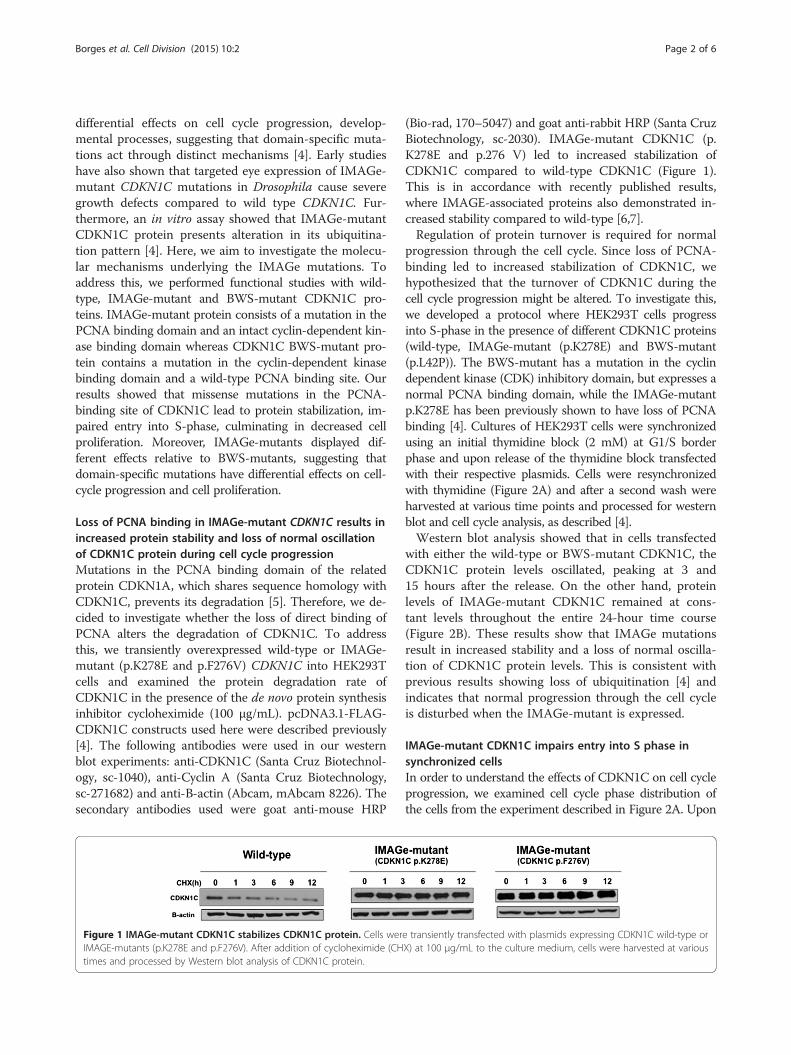
Loss of PCNA binding in IMAGe-mutant CDKN1C results inincreased protein stability and loss of normal oscillationof CDKN1C protein during cell cycle progressionMutations in the PCNA binding domain of the relatedprotein CDKN1A, which shares sequence homology withCDKN1C, prevents its degradation [5]. Therefore, we de-cided to investigate whether the loss of direct binding ofPCNA alters the degradation of CDKN1C. To addressthis, we transiently overexpressed wild-type or IMAGe-mutant (p.K278E and p.F276V) CDKN1C into HEK293Tcells and examined the protein degradation rate ofCDKN1C in the presence of the de novo protein synthesisinhibitor cycloheximide (100 μg/mL). pcDNA3.1-FLAG-CDKN1C constructs used here were described previously[4]. The following antibodies were used in our westernblot experiments: anti-CDKN1C (Santa Cruz Biotechnol-ogy, sc-1040), anti-Cyclin A (Santa Cruz Biotechnology,sc-271682) and anti-B-actin (Abcam, mAbcam 8226). Thesecondary antibodies used were goat anti-mouse HRP
Figure 1 IMAGe-mutant CDKN1C stabilizes CDKN1C protein. Cells werIMAGE-mutants (p.K278E and p.F276V). After addition of cycloheximide (CHtimes and processed by Western blot analysis of CDKN1C protein.
(Bio-rad, 170–5047) and goat anti-rabbit HRP (Santa CruzBiotechnology, sc-2030). IMAGe-mutant CDKN1C (p.K278E and p.276 V) led to increased stabilization ofCDKN1C compared to wild-type CDKN1C (Figure 1).This is in accordance with recently published results,where IMAGE-associated proteins also demonstrated in-creased stability compared to wild-type [6,7].Regulation of protein turnover is required for normal
progression through the cell cycle. Since loss of PCNA-binding led to increased stabilization of CDKN1C, wehypothesized that the turnover of CDKN1C during thecell cycle progression might be altered. To investigate this,we developed a protocol where HEK293T cells progressinto S-phase in the presence of different CDKN1C proteins(wild-type, IMAGe-mutant (p.K278E) and BWS-mutant(p.L42P)). The BWS-mutant has a mutation in the cyclindependent kinase (CDK) inhibitory domain, but expresses anormal PCNA binding domain, while the IMAGe-mutantp.K278E has been previously shown to have loss of PCNAbinding [4]. Cultures of HEK293T cells were synchronizedusing an initial thymidine block (2 mM) at G1/S borderphase and upon release of the thymidine block transfectedwith their respective plasmids. Cells were resynchronizedwith thymidine (Figure 2A) and after a second wash wereharvested at various time points and processed for westernblot and cell cycle analysis, as described [4].Western blot analysis showed that in cells transfected
with either the wild-type or BWS-mutant CDKN1C, theCDKN1C protein levels oscillated, peaking at 3 and15 hours after the release. On the other hand, proteinlevels of IMAGe-mutant CDKN1C remained at cons-tant levels throughout the entire 24-hour time course(Figure 2B). These results show that IMAGe mutationsresult in increased stability and a loss of normal oscilla-tion of CDKN1C protein levels. This is consistent withprevious results showing loss of ubiquitination [4] andindicates that normal progression through the cell cycleis disturbed when the IMAGe-mutant is expressed.
IMAGe-mutant CDKN1C impairs entry into S phase insynchronized cellsIn order to understand the effects of CDKN1C on cell cycleprogression, we examined cell cycle phase distribution ofthe cells from the experiment described in Figure 2A. Upon
e transiently transfected with plasmids expressing CDKN1C wild-type orX) at 100 μg/mL to the culture medium, cells were harvested at various
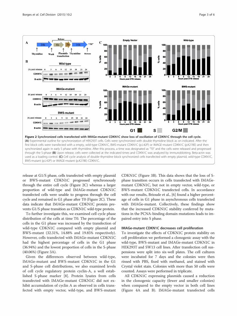
Figure 2 Synchronized cells transfected with IMAGe-mutant CDKN1C show loss of oscillation of CDKN1C through the cell cycle.(A) Experimental outline for synchronization of HEK293T cells. Cells were synchronized with double thymidine block as on indicated. After thefirst block cells were transfected with a empty, wild-type CDKN1C, BWS-mutant CDKN1C (p.L42P) or IMAGE-mutant CDKN1C (p.K278E) and thensynchronized again in early S phase with thymidine. After this process, a time was designated as “T0” and the cells were released and progressedthrough the S-phase (B) Upon release, cells were collected at the indicated times and CDKN1C was analyzed by immunoblotting. Beta-actin wasused as a loading control. (C) Cell cycle analysis of double thymidine block synchronized cells transfected with empty plasmid, wild-type CDKN1C,BWS-mutant (p.L42P) or IMAGE-mutant (p.K278E) CDKN1C.
Borges et al. Cell Division (2015) 10:2 Page 3 of 6
release at G1/S phase, cells transfected with empty plasmidor BWS-mutant CDKN1C progressed synchronouslythrough the entire cell cycle (Figure 2C) whereas a largerproportion of wild-type and IMAGe-mutant CDKN1Ctransfected cells were unable to progress through the cellcycle and remained in G1 phase after T0 (Figure 2C). Thesedata indicate that IMAGe-mutant CDKN1C protein pre-vents G1/S phase transition as CDKN1C wild-type protein.To further investigate this, we examined cell cycle phase
distribution of the cells at time T0. The percentage of thecells in the G1 phase was increased by the transfection ofwild-type CDKN1C compared with empty plasmid andBWS-mutant (32.31%, 14.88% and 19.85% respectively).However, cells transfected with IMAGe-mutant CDKN1Chad the highest percentage of cells in the G1 phase(36.94%) and the lowest proportion of cells in the S-phase(40.06%) (Figure 3A).Given the differences observed between wild-type,
IMAGe-mutant and BWS-mutant CDKN1C in the G1and S-phase cell distributions, we also examined levelsof cell cycle regulatory protein cyclin-A, a well estab-lished S-phase marker [8]. Protein lysates from cellstransfected with IMAGe-mutant CDKN1C did not ex-hibit accumulation of cyclin A as observed in cells trans-fected with empty vector, wild-type, and BWS-mutant
CDKN1C (Figure 3B). This data shows that the loss of S-phase transition occurs in cells transfected with IMAGe-mutant CDKN1C, but not in empty vector, wild-type, orBWS-mutant CDKN1C transfected cells. In accordancewith our results, Brioude et al., [6] found a higher percent-age of cells in G1 phase in asynchronous cells transfectedwith IMAGe-mutant. Collectively, these findings showthat the increased CDKN1C stability conferred by muta-tions in the PCNA-binding domain mutations leads to im-paired entry into S phase.
IMAGe-mutant CDKN1C decreases cell proliferationTo investigate the effects of CDKN1C protein stability oncell proliferation we performed a clonogenic assay with thewild-type, BWS-mutant and IMAGe-mutant CDKN1C inHEK293T and SW13 cell lines. After transfection cell sus-pensions were split into six-well plates. The cell cultureswere incubated for 7 days and the colonies were thenrinsed with PBS, fixed with methanol, and stained withCrystal violet stain. Colonies with more than 50 cells werecounted. Assays were performed in triplicate.All CDKN1C expressing plasmids caused a reduction
in the clonogenic capacity (fewer and smaller colonies)when compared to the empty vector in both cell lines(Figure 4A and B). IMAGe-mutant transfected cells

Figure 3 IMAGe-mutants have decreased progression through S-phase after synchronization in S-phase. (A) Cell cycle analyses by flowcytometry of cells at T0 transfected as described in Figure 2A. (B) Immunoblot analysis of transfected cells showed a failure of accumulation ofCyclin-A, an S phase marker in IMAGe-mutant transfected cells at time T0.
Figure 4 Clonogenic assay of the HEK293T and SW13 cells transfected with IMAGe-mutant show decreased clonogenic capacitycompared to BWS or wild-type CDKN1C. After transfection of the wild-type CDKN1C, BWS-mutants (p.L42P), IMAGE-mutant (p.K278E) and anempty vector, single cell suspensions were split into six-well in triplicate to assay for clonogenic capacity. (A) SW13 and (B) HEK293T cells results.# indicates a p < 0.05 when compared to the empty-vector condition. *p = <0.05. (C) Representative dishes of SW13 cells transfected as described. Cellswere fixed and stained with Crystal violet solution; violet staining represents a higher number of colonies.
Borges et al. Cell Division (2015) 10:2 Page 4 of 6

Borges et al. Cell Division (2015) 10:2 Page 5 of 6
displayed even fewer colonies than cells transfectedwith wild-type CDKN1C in the two cell lines studied(Figure 4A and B).CDKN1C is a negative regulator of cell proliferation and
the effects observed on the wild-type transfected cells maybe attributed to a combination of the CDK-inhibitory andPCNA domains of the CDKN1C. Moreover, the remainingeffects demonstrated by BWS-mutants probably are asso-ciated with the intact PCNA-binding domain of CDKN1C,as described previously [9]. In the IMAGe-mutant trans-fected cells, the progression through the cell cycle isimpaired as cyclical degradation of CDKN1C is lost.The G1-phase cell-cycle inhibition is increased due to in-creased stability of CDKN1C IMAGe mutants, whichmaintains an intact cyclin-dependent kinase binding do-main, resulting in decreased proliferation and the under-growth phenotype observed in patients with IMAGesyndrome. Previous reports have described the in vitroeffects of variations in CDKN1C levels on cell growth[10,11]. In vivo models have showed that mouse embry-onic growth is sensitive to the precise dosage of Cdkn1cand excess of this protein results in dose dependent em-bryonic growth retardation [12], a phenotype seen in pa-tients with IMAGe Syndrome.Further expansion of the phenotypes associated with
mutations in CDKN1C’s PCNA binding domain suggestsphenotypic heterogeneity. Recently, mutations inducingdifferent missense changes of the arginine at amino acid279 were found in individuals with IMAGe Syndromeand Silver Russell Syndrome (SRS, MIM #180860), char-acterized by prenatal and postnatal growth retardationand dysmorphic features. We hypothesize that these dif-ferences in amino acid changes (arginine to leucine inSRS versus Arginine to Proline in IMAGe Syndrome)are associated with a both differential loss of binding toPCNA and the effects of modifier genes [6]. In additionto SRS, missense mutations located in the highly con-served PCNA binding domain have also been associatedwith heterogeneous clinical phenotypes with growthrestriction and variable to no adrenal failure or skeletalabnormalities and onset of diabetes in early adulthood[13,14]. There the phenotypic spectrum associated withmissense mutations in the CDKN1C PCNA binding do-main suggests the importance of genetic modifiers in dis-ease manifestation.Our data suggests that mutations in the PCNA-binding
domain of CDKN1C leads to increased protein stability, ablock in the G1 phase and impaired S-phase entry, anddecreased cell proliferation. Our findings demonstratethat domain-specific mutations affect different aspectsof cell-cycle progression and cell proliferation, whichgive rise to the two opposing phenotypes: BWS andIMAGe syndrome. The data presented here give thefurther direct evidence of how the PCNA-binding site
mutations in CDKN1C affect the cell cycle and causeIMAGe syndrome.
AbbreviationsIMAGe: Intrauterine growth restriction, methaphyseal dysplasia, adrenalhypoplasia congenital and genital anomalies; CDKN1C: Cyclin-dependentkinase inhibitor 1C; CDK: Cyclin dependent kinase, BWS, BeckwithWiedemann syndrome.
Competing interestsThe authors declare that they have no competing interests.
Authors’ contributionsK.S.B. designed and performed experiments, analyzed data and wrote thepaper; V.A.A. constructed the plasmids and designed experiments; E.V.designed experiments and supervised the project. All authors discussed theresults and implications and commented on the manuscript at all stages. Allauthors read and approved the final manuscript.
AcknowledgementsThis work was supported by NIH grants HD044513 to EV and F31HD068136and UCLA Institutional funds to VAA and fellowship from Fundação deAmparo à Pesquisa do Estado de São Paulo (FAPESP process number 2012/09391-0) to KSB.
Author details1Department of Human Genetics, David Geffen School of Medicine at UCLA,University of California, Los Angeles, 695 Charles E. Young Drive, Los Angeles,CA 90095, USA. 2Department of Genetics, Ribeirão Preto Medical School,University of São, Ribeirão Preto, Av. Bandeirantes 3900, CEP 14049-900Ribeirão Preto, SP, Brazil. 3Department of Pathology and Laboratory Medicine,David Geffen School of Medicine at UCLA, University of California, LosAngeles, USA. 4Department of Pediatrics, David Geffen School of Medicine,University of California, Los Angeles, USA. 5Department of Urology, DavidGeffen School of Medicine, University of California, Los Angeles, USA.
Received: 29 November 2014 Accepted: 16 March 2015
References1. Matsuoka S, Edwards MC, Bai C, Parker S, Zhang P, Baldini A, et al. p57KIP2,
a structurally distinct member of the p21CIP1 Cdk inhibitor family, is acandidate tumor suppressor gene. Genes Dev. 1995;9:650–62.
2. Matsuoka S, Thompson JS, Edwards MC, Bartletta JM, Grundy P, Kalikin LM,et al. Imprinting of the gene encoding a human cyclin-dependent kinaseinhibitor, p57KIP2, on chromosome 11p15. Proc Natl Acad Sci U S A.1996;93:3026–30.
3. Romanelli V, Belinchón A, Benito-Sanz S, Martínez-Glez V, Gracia-BouthelierR, Heath KE, et al. CDKN1C (p57(Kip2)) analysis in Beckwith-Wiedemannsyndrome (BWS) patients: Genotype-phenotype correlations, novel mutations,and polymorphisms. Am J Med Genet A. 2010;152A:1390–7.
4. Arboleda VA, Lee H, Parnaik R, Fleming A, Banerjee A, Ferraz-de-Souza B,et al. Mutations in the PCNA-binding domain of CDKN1C cause IMAGesyndrome. Nat Genet. 2012;44:788–92.
5. Nishitani H, Shiomi Y, Iida H, Michishita M, Takami T, Tsurimoto T. CDKinhibitor p21 is degraded by a proliferating cell nuclear antigen-coupledCul4-DDB1Cdt2 pathway during S phase and after UV irradiation. J BiolChem. 2008;283:29045–52.
6. Brioude F, Oliver-Petit I, Blaise A, Praz F, Rossignol S, Jule ML, et al. CDKN1Cmutation affecting the PCNA-binding domain as a cause of familial RussellSilver syndrome. J Med Genet. 2013;50:823–30.
7. Hamajima N, Johmura Y, Suzuki S, Nakanishi M, Saitoh S. Increased proteinstability of CDKN1C causes a gain-of-function phenotype in patients withIMAGe syndrome. PLoS One. 2013;8:e75137.
8. Girard F, Strausfeld U, Fernandez A, Lamb NJ. Cyclin A is required for theonset of DNA replication in mammalian fibroblasts. Cell. 1991;67:1169–79.
9. Watanabe H, Pan ZQ, Schreiber-Agus N, DePinho RA, Hurwitz J, Xiong Y.Suppression of cell transformation by the cyclin-dependent kinase inhibitorp57KIP2 requires binding to proliferating cell nuclear antigen. Proc NatlAcad Sci U S A. 1998;95:1392–7.
Borges et al. Cell Division (2015) 10:2 Page 6 of 6
10. Chen B, Zhao R, Su CH, Linan M, Tseng C, Phan L, et al. CDK inhibitor p57(Kip2) is negatively regulated by COP9 signalosome subunit 6. Cell Cycle.2012;11:4633–41.
11. Zhao R, Yang HY, Shin J, Phan L, Fang L, Che TF, et al. CDK inhibitor p57(Kip2) is downregulated by Akt during HER2-mediated tumorigenicity. CellCycle. 2013;12:935–43.
12. Andrews SC, Wood MD, Tunster SJ, Barton SC, Surani MA, John RM. Cdkn1c(p57Kip2) is the major regulator of embryonic growth within its imprinteddomain on mouse distal chromosome 7. BMC Dev Biol. 2007;21(7):53.
13. Kato F, Hamajima T, Hasegawa T, Amano N, Horikawa R, Nishimura G, et al.IMAGe syndrome: clinical and genetic implications based on investigationsin three Japanese patients. Clin Endocrinol. 2014;80:706–13.
14. Kerns SL, Guevara-Aguirre J, Andrew S, Geng J, Guevara C, Guevara-AguirreM, et al. A novel variant in CDKN1C is associated with intrauterine growthrestriction, short stature, and early-adulthood-onset diabetes. J Clin EndocrinolMetab. 2014;99:2117–22.
Submit your next manuscript to BioMed Centraland take full advantage of:
• Convenient online submission
• Thorough peer review
• No space constraints or color figure charges
• Immediate publication on acceptance
• Inclusion in PubMed, CAS, Scopus and Google Scholar
• Research which is freely available for redistribution
Submit your manuscript at www.biomedcentral.com/submit