Multiplex targeted in vivo cancer detection using sensitive near-infrared SERS nanotags
9
Nano Today (2012) 7, 85—93 Available online at www.sciencedirect.com journal homepage: www.elsevier.com/locate/nanotoday RAPID COMMUNICATION Multiplex targeted in vivo cancer detection using sensitive near-infrared SERS nanotags Kaustabh Kumar Maiti a,1 , U.S. Dinish a,1 , Animesh Samanta b , Marc Vendrell a , Kiat-Seng Soh a , Sung-Jin Park a , Malini Olivo a,c , Young-Tae Chang a,b,∗ a Singapore Bioimaging Consortium, Agency for Science, Technology and Research (A*STAR), 138667 Singapore, Singapore b Department of Chemistry & MedChem Program of Life Sciences Institute, National University of Singapore, 117543 Singapore, Singapore c School of Physics, National University Ireland Galway, Ireland Received 12 December 2011; received in revised form 10 February 2012; accepted 20 February 2012 Available online 15 March 2012 KEYWORDS Raman spectroscopy; NIR Raman reporter; Multiplexing; In vivo cancer detection; SERS nanotags; Nanoparticle kinetics Summary One of the most promising advantages of surface-enhanced Raman scattering (SERS) technique for in vivo biosensing is the multiplexing potential, which is under explored due to the limited availability of near-infrared Raman reporters. Here, we report the synthesis of multiplexing capable and biocompatible SERS nanotags using highly sensitive novel NIR Raman reporters. Two new NIR Raman reporter molecules, Cy7LA, Cy7.5LA are developed to partner with recently synthesized CyNAMLA-381 reporter to construct SERS nanotags with multiplexing capability. These nanotags possess excellent SERS signal stability over a period of one month. As a proof of concept for multiplex targeted in vivo detection, we successfully demonstrated the simultaneous sensing of cancer in living mouse using these three bioconjugated nanotags. To the best of our knowledge, this is the first real demonstration of in vivo multiplex targeted detection using SERS nanotags. Further, in vivo kinetic study of these nanotags in tumor revealed their excellent sensitivity, stability and tumor specificity. These probes also show rapid clearance from the liver indicating their possible excretion. This validation renders our SERS nanotags as an ultrasensitive in vivo nanoprobe for the detection and imaging of multiple biomarkers for early diagnosis of diseases. © 2012 Elsevier Ltd. All rights reserved. ∗ Corresponding author at: Singapore Bioimaging Consortium, Agency for Science, Technology and Research (A*STAR), 138667 Singapore, Singapore. Tel.: +65 65166774; fax: +65 67791691. E-mail address: [email protected] (Y.-T.Chang). URL: http://ytchang.science.nus.edu.sg (Y.-T. Chang). 1 These authors contributed equally to this work and joint first authors. Introduction In recent years, surface-enhanced Raman scattering (SERS) has emerged as a versatile technique that can enhance the intensity of the vibrational spectra of a molecule by sev- eral order of magnitude when it is in the close proximity of nano-roughened metal surfaces such as gold/silver or when it is adsorbed on to these metallic nanoparticles. These 1748-0132/$ — see front matter © 2012 Elsevier Ltd. All rights reserved. doi:10.1016/j.nantod.2012.02.008
Transcript of Multiplex targeted in vivo cancer detection using sensitive near-infrared SERS nanotags

Nano Today (2012) 7, 85—93
Available online at www.sciencedirect.com
journa l homepage: www.e lsev ier .com/ locate /nanotoday
RAPID COMMUNICATION
Multiplex targeted in vivo cancer detection usingsensitive near-infrared SERS nanotags
Kaustabh Kumar Maiti a,1, U.S. Dinisha,1, Animesh Samantab, Marc Vendrell a,Kiat-Seng Soha, Sung-Jin Parka, Malini Olivoa,c, Young-Tae Changa,b,∗
a Singapore Bioimaging Consortium, Agency for Science, Technology and Research (A*STAR), 138667 Singapore, Singaporeb Department of Chemistry & MedChem Program of Life Sciences Institute, National University of Singapore, 117543 Singapore,Singaporec School of Physics, National University Ireland Galway, Ireland
Received 12 December 2011; received in revised form 10 February 2012; accepted 20 February 2012Available online 15 March 2012
KEYWORDSRaman spectroscopy;NIR Raman reporter;Multiplexing;In vivo cancerdetection;SERS nanotags;Nanoparticle kinetics
Summary One of the most promising advantages of surface-enhanced Raman scattering (SERS)technique for in vivo biosensing is the multiplexing potential, which is under explored due tothe limited availability of near-infrared Raman reporters. Here, we report the synthesis ofmultiplexing capable and biocompatible SERS nanotags using highly sensitive novel NIR Ramanreporters. Two new NIR Raman reporter molecules, Cy7LA, Cy7.5LA are developed to partnerwith recently synthesized CyNAMLA-381 reporter to construct SERS nanotags with multiplexingcapability. These nanotags possess excellent SERS signal stability over a period of one month. Asa proof of concept for multiplex targeted in vivo detection, we successfully demonstrated thesimultaneous sensing of cancer in living mouse using these three bioconjugated nanotags. To thebest of our knowledge, this is the first real demonstration of in vivo multiplex targeted detectionusing SERS nanotags. Further, in vivo kinetic study of these nanotags in tumor revealed their
excellent sensitivity, stability and tumor specificity. These probes also show rapid clearancefrom the liver indicating their possible excretion. This validation renders our SERS nanotags asan ultrasensitive in vivo nanoprobe for the detection and imaging of multiple biomarkers forearly diagnosis of diseases.© 2012 Elsevier Ltd. All rights re∗ Corresponding author at: Singapore Bioimaging Consortium,Agency for Science, Technology and Research (A*STAR), 138667Singapore, Singapore. Tel.: +65 65166774; fax: +65 67791691.
E-mail address: [email protected] (Y.-T. Chang).URL: http://ytchang.science.nus.edu.sg (Y.-T. Chang).
1 These authors contributed equally to this work and joint firstauthors.
I
Ihieni
1748-0132/$ — see front matter © 2012 Elsevier Ltd. All rights reserved.doi:10.1016/j.nantod.2012.02.008
served.
ntroduction
n recent years, surface-enhanced Raman scattering (SERS)as emerged as a versatile technique that can enhance the
ntensity of the vibrational spectra of a molecule by sev-ral order of magnitude when it is in the close proximity ofano-roughened metal surfaces such as gold/silver or whent is adsorbed on to these metallic nanoparticles. These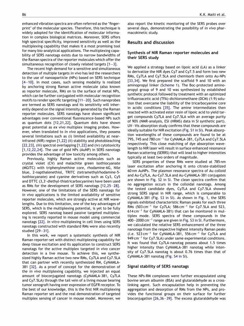
8
epwthmfbts
dt[bawmaeraagesi[[p
c(biaaHirlSeinns
Rdndst3taattRm
asm
R
St
WtR[apsttirgoUit7rlRt
l6aantsCsR6t4wn(9IhsC
S
Tb
6
nhanced vibration spectra are often referred as the ‘finger-rint’ of the molecular species. Therefore, this technique isidely adopted for the identification of molecular informa-
ion in complex biological matrices. Moreover, SERS offersigh spectral specificity, improved sensitivity as well as theultiplexing capability that makes it a most promising tool
or many bio-analytical applications. The multiplexing capa-ility of SERS nanotags exists due to narrow bandwidths ofhe Raman spectra of the reporter molecules which offer theimultaneous recognition of closely related targets [1—3].
The recent high demand for a sensitive and simultaneousetection of multiple targets in vivo has led the researcherso the use of nanoaparticle (NPs) based on SERS technique4—10]. In most cases, such sensing modality is realizedy anchoring strong Raman active molecule (also knowns reporter molecule, RM) on to the surface of metal NPs,hich can be further conjugated with molecular recognitionotifs to render specific targeting [11—20]. Such nanoprobes
re termed as SERS nanotags and its sensitivity will inher-ntly depend on the signal intensity generated by the Ramaneporter molecules. SERS nanotags have shown significantdvantages over conventional fluorescence-based NPs suchs quantum dots [12,21,22]. Quantum dots have shownreat potential as ex vivo multiplex imaging probes. How-ver, when translated to in vivo applications, they possesseveral limitations such as (i) limited availability at near-nfrared (NIR) region [1,22] (ii) stability and photobleaching22,23], (iii) spectral overlapping [1,22] and (iv) cytotoxicity1,12,22,24]. The use of gold NPs (AuNP) in SERS nanotagsrovides the advantage of low toxicity among others.
Previously, highly Raman active molecules such asrystal violet (CV) and malachite green isothiocyanateMGITC) with triphenylmethine core, rhodamine-6G, Nilelue, 2-napthalenethiol, TRITC (tetramethylrhodamine-5-sothiocyanate) and cyanine derivatives such as Cy3, Cy5nd DTTC (3,3′-diethyl-thiatricarbocyanine) have been useds RMs for the development of SERS nanotags [12,25—28].owever, one of the limitations of the SERS nanotags for
n vivo applications is the limited availability of sensitiveeporter molecules, which are strongly active at NIR wave-engths. Due to this limitation, one of the key advantages ofERS, which is the multiplexing potential, is yet to be fullyxplored. SERS nanotag based passive targeted multiplex-ng is recently reported in mouse model using commercialanotags [22]. In vitro multiplexing in cell lines using SERSanotags constructed with standard RMs were also recentlytudied [29—31].
In this work, we report a systematic synthesis of NIRaman reporter-set with distinct multiplexing capability foreep tissue excitation and its application to construct SERSanotags for the active multiplex targeted in vivo canceretection in a live mouse. To achieve this, we synthe-ized highly Raman active two new RMs, Cy7LA and Cy7.5LAhat can partner with recently synthesized RM, CyNAMLA-81 [32]. As a proof of concept for the demonstration ofhe in vivo multiplexing capability, we injected an equalmount of bioconjugated nanotags (CyNAMLA-381, Cy7LAnd Cy7.5LA) through the tail vein of a living mouse bearing
umor xenograft having over expression of EGFR receptor. Tohe best of our knowledge, this is the first NIR multiplexingaman reporter set and the real demonstration of targetedultiplex sensing of cancer in mouse model. Moreover, welavb
K.K. Maiti et al.
lso report the kinetic monitoring of the SERS probes overeveral days, demonstrating the possibility of in vivo phar-acokinetic study.
esults and discussion
ynthesis of NIR Raman reporter molecules andheir SERS study
e applied a strategy based on lipoic acid (LA) as a linkero derivatize the NIR dyes Cy7 and Cy7.5 and form two newMs, Cy7LA and Cy7.5LA and chemisorb them onto Au-NPs33,34]. We first prepared the scaffold 9 and 10 with anminopropyl linker (Scheme 1). The Boc-protected amino-ropyl group of 9 and 10 was synthesized by establishedynthetic protocol followed by treatment with an optimizedrifluoroacetic acid (TFA)-dichloromethane (DCM) (1:9) solu-ion that overcame the liability of the tricarbocyanine coren acidic conditions [35]. The amine intermediates theneacted with activated ester resin of lipoic acid to yield tar-et compounds Cy7LA and Cy7.5LA with an average purityf 90% (NMR-analysis, ESI (HRMS) data in SI synthetic part).V—Vis absorption study confirms that these compounds are
deally suitable for NIR excitation (Fig. S1 in SI). Peak absorp-ion wavelengths of these compounds are found to be at95, 745 and 780 cm−1 for CyNAMLA-381, Cy7LA and Cy7.5LAespectively. This close matching of dye absorption wave-ength to NIR laser will result in surface enhanced resonanceaman scattering (SERRS) that will lead to higher intensitiesypically at least two orders of magnitude.
SERS properties of these RMs were studied at 785 nmaser excitation after anchoring it onto citrate-stabilized0 nm AuNPs. The plasmon resonance spectra of Au colloidnd Au-Cy7LA, Au-Cy7.5LA and Au-CyNAMLA-381 conjugatesre shown in Fig. S2 in SI. The clean spectra confirm thato aggregation occurs in the colloidal nanotags. Amonghe tested candidate dyes, Cy7LA and Cy7.5LA showedtrong SERS signal in NIR range, which is comparable toyNAMLA-381 (Fig. S3 in SI). As shown in Fig. 1, the SERSignals exhibited characteristic Raman peaks for each threeMs (503 cm−1 for Cy7LA; 586 cm−1 for Cy7.5LA and 523,14 cm−1 for CyNAMLA-381) that can be monitored in mul-iplex mode. SERS spectra of these compounds in the00—2000 cm−1 range are given in Fig. S3 in SI. Furthermore,e calculated the relative SERS enhancement of the threeanotags from the respective highest intensity Raman peaksi.e. 523 cm−1 for CyNAMLA-381, 556 cm−1 for Cy7LA and49 cm−1 for Cy7.5LA) under same experimental conditions.t was found that Cy7LA-nanotag possess about 1.5 timesigher intensity than CyNAMLA-381 nanotag while inten-ity of Cy7.5LA nanotag is about 0.76 times than that ofyNAMLA-381 nanotag (Fig. S4 in SI).
ignal stability of SERS nanotags
hese NPs-RM complexes were further encapsulated usingovine serum albumin (BSA) and glutaraldehyde as a cross-
inking agent. Such encapsulation help in preventing theggregation and desorption of RMs from the NPs, and pro-ides the functional groups on their surface for furtherioconjugation [26,36—39]. The excess glutaraldehyde was
Multiplex targeted in vivo cancer detection using sensitive near-infrared SERS nanotags 87
Scheme 1 Synthesis of lipoic acid-containing amine acetylated tricarbocyanines (Cy7LA and Cy7.5LA). Reagents and conditions:(a) 1-iodopropane, CH3CN, 80 ◦C, 12 h; (b) 3-bromopropylamine hydrobromide, 120 ◦C, 12 h; (c) di-tert-butyl dicarbonate, DIEA,CHCl3, reflux, 4 h; (d) AcOH, acetic anhydride, pyridine, 110 ◦C; (e) TFA-DCM (1:9), r.t., 15 h; and (f) lipoic acid activated esterresin, CH2Cl2, r.t., 15 h.
Figure 1 Normalized SERS spectra of CyNAMLA-381, Cy7LA and Cy7.5LA after chemisorption on AuNPs. Spectra were measuredusing a Raman microscope (785 nm laser excitation, 60 �W laser power, acquisition time: 10 s) and plotted as average intensities(n = 3). The most distinctive peaks from each reporter are highlighted in yellow.

88 K.K. Maiti et al.
Figure 2 (a) Time course SERS measurement of three nanotags. SERS intensities of the highest Raman peaks (i.e. 523 cm−1
for CyNAMLA381, 503 cm−1 for Cy7LA and 949 cm−1 for Cy7.5LA), are plotted as mean ± standard deviation of 5 independentmeasurements taken from the same sample at different time points. (b) Transmission electron microscopy (TEM) images of: (1)B Cy7L
riCSfiutctsetm
S
Fw3vb[Sbmuds
Fw381 and Cy7.5LA respectively.
After the successful demonstration of multiplex detec-tion in liver, we focused on targeted sensing for xenografttumor model in living mouse formed from OSCC cells (oral
Figure 3 Multiplex SERS detection from liver site. Peaks at
SA-encapsulated CyNAMLA-381 nanotag, (2) BSA-encapsulated
emoved by requisite amount of glycine treatment. We stud-ed the long-term SERS-signal stability of CyNAMLA-381,y7LA and Cy7.5LA-AuNPs, over a period of one month. TheERS intensities of the highest Raman peaks obtained fromve independent measurements from the same sample weresed to study the stability of these nanotags at differentimes. Remarkably, these nanotags did not show any signifi-ant aggregation and exhibited stable SERS intensities overime, with a very low relative standard deviation of ∼5% ashown in Fig. 2(a). As evident from Fig. 2(b), transmissionlectron microscopy (TEM) analysis reveals that the size ofhese nanotags was around 65 nm. The observed 5 nm incre-ent in size is attributed to the BSA layer encapsulation.
ERS multiplex detection in liver and tumor site
or the demonstration of the in vivo multiplexing capability,e injected three BSA encapsulated nanotags (CyNAMLA-81, Cy7LA and Cy7.5LA) in equal amount through the tailein of a living mouse. These nanotags are generally taken upy the Kupffer cells of the reticuloendothelial system (RES)22] and as a result accumulation occurs in the liver site.ERS measurement was carried out 5 h after the injectiony focusing the laser beam externally through the skin. The
easured spectra depicted a mixture of the three individ-al spectrum from three nanotags and clearly revealed theistinct Raman shift corresponding to each RMs with leastpectral overlap in the 500—600 cm−1 window as shown in
5sCe
A nanotag, and (3) BSA-encapsulated Cy7.5LA nanotag.
ig. 3. Raman shift at 503 cm−1 is originating from Cy7LAhile 523 cm−1 and 586 cm−1 are contributed by CyNAMLA-
03 cm−1 corresponds to Cy7LA (red shade); 523 cm−1 corre-ponds to CyNAMLA-381 (green shade); 586 cm−1 corresponds toy7.5LA (blue shade). SERS spectra obtained with 785 nm laserxcitation at 30 mW power and integration time 20 s.
Multiplex targeted in vivo cancer detection using sensitive near-infrared SERS nanotags 89
Figure 4 In vivo multiplex detection in xenograft tumor: (A) SERS spectra from tumor site (peaks obtained at 503 and 586 cm−1
from two EGFR positive nanotags, Cy7LA and Cy7.5LA); (B) SERS spectra from liver site (peaks obtained at 503, 523 and 586 cm−1
from two EGFR nanotag Cy7LA, Cy7.5LA and anti-HER2 nanotag CyNAMLA-381); and (C) SERS spectra from dorsal region. SERS spectration
cmtfrom EGFR negative nanotag, it further confirms the absenceof any nonspecific binding in the tumor site.
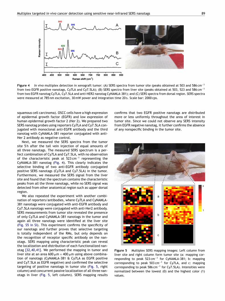
Figure 5 Multiplex SERS mapping images: Left column fromliver site and right column form tumor site (a: mapping cor-responding to peak 523 cm−1 for CyNAMLA-381; b: mapping
were measured at 785 nm excitation, 30 mW power and integra
squamous cell carcinoma). OSCC cells have a high expressionof epidermal growth factor (EGFR) and low expression ofhuman epidermal growth factor 2 (Her 2). We prepared twoSERS nanotag probes using reporters Cy7LA and Cy7.5LA con-jugated with monoclonal anti-EGFR antibody and the thirdnanotag with CyNAMLA-381 reporter conjugated with anti-Her 2 antibody as negative control.
Next, we measured the SERS spectra from the tumorsite 5 h after the tail vein injection of equal amounts ofall three nanotags. The measured SERS spectrum is a per-fect combination of Cy7LA and Cy7.5LA, with no observationof the characteristic peak at 523 cm−1 representing theCyNAMLA-381 nanotag (Fig. 4). This clearly indicates theselective binding of two anti-EGFR antibody conjugatedpositive SERS nanotags (Cy7LA and Cy7.5LA) in the tumor.Furthermore, we measured the SERS signal from the liversite and found that the spectrum contains the characteristicpeaks from all the three nanotags, while no SERS signal wasdetected from other anatomical region such as upper dorsalregion.
We also repeated the experiment with another combi-nation of reporters/antibodies, where Cy7LA and CyNAMLA-381 nanotags were conjugated with anti-EGFR antibody andCy7.5LA nanotags were conjugated with anti-Her2 antibody.SERS measurements from tumor site revealed the presenceof only Cy7LA and CyNAMLA-381 nanotags in the tumor andagain all three nanotags were identified at the liver site(Fig. S5 in SI). This experiment confirms the specificity ofour nanotags and further proves that selective targetingis totally independent of the RMs, but only depends onthe recognition of receptor specific antibody on the nan-otags. SERS mapping using characteristic peak can revealthe localization and distribution of each functionalized nan-otag [32,40,41]. We performed the mapping in tumor andliver site at an area 600 �m × 400 �m using above combina-tion of nanotags (CyNAMLA-381 & Cy7LA as EGFR positive
and Cy7.5LA as EGFR negative) and confirmed the selectivetargeting of positive nanotags in tumor site (Fig. 5, rightcolumn) and concurrent passive localization of all three nan-otags in liver (Fig. 5, left column). SERS mapping resultsccnv
time 20 s. Scale bar: 2000 cps.
onfirms that two EGFR positive nanotags are distributedore or less uniformly throughout the area of interest in
umor site. Since we could not observe any SERS intensity
orresponding to peak 503 cm−1 for Cy7LA, and c: mappingorresponding to peak 586 cm−1 for Cy7.5LA). Intensities wereormalized between the lowest (0) and the highest color (1)alues.

90
Figure 6 Kinetics study of nanotags in tumor and liver sitewith normalized intensity of 503 cm−1 for Cy7LA, 523 cm−1 forCyNAMLA-381 and 586 cm−1 for Cy7.5LA nanotags. Spectra weremb
s[CmcicL
K
WlppiCsC2iosgicttooeoip
C
Iatfoo
aoiefoic
odtfisvdiptsa
M
S
AoApNNCAwtmBC1
5itS(oa5
S
ITmlsb
easured and plotted as average intensities (n = 3) with errorar denoting relative standard deviation.
Limit of detection (LOD) study will provide an under-tanding about the sensitivity of the constructed nanotag42]. To understand a ballpark figure of LOD, we chosey7LA-nanotag as a representative candidate and weeasured the in vitro SERS intensity of anti-EGFR antibody
onjugated nanotags in a suspension of OSCC cells. Thentensity of the Raman peak of Cy7LA increased in aoncentration-dependent manner, and we estimated anOD around 54 pM (Fig. S6 in SI).
inetic study of SERS nanotags
e studied the kinetics of these SERS nanotags in tumor andiver, taking advantage of the long term stability and multi-lexing capabilities. We monitored the SERS intensity for aeriod of 8 days at both tumor and liver sites after inject-ng CyNAMLA-381 and Cy7LA nanotags as EGFR positive andy7.5LA as EGFR negative. We could observe that SERS inten-ity from 503 cm−1 and 523 cm−1 peaks (respectively fromy7LA, CyNAMLA-381) in tumor site increased initially up to4 h and then remained more or less same for 8 days, whichndicates that these nanotags are highly specific and stablever a period of time (Fig. 6). On the other hand, SERS inten-ity from all three nanotags localized in the liver site showsradual decrease in 48 h and then steeply falls to zero leveln 8 days. Our observation suggests that these nanoparti-les, taken up by Kupffer cells, might have been excretedhrough the fecal pathway, which agrees with the report onhe excretion of BSA encapsulated Au-NPs [43,44]. Also, RESf the body can also function to phagocytose these typesf nanoparticles, which is later moved into liver and gen-rally excreted as feces with bile [44]. The easy clearancef our nanotags from liver site and their excellent sensitiv-ty, stability and tumor specificity projects its potential as aromising in vivo theragnostic probe.
onclusions
n summary, we report two new lipoic acid-containing NIRctive tricarbocyanine Raman reporters Cy7LA and Cy7.5LA
o partner with our previously discovered CyNAMLA-381or in vivo multiplex targeted imaging. We believe thatur NIR RMs will help in addressing the limited availabilityf NIR Raman reporters for sensitive sensing and imagingocpm
K.K. Maiti et al.
pplications. Our nanotags showed excellent stabilityver a period of one month on shelf with negligible SERSntensity fluctuation, which is a significant achievementspecially long term monitoring of a SERS signal is requiredor both in vitro and in vivo applications. To the best ofur knowledge, this is the first demonstration of a selectiven vivo multiplex targeted detection with full multiplexingapability.
Furthermore, we studied the kinetics and localizationf these nanotags in tumor and liver over a period of 8ays. The stable SERS intensity from targeted nanotags inhe tumor suggests its preferred localization and speci-city. The signals of the nanotags in the liver decreasedteeply after 2 days, which indicates the possible excretionia fecal pathway. We believe that the study will help inesigning sensitive and biocompatible nanoprobes for in vivomaging applications and simultaneous detection of multi-le diseases. Further studies on these materials with otherargeting moieties will be carried out in due course to under-tand the long-term toxicity of the nanoprobes for practicalpplications.
aterials and methods
ynthetic materials and instrumental details
ll analytical grade chemicals (building block amines plusthers) and solvents were purchased from Sigma—Aldrich,lfa Aesar, Fluka, Merck or Acros, and used without furtherurification. Anti-EGFR (sc-120) and Anti-HER2 (sc-71667,eu 0.N.211) was supplied by Santa Cruz Biotechnology, Inc.ormal-phase chromatography was carried out using Sili-ycle 60 (particle size: 0.040—0.063 mm, 230—400 mesh).nalytical characterization was performed on a HPLC-MSith a DAD detector and a single quadrupole mass spec-
rometer (Agilent 6130 series) with an ESI probe. Analyticalethod, unless indicated: eluents: A: H2O (0.1% HCOOH),: ACN (0.1% HCOOH), gradient from 5% to 100% B in 6 min;18 (2) Luna column (4.6 mm × 50 mm, 5 �m particle size).H-NMR spectra were recorded on Bruker Advance 300 or00 NMR spectrometers, and chemical shifts are expressedn parts per million (ppm). High resolution mass spectrome-ry (HRMS) data was recorded on a Micromass VG 7035 (Masspectrometry Laboratory at National University of SingaporeNUS)). Surface plasmon absorption spectra were measuredn a SpectraMax M2 spectrophotometer (Molecular Devices),nd the data analysis was performed using GraphPad Prism.0 and Origin 6.
ERS measurements
nstrumentationhe spectral measurements were carried out in a Ramanicroscope (Renishaw InVia, UK) using an excitation wave-
ength at 785 nm (model: HPNIR785). In brief, the laserystem is coupled to a Leica microscope so that the laseream is directed to the sample through a 50× or 20×
bjective lens and a Peltier cooled CCD detector is used toollect the Raman signals. The WiRE 3.0 software packagerovided with the Raman system was employed for instru-ent control and data acquisition. Stokes shifted Raman
ear-
Bm
AwitgctttegttcsP
A
TN(Aw1cwa4rn
T
Ttsi0t
A
W(tC
A
Multiplex targeted in vivo cancer detection using sensitive n
spectra were collected in the range of 400—2000 cm−1 witha resolution of about 1 cm−1 and the exposure time was setat 10 s throughout the measurements. The shutter of thelaser was immediately closed after each measurement tominimize any possible photodamage of the sample under aprolonged illumination. Baseline correction of the measuredspectra was performed to remove the broad backgroundand fluorescence band. Prior to each measurement, theinstrument was calibrated with a silicon standard whoseRaman peak is centered at 520 cm−1.
Spectral measurementsFor the SERS measurements of CyNAML-381, Cy7LA andCy7.5LA-Au colloid mixtures, 20 �M solutions of eachreporter compounds in deionized water were mixed withAu colloid (BBInternational, UK, 2.6 × 10¹⁰ particles/mL) in a1:9 ratio (v/v). 20 �L of the reporter-Au colloid mixture solu-tions were placed on a clean glass slide with a cover slip andmeasured under the Raman microscope. The results are plot-ted as average intensities of 5 independent measurementsfrom one single sample preparation.
In vivo SERS multiplexingBalb/c nude mice from the Biological Resource Centre(Biomedical Sciences Institute, A*STAR) were anes-thetized by intraperitoneal injection of ketamine(150 mg/kg)/xylazine (10 mg/kg) at the age of 4—6weeks, and OSCC cells (5 × 10⁶ cells per site in a volumeof 150 �L) were injected subcutaneously into the rearflank. When the tumors grew to a size around ∼0.2 cm indiameter, anti-EGFR antibody-conjugated SERS nanotagsfor targeted (positive control) and anti-HER2 antibody-conjugated nanotag for non-targeted (negative control)(430 pM, 100 �L) were injected into the tail vein of themice. After 5 h, mice were anesthetized by intraperitonealinjection of ketamine and xylazine mixture solution andin vivo SERS measurements were performed from tumorsite and non-tumorogenic area, i.e. liver and dorsal regionusing Raman microscope with specified area using 30 mW,785 nm laser excitation. The integration time was set as20 s and the laser was coupled to the sample through a 20×objective lens with a beam spot of aprox. 3 �m. The animalexperiment procedures were performed in accordance witha protocol approved by the Institutional Animal Care andUse Committee (IACUC).
SERS mappingSERS mapping experiments were performed in a RenishawInVia Raman microscope system with a laser beam directedto the sample through a 20× objective lens. Antibody conju-gated nanotag (CyNAMLA-381-nanotag, Cy7LA-nanotag andCy7.5LA-nanotag) with equal concentration (450 pM) wereinjected through tail-vein. SERS-mapping in tumor and non-tumor area was conducted by selecting small area. Laserexcitation at 785 nm wavelength (focal spot of 3 �m) and30 mW power was used. Mapping measurements at 523 cm−1
for CyNAMLA-381, 503 cm−1 for Cy7LA and 586 cm−1 forCy7.5LA were carried out as raster scans in 40 �m stepsover the specified area (aprox. 600 �m × 400 �m) with 1 sintegration time.
Scd
infrared SERS nanotags 91
SA encapsulation of nanoparticles with reporterolecules
ll the reporter molecules (20 �M solutions in deionizedater) were mixed with Au colloid (2.6 × 10¹⁰ particles/mL)
n a 1:9 ratio (v/v). After 10 min incubation, colloids werereated with BSA (0.5% in deionized water) mixed with 25%lutaraldehyde (15:1) and reacted at r.t. for 4 h followed byentrifugation (8000 rpm, 5 min). Glutaraldehyde was usedo form a cross-linked organic encapsulation layer aroundhe gold and stabilize the particles, preventing the dis-ortion of the reporter molecules. In order to remove thexcess of glutaraldehyde, pellets were resuspended in 10 mMlycine with 10 mM sodium citrate (pH = 7.8) at r.t. Finally,he encapsulated Au colloids were washed 3 times by cen-rifugation (8000 rpm, 5 min), re-suspended in 1 mM sodiumitrate, and stored at 4 ◦C. For bioconjugation and long timetorage, we re-suspended the encapsulated gold colloid inBS.
ntibody conjugation of SERS nanotags
he carboxylic acids groups of BSA were activated with-(3-(dimethylamino)-propyl)-N′-ethylcarbodiimide (EDC)125 nmol) and N-hydroxysuccinimide (NHS) (125 nmol).fter incubation around 30 min, excess of EDC and NHSas removed by 3 rounds of centrifugation (8000 rpm,0 min) and re-suspended in PBS using Amicon Ultra 3 Kentrifuge microcons (Milipore). The activated particlesere then reacted with mouse monoclonal anti-EGFR ornti-HER2 at 25 ◦C for 2 h and then overnight incubation at◦C. Non-specifically bound chemicals and antibodies were
emoved by centrifugation (8000 rpm, 10 min) and the finalanotags were re-suspended in PBS and stored at 4 ◦C.
EM characterization
EM images were taken in a Jeol JEM-1010 Transmission Elec-ron Microscope. Acceleration voltage was 40—100 KV andtability was 2 ppm min−1. The magnification range of thenstrument covers from 50 to 600,000× with a resolution of.3000 nm. Images of these BSA encapsulated nanotags wereaken at a 250,000× magnification.
cknowledgments
e gratefully acknowledge National University of SingaporeNUS) (Young Investigator Award: R-143-000-353-123) andhe A*STAR Cross Council Office (CCO), Singapore (GrantCOGA02 005 2008) for the financial support.
ppendix A. Supplementary data
upplementary data associated with this arti-le can be found, in the online version, atoi:10.1016/j.nantod.2012.02.008.

9
R
[
[
[
[[[
[
[
[
[
[[[
[
[
[
[
[
[
[[
[
[
[
[
[
[
[
[
[
[
[
[
[[
ILer2iiBsCPchii2f
2
eferences
[1] S. Keren, C. Zavaleta, Z. Cheng, A. de la Zerda, O. Gheysens,S.S. Gambhir, Proc. Natl. Acad. Sci. U.S.A. 105 (2008) 5844.
[2] M. Gellner, K. Kompe, S. Schlucker, Anal. Bioanal. Chem. 394(2009) 1839.
[3] K.C. Bantz, A.F. Meyer, N.J. Wittenberg, H. Im, O. Kurtulus,S.H. Lee, N.C. Lindquist, S.H. Oh, C.L. Haynes, Phys. Chem.Chem. Phys. 13 (2011) 11551.
[4] S. Schlucker, Chemphyschem 10 (2009) 1344.[5] N.M. Sirimuthu, C.D. Syme, J.M. Cooper, Anal. Chem. 82 (2010)
7369.[6] J. Yang, Z. Wang, X. Tan, J. Li, C. Song, R. Zhang, Y. Cui,
Nanotechnology 21 (2010) 345101.[7] G. Goddard, L.O. Brown, R. Habbersett, C.I. Brady, J.C. Martin,
S.W. Graves, J.P. Freyer, S.K. Doorn, J. Am. Chem. Soc. 132(2010) 6081.
[8] D. Graham, K. Faulds, Chem. Soc. Rev. 37 (2008) 1042.[9] Q. Hu, L.L. Tay, J. Am. Chem. Soc. 129 (2007) 14.10] K.K. Strelau, R. Kretschmer, R. Moller, W. Fritzsche, J. Popp,
Anal. Bioanal. Chem. 396 (2010) 1381.11] A. Ingram, L. Byers, K. Faulds, B.D. Moore, D. Graham, J. Am.
Chem. Soc. 130 (2008) 11846.12] X. Qian, X.H. Peng, D.O. Ansari, Q. Yin-Goen, G.Z. Chen, D.M.
Shin, L. Yang, A.N. Young, M.D. Wang, S. Nie, Nat. Biotechnol.26 (2008) 83.
13] S.R. Emroy, S.M. Nie, Anal. Chem. 69 (1997) 2631.14] Y.L. Wang, J.L. Seebald, D.P. Szeto, ACS Nano 4 (2010) 4039.15] R.S. Golightly, W.E. Doering, M.J. Natan, ACS Nano 3 (2009)
2859.16] M.K. Gregas, F. Yan, J. Scaffidi, H.N. Wang, T. Vo-Dinh,
Nanomedicine 7 (2011) 115.17] J. Kneipp, H. Kneipp, B. Wittig, K. Kneipp, Nanomedicine 6
(2010) 214.18] Xu. Wang, X. Qian, J.J. Beitler, G.A. Chen, R.F. Khuri, M.M.
Lewis, C.H. Ju Shin, S. Nie, M.D. Shin, Cancer Res. 71 (2011)1526.
19] Y.H. Kim, J. Jeon, H.S. Hong, K.W. Rhim, S.Y. Lee, H. Youn,K.J. Chung, C.M. Lee, S.D. Lee, W.K. Kang, M.J. Nam, Small 7(2011) 2052.
20] Y.M. Sha, H. Xu, J.M. Natan, J. Am. Chem. Soc. 129 (2007) 14.21] M. Han, X. Gao, J.Z. Su, S. Nie, Nat. Biotechnol. 19 (2001) 631.22] C.L. Zavaleta, B.R. Smith, I. Walton, W. Doering, G. Davis, B.
Shojaei, M.J. Natan, S.S. Gambhir, Proc. Natl. Acad. Sci. U.S.A.106 (2009) 13511.
23] M.H.J.G.W. Sark van, M.T.L.P. Frederix, J.D. Heuvel Vanden,C.H. Gerriten, J. Phys. Chem. B 105 (2001) 8281.
24] I.L. Medintz, H. Mattoussi, A.R. Clapp, Int. J. Nanomedicine 3(2008) 151.
25] G. von. Maltzahn, A. Centrone, J.H. Park, R. Ramanathan,J.M. Sailor, A.T. Hatton, N.S. Bhatia, Adv. Mater. 21 (2009)3175.
26] S. Lee, H. Chon, M. Lee, J. Choo, S.Y. Shin, Y.H. Lee, I.J. Rhyu,S.W. Son, C.H. Oh, Biosens. Bioelectron. 24 (2009) 2260.
27] P.J. Huang, L.K. Chau, T.S. Yang, L.L. Tay, T.T. Lin, Adv. Funct.Mater. 19 (2009) 242.
28] S. Mahajan, J. Richardson, T. Brown, N.P. Bartlett, J. Am.Chem. Soc. 130 (2008) 15589.
29] A. Matschulat, D. Drescher, J. Kneipp, ACS Nano 4 (2010) 3259.30] D.C. Kennedy, K.A. Hoop, L.L. Tay, J.P. Pezacki, Naonoscale 2
(2010) 1413.31] S. Lee, H. Chon, S.Y. Yoon, E.K. Lee, S.I. Chang, D.W. Lim, J.
Cho, Nanoscale 4 (2012) 124.
32] A. Samanta, K.K. Maiti, K.S. Soh, X. Liao, M. Vendrell, U.S. Din-ish, S.W. Yun, R. Bhuvaneswari, H. Kim, S. Rautela, J. Chung,M. Olivo, Y.T. Chang, Angew. Chem. Int. Ed. Engl. 50 (2011)6089.
K.K. Maiti et al.
33] K.K. Maiti, U.S. Dinish, C.Y. Fu, J.J. Lee, K.S. Soh, S.W. Yun, R.Bhuvaneswari, M. Olivo, Y.T. Chang, Biosens. Bioelectron. 26(2010) 398.
34] K.K. Maiti, A. Samanta, M. Vendrell, K.S. Soh, M. Olivo, Y.T.Chang, Chem. Commun. 47 (2011) 3514.
35] J. Alonso, C. Encinas, S. Miltsov, E. Otazo, L. Rivera, M. Puyol,Dyes Pigments 71 (2006) 28.
36] H. Jang, Y.K. Kim, S.R. Ryoo, M.H. Kim, D.H. Min, Chem. Com-mun. 46 (2010) 583.
37] C.C. Lin, Y.M. Yang, Y.F. Chen, T.S. Yang, H.C. Chang, Biosens.Bioelectron. 24 (2008) 178.
38] D. Graham, D.G. Thompson, W.E. Smith, K. Faulds, Nat. Nan-otechnol. 3 (2008) 548.
39] L. Sun, B.K. Sung, C. Dentinger, B. Lutz, L. Nguyen, J. Zhang,H. Qin, M. Yamakawa, M. Cao, Y. Lu, A. Chmura, J. Zhu, X.Su, A.A. Berlin, S. Chan, B. Knudsen, Nano Lett. 7 (2007)351.
40] A. Pallaoro, G.B. Braun, N.O. Reich, M. Moskovits, Small 6(2010) 618.
41] C.D. Syme, N.M. Sirimuthu, S.L. Faley, J.M. Cooper, Chem.Commun. 46 (2010) 7921.
42] S.Y. Huh, J.A. Chung, B. Cordovez, D. Erickson, Lab Chip 9(2009) 433.
43] G. Renand, R.L. Hamilton, J.R. Havel, Hepatology 9 (1989) 380.44] K.T. Yong, I. Roy, H. Ding, E.J. Bergey, P.N. Prasad, Small 5
(2009) 1997.
Kaustabh Kumar Maiti studied chemistry atUniversity of Calcutta (India) and received hisB.Sc. in 1991, M.Sc. in organic chemistry in1993 and awarded Ph.D. from the same uni-versity in the year 2000. He worked underthe supervision of Prof. Avijit Banerji on 1,3-dipolar cycloaddition reactions of nitronesand nitrile oxides to construct differentregio- and stereoselective hetereocyclic ringsystems. He had over three years R&D expe-rience in leading Pharmaceutical Industries in
ndia, 2000—2003 (Alembic Ltd. and Sun Pharmaceuticals Industriestd.) where he worked in process development and drug discov-ry field. After this industrial training, he started his postdoctoralesearch at Pohang University of Science and Technology (POSTECH,003—2007), South Korea, with Prof. Sung-Kee Chung. He workedn the field of novel non-peptide based drug delivery system usingnositol and carbohydrate as scaffold. Before joining at Singaporeioimaging Consortium (A*STAR) as a research scientist, he did hisecond postdoctoral Research in Complex Carbohydrate Researchenter (CCRC), The University of Georgia, USA. He worked withrof. Geert-Jan Boons (2007 May—2009 April) in the field of gly-opeptide, structural aspects of peptidoglycan and LPS. Currently,is research focused on development of ultra sensitiive biocompat-ble surface enhanced Raman scattering (SERS) nanotags for in vivomaging as well as targeted cancer therapy. He published more than5 scientific papers in International journals and filed 11 patents soar.
Dinish U.S. is a bio-physicist and workingas research scientist at Bio-Optical Imag-ing Group, Singapore Bioimaging Consortium,under Agency for Science Technology andResearch (A*STAR), Singapore. He obtainedhis Ph.D. in bio-optics and imaging fromNanyang Technological University (NTU),
Singapore. He did his first post doctoralresearch at NTU in the area of nano-biophotonics. His research interests includesurface enhanced Raman scattering, optical
ear-
iiii
andimbr
Multiplex targeted in vivo cancer detection using sensitive n
coherence tomography, fluorescence imaging and nanoparticlebased multimodality optical imaging for biomedical applications.He holds 5 patents and has published over 20 international journalpapers and 25 conference proceedings.
Animesh Samanta received his B.Sc. inChemistry (Honors) in 2005 from VidyasagarUniversity in India. He obtained his M.Sc.in Bio-inrganic Chemistry from Indian Insti-tute of Technology (IIT) Madras in 2007. Hestarted his research carrier in Bioorganic andmedicinal chemistry under the guidance ofProf. Young Tae Chang from January 2008.His research interest focused on the synthe-sis of novel Near-IR dyes for the developmentof optical imaging tools either by SERS tech-
niques or by fluorescence imaging.
Marc Vendrell studied at the University ofBarcelona, where he received his B.Sc. inChemistry in 2001 and his Master Degreein Organic Chemistry in 2002. In 2007 heobtained his Ph.D. under the supervision ofProf. Albericio and Dr. Royo for his thesisin the combinatorial synthesis of peptide-heterocycle hybrids as multivalent GPCRligands. After working at the Combinato-rial Chemistry Unit in the Barcelona SciencePark as a Research Associate, he joined the
Singapore Bioimaging Consortium (SBIC) under the supervision ofProf. Young-Tae Chang at the end of 2007. His research interests arethe chemical synthesis and development of new fluorescent probesand non-invasive optical imaging tools for the study of cancer andstem cells.
Soh Kiat Seng Jason is working as researchofficer at Bio-Optical Imaging Group,Singapore Bioimaging Consortium, underAgency for Science Technology and Research(A-STAR). He obtained his B.Eng. (Hons)degree in Materials Science & Engineeringfrom Nanyang Technological University,
Singapore in 2009. He is currently involved inthe development of highly-sensitive biocom-patible SERS nanotags for in-vivo detectionand imaging of cancer in mouse model.atot
infrared SERS nanotags 93
Sung-Jin Park studied Biotechnology at theAjou University (Korea), where he receivedhis B.Sc. in 1998. In 2003, he obtained hisPh.D. in Anatomy under the supervision ofProf. Chun Myung-Hoon at the Catholic Uni-versity of Korea. After his Ph.D., he receivedthe educational training for Anatomy. In2007, he moved to ASAN medical center andworked for the development and evaluationof drug delivery system for radiosensitization.In 2009, he joined the Singapore Bioimag-
ng Consortium (SBIC) under Prof. Young-Tae Chang. His researchnterests are the development of new fluorescent probes and non-nvasive optical imaging tools for the study of stem cells andnflammations.
Malini Olivo is currently head of Bio-OpticalImaging Group, Singapore Bioimaging Con-sortium, A*STAR, Singapore. She is ScienceFoundation Ireland (SFI) Stokes Professorof Biophotonicsat the NationalUniversity ofIreland and also holds an adjunct pro-fessorship at the National University ofSingapore and Royal College of Surgeons,Ireland. She was the Principal Investigator atthe Singapore National Cancer Centre. Sheobtained Ph.D. in Bio-Medical Physics in 1990
nd since then very active in biophotonics research. She is a pio-eer in the area of clinical applications of photomedicine in opticaliagnostics and therapeutics in Singapore. Her current researchnterest is in nano-biophotonics and its applications in translationaledicine. She has published over 300 scientific papers, 1 book, 7ook chapters and 8 patents. She also secured about S$15 millionesearch grant as PI/Co-PI through various funding agencies.
Young-Tae Chang studied Chemistry atPOSTECH (Korea), and received his B.Sc. in1991 and Ph.D. in 1997. He did his postdoc-toral work with Prof. Peter Schultz at UCBerkeley and The Scripps Research Institute.In 2000, he was appointed Assistant Profes-sor at New York University and was promotedto Associate Professor in 2005. In September2007, he moved to the National University ofSingapore (NUS) and the Singapore BioimagingConsortium (SBIC) at Biopolis. He is currently
full Professor at the Department of Chemistry and the leader ofhe Medicinal Chemistry Program at NUS and Head of the Laboratoryf Bioimaging Probe Development at SBIC. He has published morehan 200 scientific papers, 3 books and filed 30 patents so far.