The hyper-IgE syndrome is not caused by a microdeletion syndrome
Upload
independentCategory
view
4download
0
1 23
international journal of stomatology& occlusion medicine ISSN 1867-2221Volume 6Number 2 J. Stomat. Occ. Med. (2013) 6:77-81DOI 10.1007/s12548-013-0077-8
Multiple odontogenic keratocysts indicatingGorlin-Goltz syndrome
G. Shirisha Rani, Sreenivasulu Pattipati,Md Rezwana Begum & M. NaveenKumar

1 23
Your article is protected by copyright and
all rights are held exclusively by Springer-
Verlag Wien. This e-offprint is for personal
use only and shall not be self-archived
in electronic repositories. If you wish to
self-archive your article, please use the
accepted manuscript version for posting on
your own website. You may further deposit
the accepted manuscript version in any
repository, provided it is only made publicly
available 12 months after official publication
or later and provided acknowledgement is
given to the original source of publication
and a link is inserted to the published article
on Springer's website. The link must be
accompanied by the following text: "The final
publication is available at link.springer.com”.

case study
Multiple odontogenic keratocysts indicating Gorlin-Goltz syndrome 77
Abstract Gorlin-Goltz syndrome, also known as ne-void basal cell carcinoma syndrome is inherited in an autosomal dominant way, although sporadic cases have been reported. This syndrome shows a high penetrance and variable expressivity and is a multisystemic process characterized by the presence of multiple pigmented basocellular carcinomas, keratocysts in the jaws, palmar and plantar pits and calcification of the falx cerebri. To-gether with these major features a great number of pro-cesses considered as minor features have also been de-scribed, such as numerous skeletal, dermatology related and neurological anomalies. Due to the importance of oral maxillofacial manifestations of this syndrome it is fundamental to know its characteristics in order to make a diagnosis and initiate an early preventive treatment. This paper reports the case of a 20-year-old male patient with Gorlin-Goltz syndrome, emphasizing the clinical and radiographic manifestations. This case report high-lights the importance of dental health professionals in the early diagnosis of Gorlin-Goltz syndrome and in a preventive multidisciplinary approach to provide a bet-ter prognosis for the patient.
Keywords: Keratocystic odontogenic tumors, Gorlin-Goltz syndrome, Seizures, Calcifications, Autosomal dominant inheritance
Introduction
The Gorlin-Goltz syndrome, also known as nevoid basal cell carcinoma syndrome (NBCCS), is an infrequent mul-tisystemic disease inherited as an autosomal dominant disorder, which has a high level of penetrance and vari-able degrees of expression. It is characterized by mul-tiple keratocystic odontogenic tumors (KCOT) in the jaws, multiple basal cell nevi carcinomas and skeletal abnormities. Other findings of this syndrome are hyper-telorism, palmar and plantar pits, calcification of the falx cerebri, central nervous system and ocular lesions, cleft lip and palate, mandibular prognathism (class III jaw relationship) and in rare cases ovarian fibroma [1, 2]. This syndrome can be diagnosed early by a dentist, as KCOTs are usually one of the first manifestations of the syndrome [1, 2].
This paper reports the case of a 20-year-old male patient with Gorlin-Goltz syndrome, emphasizing its clinical and radiographic manifestations.
Case report
A 20-year-male patient presented to the Department of Oral Medicine and Radiology with a chief complaint of swelling in lower left posterior region of the jaw for 2 months. The swelling was asymptomatic, sudden in onset, was initially the size of a peanut which gradu-ally increased and attained the present size. Dental history revealed surgical extraction of two upper front teeth 4 years previously in a private dental college due to a similar swelling. Medical history revealed an attack of seizures 1 month ago for which he was still receiving medication. Family history revealed that the patient’s
J. Stomat. Occ. Med. (2013) 6:77–81DOI 10.1007/s12548-013-0077-8
Multiple odontogenic keratocysts indicating Gorlin-Goltz syndromeA case report
G. Shirisha Rani, Sreenivasulu Pattipati, Md. Rezwana Begum, M. Naveen Kumar
S. Pattipati, MDS () Department of Oral Medicine & Radiology, St. Joseph Dental College & Hospital, Duggirala, Eluru 534004, Andhra Pradesh, Indiae-mail: [email protected]
G. Shirisha Rani, MDSDepartment of Oral Medicine & Radiology, Meghana Institute of Dental Sciences, Mallaram, Nizamabad 503003, Andhra Pradesh, India
Md. R. Begum, MDSDepartment of Oral Medicine & Radiology, Githam Dental College, Gandhinagar Campus, Rushikonda, Visakhapatnam 530045, Andhra Pradesh, India
M. Naveen Kumar, MDSDepartment of Oral & Maxillofacial Pathology, Kamineni Institute of Dental Sciences, Sreepuram, Narketpally, Nalgonda 508254, Andhra Pradesh, India
Received: 25 December 2012 / Accepted: 26 February 2013 / Published online: 23 April 2013© Springer-Verlag Wien 2013
Author's personal copy

78 Multiple odontogenic keratocysts indicating Gorlin-Goltz syndrome
case study
father had problems with jaw tumors. General physical examination showed elevation in the upper chest region which had been present since birth and extraoral exami-nation revealed mild hypertelorism (Fig. 1).
Intraoral examination revealed missing teeth #13, 18, 23, 28, 38, and 48 (Fig. 2). A mild diffuse swelling was seen in both left and right retromolar areas with the swell-ing in right retromolar area measuring approximately 1 × 1 cm2 (Fig. 3), the swelling in the left side approxi-mately 1 × 3 cm2 (Fig. 4) was oval in shape extending from the mesial surface of tooth #37 anteriorly to the retro-molar area posteriorly with obliteration of the vestibule
inferiorly. Surface and adjacent mucosa appeared nor-mal with no signs of ulcerations or discharge, on palpa-tion the lesions were not tender, bony hard, well defined and immobile. Fine needle aspiration cytology was done for the left sided lesion which showed a white cheesy col-ored fluid. Based on the history and clinical examination a provisional diagnosis of multiple odontogenic kerato-cysts was given. A differential diagnosis of multiple den-tigerous cysts and Gorlin-Goltz syndrome was given.
To rule out Gorlin-Goltz syndrome the patient was advised to undertake further investigations, such as orthopantomogram (OPG), chest radiograph, ophthal-mological consultation, ultrasonography of the abdo-men and pelvis, computed tomography (CT) of brain, magnetic resonance imaging (MRI) of the brain and an electroencephalogram (EEG).
The OPG (Fig. 5) revealed missing teeth #13 and 23 and unerupted third molars with unilocular radiolucency in right and left retromolar areas. The lesion on right side was approximately 1 × 1.5 cm2, oval in shape, extending anteriorly from the furcation area of tooth #48 to the ret-
Fig. 1 Facial photograph of the patient with signs of mild hypertelorism
Fig. 2 Intraoral photograph of the upper jaw with teeth 13, 23, 18 and 28 missing
Fig. 3 Photograph showing swelling of retromolar area with tooth 48 missing
Fig. 4 Photograph showing swelling of retromolar area with tooth 38 missing
Author's personal copy

case study
Multiple odontogenic keratocysts indicating Gorlin-Goltz syndrome 79
romolar area posteriorly and involving the whole of the distal half of tooth #48. The lesion on the left side was approximately 3 × 2 cm2, extending anteriorly from the distal root of tooth #36 to the retromolar area and the body of the mandible posteriorly and involving the whole of tooth #38 with thinning of the lower border of the mandible. The chest radiograph did not reveal any char-acteristic findings related to chest elevation and no bifid ribs were seen (Fig. 6). Ultrasound examination revealed small right renal calculi, the CT scan of the brain revealed falx and tentorial calcification and thinning of the corpus callosum (Fig. 7, 8). The EEG revealed a previous attack of seizures but MRI and chest radiograph did not reveal any signs of pathology.
Enucleation of the lesions was carried out and Car-noy’s solution was applied. The enucleated cystic linings
were sent for microscopic examination and H&E stained sections of the lesions on both sides showed a 5–6 layer thick parakeratinized stratified epithelium with cor-rugated surface (Fig. 9). Basal cells exhibited palisade nuclei and underlying connective tissue was dense with mild inflammatory infiltrates (Fig. 10) which is suggestive of odontogenic keratocysts. Based on the above findings a final diagnosis of Gorlin-Goltz syndrome was given.
Discussion
Gorlin-Goltz syndrome is an autosomal dominant dis-order with a high penetrance and a variable expressiv-ity characterized with several development defects and a predisposition to cancer [1]. The estimated prevalence
Fig. 6 Chest radiograph showing no signs of bifid rib
Fig. 7 Computed tomography axial scan showing falx and tentorial calcification
Fig. 5 Orthopantogram showing missing 13, 23 and unerupted third molars with unilocular radiolucency in right and left retromolar areas
Author's personal copy

80 Multiple odontogenic keratocysts indicating Gorlin-Goltz syndrome
case study
ranges from 1 in 57,000 to 1 in 164,000 [3–6] and occurs in all ethnic groups but most reports have been in Cau-casians. Both genders are equally affected [3, 5–7]. Clini-cal features of the syndrome arise in the first, second or third decade of life [6, 8, 9] and it has received several names such as basal cell nevus syndrome, nevoid basal cell carcinomas syndrome, multiple basal epithelioma, jaw cysts and bifid rib syndrome [3, 5]. The NBCCS was first reported by Jarish in 1894 who described a patient with multiple basal cell carcinomas, scoliosis and learn-ing disability [10]. Howell and Caro were the first to asso-ciate the basal cell nevus with other cutaneous disorders
and anomalies, while Gorlin and Goltz defined the con-dition as a syndrome comprising the principal triad of multiple basal cell nevi, jaw keratocysts and skeletal anomalies [11]. This triad was later modified by Rayner et al. who established that for the diagnosis at least cysts had to appear in combination with calcification of the falx cerebri or palmar and plantar pits [3, 5, 12]. A wide spec-trum of other neurological, ophthalmic, endocrine and genital manifestations [13] are also now known to be vari-ably associated with this triad. However, 60 % of patients with Gorlin-Goltz are known to have affected family members and 35–50 % of these represent new mutations [14, 15]. The causative gene of Gorlin-Goltz syndrome is on chromosome 9q (22.3–q31) [1, 6, 7] and the tumor suppressor gene called patched (PTCH), located on chromosome 9q22.3 has been identified as the cause. However, mutations in others genes such as patched 2 (PTCH2), smoothened (SMO) and sonic hedgehog (SHH) have been reported in cases of basal cell carcinoma and medulloblastoma [1]. Diagnostic guidelines include familial history, oral care, skin examinations, chest, skull, and panoramic radiographs, maxillofacial CT, brain MRI, and pelvic ultrasonography in women [3, 5, 16]. Diagnos-tic criteria for nevoid basal cell carcinoma were put forth by Evans et al. [8] which was modified by Kimonis et al. in 1997 [17]. According to them the diagnosis of Gorlin-Goltz syndrome can be established when two major or one major and two minor criteria are present [3, 6, 7, 18]. The following diagnostic criteria for NBCCS were taken from Shakya and Mubeen [3].
Major criteria
• More than2basal cell carcinomas (BCCs), 1BCC inpatients younger than 30 years of age or more than 10 basal cell naevi;
• Anyodontogenickeratocyst (provenbyhistology)orpolyostotic bone cyst;
• Threeormorepalmarorplanterpits;
Fig. 8 Computed tomography axial scan showing thinning of corpus callosum
Fig. 9 Histological section of the lesions showing a 5–6 layer thick parakeratinized stratifed epithelium with surface corru-gation (H&E 10×)
Fig. 10 Histological section of the lesions showing basal cells exhibiting palisade nuclei (H&E 20×)
Author's personal copy
case study
Multiple odontogenic keratocysts indicating Gorlin-Goltz syndrome 81
• Ectopiccalcificationinpatientsyoungerthan20yearsof age (lamellar or early falx cerebri calcification) and
• ApositivefamilyhistoryofNBCCS.
Minor criteria
• Congenitalskeletalanomaly(e.g.bifid,splayed,fusedor missing rib, or bifid, wedged or fused vertebra);
• Occipital-frontalcircumferencegreaterthanthe97thpercentile, with frontal bossing;
• Cardiacorovarianfibromas;• Medulloblastoma;• Lymphomesentericcysts;• Congenital malformation, such as cleft lip/palate,
polydactylism or• Eye anomalies (e.g. cataract, coloboma or microph-
thalmos).
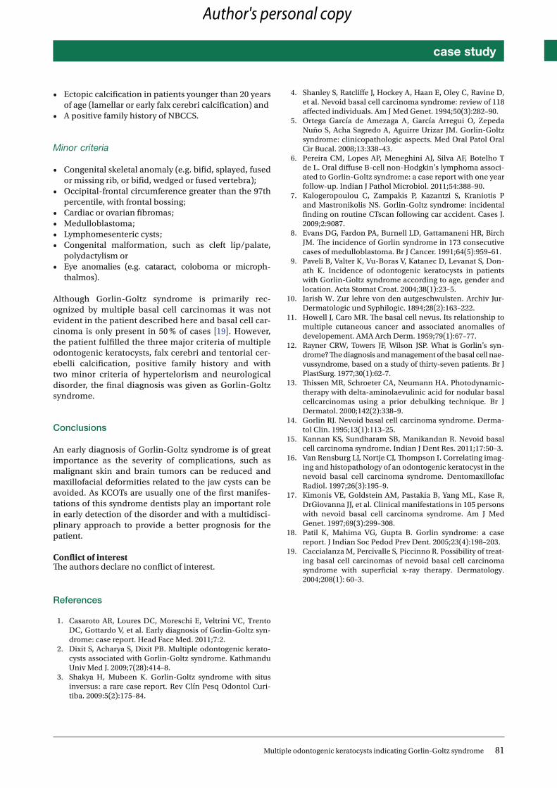
Although Gorlin-Goltz syndrome is primarily rec-ognized by multiple basal cell carcinomas it was not evident in the patient described here and basal cell car-cinoma is only present in 50 % of cases [19]. However, the patient fulfilled the three major criteria of multiple odontogenic keratocysts, falx cerebri and tentorial cer-ebelli calcification, positive family history and with two minor criteria of hypertelorism and neurological disorder, the final diagnosis was given as Gorlin-Goltz syndrome.
Conclusions
An early diagnosis of Gorlin-Goltz syndrome is of great importance as the severity of complications, such as malignant skin and brain tumors can be reduced and maxillofacial deformities related to the jaw cysts can be avoided. As KCOTs are usually one of the first manifes-tations of this syndrome dentists play an important role in early detection of the disorder and with a multidisci-plinary approach to provide a better prognosis for the patient.
Conflict of interest The authors declare no conflict of interest.
References
1. CasarotoAR,LouresDC,MoreschiE,VeltriniVC,TrentoDC, Gottardo V, et al. Early diagnosis of Gorlin-Goltz syn-drome: case report. Head Face Med. 2011;7:2.
2. Dixit S, Acharya S, Dixit PB. Multiple odontogenic kerato-cysts associated with Gorlin-Goltz syndrome. Kathmandu Univ Med J. 2009;7(28):414–8.
3. Shakya H, Mubeen K. Gorlin-Goltz syndrome with situs inversus: a rare case report. Rev Clín Pesq Odontol Curi-tiba. 2009:5(2):175–84.
4. Shanley S, Ratcliffe J, Hockey A, Haan E, Oley C, Ravine D, et al. Nevoid basal cell carcinoma syndrome: review of 118 affected individuals. Am J Med Genet. 1994;50(3):282–90.
5. Ortega García de Amezaga A, García Arregui O, Zepeda Nuño S, Acha Sagredo A, Aguirre Urizar JM. Gorlin-Goltz syndrome: clinicopathologic aspects. Med Oral Patol Oral Cir Bucal. 2008;13:338–43.
6. PereiraCM, LopesAP,MeneghiniAJ, SilvaAF,BotelhoTdeL.OraldiffuseB-cellnon-Hodgkin’slymphomaassoci-ated to Gorlin-Goltz syndrome: a case report with one year follow-up. Indian J Pathol Microbiol. 2011;54:388–90.
7. Kalogeropoulou C, Zampakis P, Kazantzi S, Kraniotis P and Mastronikolis NS. Gorlin-Goltz syndrome: incidental finding on routine CTscan following car accident. Cases J. 2009;2:9087.
8. EvansDG,FardonPA,BurnellLD,GattamaneniHR,BirchJM. The incidence of Gorlin syndrome in 173 consecutive cases of medulloblastoma. Br J Cancer. 1991;64(5):959–61.
9. PaveliB,ValterK,Vu-BorasV,KatanecD,LevanatS,Don-ath K. Incidence of odontogenic keratocysts in patients with Gorlin-Goltz syndrome according to age, gender and location. Acta Stomat Croat. 2004;38(1):23–5.
10. Jarish W. Zur lehre von den autgeschwulsten. Archiv Jur-Dermatologic und Syphilogic. 1894;28(2):163–222.
11. Howell J, Caro MR. The basal cell nevus. Its relationship to multiple cutaneous cancer and associated anomalies of developement. AMA Arch Derm. 1959;79(1):67–77.
12. Rayner CRW, Towers JF, Wilson JSP. What is Gorlin’s syn-drome? The diagnosis and management of the basal cell nae-vussyndrome, based on a study of thirty-seven patients. Br J PlastSurg. 1977;30(1):62-7.
13. Thissen MR, Schroeter CA, Neumann HA. Photodynamic-therapy with delta-aminolaevulinic acid for nodular basal cellcarcinomas using a prior debulking technique. Br J Dermatol. 2000;142(2):338–9.
14. Gorlin RJ. Nevoid basal cell carcinoma syndrome. Derma-tol Clin. 1995;13(1):113–25.
15. Kannan KS, Sundharam SB, Manikandan R. Nevoid basal cell carcinoma syndrome. Indian J Dent Res. 2011;17:50–3.
16. VanRensburgLJ,NortjeCJ,ThompsonI.Correlatingimag-ing and histopathology of an odontogenic keratocyst in the nevoid basal cell carcinoma syndrome. Dentomaxillofac Radiol. 1997;26(3):195–9.
17. KimonisVE,GoldsteinAM,PastakiaB,YangML,KaseR,DrGiovanna JJ, et al. Clinical manifestations in 105 persons with nevoid basal cell carcinoma syndrome. Am J Med Genet. 1997;69(3):299–308.
18. Patil K, Mahima VG, Gupta B. Gorlin syndrome: a case report. J Indian Soc Pedod Prev Dent. 2005;23(4):198–203.
19. Caccialanza M, Percivalle S, Piccinno R. Possibility of treat-ing basal cell carcinomas of nevoid basal cell carcinoma syndrome with superficial x-ray therapy. Dermatology. 2004;208(1): 60–3.
Author's personal copy
Copyright © 2022 FDOKUMEN