Multiple gene genealogies and phenotypic characters differentiate several novel species of...
19
Persoonia 20, 2008: 19 – 37 www.persoonia.org doi:10.3767/003158508X302212 © 2008 Nationaal Herbarium Nederland Centraalbureau voor Schimmelcultures RESEARCH ARTICLE Multiple gene genealogies and phenotypic characters differentiate several novel species of Mycosphaerella and related anamorphs on banana M. Arzanlou 1,2 , J.Z. Groenewald 1 , R.A. Fullerton 3 , E.C.A. Abeln 4 , J. Carlier 5 , M.-F. Zapater 5 , I.W. Buddenhagen 6 , A. Viljoen 7 , P.W. Crous 1,2 1 CBS Fungal Biodiversity Centre, Uppsalalaan 8, 3584 CT Utrecht, The Netherlands; corresponding author email: [email protected]. 2 Laboratory of Phytopathology, Wageningen University, Binnenhaven 5, 6709 PD Wageningen, The Netherlands. 3 The Horticulture and Food Research Institute of New Zealand, Private Bag 92169, Auckland, New Zealand. 4 TNO Quality of Life, Utrechtseweg 48, 3700 AJ Zeist, The Netherlands. 5 Centre de coopération internationale en recherche agronomique pour le développement (CIRAD), UMR BGPI, Montpellier, France. 6 College of Agricultural and Environmental Sciences, University of California, Davis, USA. 7 Department of Plant Pathology, University of Stellenbosch, P. Bag X1, Matieland 7602, South Africa. Abstract Three species of Mycosphaerella, namely M. eumusae, M. fijiensis, and M. musicola are involved in the Sigatoka disease complex of bananas. Besides these three primary pathogens, several additional species of Mycosphaerella or their anamorphs have been described from Musa. However, very little is known about these taxa, and for the majority of these species no culture or DNA is available for study. In the present study, we collected a global set of Mycosphaerella strains from banana, and compared them by means of morphology and a multi-gene nucleotide sequence data set. The phylogeny inferred from the ITS region and the combined data set containing partial gene sequences of the actin gene, the small subunit mitochondrial ribosomal DNA and the histone H3 gene revealed a rich diversity of Mycosphaerella species on Musa. Integration of morphological and molecular data sets confirmed more than 20 species of Mycosphaerella (incl. anamorphs) to occur on banana. This study reconfirmed the previously described presence of Cercospora apii, M. citri and M. thailandica, and also identified Mycosphaerella communis, M. lateralis and Passalora loranthi on this host. Moreover, eight new species identified from Musa are described, namely Dissoconium musae, Mycosphaerella mozambica, Pseudocercospora assamensis, P. indone- siana, P. longispora, Stenella musae, S. musicola, and S. queenslandica. Article info Received: 8 January 2008; Accepted: 5 March 2008; Published: 21 March 2008. Key words Mycosphaerella phylogeny Sigatoka disease complex taxonomy INTRODUCTION The genus Mycosphaerella is phylogenetically heterogeneous (Crous et al. 2007a), contains more than 3000 names (Aptroot 2006), and has been linked to more than 30 well-known ana- morphic genera (Crous et al. 2006a, b, 2007a, b, Arzanlou et al. 2007a). Species of Mycosphaerella inhabit different eco- logical niches as saprobes, plant pathogens or endophytes (Farr et al. 1995, Verkley & Starink-Willemse 2004, Crous et al. 2004b, 2006a, 2007a, b), and have a worldwide distribution from tropical and subtropical to warm and cool regions (Crous 1998, Crous et al. 2000, 2001). Plant-pathogenic species of Mycosphaerella are among the most common and destructive plant pathogens occurring on a wide range of hosts including trees, herbaceous plants, and plantation crops. The invasion of leaf and stem tissue and concomitant distortion of the host plant physiology cause considerable economic losses (Park et al. 2000, Goodwin et al. 2001, Maxwell et al. 2005, Cortinas et al. 2006, Crous et al. 2006a, b, Hunter et al. 2006). The Sigatoka disease complex, which is the most serious and economically important leaf spot disease of banana, is attrib- uted to species of Mycosphaerella. Mycosphaerella musicola (anamorph Pseudocercospora musae) which causes (yellow) Sigatoka disease, M. fijiensis (anamorph P. fijiensis) which causes the black Sigatoka disease, and M. eumusae (ana- morph P. eumusae), which causes eumusae leaf spot disease (reviewed in Jones 2000, 2003, Crous & Mourichon 2002) are the major constituents of the Sigatoka disease complex. The disease reduces the photosynthetic capacity of the plant as a consequence of necrotic leaf lesions, and induces physiological alterations of the plant, resulting in reduced crop yield and fruit quality. All three species emerged on bananas during the last century and became major constraints to commercial produc- tion worldwide. The chronology of disease records around the world and genetic structure of pathogen population suggests that South-East Asia, where the host genus Musa is indigenous, is the centre of origin for all three fungal species (Mourichon & Fullerton 1990, Carlier et al. 1996, Hayden et al. 2003, Rivas et al. 2004). Yellow Sigatoka disease was first reported on banana in Java in 1902. The disease spread rapidly to all banana-growing regions during the following 20 years, and has since reached the limits of its distribution worldwide (reviewed in Jones 2000, 2003). The fungus responsible for the disease was described as Cercospora musae. In 1941 Leach established the connec- tion between C. musae and its teleomorph, Mycosphaerella
-
Upload
independent -
Category
Documents
-
view
0 -
download
0
Transcript of Multiple gene genealogies and phenotypic characters differentiate several novel species of...

Persoonia 20, 2008: 19–37www.persoonia.org doi:10.3767/003158508X302212
© 2008 Nationaal Herbarium Nederland Centraalbureau voor Schimmelcultures
RESEARCH ARTICLE
Multiple gene genealogies and phenotypic characters differentiate several novel species of Mycosphaerella and related anamorphs on banana
M. Arzanlou1,2, J.Z. Groenewald1, R.A. Fullerton3, E.C.A. Abeln4, J. Carlier5, M.-F. Zapater5, I.W. Buddenhagen6, A. Viljoen7, P.W. Crous1,2
1 CBS Fungal Biodiversity Centre, Uppsalalaan 8, 3584 CT Utrecht, The Netherlands; corresponding author email: [email protected].
2 Laboratory of Phytopathology, Wageningen University, Binnenhaven 5, 6709 PD Wageningen, The Netherlands.
3 The Horticulture and Food Research Institute of New Zealand, Private Bag 92169, Auckland, New Zealand.
4 TNO Quality of Life, Utrechtseweg 48, 3700 AJ Zeist, The Netherlands.5 Centre de coopération internationale en recherche agronomique pour le
développement (CIRAD), UMR BGPI, Montpellier, France.6 College of Agricultural and Environmental Sciences, University of California,
Davis, USA.7 Department of Plant Pathology, University of Stellenbosch, P. Bag X1,
Matieland 7602, South Africa.
Abstract Three species of Mycosphaerella, namely M. eumusae, M. fijiensis, and M. musicola are involved in the Sigatoka disease complex of bananas. Besides these three primary pathogens, several additional species of Mycosphaerella or their anamorphs have been described from Musa. However, very little is known about these taxa, and for the majority of these species no culture or DNA is available for study. In the present study, we collected a global set of Mycosphaerella strains from banana, and compared them by means of morphology and a multi-gene nucleotide sequence data set. The phylogeny inferred from the ITS region and the combined data set containing partial gene sequences of the actin gene, the small subunit mitochondrial ribosomal DNA and the histone H3 gene revealed a rich diversity of Mycosphaerella species on Musa. Integration of morphological and molecular data sets confirmed more than 20 species of Mycosphaerella (incl. anamorphs) to occur on banana. This study reconfirmed the previously described presence of Cercospora apii, M. citri and M. thailandica, and also identified Mycosphaerella communis, M. lateralis and Passalora loranthi on this host. Moreover, eight new species identified from Musa are described, namely Dissoconium musae, Mycosphaerella mozambica, Pseudocercospora assamensis, P. indonesiana, P. longispora, Stenella musae, S. musicola, and S. queenslandica.
Article info Received: 8 January 2008; Accepted: 5 March 2008; Published: 21 March 2008.
Key words
MycosphaerellaphylogenySigatoka disease complextaxonomy
InTRoduCTIon
The genus Mycosphaerella is phylogenetically heterogeneous (Crous et al. 2007a), contains more than 3000 names (Aptroot 2006), and has been linked to more than 30 well-known ana-morphic genera (Crous et al. 2006a, b, 2007a, b, Arzanlou et al. 2007a). Species of Mycosphaerella inhabit different eco-logical niches as saprobes, plant pathogens or endophytes (Farr et al. 1995, Verkley & Starink-Willemse 2004, Crous et al. 2004b, 2006a, 2007a, b), and have a worldwide distribution from tropical and subtropical to warm and cool regions (Crous 1998, Crous et al. 2000, 2001). Plant-pathogenic species of Mycosphaerella are among the most common and destructive plant pathogens occurring on a wide range of hosts including trees, herbaceous plants, and plantation crops. The invasion of leaf and stem tissue and concomitant distortion of the host plant physiology cause considerable economic losses (Park
et al. 2000, Goodwin et al. 2001, Maxwell et al. 2005, Cortinas et al. 2006, Crous et al. 2006a, b, Hunter et al. 2006).
The Sigatoka disease complex, which is the most serious and economically important leaf spot disease of banana, is attrib-uted to species of Mycosphaerella. Mycosphaerella musicola (anamorph Pseudocercospora musae) which causes (yellow) Sigatoka disease, M. fijiensis (anamorph P. fijiensis) which causes the black Sigatoka disease, and M. eumusae (ana-morph P. eumusae), which causes eumusae leaf spot disease (reviewed in Jones 2000, 2003, Crous & Mourichon 2002) are the major constituents of the Sigatoka disease complex. The disease reduces the photosynthetic capacity of the plant as a consequence of necrotic leaf lesions, and induces physiological alterations of the plant, resulting in reduced crop yield and fruit quality. All three species emerged on bananas during the last century and became major constraints to commercial produc-tion worldwide. The chronology of disease records around the world and genetic structure of pathogen population suggests that South-East Asia, where the host genus Musa is indigenous, is the centre of origin for all three fungal species (Mourichon & Fullerton 1990, Carlier et al. 1996, Hayden et al. 2003, Rivas et al. 2004).
Yellow Sigatoka disease was first reported on banana in Java in 1902. The disease spread rapidly to all banana-growing regions during the following 20 years, and has since reached the limits of its distribution worldwide (reviewed in Jones 2000, 2003). The fungus responsible for the disease was described as Cercospora musae. In 1941 Leach established the connec-tion between C. musae and its teleomorph, Mycosphaerella

20 Persoonia – Volume 20, 2008
Spe
cies
A
cces
sion
num
ber1
Sou
rce
O
rigin
G
enB
ank
num
bers
(IT
S, A
CT,
HIS
, mtS
SU
)
Cer
cosp
ora
apii
CP
C 1
2682
; CB
S 1
1939
5 M
usa
cv. C
aven
dish
W
este
rn B
angl
ades
h E
U51
4222
, —, —
, —
CP
C 1
2683
M
usa
cv. C
aven
dish
W
este
rn B
angl
ades
h E
U51
4223
, —, —
, —
CP
C 1
2684
M
usa
cv. C
aven
dish
W
este
rn B
angl
ades
h E
U51
4224
, —, —
, —D
isso
coni
um m
usae
X
1021
; CB
S 1
2245
3 M
usa
cv. N
endr
an (
Pla
ntai
n) A
AB
In
dia
EU
5142
25, E
U51
4296
, EU
5143
49, E
U51
4402
X
1022
; CB
S 1
2245
4 M
usa
cv. N
endr
an (
Pla
ntai
n) A
AB
In
dia
EU
5142
26, E
U51
4297
, EU
5143
50, E
U51
4403
Myc
osph
aere
lla c
itri
X12
6; C
BS
122
455
Citr
us s
p.
US
A, F
lorid
a E
U51
4227
, —, —
, —
X74
2; C
BS
122
456
Mus
a cv
. SH
343
6 A
AA
A
Tong
a E
U51
4228
, —, —
, —
X74
3 M
usa
cv. S
H 3
362
AA
To
nga
EU
5142
29, —
, —, —
Myc
osph
aere
lla c
olom
bien
sis
X24
M
usa
culti
var
Moz
ambi
que
EU
5142
30, —
, —, —
X
215
—
Sou
th A
fric
a E
U51
4231
, EU
5142
98, E
U51
4351
, EU
5144
04M
ycos
phae
rella
com
mun
is
X10
23
Mus
a cv
. Val
ery:
Cav
aend
ish
Trin
idad
E
U51
4232
, EU
5142
99, E
U51
4352
, EU
5144
05M
ycos
phae
rella
eum
usae
S
1030
B
Mus
a cu
ltiva
r M
aurit
ius
EU
5142
33, E
U51
4300
, EU
5143
53, E
U51
4406
S
1037
B
Mus
a cu
ltiva
r M
aurit
ius
EU
5142
34, E
U51
4301
, EU
5143
54, E
U51
4407
S
1037
C
Mus
a cu
ltiva
r M
aurit
ius
EU
5142
35, E
U51
4302
, EU
5143
55, E
U51
4408
S
1037
G
Mus
a cu
ltiva
r M
aurit
ius
EU
5142
36, —
, —, —
S
1037
H
Mus
a cu
ltiva
r M
aurit
ius
EU
5142
37, —
, —, —
X
208;
CIR
AD
115
6; C
PC
457
9 M
usa
culti
var
Mau
ritiu
s E
U51
4238
, —, —
, —
X20
9; C
BS
114
825;
CIR
AD
115
7; C
PC
458
0 M
usa
culti
var
Mau
ritiu
s E
U51
4239
, —, —
, —
X86
5; C
IRA
D 5
35
Mus
a cu
ltiva
r In
dia
EU
5142
40, E
U51
4303
, EU
5143
56, E
U51
4409
X
866;
CB
S 1
2137
7; C
IRA
D 4
58
Mus
a cu
ltiva
r M
alay
sia
EU
5142
41, E
U51
4304
, EU
5143
57, E
U51
4410
X
867;
CB
S 1
2137
8; C
IRA
D 4
59
Mus
a cu
ltiva
r M
alay
sia
EU
5142
42, E
U51
4305
, EU
5143
58, E
U51
4411
X
869;
CB
S 1
2138
0; C
IRA
D 5
63
Mus
a cu
ltiva
r S
ri La
nka
EU
5142
43, E
U51
4306
, EU
5143
59, E
U51
4412
X
870;
CB
S 1
2138
1; C
IRA
D 4
85
Mus
a cu
ltiva
r T
haila
nd
EU
5142
44, E
U51
4307
, EU
5143
60, E
U51
4413
X
873;
CB
S 1
2138
2; C
IRA
D 4
87
Mus
a cu
ltiva
r T
haila
nd
EU
5142
45, E
U51
4308
, EU
5143
61, E
U51
4414
X
875;
CIR
AD
671
M
usa
culti
var
Vie
tnam
E
U51
4246
, EU
5143
09, E
U51
4362
, EU
5144
15
X87
6; C
BS
121
383;
CIR
AD
744
M
usa
culti
var
Mau
ritiu
s E
U51
4247
, EU
5143
10, E
U51
4363
, EU
5144
16M
ycos
phae
rella
fijie
nsis
C
IRA
D 8
6; C
BS
120
258;
C86
a M
usa
culti
var
Cam
eroo
n E
U51
4248
, —, —
, —
X84
M
usa
culti
var
Col
ombi
a E
U51
4249
, EU
5143
11, E
U51
4364
, EU
5144
17
X92
M
usa
culti
var
Col
ombi
a E
U51
4250
, EU
5143
12, E
U51
4365
, EU
5144
18
X10
4 M
usa
culti
var
Col
ombi
a E
U51
4251
, EU
5143
13, E
U51
4366
, EU
5144
19
X11
0 M
usa
culti
var
Col
ombi
a E
U51
4252
, EU
5143
14, E
U51
4367
, EU
5144
20
X84
3; C
IRA
D 1
1 M
usa
culti
var
Hon
dura
s E
U51
4253
, EU
5143
15, E
U51
4368
, EU
5144
21
X84
7; C
BS
121
362;
CIR
AD
364
M
usa
culti
var
Taiw
an
EU
5142
54, E
U51
4316
, EU
5143
69, E
U51
4422
X
850;
CIR
AD
355
M
usa
culti
var
Ivor
y C
oast
E
U51
4255
, EU
5143
17, E
U51
4370
, EU
5144
23M
ycos
phae
rella
late
ralis
S
1024
M
usa
culti
var
Mau
ritiu
s E
U51
4256
, —, —
, —M
ycos
phae
rella
moz
ambi
ca
X34
; CB
S 1
2246
4 M
usa
culti
var
Moz
ambi
que
EU
5142
57, E
U51
4318
, EU
5143
71, E
U51
4424
X
884;
CB
S 1
2139
1; U
Q43
8 M
usa
culti
var
Aus
tral
ia
EU
5142
58, E
U51
4319
, EU
5143
72, E
U51
4425
Myc
osph
aere
lla m
usae
X
398;
CB
S 1
2245
8 M
usa
cv. C
aven
dish
AA
A
Tong
a E
U51
4259
, EU
5143
20, E
U51
4373
, EU
5144
26
X81
3 M
usa
culti
var
Mal
awi
EU
5142
60, E
U51
4321
, EU
5143
74, E
U51
4427
X
814;
CB
S 1
2245
9 M
usa
culti
var
Mal
awi
EU
5142
61, E
U51
4322
, EU
5143
75, E
U51
4428
X
818;
CB
S 1
2246
0 M
usa
culti
var
Mal
awi
EU
5142
62, —
, —, —
X
819;
CB
S 1
2246
1 M
usa
culti
var
Mal
awi
EU
5142
63, —
, —, —
X
879;
CB
S 1
2138
6; C
IRA
D 6
4 M
usa
culti
var
Mal
awi
EU
5142
64, E
U51
4323
, EU
5143
76, E
U51
4429
Myc
osph
aere
lla m
usic
ola
X42
; CB
S 1
1663
4; IM
I 123
823
Mus
a cu
ltiva
r C
uba
EU
5142
65, —
, —, —
X
63
Mus
a cu
ltiva
r W
indw
ard
Isla
nds
EU
5142
66, E
U51
4324
, EU
5143
77, E
U51
4430
X
67
Mus
a cu
ltiva
r W
indw
ard
Isla
nds
EU
5142
67, E
U51
4325
, EU
5143
78, E
U51
4431
X
588
Mus
a cv
. Will
iam
s A
ustr
alia
E
U51
4268
, EU
5143
26, E
U51
4379
, EU
5144
32
X58
9 M
usa
cv. W
illia
ms
Aus
tral
ia
EU
5142
69, E
U51
4327
, EU
5143
80, E
U51
4433
X
596
Mus
a cv
. SH
-336
2 A
A
Aus
tral
ia
EU
5142
70, E
U51
4328
, EU
5143
81, E
U51
4434
X
602
Mus
a cv
. Lak
atan
A
ustr
alia
E
U51
4271
, EU
5143
29, E
U51
4382
, EU
5144
35
X85
7; C
BS
121
371;
UQ
430
Mus
a cu
ltiva
r A
ustr
alia
E
U51
4272
, EU
5143
30, E
U51
4383
, EU
5144
36
Tab
le 1
Is
olat
es o
f Myc
osph
aere
lla o
r its
ana
mor
phs
used
for
DN
A a
naly
sis
and
mor
phol
ogic
al s
tudi
es.
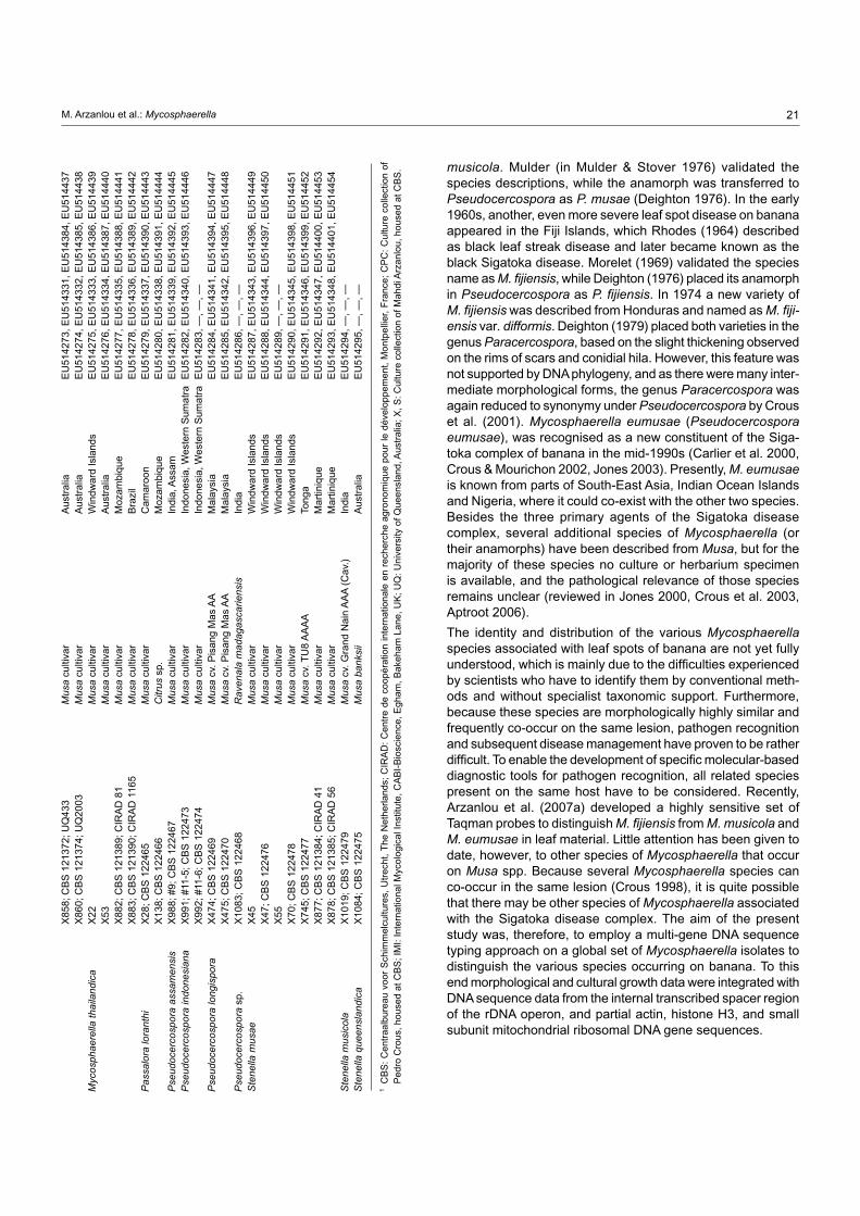
21M. Arzanlou et al.: Mycosphaerella
musicola. Mulder (in Mulder & Stover 1976) validated the species descriptions, while the anamorph was transferred to Pseudocercospora as P. musae (Deighton 1976). In the early 1960s, another, even more severe leaf spot disease on banana appeared in the Fiji Islands, which Rhodes (1964) described as black leaf streak disease and later became known as the black Sigatoka disease. Morelet (1969) validated the species name as M. fijiensis, while Deighton (1976) placed its anamorph in Pseudocercospora as P. fijiensis. In 1974 a new variety of M. fijiensis was described from Honduras and named as M. fijiensis var. difformis. Deighton (1979) placed both varieties in the genus Paracercospora, based on the slight thickening observed on the rims of scars and conidial hila. However, this feature was not supported by DNA phylogeny, and as there were many inter-mediate morphological forms, the genus Paracercospora was again reduced to synonymy under Pseudocercospora by Crous et al. (2001). Mycosphaerella eumusae (Pseudocercospora eumusae), was recognised as a new constituent of the Siga-toka complex of banana in the mid-1990s (Carlier et al. 2000, Crous & Mourichon 2002, Jones 2003). Presently, M. eumusae is known from parts of South-East Asia, Indian Ocean Islands and Nigeria, where it could co-exist with the other two species. Besides the three primary agents of the Sigatoka disease complex, several additional species of Mycosphaerella (or their anamorphs) have been described from Musa, but for the majority of these species no culture or herbarium specimen is available, and the pathological relevance of those species remains unclear (reviewed in Jones 2000, Crous et al. 2003, Aptroot 2006).
The identity and distribution of the various Mycosphaerella species associated with leaf spots of banana are not yet fully understood, which is mainly due to the difficulties experienced by scientists who have to identify them by conventional meth-ods and without specialist taxonomic support. Furthermore, because these species are morphologically highly similar and frequently co-occur on the same lesion, pathogen recognition and subsequent disease management have proven to be rather difficult. To enable the development of specific molecular-based diagnostic tools for pathogen recognition, all related species present on the same host have to be considered. Recently, Arzanlou et al. (2007a) developed a highly sensitive set of Taqman probes to distinguish M. fijiensis from M. musicola and M. eumusae in leaf material. Little attention has been given to date, however, to other species of Mycosphaerella that occur on Musa spp. Because several Mycosphaerella species can co-occur in the same lesion (Crous 1998), it is quite possible that there may be other species of Mycosphaerella associated with the Sigatoka disease complex. The aim of the present study was, therefore, to employ a multi-gene DNA sequence typing approach on a global set of Mycosphaerella isolates to distinguish the various species occurring on banana. To this end morphological and cultural growth data were integrated with DNA sequence data from the internal transcribed spacer region of the rDNA operon, and partial actin, histone H3, and small subunit mitochondrial ribosomal DNA gene sequences.
X
858;
CB
S 1
2137
2; U
Q43
3 M
usa
culti
var
Aus
tral
ia
EU
5142
73, E
U51
4331
, EU
5143
84, E
U51
4437
X
860;
CB
S 1
2137
4; U
Q20
03
Mus
a cu
ltiva
r A
ustr
alia
E
U51
4274
, EU
5143
32, E
U51
4385
, EU
5144
38M
ycos
phae
rella
thai
land
ica
X22
M
usa
culti
var
Win
dwar
d Is
land
s E
U51
4275
, EU
5143
33, E
U51
4386
, EU
5144
39
X53
M
usa
culti
var
Aus
tral
ia
EU
5142
76, E
U51
4334
, EU
5143
87, E
U51
4440
X
882;
CB
S 1
2138
9; C
IRA
D 8
1 M
usa
culti
var
Moz
ambi
que
EU
5142
77, E
U51
4335
, EU
5143
88, E
U51
4441
X
883;
CB
S 1
2139
0; C
IRA
D 1
165
Mus
a cu
ltiva
r B
razi
l E
U51
4278
, EU
5143
36, E
U51
4389
, EU
5144
42P
assa
lora
lora
nthi
X
28; C
BS
122
465
Mus
a cu
ltiva
r C
amar
oon
EU
5142
79, E
U51
4337
, EU
5143
90, E
U51
4443
X
138;
CB
S 1
2246
6 C
itrus
sp.
M
ozam
biqu
e E
U51
4280
, EU
5143
38, E
U51
4391
, EU
5144
44P
seud
ocer
cosp
ora
assa
men
sis
X98
8; #
9; C
BS
122
467
Mus
a cu
ltiva
r In
dia,
Ass
am
EU
5142
81, E
U51
4339
, EU
5143
92, E
U51
4445
Pse
udoc
erco
spor
a in
done
sian
a
X99
1; #
11-5
; CB
S 1
2247
3 M
usa
culti
var
Indo
nesi
a, W
este
rn S
umat
ra
EU
5142
82, E
U51
4340
, EU
5143
93, E
U51
4446
X
992;
#11
-6; C
BS
122
474
Mus
a cu
ltiva
r In
done
sia,
Wes
tern
Sum
atra
E
U51
4283
, —, —
, —P
seud
ocer
cosp
ora
long
ispo
ra
X47
4; C
BS
122
469
Mus
a cv
. Pis
ang
Mas
AA
M
alay
sia
EU
5142
84, E
U51
4341
, EU
5143
94, E
U51
4447
X
475;
CB
S 1
2247
0 M
usa
cv. P
isan
g M
as A
A
Mal
aysi
a E
U51
4285
, EU
5143
42, E
U51
4395
, EU
5144
48P
seud
ocer
cosp
ora
sp.
X10
83; C
BS
122
468
Rav
enal
a m
adag
asca
riens
is
Indi
a E
U51
4286
, —, —
, —S
tene
lla m
usae
X
45
Mus
a cu
ltiva
r W
indw
ard
Isla
nds
EU
5142
87, E
U51
4343
, EU
5143
96, E
U51
4449
X
47; C
BS
122
476
Mus
a cu
ltiva
r W
indw
ard
Isla
nds
EU
5142
88, E
U51
4344
, EU
5143
97, E
U51
4450
X
55
Mus
a cu
ltiva
r W
indw
ard
Isla
nds
EU
5142
89, —
, —, —
X
70; C
BS
122
478
Mus
a cu
ltiva
r W
indw
ard
Isla
nds
EU
5142
90, E
U51
4345
, EU
5143
98, E
U51
4451
X
745;
CB
S 1
2247
7 M
usa
cv. T
U8
AA
AA
To
nga
EU
5142
91, E
U51
4346
, EU
5143
99, E
U51
4452
X
877;
CB
S 1
2138
4; C
IRA
D 4
1 M
usa
culti
var
Mar
tiniq
ue
EU
5142
92, E
U51
4347
, EU
5144
00, E
U51
4453
X
878;
CB
S 1
2138
5; C
IRA
D 5
6 M
usa
culti
var
Mar
tiniq
ue
EU
5142
93, E
U51
4348
, EU
5144
01, E
U51
4454
Ste
nella
mus
icol
a
X10
19; C
BS
122
479
Mus
a cv
. Gra
nd N
ain
AA
A (
Cav
.)
Indi
a E
U51
4294
, —, —
, —S
tene
lla q
ueen
slan
dica
X
1084
; CB
S 1
2247
5 M
usa
bank
sii
Aus
tral
ia
EU
5142
95, —
, —, —
1 C
BS
: C
entr
aalb
urea
u vo
or S
chim
mel
cultu
res,
Utr
echt
, T
he N
ethe
rland
s; C
IRA
D:
Cen
tre
de c
oopé
ratio
n in
tern
atio
nale
en
rech
erch
e ag
rono
miq
ue p
our
le d
ével
oppe
men
t, M
ontp
ellie
r, F
ranc
e; C
PC
: C
ultu
re c
olle
ctio
n of
P
edro
Cro
us, h
ouse
d at
CB
S; I
MI:
Inte
rnat
iona
l Myc
olog
ical
Inst
itute
, CA
BI-
Bio
scie
nce,
Egh
am, B
akeh
am L
ane,
UK
; UQ
: Uni
vers
ity o
f Que
ensl
and,
Aus
tral
ia; X
, S: C
ultu
re c
olle
ctio
n of
Mah
di A
rzan
lou,
hou
sed
at C
BS
.

22 Persoonia – Volume 20, 2008
MATERIALS And METHodS
Isolates
Isolates (Table 1) were obtained by isolation from infected symptomatic banana leaves, or supplied as pure cultures by the following departments and institutes: The Horticulture and Food Research Institute of New Zealand, Auckland, New Zealand; Centre de coopération internationale en recherché agronomique pour le développement (CIRAD, Montpellier, France); University of Florida, Tropical Research & Education Centre (USA); Forestry and Agricultural Biotechnology Institute (FABI, Pretoria, South Africa). Isolates were recovered from infected banana leaves as single ascospores or conidia. Ger-minating spores were examined 24 h after germination on 2 % malt extract agar (MEA; Sigma-Aldrich Chemie, Zwijndrecht, The Netherlands) plates under a stereomicroscope, and single-spore cultures were established on fresh MEA plates following the protocol of Crous (1998).
DNA phylogeny
Genomic DNA was isolated from fungal mycelia grown on MEA, using the FastDNA kit (BIO101, Carlsbad, CA, USA) accord-ing to the manufacturer’s protocol. The primers ITS1 and ITS4 (White et al. 1990) were used to amplify part of the internal transcribed spacer region (ITS) of the nuclear ribosomal RNA operon, including the 3’ end of the 18S rRNA gene, the first ITS region, the 5.8S rRNA gene, the second ITS region, and the 5’ end of the 28S rRNA gene. A part of the actin gene (ACT) was amplified with primers ACT-512F and ACT-783R (Carbone & Kohn 1999), a part of the small subunit mitochondrial ribosomal DNA (mtSSU) with primers MNS1 and MNS2 (Li et al. 1994), and a part of the histone H3 (HIS) gene with primers CYLH3F and CYLH3R (Crous et al. 2004b). Amplification reactions were performed with each primer set in a total reaction volume of 25 µl, which was composed of 1 × PCR Buffer (Applied Biosys-tems, Foster City, USA), variable MgCl
2 concentrations, 60 µM
dNTPs, 0.2 µM of each forward and reverse primer, 1.5 U of Taq DNA polymerase (Roche Diagnostics, Indianapolis, USA) and 1–10 ng of genomic DNA. PCR cycle conditions were 5 min of 95 °C, followed by 36 cycles of 94 °C for 30 s, 55 °C for 30 s, 72 °C for 60 s, and a final elongation at 72 °C for 7 min. Amplicons were sequenced using both PCR primers with a DYEnamic ET Terminator Cycle Sequencing kit (Amersham Biosciences, Roosendaal, the Netherlands) according to the manufacturer’s recommendations, and sequences were ana-lysed on an ABI Prism 3700 DNA Sequencer (Perkin-Elmer, Norwalk, Foster City, CA).
The resulting nucleotide sequences were analysed and auto-matically aligned using BioNumerics v. 4.5 (Applied Maths, Kort-rijk, Belgium) followed by manual improvement by eye where necessary. Phylogenetic analyses were performed with PAUP (Phylogenetic Analysis Using Parsimony) v. 4.0b10 (Swofford 2003), using the neighbour-joining algorithm with the uncor-rected (“p”), the Kimura 2-parameter and the HKY85 substitu-tion models. Alignment gaps longer than 10 bases were coded as single events for the phylogenetic analyses; the remaining gaps were treated as missing data. Any encountered ties were randomly broken. Phylogenetic relationships were also inferred
with the parsimony algorithm using the heuristic search op-tion with simple (ITS alignment) or 100 random taxa additions (combined alignment) and tree bisection and reconstruction (TBR) as the branch-swapping algorithm; alignment gaps were treated as missing (combined alignment) or as a fifth character state (ITS alignment) and all characters were unordered and of equal weight. Branches of zero length were collapsed and all multiple, equally parsimonious trees were saved. Other measures calculated included tree length, consistency index, retention index and rescaled consistency index (TL, CI, RI and RC, respectively). The robustness of the obtained trees was evaluated by 10 000 000 fast stepwise (ITS alignment) or 1000 bootstrap heuristic bootstrap replications (combined alignment). Sequences were deposited in GenBank (Table 1) and the alignments in TreeBASE (www.treebase.org).
Morphology
Growth rates and colony morphology were recorded from colo-nies grown on MEA plates after 30 d incubation in darkness at 24 °C. Colony colours (surface and reverse) were assessed
after growth on MEA and oatmeal agar (OA, Gams et al. 2007) using the colour charts of Rayner (1970). Microscopic obser-vations were made from colonies cultivated on MEA and OA. Preparations were mounted in lactic acid and studied under a light microscope (× 1000 magnification). The 95 % confidence intervals were derived from 30 observations of spores formed on MEA or OA, with extremes given in parentheses. All cultures obtained in this study are maintained in the culture collection of the Centraalbureau voor Schimmelcultures (CBS) in Utrecht, the Netherlands or the working collections of Pedro Crous (CPC) or Mahdi Arzanlou (X, S numbers) at CBS (Table 1). Nomenclatural novelties and descriptions were deposited in MycoBank (www.MycoBank.org) (Crous et al. 2004a).
RESuLTS
DNA phylogeny
Two alignments of DNA sequences were subjected to phyloge-netic analyses. The first alignment consisted of ITS sequences generated in this study as well as sequences obtained from the NCBI GenBank nucleotide sequence database. The ITS align-ment consisted of a total number of 113 sequences (including one outgroup); 508 characters including alignment gaps were subjected to the analyses. Of these characters, 224 were par-simony-informative, 42 variable and parsimony-uninformative, and 242 were constant. Trees supporting the same clades were obtained irrespective of the analysis method used. The parsimony analysis yielded 11 780 equally most parsimonious trees that mainly differed in the order of taxa at the terminal nodes; one of the trees is presented in Fig. 1 (TL = 861 steps; CI = 0.569; RI = 0.934; RC = 0.532).
The sequence data in the second alignment were analysed as one combined set consisting of 1648 characters (incl. align-ment gaps) (number of included characters: ITS: 509, ACT: 188, HIS: 375, mtSSU: 576). This second alignment included 54 sequences (including the outgroup) and of the 1648 char-acters, 517 were parsimony-informative, 93 were variable and

23M. Arzanlou et al.: Mycosphaerella
10 changes
Davidiella tassiana DQ289800X1023
AY725537
AY725535
EF394854 Dissoconium australiensis
X1021
X1022
AF309625
S1024
AF309624
DQ267587
DQ302984
DQ303075 Ps. epispermogoniana
DQ267588
AY424802X398
X818
X819
X813
X879
AY923761
X814
EU041796 Ramichloridium biverticillatum
EU041801 Ramichloridium musae
DQ530216 Mycosphaerella pseudovespa
EU041783 Periconiella velutina
DQ632677
X1084
X55
X877, X878
X70, X745
X45, X47
EF535708 Stenellopsis liriopes
X1019 Stenella musicolaX126
AY752146
X743
X742
AF181703
AF181704
DQ632684
X215 M. colombiensis
X24 M. colombiensis
X53 M. thailandica
X883 M. thailandica
X22 M. thailandica
X882 M. thailandica
AY752156 M. thailandica
AF309612 M. colombiensis
EF394849 M. thailandica
CPC 12684
CPC 12682
AY266162
CPC 12683
AF297230 Cercospora nicotianae
AY840519 Cercospora apii
AY152590 Mycosphaerella laricina
AY752163
AY752162
AY348311X138
X28
DQ676520 Passalora sp.
AF362058 Mycosphaerella confusa
X34
X884
EU042175
AY509744
AY626981
DQ267577
X847
X850
CIRAD 86
X92
X84
AY266150, AY923765
X843X104, X110
DQ289829 Pseudocercospora vitis
X988 Pseudocercospora assamensis
X1083 Pseudocercospora sp.
DQ885903 Passalora schizolobii
DQ267602 Pseudocercospora paraguayensis
AF309595 Pseudocercospora basiramifera
X991
X992
X865
X870
AY923758
S1037G, S1037H
X208, X209
AY646484, AY646483 Mycosphaerella sp.
X474, X475 Pseudocercospora longispora
X42, AY266148
X67
X602, X63
X589, X596
X858, X860
X588, X857
Mycosphaerella communis
Dissoconium musae
Mycosphaerella lateralis
Mycosphaerella marksii
Mycosphaerella musae
Stenella queenslandica
Stenella musae
Mycosphaerella citri
Mycosphaerella colombiensis /
Mycosphaerella thailandica
Cercospora apii
Passalora sp.
Passalora loranthi
Mycosphaerella mozambica
Mycosphaerella aurantia
Mycosphaerella africana
Mycosphaerella fijiensis
Mycosphaerella eumusae
Mycosphaerella musicola
100
89
84
100
81
65
100 74
97
86
100
100
100
100
100
86
93
100
89
95
97
69
74
95
67
66
98
97
100
Pseudocercospora indonesiana
parsimony-uninformative, and 1038 were constant. Trees supporting the same clades were obtained irrespective of the analysis method used. The parsimony analysis yielded eight equally most parsimonious trees that mainly differed in the order of taxa at the terminal nodes; one of the trees is presented in Fig. 2 (TL = 1513 steps; CI = 0.654; RI = 0.901; RC = 0.589). Similar to the results obtained for the ITS alignment, the same lineages were found with the combined alignment. The ACT and HIS data were found to be more variable within species than the ITS and mtSSU data (data not shown for individual loci, variation within clades in Fig. 2). The phylogenetic results obtained are discussed where applicable in the descriptive notes below.
Taxonomy
The results of this study showed a rich diversity of Mycosphaerella spp. on Musa. Phylogenetic analyses revealed that more than 20 species of Mycosphaerella or its anamorphs occur on banana, including species known from hosts other than banana, namely Cercospora apii, Mycosphaerella citri, M. com munis, M. lateralis, M. thailandica, and Passalora loranthi (Fig. 1). Furthermore, eight species proved to be morphologically and phylogenetically distinct from the species presently known from banana. These new species are described below.
Fig. 1 One of 11 780 equally most parsimonious trees obtained from a heuristic search with simple taxon additions of the ITS sequence alignment. The scale bar shows 10 changes, and bootstrap support values (65 % and higher) from 10 000 000 fast stepwise replicates are shown at the nodes. Thickened lines indicate the strict consensus branches. The tree was rooted to sequences of Davidiella tassiana strain CPC 11600 (GenBank accession number DQ289800). M. = Mycosphaerella and Ps. = Pseudocercospora.

24 Persoonia – Volume 20, 2008
10 changes
Davidiella tassiana
X1023 Mycosphaerella communis
X1021
X1022
X34
X884
X28
X138
X53 M. thailandica
X215 M. colombiensis
X22 M. thailandica
X882 M. thailandica
X883 M. thailandica
X879
X398
X814
X813
X878
X47
X70
X745
X877
X45
X847
X850
X92
X84
X110
X843
X104
X475
X474
X588
X602
X589
X596
X860
X858
X857
X63
X67
X988 Pseudocercospora assamensis
X991 Pseudocercospora indonesiana
X865
X870
X866
X875
X867
S1037B
X869
X876
X873
S1030B
S1037C
Dissoconium musae
Passalora loranthi
Mycosphaerella mozambica
M. thailandica /
M. colombiensis
Mycosphaerella musae
Stenella musae
Mycosphaerella fijiensis
Pseudocercospora longispora
Mycosphaerella musicola
Mycosphaerella eumusae
100
100
100
67
100
90
100
100
100
65
100
65
100
100
100
100
100
65
87
67
83
100
86
83
Cercospora apii Fresen., Beitr. Mykol. 3: 91. 1863
= Cercospora hayi Calp., Studies on the Sigatoka disease of bananas and its fungus pathogen, Atkins Garden and Research Laboratory, Cuba: 63. 1955.
Specimens examined. Cuba, Musa paradisiaca var. sapientum, 1955, L. Calpouzos, holotype FH, ex-type culture ATCC 12234. – IndIa, Bangladesh, Musa cv. Cavendish, Oct. 2005, I. Buddenhagen, CBS H-20035, culture CBS 119395.
Notes — In their treatment of the genus Cercospora, Crous & Braun (2003) considered C. hayi to be a synonym of the older name, Cercospora apii, which is known to have a wide host range. Based on a comparison of DNA sequence data with the ex-type strain of C. apii (GenBank AY840519; Groenewald et al. 2006), this synonymy appears to be correct.
Dissoconium musae Arzanlou & Crous, sp. nov. — MycoBank MB505972; Fig. 3, 4
Dissoconio communi simile, sed coloniis in vitro tarde crescentibus (usque ad 10 mm diam post 30 dies ad 24 °C in agaro maltoso).
Etymology. Named after its host plant, Musa.
In vitro on MEA: Mycelium submerged and superficial; sub-merged hyphae hyaline to subhyaline, thin-walled, smooth, forming a dense network with numerous anastomoses, 2–3 µm wide; aerial hyphae subhyaline, smooth, 2–3 µm wide.
Fig. 2 One of eight equally most parsimonious trees obtained from a heuristic search with 100 random taxon additions of the combined (ITS, ACT, HIS, mtSSU) sequence alignment. The scale bar shows 10 changes, and boot-strap support values (65 % and higher) from 1000 replicates are shown at the nodes. Thickened lines indicate the strict consensus branches. The tree was rooted to sequences of Davidiella tassiana strain CPC 11600 (GenBank accession number DQ289800, DQ289867, EF679665, EU514455, respec-tively). M. = Mycosphaerella.

25M. Arzanlou et al.: Mycosphaerella
Conidiophores arising orthotropically from vegetative hyphae, often reduced to conidiogenous cells and continuous with sup-porting hyphae, thin-walled, smooth, pale brown, unbranched, straight, subulate to lageniform, tapering towards the apex, (10–)19–25(–53) × (2.5–)3–5 µm. Conidiogenous cells termi-nal, proliferating sympodially (but appearing as annellides under the light microscope), giving rise to a short conidium-bearing rachis, loci somewhat darkened and thickened. Conidia form-
ing in sympodial order in pairs on a conidiogenous cell; the primary conidium is 2-celled, while the secondary conidium is aseptate; primary conidia pale olivaceous-brown, thin-walled, smooth, ellipsoidal to obclavate, 1-septate, apex obtuse, base obconically-truncate, (11–)22–26(–35) × (3–)4–5 µm, hilum unthickened; about 1 µm diam. Secondary conidia 1-celled, pale olivaceous-brown, pyriform to turbinate, 4–5 × 3–4 µm, base truncate, flat, unthickened, about 0.5 µm diam. Both conidial
Fig. 3 Dissoconium musae (CBS 122453). a–d. Conidiophores with sympodially proliferating conidiogenous cells, which produce primary and secondary conidia in pairs; e–g. primary conidia with truncate base; h–l. anastomoses between hyphae, primary and secondary conidia and primary conidia. — Scale bar = 10 µm.
a
ge
cb
f
i
h
j k l
d

26 Persoonia – Volume 20, 2008
types are discharged forcibly in pairs and then anastomose on the agar surface. Anastomosis between primary conidia occurs as well and primary conidia may show multiple anastomoses. Primary conidia germinate from both ends and produce sev-eral conidiogenous cells and conidia (microcyclic conidiation). Germination of secondary conidia was not observed. Cultural characteristics — Colonies on MEA slow-growing, reaching 10 mm diam after 30 d at 24 °C, erumpent, unevenly folded, with sparse aerial mycelium, colonies with granulate margin; surface hazel to isabelline in centre, and vinaceous-buff in outer region; brown-vinaceous in reverse. Colonies on OA reaching 25 mm diam after 30 d at 24 °C, effuse, with moder-ate aerial mycelium, later become powdery in centre, surface hazel; olivaceous in reverse.
Specimen examined. IndIa, Tamil Nadu, Tiruchirapally, Musa cv. Nendran (Plantain) AAB, 2005, I. Buddenhagen, holotype CBS H-20036, culture ex-type X1021 = CBS 122453.
Notes — The genus Dissoconium is characterised by pro-ducing pairs of forcibly discharged primary and secondary conidia on sympodially proliferating conidiogeneous cells. Sympodial proliferation of the conidiogenous cells gives rise to a conidium-bearing rachis, which resembles that encountered in the genus Ramichloridium. The recent revision of the genus Ramichloridium and allied genera (Arzanlou et al. 2007b) revealed that R. apiculatum, the type species of the genus, is phylogenetically close to the species in the genus Dissoconium. However, Dissoconium is morphologically distinct from Ramichloridium by producing two types of forcibly discharged conidia. So far, seven species of Dissoconium have been de-scribed from different substrates (de Hoog et al. 1991, Jackson et al. 2004). Dissoconium musae is phylogenetically distinct from the other species of this genus, but morphologically similar to D. commune and D. dekkeri (teleomorph: Mycosphaerella lateralis), from which it differs based on its slower growth rate in culture.
Mycosphaerella eumusae Crous & Mour., Sydowia 54: 36. 2002
Anamorph. Pseudocercospora eumusae Crous & Mour., Sydowia 54: 36. 2002.
Specimen examined. ReunIon, on leaves of Musa sp., 2001, J. Carlier, PREM 57314 (holotype of teleomorph), PREM 57315 (holotype of anamorph), cultures ex-type (CIRAD 1156, 1157 = CPC 4579, 4580 = CBS 114824, CBS 114825).
Notes — Based on the DNA sequence data obtained in this study (Fig. 2), it appears that M. eumusae is heterogeneous as presently circumscribed. Further studies would be required to determine if the phylogenetic variation also correlates with differences in morphology.
Mycosphaerella fijiensis M. Morelet, Ann. Soc. Sci. Nat. Archéol. Toulon Var 21: 105. 1969
= Mycosphaerella fijiensis var. difformis J.L. Mulder & R.H. Stover, Trans. Brit. Mycol. Soc. 67: 82. 1976. Anamorph. Pseudocercospora fijiensis (M. Morelet) Deighton, Mycol. Pap. 140: 144. 1976. Basionym. Cercospora fijiensis M. Morelet, Ann. Soc. Sci. Nat. Archéol. Toulon Var 21: 105. 1969. ≡ Paracercospora fijiensis (M. Morelet) Deighton, Mycol. Pap. 144: 51. 1979. = Cercospora fijiensis var. difformis J.L. Mulder & R.H. Stover, Trans. Brit. Mycol. Soc. 67: 82. 1976. ≡ Paracercospora fijiensis var. difformis (J.L. Mulder & R.H. Stover) Deighton, Mycol. Pap. 144: 52. 1979.
Specimens examined. HawaII, on leaves of Musa sp., D.S. Meredith & J.S. Lawrence, holotype IMI 136696. – CameRoon, date and collector unknown, epitype designated here CBS H-20037, culture ex-epitype CIRAD 86 = CBS 120258.
Note — The specimen and associated strain designated here as epitype, represent the strain that was selected by the Mycosphaerella consortium to obtain the full genome sequence of M. fijiensis (www.jgi.doe.gov/sequencing/why/CSP2006/mycosphaerella.html).
Mycosphaerella mozambica Arzanlou & Crous, sp. nov. — MycoBank MB505973; Fig. 5, 6
Anamorph. Ramichloridium-like.
Ascosporae rectae vel curvatae, fusoideo-ellipsoideae utrinque obtusae, ad septum medianum vix constrictae, (9–)10–11(–12) × 3–3.5(–4) µm.
Etymology. Named after the country of origin, Mozambique.
In vivo: Leaf spots amphigenous, irregular to subcircular,1–7 mm diam, grey to pale brown on adaxial surface, grey on abaxial surface, with dark brown margins. Ascomata amphigenous, intermingled among those of M. musicola, dark brown, subepi-dermal, becoming erumpent, globose, 70–90 µm diam; wall consisting of 2–3 layers of medium brown textura angularis. Asci aparaphysate, fasciculate, bitunicate, subsessile, obovoid to broadly ellipsoid, straight to slightly curved, 8-spored, 28–35 × 7–9 µm. Ascospores bi- to tri-seriate, overlapping, hyaline, non-guttulate, thin-walled, straight to curved, fusoid-ellipsoidal with obtuse ends, widest in middle of apical cell, medianly 1-septate, not to slightly constricted at the septum, tapering
Fig. 4 Dissoconium musae (CBS 122453). — Scale bar = 10 µm.

27M. Arzanlou et al.: Mycosphaerella
Fig. 5 Mycosphaerella mozambica (CBS 122464). a. Verruculose hyphae; b–e. unbranched or loosely branched conidiophores with sympodially proliferat-ing conidiogenous cells; f–g. sympodially proliferating conidiogenous cells give rise to short conidium-bearing rachis; h. conidia with truncate base. — Scale bars = 10 µm.
a
ge
db
f h
c
Fig. 6 Mycosphaerella mozambica (CBS 122464). a. Ascus with biseriate ascospores; b. ascospore germination pattern; c. conidiophores with sym-podially proliferating conidiogenous cells, which give rise to short conidium-bearing rachis; d. conidia. — Scale bar = 10 µm.
a
b
dc
towards both ends, but more prominently towards the lower end, (9–)10–11(–12) × 3–3.5(–4) µm; ascospores becoming distorted upon germination after 24 h on MEA, becoming con-stricted at the septum, 6–7 µm wide with irregular, wavy germ tubes, growing 90 ° to the long axis, and not arising from the polar ends of the spore.
In vitro on MEA: Mycelium submerged and superficial; sub-merged hyphae hyaline to subhyaline, thin-walled, smooth or slightly rough, 2–4 µm wide; aerial hyphae pale olivaceous, smooth or finely verruculose. Conidiophores arising from un-branched or loosely branched hyphae, occasionally reduced to conidiogenous cells or integrated, hyaline, subcylindrical, 2–2.5 µm wide and up to 35 µm long. Conidiogenous cells integrated, terminal, polyblastic, sympodial, loci aggregated, flat, not protu-berant (not denticle-like), unthickened, but somewhat darkened. Conidia solitary, obovoid, ellipsoidal, obclavate 0(–1)-septate, hyaline, thin-walled, smooth, (5–)9–12(–22) × 2–2.5(–3) µm; hilum truncate, flat, broad, unthickened, slightly darkened, about 1 µm diam. Although rarely observed, older conidia can become elongated, obclavate, and up to 4-septate. Cultural characteristics — Colonies on MEA reaching 45 mm diam after 30 d at 24 °C; erumpent, folded, with moderate velvety to hairy aerial mycelium, with smooth, entire margins; surface pale vinaceous to mouse-grey; brown-vinaceous in reverse. Colonies on OA reaching 51 mm diam after 30 d at 24 °C; effuse, with sparse aerial mycelium and entire edge; surface vinaceous-buff to vinaceous, and pale vinaceous in reverse.

28 Persoonia – Volume 20, 2008
Specimens examined. mozambIque, Chimoio, Bairro, on leaf of Musa cv. 2003, A. Viljoen, holotype CBS H-20039, culture ex-type X34 = CBS 122464; CBS H-20040, CBS H-20041, CBS H-20042.
Notes — Sympodially proliferating conidiogenous cells are somewhat confusing with other morphologically similar genera such as Ramichloridium and Veronaea. The type species and most of the taxa referred to these genera are dematiaceous. The scars in Ramichloridium are subhyaline and slightly prominent. Veronaea has pigmented, truncate, flat loci and conidia with truncate bases. A recent revision of Ramichloridium and allied genera (Arzanlou et al. 2007b) revealed the type species of Ramichloridium, R. apiculatum, to be allied to the Dissoconium clade in Capnodiales, while the type species of Veronaea, V. botryosa, resides in Chaetothyiales. Mycosphaerella mozambica appeared to occur quite commonly on the banana samples investigated from Mozambique. Based on DNA sequence data, the ex-type strain appears similar to an isolate collected in Australia (CBS 121391 = X884). Unfortunately, however, the latter strain was sterile, so this could not be confirmed based on morphology.
Mycosphaerella musae (Speg.) Syd. & P. Syd., Philipp. J. Sci., C 8: 482. 1913
Basionym. Sphaerella musae Speg., Anales Mus. Nac. Hist. Nat. Buenos Aires 19: 354. 1909. = Sphaerella musae Sacc., Atti Accad. Sci. Veneto-Trentino-Istriana, Ser. 3, 10: 67. 1917, homonym.
Specimen examined. aRgentIna, Jujuy, Orán, on leaves of Musa sapientum, Mar. 1905, holotype LPS, slide ex-type IMI 91165.
Notes — Mycosphaerella musae is reported to be the causal organism of Mycosphaerella speckle disease. However, as shown in the present study (Fig. 1), several distinct species appear to be able to induce these symptoms. Further collections would thus be required to recollect this species. All cultures examined in the present study were sterile.
Mycosphaerella musicola R. Leach ex J.L. Mulder, Trans. Brit. Mycol. Soc. 67: 77. 1976
Basionym. Mycosphaerella musicola R. Leach, Trop. Agric. (Trinidad) 18: 92. 1941 (nom. nud.). Anamorph. Pseudocercospora musae (Zimm.) Deighton, Mycol. Pap. 140: 148. 1976. Basionym. Cercospora musae Zimm., Centralbl. Bakteriol. Parasitenk. 2. Abt. 8: 219. 1902. = Cercospora musae Massee, Bull. Misc. Inform. Kew 28: 159. 1914.
Specimens examined. JamaICa, on leaves of Musa sapientum, Jan. 1959, R. Leach, holotype IMI 75804a. – Cuba, on leaves of Musa sp., epitype desig-nated here CBS H-20038, culture ex-epitype IMI 123823 = CBS 116634.
Pseudocercospora assamensis Arzanlou & Crous, sp. nov. — MycoBank MB505974; Fig. 7, 8
Pseudocercosporae musae similis, sed conidiis longioribus et angustioribus, (30–)59–70(–83) × 2–3 µm.
Etymology. Named after the locality of origin, India, Assam.
In vitro on MEA: Mycelium submerged and superficial; sub-merged hyphae smooth, branched, septate, medium brown, 2.5–4 µm wide; aerial hyphae thin-walled, smooth, medium brown. Conidiophores solitary, arising from superficial hyphae, medium brown, thin-walled, smooth, unbranched or branched above, 0–1-septate, subcylindrical, straight, up to 20 µm long, 2–3 µm wide. Conidiogenous cells integrated, terminal, or conidiophores reduced to conidiogenous cells, subcylindrical, tapering to truncate or bluntly rounded apices, medium brown, smooth, proliferating sympodially; conidial scars inconspicu-ous. Conidia solitary, pale brown, smooth, subcylindrical, with truncate bases and bluntly rounded apices, thin-walled with irregular swellings in older conidia, straight or curved, pluri-septate, (30–)59–70(–83) × 2–3 µm; hila about 1 µm wide, neither thickened nor darkened-refractive; microcyclic conidi-ation observed. Cultural characteristics — Colonies on MEA reaching 47 mm diam after 30 d at 24 °C. Colonies elevated at the centre, with
Fig. 7 Pseudocercospora assamensis (CBS 122467). a. Conidiophore with sympodial and percurrent growth of conidiogenous cell; b–c. conidia. — Scale bar = 10 µm.
a b c

29M. Arzanlou et al.: Mycosphaerella
Fig. 8 Pseudocercospora assamensis (CBS 122467). — Scale bar = 10 µm.
abundant aerial mycelium, and entire, smooth margin; surface pale mouse-grey to mouse-grey, olivaceous in reverse. Colo-nies on OA reaching 35 mm diam after 30 d at 24 °C; effuse, with moderate, velvety aerial mycelium, and entire, smooth margins; surface pale mouse-grey, and iron-grey in reverse.
Specimen examined. IndIa, Assam, Naojan, on leaf of Musa cv. Nanderan (Plantain), 2005, I. Buddenhagen, holotype CBS H-20044, culture ex-type X988 = CBS 122467.
Notes — Based on its characteristic conidial shape and dimensions, P. assamensis appears distinct from those spe-cies presently known from this host. Pseudocercospora musae conidia are shorter and above all wider (10–80 × 2–6 µm; Carlier et al. 2000) than in P. longispora. Pseudocercospora longispora has much longer and somewhat wider conidia.
Pseudocercospora indonesiana Arzanlou & Crous, sp. nov. — MycoBank MB505975; Fig. 9, 10
Pseudocercosporae longisporae similis, sed conidiis modice brunneis, hyphis tenuitunicatis, modice brunneis, non inflatis et non monilioidibus-muriformi-bus, coloniis in vitro celeriter crescentibus (usque ad 27 mm diam post 30 dies ad 24 °C in agaro maltoso).
Etymology. Named after its country of origin, Indonesia.
In vitro on MEA: Mycelium submerged and superficial; sub-merged hyphae thin-walled, smooth, branched, septate, medium brown, 2.5–4 µm wide; aerial hyphae, thin-walled, smooth, medium brown. Conidiophores solitary, arising from superficial hyphae, medium brown, smooth, unbranched, 0–2-septate, subcylindrical, straight, up to 30 µm long, 2–2.5 µm wide. Conidiogenous cells integrated, terminal, subcylindrical, tapering to truncate or bluntly rounded apices, medium brown, smooth, proliferating sympodially, frequently reduced to conidiogenous loci; conidial scars inconspicuous. Conidia solitary, pale brown, smooth, subcylindrical, bases truncate, apices bluntly rounded, thin-walled, straight or curved, guttulate, 3–7-septate,
Fig. 9 Pseudocercospora indonesiana (CBS 122473). a–d. Conidia; e. intercalary conidiogenous cell. — Scale bar = 10 µm.
a b dc e

30 Persoonia – Volume 20, 2008
Fig. 10 Pseudocercospora indonesiana (CBS 122473). — Scale bar = 10 µm.
(40–)78–95(–120) × 2–3 µm; hila unthickened, neither dark-ened nor refractive. Cultural characteristics — Colonies on MEA reaching 27 mm diam after 30 d at 24 °C. Colonies low convex, with abun-dant aerial mycelium, and entire, smooth margin; surface pale mouse-grey to mouse-grey; in reverse dark mouse-grey. Colo-nies on OA reaching 35 mm diam after 47 d at 24 °C; effuse, with moderate aerial mycelium, and entire, smooth margins; surface pale mouse-grey; in reverse olivaceous-black.
Specimen examined. IndonesIa, Western Sumatra, Kumango, on leaf of Musa cv. Buai, 2004, I. Buddenhagen, holotype CBS H-20045, culture ex-type X992 = CBS 122473.
Notes — Pseudocercospora indonesiana is phylogenetically distinct from the other species of Pseudocercospora occurring on Musa. Morphologically it has longer conidia than P. musae (teleomorph M. musicola) and P. assamensis, though they are very similar to those of P. longispora; it can, however, be distinguished from the latter by having medium brown conidia (those of P. longispora being pale brown), and its faster growth rate on MEA and OA.
Pseudocercospora longispora Arzanlou & Crous, sp. nov. — MycoBank MB505976; Fig. 11, 12
Pseudocercosporae musae similis, sed conidiis longioribus, 82–120 × 2.5–4 µm.
Etymology. Named after its characteristically long conidia.
In vitro on OA: Mycelium submerged and superficial; submerged hyphae smooth, branched, septate, medium brown, thin-walled, 2–3 µm wide; aerial hyphae smooth, medium brown; hyphal cells become thick-walled, swollen, forming dark-brown moni-lioid, muriform cells, 5–17 × 7–12 µm. Conidiophores solitary, arising from superficial hyphae; conidiophores medium brown, smooth, unbranched or branched above, 0–2-septate, subcylin-drical, straight, up to 30 µm long, 2–3 µm wide. Conidiogenous cells integrated, terminal, subcylindrical, tapering to truncate or
Fig. 11 Pseudocercospora longispora (CBS 122469). a–e. Conidia. — Scale bar = 10 µm.
a b dc e

31M. Arzanlou et al.: Mycosphaerella
bluntly rounded apices, medium brown, smooth, forming conidia by sympodial proliferation, rarely by means of percurrent pro-liferation; conidial scars inconspicuous. Conidia solitary, pale brown, thin-walled, smooth, cylindrical to subcylindrical, widest in the middle of conidium, tapering towards the apex, bases truncate, straight, multi-septate, 82–120 × 2.5–4 µm; hila about 1 µm diam, neither thickened nor darkened-refractive. Cultural characteristics — Colonies reaching 15 mm diam after 30 d at 24 °C. Colonies erumpent, with moderate aerial mycelium, and entire, smooth edges; surface buff to rosy-buff, mouse-grey to dark grey; in reverse dark mouse-grey. Colonies on OA reaching 15 mm diam after 30 d at 24 °C, effuse, with abundant aerial mycelium, and entire, smooth margins; surface pale mouse-grey; in reverse dark mouse-grey.
Specimen examined. malaysIa, Felcra Plantation, Melaka, Musa cv. Pisang Byok AAA/AAB, July 1988, D.R. Jones, holotype CBS H-20043, culture ex-type X475 = CBS 122470.
Notes — Pseudocercospora longispora resembles P. musae (teleomorph Mycosphaerella musicola) in its colony morphology on MEA and OA. However, in P. musae conidia are much shorter (10–80 × 2–6 µm; Carlier et al. 2000) than in P. longispora.
Stenella musae Arzanlou & Crous, sp. nov. — MycoBank MB505977; Fig. 13, 14a
Conidiophora ex hyphis superficialibus oriunda, modice brunnea, tenui-tunicata, verruculosa vel verrucosa, 0–3-septata, subcylindrica, recta vel geniculata-sinuosa, non ramosa, ad 30 µm longa et 2–2.5 µm lata. Cellulae conidiogenae integratae, terminales, interdum intercalares, modice brunneae, verruculosae, subcylindricae, apicem versus attenuatae, sympodiales, locis truncatis, subdenticulatis, 1–1.5 µm diam, inspissatis et fuscatis-refringenti-bus praeditae. Conidia solitaria, dilute brunnea, verruculosa, tenuitunicata, subcylindrica vel obclavata, recta vel curvata, 0–7-septata, (7–)27–40(–70) × 1.5–3 µm, hilo inspissato obscuriore refringente, 1–1.5 µm diam praedita.
Etymology. Named after its host, Musa.
In vitro on MEA: Mycelium submerged and superficial; sub-merged hyphae smooth to verrucose, thin-walled, subhyaline to medium brown, 2–3 µm wide, with thin septa; aerial hyphae coarsely verrucose, olivaceous-brown to medium brown, rather
Fig. 12 Pseudocercospora longispora (CBS 122469). — Scale bar = 10 µm.
Fig. 13 Stenella musae (CBS 122477). a–d. Conidiophores with sympodially proliferating conidiogenous cells; e–f. conidia. — Scale bar = 10 µm.
a b
dc e f

32 Persoonia – Volume 20, 2008
b
Fig. 14 a. Stenella musae (CBS 122477); b. Stenella queenslandica (CBS 122475); c. Stenella musicola (CBS 122479). — Scale bar = 10 µm.
a
c
thick-walled, 2–2.5 µm wide, with thin septa. Conidiophores arising from superficial hyphae, medium brown, rather thick-walled, finely verrucose to verruculose, 0–3-septate, sub-cylindrical, straight to geniculate-sinuous, unbranched, up to 30 µm long, 2–2.5 µm wide. Conidiogenous cells integrated, terminal, sometimes intercalary, unbranched, medium brown, finely verruculose, subcylindrical, tapering towards flat-tipped, subdenticulate apical loci, 1–1.5 µm diam, proliferating sym-podially; loci thickened, darkened, refractive. Conidia solitary, thin-walled, pale brown, finely verrucose, subcylindrical to ob-clavate, with subobtuse apex, and long obconically subtruncate
to obconically subtruncate base, straight to curved, 0–7-sep-tate, (7–)27–40(–70) × 1.5–3 µm; hilum thickened, darkened, refractive, 1–1.5 µm diam. Cultural characteristics — Colonies on MEA reaching 30 mm diam after 30 d at 24 °C. Colonies erumpent, unevenly folded, with moderate aerial mycelium, and entire, smooth margin; surface pale mouse-grey to mouse-grey; in reverse dark mouse-grey. Colonies on OA reaching 48 mm diam after 30 d at 24 °C; effuse, with moderate aerial mycelium, and entire margins; surface pale mouse-grey to mouse-grey, and dark mouse-grey in reverse.
Specimens examined. tonga, ACIAR Plot, Tongatapu, Musa cv. TU8 AAAA, Mar. 1990, R.A. Fullerton, holotype CBS H-20047, culture ex-type X745 = CBS 122477. – wIndwaRd Islands, St Lucia, on Musa cv., 2003, E. Reid, culture X47 = CBS 122476.
Notes — Stover (1994) discussed and illustrated a Stenella sp. from banana, and named it ‘Cercospora nonvirulentum’, which was considered as a prevalent co-inhabitant with Black Leaf Streak and Sigatoka. Mycosphaerella musae is the causal agent of Mycosphaerella Speckle disease of banana (Carlier et al. 2000). A comparison made between strains isolated from Mycosphaerella Speckle disease symptoms (presumed M. musae), and ‘Cercospora nonvirulentum’ isolates in culture, suggested that the two species are identical, both producing brown, verruculose conidia with thickened scars on agar me-dium (Stover 1994). An inoculation assay carried out by using a mixture of conidia and mycelium of ‘Cercospora nonvirulentum’ on banana ‘Cavendish Valery’ leaves resulted in leaf spot symptoms after 70 d incubation, resembling those obtained using ascospores derived from ‘M. musae’ strains. Because ‘Cercospora nonvirulentum’ was never validly published, it is difficult to make a comparison with Stenella musae. However, based on the description provided by Stover (1994), S. musae has shorter conidia (7–70 × 1.5–3 µm) than ‘Cercospora nonvirulentum’ (55–200 × 2.6–3.2 µm). A further complication lies in the fact that several phylo-genetically distinct species of Mycosphaerella have in the past been isolated from Mycosphaerella Speckle disease symptoms of banana. All the ‘M. musae’ isolates examined in this study were sterile, and thus could not be used for morphological comparison. Mycosphaerella musae was originally described from Musa sapientum leaves collected in Argentina. An exami-nation of the type (IMI 91165) shows ascospores to be straight to slightly curved, fusoid-ellipsoidal with narrowly obtuse ends, being widest at the median septum (Fig. 15). Further collections would thus be required to clarify the identity of this species.
Fig. 15 Ascospores of Mycosphaerella musae (IMI 91165). — Scale bar = 10 µm.

33M. Arzanlou et al.: Mycosphaerella
Stenella musicola Arzanlou & Crous, sp. nov. — MycoBank MB505978; Fig. 14c, 16
Stenellae musae similis, sed conidiophoris leviter longioribus et latioribus, (18–)30–36(–45) × (2–)2.5–3(–4) µm, conidiis saepe longioribus, (7–)37–57(–120) × 2–4 µm. A Stenellae queenslandica conidiophoris 0–2-septatis et conidiis 2–4 µm latis differt.
Etymology. Named after its host, Musa.
In vitro on MEA: Mycelium submerged and superficial; sub-merged hyphae smooth to verrucose, thin-walled, subhyaline to olivaceous brown, 2–3 µm wide, with thin septa; aerial hyphae coarsely verrucose, olivaceous-brown, rather thick-
Fig. 16 Stenella musicola (CBS 122479). a–e. Conidiophores with sympodially proliferating conidiogenous cells and darkened, thickened loci; f–g. hyphal anastomoses; h–i. conidia. — Scale bar = 10 µm.
a
g
ec
b
f ih
d

34 Persoonia – Volume 20, 2008
walled, 2–2.5 µm wide, with thin septa. Conidiophores arising from superficial hyphae, pale brown, rather thick-walled, finely verruculous, 0–2-septate, occasionally continuous with sup-porting hyphae, subcylindrical, straight to geniculate-sinuous, unbranched, (18–)30–36(–45) × (2–)2.5–3(–4) µm. Conidiogenous cells integrated, terminal, sometimes intercalary, un-branched, pale brown, smooth or finely verruculose, cylindrical to subcylindrical, sometimes swollen at the apex, with flat-tipped apical loci, proliferating sympodially; 1–1.5 µm diam, loci thickened, darkened, refractive. Conidia solitary, rarely in unbranched chains, medium brown, thin-walled, finely ver-ruculose subcylindrical to obclavate, with subobtuse apex, and long obconically subtruncate to obconically subtruncate base, straight to curved, 0–pluri-septate, (7–)37–57(–120) × 2–4 µm; hilum thickened, darkened, refractive, 1–1.5 µm wide. Cultural characteristics — Colonies on MEA reaching 28 mm diam after 30 d at 24 °C; effuse, slightly raised at the centre, with moderate, velvety to hairy aerial mycelium; folded, with entire smooth margin; surface pale mouse-grey to mouse-grey; in reverse dark mouse-grey. Colonies on OA reaching 39 mm diam after 30 d at 24 °C; effuse, with moderate velvety to hairy aerial mycelium, and entire, smooth margins; surface pale mouse-grey to mouse-grey, and olivaceous in reverse.
Specimen examined. IndIa, Tamil Nadu, Tiruchirapally, on leaf of Musa cv. Grand Nain AAA (Cav.), 2005, I. Buddenhagen, holotype CBS H-20046, culture ex-type X1019 = CBS 122479.
Notes — Stenella musicola morphologically also resembles S. citrigrisea (teleomorph Mycosphaerella citri), which is known from Citrus (Pretorius et al. 2003). It differs from the later species, however, based on its conidial dimensions. In S. musicola conidia range from (7–)37–57(–120) × 2–4 µm, while in S. citrigrisea conidia are longer and narrower, namely 25–200 × 1.5–3 µm. The three new Stenella species on Musa spp. are morphologically very similar and only gradually differentiated in the size and septation of the conidiophores and conidia.
Stenella queenslandica Arzanlou & Crous, sp. nov. — Myco-Bank MB505979; Fig. 14b, 17
Stenellae musae similis, sed conidiis longioribus, 51–83 × 2–2.5 µm. A Stenella musicola conidiophoris 1–4-septatis et conidiis saepe longioribus et angustioribus, 51–83 × 2–2.5 µm, differt.
Etymology. Named after Queensland, the state in Australia where this fungus was collected.
In vitro on MEA: Mycelium submerged and superficial; sub-merged hyphae smooth, thin-walled, subhyaline to olivaceous-brown, 2–3 µm wide, with thin septa; aerial hyphae coarsely verrucose, olivaceous-brown, rather thick-walled, 2–2.5 µm wide, with thin septa. Conidiophores arising from superficial hyphae, pale brown, thin-walled, finely verrucose, 1–4-septate, occasionally reduced to conidiogenous cells, subcylindrical, straight to geniculate-sinuous, unbranched, up to 40 µm long and 2–3 µm wide. Conidiogenous cells integrated, terminal, some- times intercalary, unbranched, pale brown, smooth or finely verruculose, cylindrical, tapering to a bluntly rounded apex with flat-tipped apical loci that proliferate sympodially; loci thickened, darkened, refractive about 1 µm diam. Conidia soli-tary, medium brown, thin-walled, verruculose, subcylindrical to obclavate, with subobtuse to obtuse apex and long obconi-cally subtruncate to obconically subtruncate base, straight to curved, 0–multi-septate, 51–83 × 2–2.5 µm; hilum thickened, darkened, refractive, 0.5–1 µm wide. Cultural characteristics — Colonies on MEA reaching 24 mm diam after 30 d at 24 °C. Colonies effuse, slightly elevated at the centre with abundant aerial mycelium, and entire, smooth margins; surface mouse-grey to dark mouse-grey; dark mouse-grey in reverse. Colonies on OA reaching 41 mm diam after 30 d at 24 °C, colonies effuse, with moderate aerial mycelium, and entire, smooth margin; surface olivaceous-grey; iron-grey in reverse.
Specimen examined. austRalIa, Queensland, Mount Lewis, Mount Lewis Road, 16° 34' 47.2" S, 145° 19' 7" E, 538 m alt., on Musa banksii leaf, Aug. 2006, P.W. Crous, W. Gams & B. Summerell, holotype CBS H-20050, culture ex-type CBS 122475.
Fig. 17 Stenella queenslandica (CBS 122475). a. Conidiophore with terminal conidiogenous cell; b–d. conidia. — Scale bar = 10 µm.
a cb d

35M. Arzanlou et al.: Mycosphaerella
Notes — The ITS sequence of Stenella queenslandica is identical to that of Mycosphaerella obscuris (Burgess et al. 2007), a pathogen of Eucalyptus known from Vietnam and Indo-nesia. However, the latter fungus is a species of Teratosphaeria with a Readeriella anamorph (CBS 119973), which appears to be a synonym of T. suttonii (Crous & Wingfield 1997, Crous et al. 2007a, b), and the deposited sequences (DQ632676, DQ632677) belong to another species.
dISCuSSIon
The present study is the first multi-gene DNA phylogenetic study of a global set of Mycosphaerella isolates associated with the Sigatoka disease complex of banana. Considering that Siga-toka diseases are the economically most important diseases of banana and the main constraint for banana production worldwide (reviewed in Jones 2000), there was a huge paucity of knowledge relating to the identity of other Mycosphaerella species occurring on banana. Even though several species of Mycosphaerella have in the past been described from Musa, the majority has never been known from culture (Pont 1960, Stover 1963, 1969, 1977, 1980, 1994, Mulder & Stover 1976, Pons 1987, Crous et al. 2003, Aptroot 2006, Arzanlou et al. 2007a). The integration of DNA analyses and morphology in the present study revealed more than 20 species of Mycosphaerella to occur on banana. Five of these species were shown to have wider host ranges than banana only, and we described a further eight new species of Mycosphaerella from various Musa collections.
The three primary agents of the Sigatoka disease complex, M. eumusae, M. fijiensis, and M. musicola can be distinguished based on their conidial morphology and ascospore germina-tion patterns (reviewed in Jones 2000, Crous & Mourichon 2002). Conidia of M. fijiensis are medium brown, and have a characteristic thickening along the basal rim of the hilum, which is absent in M. musicola and M. eumusae. These two species have medium and pale brown conidia, respectively. Ascospores of M. fijiensis and M. musicola germinate from both polar ends, do not become distorted (4–5 µm wide), with a germ tube parallel to the long axis of the spore. However, in M. musicola a mucoid sheath surrounds the germinating ascospores, and the germ tubes are more irregular in width than in M. fijiensis. Ascospores of M. eumusae show some distortion upon germi-nation (5–6 µm wide), and frequently germinate by means of 3–4 germ tubes, which grow parallel or lateral to the long axis of the spore (Fig. 18, 19). Thus, all of these species can be identified based on a combination of morphology and cultural characteristics, but proper identification remains problematic to the non-specialist. Hence the DNA barcodes generated in this study, along with the Taqman probes (Arzanlou et al. 2007a) is an alternative method of identification.
Besides the three primary agents of the Sigatoka complex disease, which have Pseudocercospora anamorphs, three ad-ditional Pseudocercospora species were described from Musa in the present study. One of these, Pseudocercospora longispora, has in the past been confused with P. musae (teleomorph M. musicola) and has been isolated from similar Sigatoka disease lesions. Although these species can be distinguished
based on differences in conidial size and shape, these char-acters overlap among the various Pseudocercospora species, making explicit identification solely possible by means of ad-ditional markers such as DNA sequence data (Fig. 2).
Much confusion still surrounds the identity of M. musae. Ac-cording to Stover (1994), M. musae is identical to a Stenella species called ‘Cercospora nonvirulentum’. This species was considered as a prevalent co-inhabitant with black Sigatoka and yellow Sigatoka. A comparison made between isolates isolated from Mycosphaerella Speckle disease symptoms, revealed several phylogenetically distinct species to be associated with this disease. In the present study we treated four Stenella species, three of which proved to be new on banana. None of these three new species fit with the description provided for ‘Cercospora nonvirulentum’ by Stover (1994), which appears to represent yet another undescribed species of Stenella. Fur-ther collections would thus be required to resolve the status of ‘Cercospora nonvirulentum’ and M. musae.
Data obtained in the present study revealed three species of Dissoconium on Musa, of which one is described as new. The recent revision of the genus Ramichloridium and allied genera (Arzanlou et al. 2007b) revealed that R. apiculatum, type spe-
Fig. 18 a–c. Conidia in Pseudocercospora eumusae, P. fijiensis, and P. musae, respectively; d–f. ascospore germination pattern in M. eumusae, M. fijiensis and M. musicola, respectively. — Scale bar = 10 µm.
a
b
e
fd
c

36 Persoonia – Volume 20, 2008
cies of Ramichloridium, has phylogenetic affinity with the genus Dissoconium. However, the latter genus is morphologically distinct from Ramichloridium by producing forcibly discharged pairs of primary and secondary conidia. Thus far seven species of Dissoconium have been described from different substrates, and as in the Pseudocercospora species occurring on Musa, identification is best achieved by means of molecular sequence data.
It is interesting to note that up to six species have been reported during the course of the present study as occurring on hosts other than Musa. Although our present data suggest the causal agents of Sigatoka to be highly specific to banana, no informa-tion is presently available to elucidate the ecology and possible pathology of the wide host range species, and inoculation stud-ies would now be required to fully resolve their status as foliar pathogens of banana. The possibility exists that some of the species described here as new have been described previously on hosts other than banana. However, none of the sequences presently in GenBank, or in the MycoBank database, match any known comparable species.
From the data presented in this study, it is clear that the Sigatoka disease complex is caused by a multitude of Mycosphaerella species. However, the exact contribution of each of these spe-cies to the disease complex remains unclear. The multi-locus DNA sequence data set established in this study can be used to develop species-specific molecular detection tools, which is a good alternative for traditional diagnostics. These tools can subsequently be implemented in disease management programmes.
Acknowledgements The work of Mahdi Arzanlou was funded by the Ministry of Science, Research and Technology of Iran, which we gratefully acknowledge. Several colleagues from different countries provided material without which this work would not have been possible. Errol Reid from the Windward Islands, Yasmina Jaufeerally-Fakim from Mauritius, Gert Kema
from the Netherlands and Randy Christopher Ploetz from Florida, USA. We thank Marjan Vermaas for preparing the photographic plates, and Arien van Iperen for taking care of the cultures. Dr Lute-Harm Zwiers and Prof. dr P.J.G.M. de Wit are thanked for their critical review of this article, and Prof. dr Walter Gams for providing the Latin diagnoses.
REFEREnCES
Aptroot A. 2006. Mycosphaerella and its anamorphs: 2. Conspectus of Mycosphaerella. CBS Biodiversity Series 5: 1–231.
Arzanlou M, Abeln ECA, Kema GHJ, Waalwijk C, Carlier J, Vries I de, Guzmán M, Crous PW. 2007a. Molecular diagnostics for the Sigatoka disease com-plex of banana. Phytopathology 97: 1112–1118.
Arzanlou M, Groenewald JZ, Gams W, Braun U, Shin H-D, Crous PW. 2007b. Phylogenetic and morphotaxonomic revision of Ramichloridium and allied genera. Studies in Mycology 58: 57–93.
Burgess TI, Barber PA, Sufaati S, Xu D, Hardy GEStJ, Dell B. 2007. Myco-sphaerella spp. on Eucalyptus in Asia; new species; new hosts and new records. Fungal Diversity 24: 135–157.
Carbone I, Kohn LM. 1999. A method for designing primer sets for speciation studies in filamentous ascomycetes. Mycologia 91: 553–556.
Carlier J, Lebrun MH, Zapater MF, Dubois C, Mourichon X. 1996. Genetic structure of the global population of bananas black leaf streak fungus Mycosphaerella fijiensis. Molecular Ecology 5: 499–510.
Carlier J, Zapater MF, Lapeyre F, Jones DR, Mourichon X. 2000. Septoria leaf spot of banana: a newly discovered disease caused by Mycosphaerella eumusae (anamorph Septoria eumusae). Phytopathology 90: 884–890.
Cortinas MN, Crous PW, Wingfield BD, Wingfield MJ. 2006. Multi-gene phylogenies and phenotypic characters distinguish two species within the Colletogloeopsis zuluensis complex associated with Eucalyptus stem cankers. Studies in Mycology 55: 133–146.
Crous PW. 1998. Mycosphaerella spp. and their anamorphs associated with leaf spot diseases of Eucalyptus. Mycologia Memoirs 21: 1–170.
Crous PW, Aptroot A, Kang JC, Braun U, Wingfield MJ. 2000. The genus Mycosphaerella and its anamorphs. Studies in Mycology 45: 107–121.
Crous PW, Braun U. 2003. Mycosphaerella and its anamorphs. 1. Names pub-lished in Cercospora and Passalora. CBS Biodiversity Series 1: 1–571.
Crous PW, Braun U, Groenewald JZ. 2007a. Mycosphaerella is polyphyletic. Studies in Mycology 58: 1–32.
Fig. 19 Pseudocercospora fijiensis. a–c. Conidiophores with sympodially proliferating conidiogenous cells; d–f. obclavate conidia with darkened hilum. — Scale bar = 10 µm.
a
ecb fd

37M. Arzanlou et al.: Mycosphaerella
Crous PW, Gams W, Stalpers JA, Robert V, Stegehuis G. 2004a. MycoBank: an online initiative to launch mycology into the 21st century. Studies in Mycology 50: 19–22.
Crous PW, Groenewald JZ, Aptroot A, Braun U, Xavier M, Carlier J. 2003. Integrating morphological and molecular data sets in Mycosphaerella, with specific reference to species occurring on Musa. In: Jacome L, Lepoivre P, Marin D, Ortiz R, Romero R, Escalant JV (eds), Mycosphaerella leaf spot diseases of bananas: present status and outlook. Proceedings of the Second International Workshop on Mycosphaerella leaf spot diseases of bananas, San José, Costa Rica: 43–57. INIBAP, France.
Crous PW, Groenewald JZ, Pongpanich K, Himaman W, Arzanlou M, Wingfield MJ. 2004b. Cryptic speciation and host specificity among Myco-sphaerella spp. occurring on Australian Acacia species grown as exotics in the tropics. Studies in Mycology 50: 457–469.
Crous PW, Kang JC, Braun U. 2001. A phylogenetic redefinition of anamorph genera in Mycosphaerella based on ITS rDNA sequence and morphology. Mycologia 93: 1081–1101.
Crous PW, Mourichon X. 2002. Mycosphaerella eumusae and its anamorph Pseudocercospora eumusae spp. nov.: causal agent of eumusae leaf spot disease of banana. Sydowia 54: 35–43.
Crous PW, Summerell BA, Carnegie AJ, Mohammed C, Himaman W, Groe-newald JZ. 2007b. Foliicolous Mycosphaerella spp. and their anamorphs on Corymbia and Eucalyptus. Fungal Diversity 26: 143–185.
Crous PW, Verkley GJM, Groenewald JZ. 2006a. Eucalyptus microfungi known from culture. 1. Cladoriella and Fulvoflamma genera nova, with notes on some other poorly known taxa. Studies in Mycology 55: 53–63.
Crous PW, Wingfield MJ. 1997. Colletogloeopsis, a new coelomycete genus to accommodate anamorphs of Mycosphaerella occurring on Eucalyptus. Canadian Journal of Botany 75: 667–674.
Crous PW, Wingfield MJ, Mansilla JP, Alfenas AC, Groenewald JZ. 2006b. Phylogenetic reassessment of Mycosphaerella spp. and their anamorphs occurring on Eucalyptus. II. Studies in Mycology 55: 99–131.
Deighton FC. 1976. Studies on Cercospora and allied genera. VI. Pseudo-cercospora Speg., Pantospora Cif. and Cercoseptoria Petr. Mycological Papers 140: 1–168.
Deighton FC. 1979. Studies on Cercospora and allied genera. VII. New spe-cies and redispositions. Mycological Papers 144: 1–56.
Farr DF, Bills GF, Chamuris GP, Rossman AY. 1995. Fungi on plants and plant products in the United States. APS Press, St Paul, MN, USA.
Gams W, Verkley GJM, Crous PW. 2007. CBS course of mycology, 5th ed. Centraalbureau voor Schimmelcultures, Utrecht, Netherlands.
Goodwin SB, Dunkle DL, Zismann VL. 2001. Phylogenetic analysis of Cerco-spora and Mycosphaerella based on the internal transcribed spacer region of ribosomal DNA. Phytopathology 91: 648–658.
Groenewald M, Groenewald JZ, Braun U, Crous PW. 2006. Host range of Cercospora apii and C. beticola and description of C. apiicola, a novel species from celery. Mycologia 98: 275–285.
Hayden HL, Carlier J, Aitken EAB. 2003. Population differentiation in the banana leaf spot pathogen Mycosphaerella musicola, examined at a global scale. Plant Pathology 52: 713–719.
Hoog GS de, Hijwegen T, Batenburg van der Vegte WH. 1991. A new species of Dissoconium. Mycological Research 95: 679–682.
Hunter GC, Wingfield BD, Crous PW, Wingfield MJ. 2006. A multi-gene phylogeny for species of Mycosphaerella occurring on Eucalyptus leaves. Studies in Mycology 55: 147–161.
Jackson SL, Maxwell A, Neumeister-Kemp HG, Dell B, Hardy GEStJ. 2004. Infection, hyperparasitism and conidiogenesis of Mycosphaerella lateralis on Eucalyptus globulus in Western Australia. Australasian Plant Pathology 33: 49–53.
Jones DR. 2000. Diseases of banana, abaca and enset. CAB International, Wallingford, Oxon, UK.
Jones DR. 2003. The distribution and importance of the Mycosphaerella leaf spot diseases of banana. In: Jacome L, Lepoivre P, Marin D, Ortiz R, Romero R, Escalant JV (eds), Mycosphaerella leaf spot diseases of bananas: present status and outlook. Proceedings of the Second Interna-tional Workshop on Mycosphaerella leaf spot diseases of bananas, San José, Costa Rica: 25–42. INIBAP, France.
Leach R. 1941. Banana leaf spot Mycosphaerella musicola, the perfect stage of Cercospora musae Zimm. Tropical Agriculture (Trinidad) 18: 91–95.
Li KN, Rouse DI, German TL. 1994. PCR primers that allow intergeneric differentiation of ascomycetes and their application to Verticillium spp. Applied and Environmental Microbiology 60: 4324–4331.
Maxwell A, Jackson SL, Dell B, Hardy GEStJ. 2005. PCR-identification of Mycosphaerella species associated with leaf diseases of Eucalyptus. Mycological Research 109: 992–1004.
Morelet M.1969. Micromycetes du var et d’ailleurs (2ème Note). Annales de la Societe des Sciences Naturelles et d’Archèologie de Toulon et du Var 21:104–106.
Mourichon X, Fullerton RA. 1990. Geographical distribution of the two spe-cies Mycosphaerella musicola Leach (Cercospora musae) and M. fijiensis Morelet (C. fijiensis), respectively agents of Sigatoka disease and black leaf streak disease in bananas and plantains. Fruits 45: 213–218.
Mulder JL, Stover RH. 1976. Mycosphaerella species causing banana leaf spot. Transactions of the British Mycological Society 67: 77–82.
Park RF, Keane PJ, Wingfield MJ, Crous PW. 2000. Fungal diseases of eucalypt foliage. In: Keane PJ, Kile GA, Podger FD, Brown BN (eds), Diseases and pathogens of eucalypts: 153–259. CSIRO Publishing, Colling- wood, Australia.
Pons, N.1987. Notes on Mycosphaerella fijiensis var. difformis. Transactions of the British Mycological Society 89: 120–124.
Pont W. 1960. Three leaf speckle diseases of the banana in Queensland. Queensland Journal of Agricultural Science 17: 271–309.
Pretorius MC, Crous PW, Groenewald JZ, Braun U. 2003. Phylogeny of some cercosporoid fungi from Citrus. Sydowia 55: 286–305.
Rayner RW. 1970. A mycological colour chart. CMI and British Mycological Society. Kew, UK.
Rhodes PL. 1964. A new banana disease in Fiji. Commonwealth Phytopatho-logical News 10: 38–41.
Rivas GG, Zapater MF, Abadie C, Carlier J. 2004. Founder effects and stochastic dispersal at the continental scale of the fungal pathogen of bananas Mycosphaerella fijiensis. Molecular Ecology 13: 471–482.
Stover RH. 1963. Leaf spot of bananas caused by Mycosphaerella musicola: associated ascomycetous fungi. Canadian Journal of Botany 41: 1481– 1485.
Stover RH. 1969. The Mycosphaerella species associated with banana leaf spots. Tropical Agriculture (Trinidad) 46: 325–331.
Stover RH. 1977. A non-virulent benomyl tolerant Cercospora from leaf spots caused by Mycosphaerella fijiensis var. difformis and M. musicola. Transactions of the British Mycological Society 69: 500–502.
Stover RH. 1980. Sigatoka leaf spots of banana and plantain. Plant Disease 64: 750–755.
Stover RH. 1994. Mycosphaerella musae and Cercospora non-virulentum from Sigatoka leaf spots are identical. Fruits 49: 187–190.
Swofford DL. 2003. PAUP*. Phylogenetic Analysis Using Parsimony (*and other Methods). Version 4. Sinauer Associates, Sunderland, Massachu-setts.
Verkley GJM, Starink-Willemse M. 2004. A phylogenetic study of some Septoria species pathogenic to Asteraceae based on ITS ribosomal DNA sequences. Mycological Progress 3: 315–322.
White TJ, Bruns T, Lee S, Taylor J. 1990. Amplification and direct sequencing of fungal ribosomal RNA genes for phylogenetics. In: Innis MA, Gelfand DH, Sninsky JJ, White TJ (eds), PCR Protocols: a guide to methods and applications: 315–322. Academic Press, San Diego, California, USA.