Development of an Algorithm To Differentiate Uterine ...
20
Page 1/20 Development of an Algorithm To Differentiate Uterine Sarcoma From Fibroid Using MRI and LDH Levels Ayako Suzuki Kindai University Faculty of Medicine Aki Kido Kyoto University Mitsuru Matsuki Kindai University Faculty of Medicine Yasushi Kotani ( [email protected] ) Kindai University Faculty of Medicine Kosuke Murakami Kindai University Faculty of Medicine Yukio Yamanishi Japanese Red Cross Wakayama Medical Center Isao Numoto Kindai University Faculty of Medicine Hidekatsu Nakai Kindai University Faculty of Medicine Tomoyuki Otani Kindai University Faculty of Medicine Ikuo Konishi Kyoto University Masaki Mandai Kyoto University Noriomi Matsumura Kindai University Faculty of Medicine Research Article Keywords: algorithm, uterine sarcoma, broid, MRI Posted Date: November 30th, 2021 DOI: https://doi.org/10.21203/rs.3.rs-1085919/v1
-
Upload
khangminh22 -
Category
Documents
-
view
7 -
download
0
Transcript of Development of an Algorithm To Differentiate Uterine ...

Page 1/20
Development of an Algorithm To DifferentiateUterine Sarcoma From Fibroid Using MRI and LDHLevelsAyako Suzuki
Kindai University Faculty of MedicineAki Kido
Kyoto UniversityMitsuru Matsuki
Kindai University Faculty of MedicineYasushi Kotani ( [email protected] )
Kindai University Faculty of MedicineKosuke Murakami
Kindai University Faculty of MedicineYukio Yamanishi
Japanese Red Cross Wakayama Medical CenterIsao Numoto
Kindai University Faculty of MedicineHidekatsu Nakai
Kindai University Faculty of MedicineTomoyuki Otani
Kindai University Faculty of MedicineIkuo Konishi
Kyoto UniversityMasaki Mandai
Kyoto UniversityNoriomi Matsumura
Kindai University Faculty of Medicine
Research Article
Keywords: algorithm, uterine sarcoma, �broid, MRI
Posted Date: November 30th, 2021
DOI: https://doi.org/10.21203/rs.3.rs-1085919/v1

Page 2/20
License: This work is licensed under a Creative Commons Attribution 4.0 International License. Read Full License

Page 3/20
AbstractThis study aimed to establish an evaluation method for detecting uterine sarcoma with 100% sensitivityusing MRI and serum LDH levels. One evaluator reviewed the MRI images and LDH values of a total of1801 cases, including 36 cases of uterine sarcoma and 1765 cases of uterine �broids. The reproducibilityof the algorithm was also examined using a test set of 61 cases, including 14 cases of uterine sarcoma,by four evaluators with different imaging experience and abilities. From the MRI images and LDH valuesof 1801 cases of uterine sarcoma and uterine �broid, we found that all sarcomas were included in thegroup with high T2WI and either high T1WI, unclear margin, or high LDH value. In addition, when caseswith DWI were examined, all sarcomas had high DWI. Among the 36 sarcoma cases, the group withpositive �ndings in T2WI, T1WI, margin, and serum LDH levels all had a poor prognosis (p = 0.015). Thereproducibility of the algorithm was examined by four evaluators, and the sensitivity of sarcomadetection ranged from 71–93%. We established an algorithm that is not uterine sarcoma if tumors in themyometrium with low T2WI and DWI.
IntroductionUterine �broids are a frequent benign disease with a predilection for sexual maturity. Myomectomy is thesurgical treatment of choice for uterine �broids to preserve fertility, and in recent years laparoscopicmyomectomy is often performed. A power morcellator has been commonly used during laparoscopicmyomectomy1. However, if uterine sarcoma, which is latent in a very rare frequency, cannot bedistinguished preoperatively, surgery using a morcellator in the preoperative diagnosis of uterine �broidsmay lead to dissemination of sarcoma cells in the abdominal cavity, which may be lethal1. For thisreason, the US Food and Drug Administration guidelines recommend against the use of laparoscopicpower morcellation for tumors diagnosed as uterine �broids1–4.
On the other hand, many reports suggest that uterine sarcoma can be differentiated from leiomyoma bymagnetic resonance imaging (MRI). Characteristic �ndings of uterine sarcoma include high T2 weightedImage (WI), diffusion weighted image (DWI), and T1WI, and unclear margins5. Recently, there have beenstudies to differentiate atypical degenerative myoma from sarcoma, based on the consensus that it iseasy to differentiate typical myoma from sarcoma6. Some studies have tried to quantify signal intensityto �nd the cut-off, and others have tried to introduce machine learning7,8. In addition to imaging �ndings,it is well known that Lactate Dehydrogenase (LDH) levels are often elevated in uterine sarcoma9.
However, most of the studies have conducted retrospective studies with small sample sizes. Theusefulness of the procedure has not been examined from the viewpoint of excluding uterine sarcoma andsafely performing surgery as uterine �broids. In this sense, we thought that the research needed ingynecological practice is to determine the negative �ndings to determine that the disease can be safelytreated as a myoma, rather than to look for positive �ndings of sarcoma to improve the percentageaccuracy of differentiation from degenerative myoma.
Page 4/20
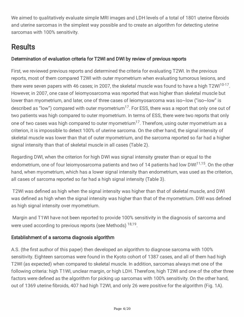
We aimed to qualitatively evaluate simple MRI images and LDH levels of a total of 1801 uterine �broidsand uterine sarcomas in the simplest way possible and to create an algorithm for detecting uterinesarcomas with 100% sensitivity.
ResultsDetermination of evaluation criteria for T2WI and DWI by review of previous reports
First, we reviewed previous reports and determined the criteria for evaluating T2WI. In the previousreports, most of them compared T2WI with outer myometrium when evaluating tumorous lesions, andthere were seven papers with 46 cases; in 2007, the skeletal muscle was found to have a high T2WI10-17.However, in 2007, one case of leiomyosarcoma was reported that was higher than skeletal muscle butlower than myometrium, and later, one of three cases of leiomyosarcoma was iso~low (“iso~low” isdescribed as “low”) compared with outer myometrium17. For ESS, there was a report that only one out oftwo patients was high compared to outer myometrium. In terms of ESS, there were two reports that onlyone of two cases was high compared to outer myometrium17. Therefore, using outer myometrium as acriterion, it is impossible to detect 100% of uterine sarcoma. On the other hand, the signal intensity ofskeletal muscle was lower than that of outer myometrium, and the sarcoma reported so far had a highersignal intensity than that of skeletal muscle in all cases (Table 2).
Regarding DWI, when the criterion for high DWI was signal intensity greater than or equal to theendometrium, one of four leiomyosarcoma patients and two of 14 patients had low DWI11,15. On the otherhand, when myometrium, which has a lower signal intensity than endometrium, was used as the criterion,all cases of sarcoma reported so far had a high signal intensity (Table 3).
T2WI was de�ned as high when the signal intensity was higher than that of skeletal muscle, and DWIwas de�ned as high when the signal intensity was higher than that of the myometrium. DWI was de�nedas high signal intensity over myometrium.
Margin and T1WI have not been reported to provide 100% sensitivity in the diagnosis of sarcoma andwere used according to previous reports (see Methods) 18,19.
Establishment of a sarcoma diagnosis algorithm
A.S. (the �rst author of this paper) then developed an algorithm to diagnose sarcoma with 100%sensitivity. Eighteen sarcomas were found in the Kyoto cohort of 1387 cases, and all of them had highT2WI (as expected) when compared to skeletal muscle. In addition, sarcomas always met one of thefollowing criteria: high T1WI, unclear margin, or high LDH. Therefore, high T2WI and one of the other threefactors were de�ned as the algorithm for picking up sarcomas with 100% sensitivity. On the other hand,out of 1369 uterine �broids, 407 had high T2WI, and only 26 were positive for the algorithm (Fig. 1A).

Page 5/20
Furthermore, when the Kindai cohort I was evaluated in the same way, among 293 cases, nine cases ofsarcoma were algorithm positive, and 29 cases of myoma were algorithm positive (Fig. 1B). In the Kyotocohort and Kindai cohort I, the overall percentage of algorithm-positive cases was 4.5%, including 2.9%for myoma and 1.6% for sarcoma (Fig. 1C). The sensitivity of the sarcoma diagnosis was 100%,speci�city 97%, positive predictive value 36%, and negative predictive value 100%.
Finally, Kindai cohort II was also analyzed. Of the 10 cases diagnosed as sarcoma by the pathologyreport, nine were algorithm positive, but one was algorithm negative. The pathology review of the onecase changed the diagnosis to myoma. On the other hand, of 102 randomly selected myomas, nine werealgorithm positive (Fig. 1D).
Analysis of algorithm positive cases
We identi�ed positive cases from all four cohorts (Kyoto cohort, Kindai cohort I and II) (n = 94). We foundthat (i) the group with positive results for all four algorithm factors had only leiomyosarcoma or high-grade ESS and no myoma. (ii) The group with positive results for the three imaging factors of thealgorithm had mostly low-grade ESS and only one myoma. (iii) The remaining group had more myomas(Fig. 2A). Prognostic analysis showed that the prognosis of group (i) was poor (p = 0.015). In addition,when we examined the 40 cases with available DWI images among the algorithm-positive cases,sarcomas were found to be high DWI in all cases (Fig. 2C).
Evaluation of inter-examiner agreement
Next, using the algorithm established by A.S. (reader a), we examined whether other readers coulddiagnose sarcoma as algorithm positive in a test set of 61 cases, including 14 cases of sarcoma [Fig. 3,Table 4, S1-61 (Age, Pathology, Final results of T2WI, T1WI, DWI, and margin discussed by �ve doctors,LHD value)]. As a result, readers b, c, and d diagnosed sarcoma as 100% high in both T2WI and DWI,while the match rate with reader a for T1WI and margin was only 43–86%. For readers b, c, and d, thepositive rate was 93% (13/14), which was the same even when low DWI was excluded. In contrast,readers b, c, and d in leiomyoma had lower concordance rates with reader a than sarcoma, ranging from53% to 62% for T2WI and 62% to 83% for DWI. Reader e had a low algorithm positivity rate of 71%(10/14) for sarcoma but a high concordance rate of 93% (13/14) with the original diagnostic report.
The sarcoma for which readers b, c, d, and e failed to show a positive algorithm was diagnosed assarcoma by preoperative biopsy because of a vaginal bleeding tumor at the time of initial diagnosis. Inaddition, the sarcoma for which readers c and d failed to show a positive algorithm had an LDH level of217 IU/L (normal value ≤ 222 IU/L) at the time of MRI scan but rose to 268 IU/L before surgery andeventually showed a positive algorithm. Therefore, when combined with the clinical information, theresults of readers b, c, and d were su�cient to suspect sarcoma preoperatively in all sarcoma cases.Reader e showed that if “high T2WI or high DWI” was considered as “high T2WI”, the preoperativeincrease in LDH level would have resulted in a positive algorithm in all cases. If “high T2WI or high DWI”is considered to be “high T2WI”, all patients would have been positive for the algorithm due to increased

Page 6/20
preoperative LDH levels. In actual clinical practice, in two of the three cases of myoma in the readingreport, the attending physician judged the mass to be myoma, and over the next few months, the massgrew and was subsequently diagnosed as sarcoma.
Treatment strategies for intramyometrial masses using sarcoma diagnostic algorithms
Finally, based on the above data and taking into account the reproducibility of the diagnosis, we havepresented our ideas on how to use sarcoma diagnostic algorithms to safely treat intramuscular masses(Fig. 4).
DiscussionTo establish a reproducible evaluation method for MRI, it is essential to make the evaluation criteriasimple and clear. In many papers, the criterion for evaluating T2WI to diagnose uterine sarcoma has beenwhether or not the signal intensity is higher than that of the outer myometrium (Table 2). However, whenthe signal intensity of T2WI is compared with that of the outer myometrium, it is often low in sarcoma17.Furthermore, we recently found that most of the low-grade ESS are low T2WI when compared to outermyometrium (data not shown, paper in submission). In addition, when the criterion for DWI isendometrium, sarcoma that is often lower than that has been reported13,15. Similarly, in the present study,we found sarcomas with low T2WI compared to outer myometrium and low DWI compared to theendometrium (S 1-61). These criteria have been used in previous reports because many studies have triedto clarify the difference between degenerated leiomyoma and sarcoma, which have relatively high T2WIand DWI signal intensity, rather than aiming for 100% sensitivity in sarcoma detection. The T2WI signalintensity of the myometrium varies greatly with the menstrual cycle, while that of skeletal muscle isconstant and lower than that of the myometrium8,20. Therefore, we used skeletal muscle as the referencefor T2WI and myometrium as the reference for DWI and were able to detect sarcoma with 100%sensitivity in four patients, except for reader e (Fig. 3). Considering the risk of parasitic myoma, a powermorcellator may be acceptable while using a containment system1–4.
In the present study, there were no cases of sarcoma in the reader group when both T2WI and DWI werehigh, but T1WI was low, the margin was clear, and LDH was low; however, in the reader b, c, and d groups,there was one case of sarcoma in each group (Fig. 3). These “mixed” sarcoma cases were cases thatcould have been diagnosed with suspected sarcoma preoperatively due to the clinical course, but it wouldbe dangerous to assume that sarcoma is not included in this category at this time. Therefore, minimallyinvasive surgery (MIS) using a power morcellator should be avoided as much as possible afterdiscussing this with the patient. If it is done, it should be done carefully using a containment system. Andif both T2WI and DWI are high, with either high T1WI, unclear margin, or high LDH, sarcoma should besuspected as algorithm positive (Fig. 1). In such cases, guidelines and MIS should not be performed as a“case of suspected malignancy” 1–4. In particular, if all three imaging �ndings are positive, the possibilityof sarcoma is extremely high, and if LDH is high, the prognosis may be poor (Fig. 2). Consistent with ourresults, it has been reported that elevated LDH expression in uterine sarcoma correlates with poor

Page 7/20
prognosis23. Thus, our simple algorithm can be reproduced even by non-specialists in gynecological MRIimaging and is directly relevant to medical treatment policy (Fig. 4).
The limitations of this study are: (i) it is a retrospective study, (ii) the sample size is relatively small toverify the high sensitivity of detection, as there were only 36 cases of sarcoma in the cohort, while therewere 1801 cases in the entire cohort, and (iii) the data are heterogeneous, as we used images from anolder era. (iv) Smooth muscle tumour of uncertain malignant potential was excluded, and (v) thesensitivity of the algorithm for sarcoma detection was not 100% in the study of interobserver agreement.In the present study, even when limited to sarcoma, the rate of agreement in evaluating individual factorswas low, especially for margin (Table 4). One of the reasons for this result was that the criteria werecommunicated to the narrative and evaluated based on it instead of using actual MRI images in thetraining set. In Japan, the number of MRIs per capita is much larger than in other countries, and almostall cases of surgery for the diagnosis of uterine �broids or uterine sarcoma are performed preoperativelywith MRI without contrast22.
In conclusion, we have proposed the �rst diagnostic algorithm to identify sarcoma close to 100%sensitivity. Once this algorithm is established and commonly used, it is expected to minimize the risk oflatent uterine sarcoma and allow safe treatment, including MIS.
Materials And MethodsStudy population
In this study, we enrolled patients who underwent surgery for tumors larger than 3 cm in the myometriumand underwent MRI before treatment and were pathologically diagnosed as leiomyosarcoma, low or high-grade endometrial stromal sarcoma (ESS), or leiomyoma. The Kyoto cohort was conducted at Kyotouniversity hospital from January 1986 to March 2005. The Kyoto cohort consisted of sarcoma (n = 18)and leiomyoma (n = 1369) treated at Kyoto university hospital between January 1986 and March 2005.The leiomyoma group included one case of postoperative parasitic leiomyoma. In the Kyoto cohort andKindai cohort I, all patients who ful�lled the criteria for leiomyoma and sarcoma were included in thestudy. Ten patients who were treated at Kindai university hospital from January 2014 to December 2018and diagnosed with sarcoma (1 case later became leiomyoma as described in Results) were included inthe study, including one case that later became a leiomyoma) and randomly selected leiomyomas of thesame period (n = 102).
This study was conducted under the auspices of Kindai University (approval number R02-036) and KyotoUniversity (approval number G288). All research was performed in accordance with Ethical Guidelines forMedical and Health Research Involving Human Subjects. All study subjects provided informed consentand assent in principle, but those who could not be contacted were given the opportunity to refuse toparticipate in the study by opting out. Those who refused to give consent were excluded from the study.
MRI protocol

Page 8/20
Kyoto University
Magnetic resonance imaging scanning was conducted using a 1.5-T MR imaging system (Signa; GEMedical Systems, Milwaukee, USA. or Symphony; Siemens Healthineers, Erlangen, Germany) equippedwith a phased-array coil. Routine clinical sequences included axial and sagittal T2WI (Fast Spin Echo)and sagittal T1WI (spin echo). Detailed parameters are shown in Table 1. Before MRI examination, 20 mgof butyl scopolamine (Buscopan; Nippon Boehringer Ingelheim, Tokyo, Japan) was administeredintramuscularly to reduce bowel motion, unless contraindicated.
Kindai University
Magnetic resonance imaging scanning was conducted using 1.5-T MR imaging systems (Signa HD xt; GEHealthcare, Milwaukee, USA, or Intera Achieva, Philips Healthcare, Best, the Netherlands) equipped withphased array coils. Routine clinical sequences included T1-weighted spin-echo (SE) or fast SE (FSE)images in axial or sagittal planes and T2-weighted FSE images in axial and sagittal planes. Detailedparameters are shown in Table 1.
Review of the literature to determine the evaluation criteria for T2WI or DWI
We searched PubMed for “uterine sarcoma MRI” and identi�ed 384 articles as of February 2021. Amongthese articles, 13 original papers that evaluated uterine leiomyosarcoma or ESS by specifying what theycompared to in T2WI or DWI were included in the analysis.
The setting of evaluation criteria
On T2WI, the lesions were classi�ed according to the Oguchi type classi�cation, which we proposedpreviously23. In T2WI, tumorous lesions other than degenerated areas were evaluated, and if the signalintensity was clearly higher than that of skeletal muscle, the case was classi�ed as high T2WI, and if itwas lower than that of skeletal muscle, as low T2WI. Tumors with high T2WI are consistent with tumorsclassi�ed as type 4 or 5 in the Oguchi type classi�cation, which we proposed previously23. On T1, thesignal intensity (SI) of skeletal muscle was set as the reference standard18. Therefore, ‘high SI’ wasde�ned as higher SI than skeletal muscle, and ‘low SI’ was similar to or lower SI than skeletal muscle. OnDWI, the reference standard of SI was normal uterine outer myometrium and evaluated in b = 1000s/mm2. Then, ‘high SI’ was de�ned in case of similar to or higher than normal uterine outer myometrium.If SI was high because of hemorrhage, it was evaluated as ‘high SI’. Regarding tumor border evaluation, itwas de�ned as ‘clear’ when the tumor margin was de�nitely traced24. The LDH value was determined bywhether the value at the time of the MRI scan and the most recent value was higher than the standardinstitutional value. When multiple uterine �broids were found, the lesion larger than 3 cm and with highT2WI signal intensity was adopted.
Evaluation method of inter-examiner agreement
Page 9/20
Author A.S. (reader a), an obstetrician and gynecologist with more than 20 years of experience, who hasknowledge in this �eld and has written a review paper, evaluated the images of all study cases anddeveloped the diagnostic algorithm5. For the test set to evaluate the inter-examiner agreement, werandomly selected 31 leiomyomas with high T2WI (including 14 algorithm-positive cases) and 16leiomyomas with low T2WI (including 16 algorithm-positive cases), in addition to 14 sarcoma cases withavailable DWI images in the Kindai cohort I and II. A.S. informed the other examiners of the narrativemethod for T2WI, T1WI, and DWI.
Reader b was a diagnostic radiologist with more than 20 years of experience, specializing in gynecology.Reader c was a fellow in diagnostic radiology with less than 5 years of experience. Reader d was agynecologist with 18 years of experience. Reader e was an obstetrician with 15 years of experience.Reader a and d knew the clinical information. Reader b, c, and e were not involved in the diagnosis ortreatment of any of the cases in the Kindai cohort. Reader b, c, and e had all the clinical information,including pathology results, LDH levels, and age. All clinical information, including pathology results, LDHlevels, and age, were blinded.
Pathology review
In this cohort, a case of uterine sarcoma, which was thought to be incorrectly pathologically diagnosed,was reviewed by Dr. Sachiko Minamiguchi, a pathologist specializing in gynecologic oncology at adifferent institution from A.S. and who was not a co-author of this article. The pathology was reviewed byDr. Sachiko Minamiguchi, a pathologist specializing in gynecologic tumors who belongs to a differentinstitution from A.S. and is not a co-author of this paper.
Statistical analysis
Survival analysis was performed by log-rank test. Statistical analysis was performed using GraphpadPrism version 9.0, and p < 0.05 was considered statistically signi�cant.
DeclarationsAcknowledgement
Dr. Sachiko Minamiguchi of Kyoto University Department of Diagnostic Pathology performed thepathology review. Dr. Takefumi Hamakawa, Department of Radiology, Kindai University, evaluated theimages as a volunteer for the interobserver agreement. We thank them for their cooperation.
Author contributions
Ayako Suzuki: Concept and design, data collection, data analysis, article writing, approved the �nalversion.
Aki Kido: Data collection, data analysis, article writing, approved the �nal version.
Page 10/20
Mitsuru Matsuki: Data collection, data analysis, article writing, approved the �nal version.
Yasushi Kotani: Data collection, data analysis, article writing, approved the �nal version.
Kosuke Murakami: Data analysis, article writing, approved the �nal version.
Yukio Yamanishi: Data collection, data analysis, article writing, approved the �nal version.
Isao Numoto: Data analysis, article writing, approved the �nal version.
Hidekatsu Nakai: Data analysis, article writing, approved the �nal version.
Tomoyuki Otani: Data collection, data analysis, article writing, approved the �nal version.
Ikuo Konishi: Supervision, article writing, approved the �nal version.
Masaki Mandai: Supervision, article writing, approved the �nal version.
Noriomi Matsumura: Data analysis, article writing, approved the �nal version.
Declaration of Competing Interest
The authors report no con�ict of interest.
References1. Glaser, L. M., Friedman, J., Tsai, S., Chaudhari, A. & Milad, M. Laparoscopic myomectomy and
morcellation: A review of techniques, outcomes, and practice guidelines. Best Pract. Res. Clin. Obstet.Gynaecol, 46, 99–112 (2018).
2. Parker, W. H. et al. U.S. Food and Drug Administration's Guidance Regarding Morcellation ofLeiomyomas: Well-Intentioned, But Is It Harmful for Women? Obstet. Gynecol, 127, 18–22 (2016).
3. Halaska, M. J. et al. ESGO Council, European Society of Gynecological Oncology Statement onFibroid and Uterine Morcellation. Int. J. Gynecol. Cancer, 27, 189–192 (2017).
4. ACOG Committee Opinion No. 770: Uterine Morcellation for Presumed Leiomyomas. Obstet. Gynecol,133, e238–e248 (2019).
5. Suzuki, A. et al. Differential Diagnosis of Uterine Leiomyoma and Uterine Sarcoma using MagneticResonance Images: A Literature Review. Healthcare, 7, 158 (2019).
�. Bi, Q. et al. Utility of Clinical Parameters and Multiparametric MRI as Predictive Factors forDifferentiating Uterine Sarcoma From Atypical Leiomyoma. Acad. Radiol, 25, 993–1002 (2018).
7. Malek, M. et al. Investigating the diagnostic value of quantitative parameters based on T2-weightedand contrast-enhanced MRI with psoas muscle and outer myometrium as internal references fordifferentiating uterine sarcomas from leiomyomas at 3T MRI. Cancer Imaging, 19, 20 (2019).
Page 11/20
�. Nakagawa, M. et al. Machine Learning to Differentiate T2-Weighted Hyperintense UterineLeiomyomas from Uterine Sarcomas by Utilizing Multiparametric Magnetic Resonance QuantitativeImaging Features. Acad. Radiol, 26, 1390–1399 (2019).
9. Glorie, N., Baert, T., Bosch, V. A. N. D. E. N., Coosemans, A. N. & T. & Circulating Protein Biomarkers toDifferentiate Uterine Sarcomas from Leiomyomas. Anticancer Res, 39, 3981–3989 (2019).
10. Tanaka, Y. O., Nishida, M., Tsunoda, H., Okamoto, Y. & Yoshikawa, H. Smooth muscle tumors ofuncertain malignant potential and leiomyosarcomas of the uterus: MR �ndings. J. Magn. Reson.Imaging, 20, 998–1007 (2004).
11. Tamai, K. et al. The utility of diffusion-weighted MR imaging for differentiating uterine sarcomasfrom benign leiomyomas. Eur. Radiol, 18, 723–730 (2008).
12. Namimoto, T. et al. Combined use of T2-weighted and diffusion-weighted 3-T MR imaging fordifferentiating uterine sarcomas from benign leiomyomas. Eur. Radiol, 19, 2756–2764 (2009).
13. Tasaki, A. et al. Differential diagnosis of uterine smooth muscle tumors using diffusion-weightedimaging: correlations with the apparent diffusion coe�cient and cell density. Abdom. Imaging, 40,1742–1752 (2015).
14. Lin, G. et al. Comparison of the diagnostic accuracy of contrast-enhanced MRI and diffusion-weighted MRI in the differentiation between uterine leiomyosarcoma / smooth muscle tumor withuncertain malignant potential and benign leiomyoma. J. Magn. Reson. Imaging, 43, 333–342 (2016).
15. Li, H. M. et al. Diffusion-Weighted Imaging for Differentiating Uterine Leiomyosarcoma FromDegenerated Leiomyoma. J. Comput. Assist Tomogr, 41, 599–606 (2017).
1�. Takeuchi, M., Matsuzaki, K. & Harada, M. Clinical utility of susceptibility-weighted MR sequence forthe evaluation of uterine sarcomas. Clin. Imaging, 53, 143–150 (2019).
17. Lakhman, Y. et al. Differentiation of Uterine Leiomyosarcoma from Atypical Leiomyoma: DiagnosticAccuracy of Qualitative MR Imaging Features and Feasibility of Texture Analysis. Eur. Radiol, 27,2903–2915 (2017).
1�. Ando, T. et al. Uterine smooth muscle tumours with hyperintense area on T1 weighted images:differentiation between leiomyosarcomas and leiomyomas. Br. J. Radiol, 91, 20170767 (2018).
19. Cornfeld, D. et al. MRI appearance of mesenchymal tumors of the uterus. Eur. J. Radiol, 74, 241–249(2010).
20. Haynor, D. R. et al. Changing appearance of the normal uterus during the menstrual cycle: MRstudies. Radiology, 161, 459–462 (1986).
21. Song, K. J. et al. Expression and prognostic value of lactate dehydrogenase-A and -D subunits inhuman uterine myoma and uterine sarcoma. Med. (Baltim), 97, e0268 (2018).
22. Yamashita, Y. et al. The essence of the Japan Radiological Society/Japanese College of RadiologyImaging Guideline. Jpn. J. Radiol, 34, 43–79 (2016).
23. Oguchi, O. et al. Prediction of histopathologic features and proliferative activity of uterine leiomyomaby magnetic resonance imaging prior to GnRH analogue therapy: correlation between T2-weighted

Page 12/20
images and effect of GnRH analogue. J. Obstet. Gynaecol, 21, 107–117 (1995).
24. Cornfeld, D. et al. MRI appearance of mesenchymal tumors of the uterus. Eur. J. Radiol, 74, 241–249(2010).
25. Fukunishi, H. et al. Unsuspected uterine leiomyosarcoma: magnetic resonance imaging �ndingsbefore and after focused ultrasound surgery. Int. J. Gynecol. Cancer, 17, 724–728 (2007).
2�. Thomassin-Naggara, I. et al. How to differentiate benign from malignant myometrial tumours usingMR imaging. Eur. Radiol, 23, 2306–2314 (2013).
27. Sato, K., Yuasa, N., Fujita, M. & Fukushima, Y. Clinical application of diffusion-weighted imaging forpreoperative differentiation between uterine leiomyoma and leiomyosarcoma. Am. J. Obstet Gynecol.210, 368.e1-368.e8 (2014)
2�. Sumi, A. et al. Assessment of MR Imaging as a Tool to Differentiate between the Major HistologicalTypes of Uterine Sarcomas. Magn. Reson. Med. Sci, 14, 295–304 (2015).
TablesDue to technical limitations, table 1 is only available as a download in the Supplemental Files section.

Page 13/20
Table 2The signal intensity of uterine sarcoma by T2WI.
comparison with histology high low year Reference
outer myometrium LMS 9 0 2004 Tanaka YO et al.10
0 1 2007 Lakhman Y et al.17
6 0 2008 Tamai K et al.11
3 1 2010 Cornfeld D et al.24
4 0 2009 Namimoto T et al.12
6 0 2015 Tasaki A et al.13
6 0 2016 Lin G et al.14
9 10 2017 Lakhman Y et al.17 (a)
11 8 2017 Lakhman Y et al.17 (b)
16 0 2017 Li HM et al.15
3 0 2019 Takeuchi M et al.16
ESS 1 1 2008 Tamai K et al.11
1 1 2010 Cornfeld D et al.24
2 0 2009 Namimoto T et al.12
1 0 2019 Takeuchi M et al.16
skeletal muscle LMS 1 0 2007 Fukunishi H et al.25
3 0 2013 Thomassin-Naggara I et al.26
ESS 11 0 2013 Thomassin-Naggara I et al.26

Page 14/20
Table 3The signal intensity of uterine sarcoma by DWI.
comparison with histology high low year Reference
endometrium LMS 4 1 2008 Tamai K et al.11
5 0 2014 Sato K et al.27
14 2 2017 Li HM et al.15
ESS 2 0 2008 Tamai K et al.11
myometrium LMS 6 0 2015 Tasaki A et al.13
6 0 2016 Lin G et al.14
3 0 2019 Takeuchi M et al.16
ESS 1 0 2019 Takeuchi M et al.16
skeletal muscle LMS 8 0 2015 Sumi A et al.28
ESS 6 0 2015 Sumi A et al.28
Due to technical limitations, table 4 is only available as a download in the Supplemental Files section.
Figures
Page 15/20
Figure 1
Differentiation of uterine sarcoma from uterine �broids by MRI images and LDH levels. A) Analysis of theKyoto cohort. All 18 sarcomas were T2 high, and the others were either T1 high, unclear margin, or highLDH (sarcoma diagnostic algorithm). Of the 1369 myomas, 407 were high T2. B) Kindai cohort I analysis.All nine sarcomas were algorithm positive, and 23 of 293 myomas were algorithm positive. c) Total of theKyoto cohort and Kindai cohort I. Of 1689 cases, 4.5% were algorithm positive. d) Analysis of Kindaicohort II. D) Analysis of Kindai cohort II; one case was originally diagnosed as sarcoma but was found tobe myoma by pathology review after algorithm negative. All other nine sarcomas were algorithm positive,and of 102 randomly selected uterine �broids, nine were algorithm positive. Black in the heatmapindicates T2; high, T1; high, margin; unclear, LDH; high. The type of histology is indicated by color.

Page 16/20
Figure 2
Analysis of algorithm-positive cases. A) Algorithm-positive cases in Kyoto cohort, Kindai cohort I, andKindai cohort II (n = 94). The cases were divided into three groups: (i) all four factors of the algorithmwere positive, (ii) three factors of the image were positive, and (iii) others. (ii) All but one were sarcomas;(iii) most were myomas, but some were sarcomas; B) Prognosis differed among the three groups, with (i)having the poorest prognosis; C) Forty of the algorithm-positive cases had available DWI, and DWI washigh for all sarcomas. All sarcomas were DWI high. Heatmap shows cases with DWI high in black.
Page 17/20
Figure 3
Veri�cation of reproducibility of image evaluation. We selected sarcoma cases with available DWI fromKindai cohort I and II and randomly selected algorithm positive myoma, T2 high only myoma, and T2 lowmyoma, respectively. Algorithm + DWI was determined by taking DWI into account. The algorithm + DWIwas determined by considering the DWI and a positive algorithm, but a low DWI was marked as negativein white. >50 y.o.; cases older than 50 years. The patient was followed up in the outpatient clinic forseveral months before surgery was performed. $; Before surgery, LDH level was elevated, and both readerc and d were algorithmically positive. #; Tumor protruded vaginally, and sarcoma could be diagnosedpreoperatively.

Page 18/20
Figure 4
Evaluation method and treatment algorithm for intramuscular uterine masses. Red; High T2WI, high DWI,high T1WI, unclear margin, high LDH; green indicates low; histology: white for leiomyoma, grey for s/osarcoma, black for sarcoma, For histology, white indicates leiomyoma, grey indicates s/o sarcoma, andblack indicates sarcoma. MIS; minimally invasive surgery. (i) Normally, a low T2 or DWI indicates nosarcoma, but when evaluated by a non-gynecologic MRI specialist, either or both may be positive.However, when evaluated by a non-gynecological MRI specialist, if either of them is positive, it is safe totreat it as a case where both are positive. (ii) If both T2WI and DWI are high and the other factors arenegative, the result will be negative for the algorithm. However, the reproducibility of that assessment isnot 100% and does not completely eliminate the suspicion of sarcoma. Morcellators should be avoided orat least used within a bag.
Supplementary Files

Page 19/20
This is a list of supplementary �les associated with this preprint. Click to download.
1.docx
2.docx
3.docx
4.docx
5.docx
6.docx
7.docx
8.docx
9.docx
10.docx
11.docx
12.docx
13.docx
14.docx
15.docx
16.docx
17.docx
18.docx
19.docx
20.docx
21.docx
22.docx
23.docx
24.docx
25.docx
26.docx
27.docx
28.docx
29.docx
30.docx
31.docx
32.docx
33.docx
Page 20/20
34.docx
35.docx
36.docx
37.docx
38.docx
39.docx
40.docx
41.docx
42.docx
43.docx
44.docx
45.docx
46.docx
47.docx
48.docx
49.docx
50.docx
51.docx
52.docx
53.docx
54.docx
55.docx
56.docx
57.docx
58.docx
59.docx
60.docx
61.docx
Table1.jpg
Table4.jpg