mRNAs for activin receptors II and IIB are expressed in mouse oocytes and in the epiblast of...
9
ELSEVIER Mechanisms of Development 49 (1995) 3- 11 mRNAs for activin receptors II and IIB are expressed in mouse oocytes and in the epiblast of pregastrula and gastrula stage mouse embryos Katia Manovaa”, Victor De Leon”, Michael Anfelesa, Sundeep Kalantrya, Marianna GiarrC”, Liliana Attisanob, Jeffrey Wrana , Rosemary F. Bachvarova*a aDepartment of Cell Biology and Anaiomy, Cornell University Medical College, 1300 York Avenue, New York, NY 10021, USA bCeN Biology and Genetics Program, Memorial Sloan-Kettering Cancer Center, 1275 York Avenue, New York, NY 10021. USA Received I I July 1994; accepted 15 August 1994 Abstract Activin is a potent inducer of mesoderm in frog embryos. We showed previously that in the mouse, activin /3A is expressed in the uterine decidua near the embryo before and during the first appearance of mesoderm (E4.5-E6.5). Here, using Northern blot- ting and in situ hybridization, we show that mouse oocytes, E6.5 and E7.5 embryos, and E6.5 and E7.5 decidua contain mRNAs for both activin receptors type II and IIB. The expression of activin receptor type IIB is particularly strong in embryonic ectoderm apparent at ES.5 and continuing through E8.5. These results support the hypothesis that activin derived from the decidua promotes development of mesoderm in the period E5.5-E6.5. Keyword: Mouse embryos; Activin receptor 1. Introduction In the amphibian embryo, mesoderm is induced in the equatorial zone by factors emanating from the vegetal region (see Smith, 1989). The TGF-P family member activin is a potent mesoderm inducer in the amphibian system. Animal caps from Xenopus blastulae treated with activin produce a variety of mesodermal tissues, including anterior and dorsal derivatives such as head structures and notochord (Thomsen et al., 1990) and express marker genes for these tissues (Green et al., 1992). Activin and activin-like activity are present in the frog egg (Asashima et al., 1991; Fukui et al., 1994) and is thus potentially present during mesoderm induction before gastrulation. Its mRNAs are highly expressed in follicle cells (Dohrmann et al., 1993; Rebagliati and * Corresponding author, Tel.: 212 746 6161; Fax: 212 746 8175. ’ Current address: Molecular Biology Program, Sloan-Kettering Institute, 1275 York Avenue, New York, NY 10021. USA. Dawid, 1993), suggesting that activin A or B in early em- bryos may be derived by uptake of protein into the oocyte from material secreted by surrounding follicle cells. At least two types of activin receptors are present in the early Xenopus embryo and at least one is distri- buted throughout the embryo (Kondo et al., 1991; Nishimatsu et al., 1992; Hemmati-Brivanlou et al., 1992). Injection of activin mRNA or activin receptor mRNA into early embryos is able to induce a partial second axis (Thomsen et al., 1990; Kondo et al., 1991; Mathews et al., 1992). Injection of mRNA encoding a truncated activin receptor, which is predicted to inhibit endogenous activin receptor activity in a dominant neg- ative fashion, blocks mesoderm formation (Hemmati- Brivanlou and Melton, 1992). However, the truncated receptor may block the action of a wide range of TGF- &like factors and thus a crucial role for activin itself has not yet been demonstrated. In chick embryos, the hypoblast plays a central role in induction of the primitive streak (Eyal-Giladi, 1984). Activin is expressed in the chick blastoderm and can in- 0925-4773/95/%09.50 0 1995 Elsevier Science Ireland Ltd. All rights reserved SSDI 0925-4773(94)00295-X
Transcript of mRNAs for activin receptors II and IIB are expressed in mouse oocytes and in the epiblast of...

ELSEVIER Mechanisms of Development 49 (1995) 3- 11
mRNAs for activin receptors II and IIB are expressed in mouse oocytes and in the epiblast of pregastrula and gastrula stage mouse
embryos
Katia Manovaa”, Victor De Leon”, Michael Anfelesa, Sundeep Kalantrya, Marianna GiarrC”, Liliana Attisanob, Jeffrey Wrana , Rosemary F. Bachvarova*a
aDepartment of Cell Biology and Anaiomy, Cornell University Medical College, 1300 York Avenue, New York, NY 10021, USA bCeN Biology and Genetics Program, Memorial Sloan-Kettering Cancer Center, 1275 York Avenue, New York, NY 10021. USA
Received I I July 1994; accepted 15 August 1994
Abstract
Activin is a potent inducer of mesoderm in frog embryos. We showed previously that in the mouse, activin /3A is expressed in the uterine decidua near the embryo before and during the first appearance of mesoderm (E4.5-E6.5). Here, using Northern blot- ting and in situ hybridization, we show that mouse oocytes, E6.5 and E7.5 embryos, and E6.5 and E7.5 decidua contain mRNAs for both activin receptors type II and IIB. The expression of activin receptor type IIB is particularly strong in embryonic ectoderm apparent at ES.5 and continuing through E8.5. These results support the hypothesis that activin derived from the decidua promotes development of mesoderm in the period E5.5-E6.5.
Keyword: Mouse embryos; Activin receptor
1. Introduction
In the amphibian embryo, mesoderm is induced in the equatorial zone by factors emanating from the vegetal region (see Smith, 1989). The TGF-P family member activin is a potent mesoderm inducer in the amphibian system. Animal caps from Xenopus blastulae treated with activin produce a variety of mesodermal tissues, including anterior and dorsal derivatives such as head structures and notochord (Thomsen et al., 1990) and express marker genes for these tissues (Green et al., 1992). Activin and activin-like activity are present in the frog egg (Asashima et al., 1991; Fukui et al., 1994) and is thus potentially present during mesoderm induction before gastrulation. Its mRNAs are highly expressed in follicle cells (Dohrmann et al., 1993; Rebagliati and
* Corresponding author, Tel.: 212 746 6161; Fax: 212 746 8175. ’ Current address: Molecular Biology Program, Sloan-Kettering
Institute, 1275 York Avenue, New York, NY 10021. USA.
Dawid, 1993), suggesting that activin A or B in early em- bryos may be derived by uptake of protein into the oocyte from material secreted by surrounding follicle cells. At least two types of activin receptors are present in the early Xenopus embryo and at least one is distri- buted throughout the embryo (Kondo et al., 1991; Nishimatsu et al., 1992; Hemmati-Brivanlou et al., 1992). Injection of activin mRNA or activin receptor mRNA into early embryos is able to induce a partial second axis (Thomsen et al., 1990; Kondo et al., 1991; Mathews et al., 1992). Injection of mRNA encoding a truncated activin receptor, which is predicted to inhibit endogenous activin receptor activity in a dominant neg- ative fashion, blocks mesoderm formation (Hemmati- Brivanlou and Melton, 1992). However, the truncated receptor may block the action of a wide range of TGF- &like factors and thus a crucial role for activin itself has not yet been demonstrated.
In chick embryos, the hypoblast plays a central role in induction of the primitive streak (Eyal-Giladi, 1984). Activin is expressed in the chick blastoderm and can in-
0925-4773/95/%09.50 0 1995 Elsevier Science Ireland Ltd. All rights reserved SSDI 0925-4773(94)00295-X

4 K. Manova et al. /Mechanisms of Development 49 (1995) 3-l I
duce axial structures in chick epiblast (Mitrani et al., 1990; Ziv et al., 1992).
In mammalian embryos, mesoderm first appears at the primitive streak on the posterior side of the egg cy- linder. The source of inducing factors is unknown, but one candidate is the visceral endoderm which may be in a broad sense homologous to the vegetal hemisphere of the frog. However, in mammals, activin PA mRNA is not detectably expressed in visceral endoderm but rather is highly expressed in some uterine decidual cells close to the embryo from embryonic day 4.5 (E4.5) through E6.5 (Manova et al., 1992; Albano et al., 1994) in- cluding the two days before and during appearance of the primitive streak at E6.5. Activin /3B mRNA is also present in the decidua at E5.5-E7.5 but is located around the ectoplacental cone further from the embryo- nic region (Albano et al., 1994). Interestingly, activin protein is present in the mouse egg and early embryo (Albano et al., 1993). The most likely source of this pro- tein is the follicle cells of the ovary in which activin mRNAs are very abundant (Meunier et al., 1988). As in the amphibian, activin may be secreted by follicle cells and taken up by the oocyte. In addition, mouse oocytes, morulae and blastocysts contain small amounts of mRNA detectable by PCR (Albano et al., 1993) that could be a source of some of the activin present in blastocysts.
Activin acting on whole E6.4 mouse embryos can in- duce expression of the organiser marker gene goosecoid throughout the embryonic ectoderm (Blum et al., 1992). Activin stimulates expression of the mesoderm marker Brachyury (Vidricaire et al., 1994) and inhibits long term differentiation (van den Eijnden-van Raaij et al., 1991) in P19 EC cells.
If activin is playing a role in the induction and forma- tion of mesoderm in the mouse embryo, the expected target tissue is the embryonic ectoderm which forms the primitive streak and contains premesoderm cells. Ac- tivin interacts with two classes of membrane receptors, type I and type II receptors, distinguishable by their ligand-binding properties (Mathews and Vale, 1991; Attisano et al., 1992, 1993). Both receptors are trans- membrane protein serine/threonine kinases which together form a heteromeric complex essential for signal transduction (Attisano et al., 1993; ten Dijke et al., 1993, 1994; Carcamo et al., 1994). In order to pursue the potential role of activin in the mouse embryo, we have characterized the expression patterns of the mRNAs for the two forms of type II receptors, activin receptors II and IIB (Mathews and Vale 1991; Attisano et al., 1992). We find expression of both receptors within the embryo consistent with a role for activin before and during gastrulation. In addition, we demonstrate expression of the receptors on ovarian oocytes, which may mediate uptake of activin into the oocytes.
2. Results
2.1. Activin receptors II and IIB are expressed in ovaries. oocytes and testes
Northern blots of total RNA from ovaries were prepared and probed with cDNA sequences for the two activin receptors. As expected (Feng et al., 1993) mRNAs of approximately 3 and 6 kb were found for activin receptor II (Fig. 1, lane 1). The molecular weights of activin IIB mRNAs have not been observed accurately before; these were found to be approximately 4 and 7-8 kb (the upper band is a doublet ) (Fig. 1, lane 1). The concentration of mRNAs for both receptors was similar in RNA from 10 day to adult ovaries (not shown). As published by others (Kaipia et al., 1992,
Fig. I. Northern blots of RNA hybridized to the activin receptor II
probe (upper panel), washed and rehybridized to the activin receptor
IIB probe (lower panel). Lanes are taken from four different blots. The
positions of 28s and 18s RNAs are indicated by arrows. The positions
of the activin receptor RNAs are indicated by dots. Lane I. IO pg of
RNA from I month ovary. Lane 2. IO pg of RNA from 3 months tes-
tis. Lane 3. RNA from about 1000 full-grown oocytes. Lane 4, RNA
from approximately 1000 superovulated eggs (this lane was distorted
by a bubble). No ethidium stain of rRNA was visible in lanes 3 and
4. Lane 5. RNA from 50 E6.5 embryos. Lane 6, about 7 yg of RNA
from E7.5 embryos, Lane 7, 15 pg of RNA from ER.Sextraembryonic
portions. Lane 8, I5 pg RNA from E8.5embryonic portions. Lane 9.
10 pg E6.5-decidual RNA (embryos removed). Lane 10. IO pg E7.5-
decidual RNA (embryos removed).

K. Manova et al. /Mechanisms of Development 49 (1995) 3-11 5
Fig. 2. High magnification views of sections of prepubertal ovary hybridized to activin receptor probes. (A) Activin receptor II probe. One large pale labeled oocyte is visible within a multi-layered follicle. Grains appear as black dots. (B) Activin receptor IIB probe. One 2-3 layered and one small antral follicle contain labeled oocytes. Scale bar = 20 am.
1993; de Winter et al., 1992), these mRNAs are also found in testes (Fig. 1, lane 2).
RNAs isolated from full-grown oocytes and ovulated eggs were also run on Northern blots and all samples contained detectable amounts of both receptor mRNAs (Fig. 1, lanes 3 and 4). Growing oocytes also expressed mRNAs for both receptors (data not shown).
In situ hybridization on ovary sections using the probe for activin receptor II revealed a higher grain den- sity over the oocytes (Fig. 2A) with quite homogeneous labeling over the somatic tissue. Since the Northern blots indicate higher expression in ovaries than can be accounted for by expression only in oocytes, we inter- pret these results as a significant level of expression in most cells. Similar results were obtained with the probe for activin receptor IIB, except that the density of label was lower over theta and interstitial cells than over follicle cells (Fig. 2B).
In situ hybridization on sections of testis showed ex- pression of activin receptor II in late pachytene sper- matocytes and early spermatids as already reported (Kaipia et al., 1992) (Fig. 3A). Activin receptor IIB mRNA was found in type A spermatogonia and in Sertoli cells, most clearly seen in prepubertal testes (Fig. 3C) (similar to previously reported results (Kaipia et al., 1993)).
2.2. Activin receptors II and IIB are expressed in embryos and uterine decidua around E6.5
Northern blotting of RNA extracted from isolated E6.5 and E7.5 embryos revealed mRNAs for both ac- tivin receptors II and IIB (Fig. 1, lanes 5 and 6); at E8.5, both RNAs were present in extraembryonic and em- bryonic regions (Fig. 1, lanes 7 and 8); both were also present in decidual RNA from 6.5 and 7.5-day pregnant
Fig. 3. In situ hybridization of sectioned tissue to activin receptor pro- bes (A) Dark field view of testis from a 27-day-old mouse. hybridized to an activin receptor II probe. (C) Dark field view of testis from a l3- day-old mouse hybridized to the activin receptor IIB probe. (B and D) Bright and dark field images of an approximately sagittal section of an E6.5 embryo hybridized to the activin receptor II full length probe. Arrows indicate the embryonic region. Scale bars = 100 pm.

6 K. Manova er al. /Mechanisms of Development 49 (1995) 3- 1 I
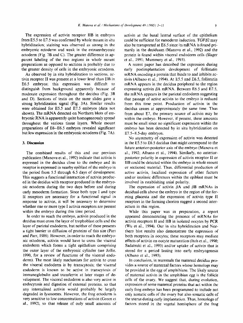
Fig. 4. Expression of activin receptor IIB in early postimplantation embryos. Sections of embryos subjected to in situ hybridization with the activin
receptor IIB probe. (A-C) Sections of E5.5. E6.5 and E7.5 embryos, respectively: (D-F) corresponding dark tield views. (A and D) Embryonic
and extraembryonic ectoderm are labeled, including mural trophoblast (small arrow). Scale bar = 50 pm. (B and E) Tangential section through
the quite strongly labeled embryonic ectoderm (arrows) showing the layer of unlabeled visceral endoderm around the upper two thirds of the egg
cylinder (arrow head). The bright areas below are due to a blood artifact. Scale bar = 100 pm. (C and F) Frontal section showing highly labeled
embryonic ectoderm and mesoderm. moderately labeled extraembryonic ectoderm and the external layer of unlabeled visceral endoderm
(arrowhead). Scale bar = 100 pm. a. amnion; c, chorion.
mice (Fig. 1, lanes 9 and 10). Comparison of the intensi- ty of the receptor mRNA bands with the reaction in ovarian RNA on the same blots revealed more activin IIB receptor RNA in embryos relative to ovary, and more activin receptor II in decidua relative to ovary.
In situ hybridization to both receptor probes was car- ried out on embryos sectioned within the uterine decidua. Stronger expression was observed in
pregastrula and gastrulating embryos using the probe for activin receptor IIB; these results are presented first. At E4.5, significant labeling was observed in the inner cell mass and trophectoderm of one of two embryos
examined (not shown). At E5.5, labeling with the same probe was quite high over all ectodermal tissues: em- bryonic and extraembryonic ectoderm, ectoplacental cone and early developing giant trophoblast cells (Fig.

Manova ei al. /Mechanisms qf Development 49 (1995) 3-11
Fig. 5. Expression of activin receptor IIB in the E8.5 embryo. Bright and corresponding dark field images are shown in each case. (A and B) Section
of head showing thick neuroepithelium on the surface. mesenchyme and foregut tube in the center. All embryonic tissues are labeled. (C and D)
Section of the wall of the implantation site showing labeled giant trophoblast cells (arrows) lining the inner surface of the decidua. (E and F) Section
of the chorion (arrows). Scale bar = 200 +m.
Fig. 6. Expression of activin receptor IIB in the E13.5 embryo. (A and B) Bright and corresponding dark field image of a cross section of the trunk
of an E13.5 embryo. sp co. spinal cord: k, kidney. (C and D) Bright lield and corresponding dark field images of another region of the same section. i, cross sections of intestine. Scale bar in C = 200 pm for all panels.

8 K. Manova et al. /Mechanisms of Development 49 (1995) 3-l I
4A and D). At E6.5, expression continued over most ectoderm derived tissues; it was relatively high in the embryonic ectoderm, moderate in the extraembryonic ectoderm and high over the ectoplacental cone (Fig. 4B and E). Labeling was clearly absent in the visceral en- doderm as well as in giant trophoblast cells. At E7.5, the grain density was high over embryonic ectoderm, in- cluding the neural plate, and over embryonic mesoderm, quite high over the allantois and moderate over the am- nion, chorion and the ectoplacental cone (Fig. 4C and
F). The moderate to low grain density seen in the decidua together with the Northern blot results indicate that there is widespread but low expression in this tissue. At E8.5, expression was strong in nervous tissue and mesoderm (Fig. 5A and B); the chorion of the forming placenta and nearby secondary giant trophoblast cells were also labeled (Fig. 5C-F). Embryos at E13.5 were examined and distinct labeling was observed in a num- ber of tissues, including the brain and spinal cord, kid- ney tubules and intestinal epithelium (Fig. 6A-D).
Fig. 7. Whole mount in situ hybridization. (A) E6-E6.5 embryos hybridized to the activin receptor II probe. Staining appears intense in the embryo-
nic ectoderm and low in the extraembryonic ectoderm. (B) E6-E6.5 embryos hybridized to the activin receptor IIB probe. Staining appears more
intense in the embryonic ectoderm and less in the extraembryonic ectoderm. (C) E7.5 embryos hybridized to the activin receptor IlB probe. Staining is seen primarily in the embryonic ectoderm and mesoderm. Sense probes showed little stain. Scale bar in C = 250 Frn for panel C and 125 pm
for panels A and B.
K. Manova et al. /Mechanisms of Development 49 ( 1995) 3-11 9
The expression of activin receptor IIB in embryos from E5.5 to E7.5 was confirmed by whole mount in situ hybridization; staining was observed as strong in the embryonic ectoderm and weak in the extraembryonic ectoderm (Fig. 7B and C). The greater difference in ap- parent labeling of the two regions in whole mount preparations as opposed to sections is probably due to the greater density of cells in the embryonic ectoderm.
As observed by in situ hybridization to sections, ac- tivin receptor II was present at a lower level than IIB in E6.5 embryos; this expression was difficult to distinguish from background apparently because of moderate expression throughout the decidua (Fig. 3B and D). Sections of testis on the same slides gave a strong hybridization signal (Fig. 3A). Similar results were obtained for E5.5 and E7.5 embryos (data not shown). The mRNA detected on Northern blots of em- bryonic RNA is apparently quite homogeneously spread throughout the various tissue layers. Whole mount preparations of E6-E6.5 embryos revealed significant but low expression in the embryonic ectoderm (Fig. 7A).
3. Discussion
The combined results of this and our previous publication (Manova et al., 1992) indicate that activin is expressed in the decidua close to the embryo and its receptor is expressed in nearby epiblast of the embryo in the period from 5.5 through 6.5 days of development. This suggests a functional interaction of activin produc- ed in the decidua with receptor produced in the embryo- nic ectoderm during the two days before and during early mesoderm formation. Since both type I and type II receptors are necessary for a functional signal in response to activin, it will be necessary to determine whether one or more type I activin receptors are present within the embryo during this time period.
In order to reach the embryo, activin produced in the decidua must cross the layer of trophoblast cells and the layer of parietal endoderm, but neither of these presents a tight barrier to diffusion of proteins of this size (Parr and Parr, 1986). However, in order to reach the embryo- nic ectoderm, activin would have to cross the visceral endoderm which forms a tight epithelium comprising the outer layer of the embryonic cylinder (see Jollie. 1990, for a review of functions of the visceral endo- derm). The most likely mechanism for activin to cross the visceral endoderm is by transcytosis; the visceral endoderm is known to be active in transcytosis of immunoglobulin and transferrin at later stages of de- velopment. The visceral endoderm is also very active in endocytosis and digestion of external proteins, so that any internalized activin would probably be largely degraded in lysosomes. However, the frog ectoderm is very sensitive to low concentrations of activin (Green et al., 1992) so that release of only small amounts of
activin at the basal lateral surface of the epithelium could be sufficient for mesoderm induction. TGFP2 may also be transported at E6.5 since its mRNA is found pri- marily in the deciduum (Manova et al., 1992) and the protein is found within visceral endoderm cells (Slager et al., 1991; Mummery et al., 1993).
A recent paper has described the expression during early postimplantation development of follistatin mRNA encoding a protein that binds to and inhibits ac- tivin (Albano et al., 1994). At E5.5 and E6.5, follistatin mRNA appears in the decidua peripheral to the region expressing activin @A mRNA. Between E6.5 and E7.5, the mRNA appears in the parietal endoderm suggesting that passage of active activin to the embryo is reduced from this time point. Production of activin in the decidua ceases at approximately the same time. Thus from about E7, the primary source of activin may be within the embryo. However, if present, these amounts must be small since no significant expression within the embryo has been detected by in situ hybridization on E7.5-8.5day embryos.
No asymmetry of expression of activin was detected in the E5.5 to E6.5 decidua that might correspond to the future anterior-posterior axis of the embryo (Manova et al., 1992; Albano et al., 1994). Similarly, no anterior- posterior polarity in expression of activin receptor II or IIB could be detected within the embryo in whole mount or sectioned material. Thus, differential production of active activin, localized expression of other factors and/or intrinsic differences within the epiblast must be involved in establishing axial polarity.
The expression of activin @A and /3B mRNAs in decidual cells above the embryo in the region of the for- ming placenta and the expression of activin type II receptors in the forming chorion suggest a second inter- action in this region.
While this paper was in preparation, a report appeared demonstrating the presence of mRNAs for activin receptors II and IIB in isolated oocytes by PCR (Wu et al., 1994). Our in situ hybridization and Nor- thern blot results also demonstrate the expression of both receptors in oocytes; these receptors may mediate effects of activin on oocyte maturation (Itoh et al., 1990; Sadatsuki et al., 1993) and/or uptake of activin that is stored for a period lasting into early embryogenesis (Albano et al., 1993).
In conclusion, in mammals the maternal decidua pro- vides a source of maternal factors whose homologs may be provided in the egg of amphibians. The likely source of maternal activin in the amphibian egg is the follicle cells of the ovary. We suggest that, during evolution, expression of some maternal proteins that act within the early frog embryo has been programmed to include not only somatic cells of the ovary but also somatic cells of the uterus during early implantation. Thus, homologs of factors stored in the vegetal hemisphere of the frog

10 K. Manova et al. /Mechanisms of Development 49 (1995) 3-11
embryo may be transmitted from the uterus through the visceral endoderm of the mammalian embryo.
4. Experimental procedures
Mice used in these studies were derived from mating ICR females to CB6Fi males. Embryos were obtained from timed pregnancies and age estimated by morpholo- gy to the nearest half day
A cDNA plasmid containing a 2.6-kb insert encoding the entire mouse activin receptor II coding sequence was kindly provided by W. Vale (Mathews and Vale. 1991). An alternative 616-nucleotide probe was prepared from a plasmid with an insert spanning the end of the coding sequence (nucleotides 1209-1824 of the published se- quence (Mathews and Vale 1991)); this was obtained by RT-PCR from testis RNA. Similar results were obtain- ed with both probes. Activin receptor IIB was cloned as described (Attisano et al., 1992) and a plasmid with a 1700N insert including the whole coding sequence was used to prepare probes. A probe from an insert lacking the kinase domain gave similar results.
Oocytes and eggs were collected as described (Manova et al., 1990). RNA was prepared according to Chomczynski and Sacchi (1987). Northern blotting was carried out as described (Manova et al., 1990) with 1 pg ethidium added to each sample. Blots were hybridized at 55-60°C for the activin receptor II probes and at 60-65°C for the activin receptor IIB probes.
In situ hybridization to sections was carried out as described (Manova et al., 1992) using [33P]-UTP rather than [35S]-UTP to label the probes in most experi- ments, and a temperature of 55-60°C for hybridization of activin receptor II and 60-65” for activin receptor IIB. Whole mount in situ hybridization for Fig. 7A and B was carried out essentially according to Wilkinson (1992) except antibody treatment and staining were according to Conlon and Rossant (1992). For Fig. 7C, prehybridization and hybridization treatments were according to Conlon and Rossant (1992) and post- hybridization treatment according to Frank and Harland (1991).
Acknowledgments
We thank W. Vale for the gift of a 2.6-kb cDNA probe for activin receptor II. This research was sup-
ported by NIH grant HD 06910.
References
Albano. R.M.. Groome. N. and Smith J.C. (1993) Development 117.
71 l-723.
Albano. R.M.. Arkell. R., Beddington. R.S.P. and Smith. J.C. (1994)
Development 120. 803-8 13. Asashima, M.. Nakano. H.. Uchiyama. H., Sugino. H.. Nakamura. T..
Eto, Y . Ejima. D.. Nishimatsu, S.. Ueno, N. and Kinoshita, K.
(1991) Proc. Nat. Acad. Sci. USA 88. 6511-6514.
Attisano. L., Wrana. J.L.. Cheifetz, S. and Massague, J. (1992) Cell 68. 97-108.
Attisano. L., Carcamo, J., Ventura, F.. Weis, F.M.B.. Massague. J.
and Wrana. J.L. (1993) Cell 75. 671-680. Blum, M.. Gaunt. S.J., Cho. K.W.Y., Steinbeisser. H., Blumberg, B.,
Bittner, D. and De Robertis. E.M. (1992) Gastrulation in the
mouse: The role of the homeobox gene goosecoid. Cell 69.
1097-l 106.
Carcamo, J., Weis. F.M.B., Ventura, F.. Wieser. R.. Wrana. J.. At-
tisano. L. and Massague. J. (1994) Type I receptors specify growth-
inhibitory and transcriptional responses to transforming growth
factor 0 and activin. Mol. Cell. Biol. 14. 3810-3821.
Chomczynski, P. and Sacchi, V. (1987) Single-step method of RNA
isolation by acid guanidinium thiocyanate-phenol-chloroform ex-
traction Anal. Biochem. 162. 156-159.
Conlon, R.A. and Rossant, J. (1992) Exogenous retinoic acid rapidly
induces anterior ectopic expression of murine HOX-2 genes in vivo.
Development 116, 357-368
de Winter, J.P.. Themmen. A.P.N., Hoogerbrugge. J.W.. Kiaij. I.A.,
Grootegoed, J.A. and de Jong. F.H. (1992) Activin receptor
mRNA expression in rat testicular cell types. Mol. Cell Endocrin.
83. l-8.
Dohrmann. C.E.. Hemmati-Brivanlou, A., Thomsen, G.H.. Fields. A..
Woolf. T.M. and Melton. D.A. (1993) Dev. Biol. 157, 474-483.
Eyal-Giladi. H. (1984) Cell Differ. 14, 245-255.
Feng. Z.M.. Mad&an. M.B. and Chen, C.L. ( 1993) Endocrinol. 132,
2592-2600.
Frank, D. and Harland, R.M. (1991) Development 113. 1387-1393.
Fukui. A., Nakamura, T., Uchiyama. H.. Sugino, K., Sugino, H. and Asashima, M. (1994) Dev. Biol. 163, 279-281.
Green, J.B.A.. New, H.V. and Smith. J.C. (1992) Cell 71. 731-740.
Hemmati-Brivanlou, A. and Melton, D.A. (1992) Nature 359,609-614.
Hemmati-Brivanlou. A., Wright, D.A. and Melton. D.A. (1992) Dev.
Dyn. 194. I-11.
Itoh. M.. Igarashi. M.. Yamada. K., Hasegawa, Y., Seki. M.. Eto, Y.
and Shibai. H. (1990) Biochem. Biophys. Res. Commun. 166,
I479- 1484.
Jollie. W.P. (1990) Teratol. 41. 361-381.
Kaipia. A., Pentila, T.-L., Shimasaki. S.. Ling, N.. Parvinen. M. and
Toppari. J. ( 1992) Endocrinol. 13 I. 2703-2710.
Kaipia. A., Parvinen. M. and Toppari. J. (1993) Endocrinol. 132.
477-479.
Kondo, M., Tashiro, K.. Fujii. G., Asano. M.. Miyoshi, R.. Yamada.
R.. Muramatsu. M. and Shiokawa. K. (1991) Biochem. Biophys.
Res. Commun. 181. 684-690.
Manova. K.. Nocka. K., Besmer. P. and Bachvarova, R.F. (1990) De-
velopment 110. 1057-1069.
Manova. K.. Paynton. B.V.. and Bachvarova, R.F. (1992) Mech. Dev.
36. 141-152.
Mathews, L.S. and Vale, W.W. (1991) Cell 65, 973-982.
Mathews. L.S.. Vale, W.W. and Kintner, C.R. (1992) Science 255,
1702- 1705.
Meunier, H.. Cajander. S.B.. Roberts, V.J., Rivier. C., Sawchenko, P.E., Hsueh. A.J.W. and Vale. W. (1988) Mol. Endocrinol. 2.
1352-1363.
Mitrani, E.. Ziv. T., Thomsen, G.. Shimoni. Y.. Melton, D.A. and
Bril, A. (1990) Cell 63. 495-501.
Mummery, C.L.. van den Eijnden-van Raaij. A.J.M. (1993) Int. J.
Dev. Biol. 37, 169-182.
Nishimatsu, S.. Iwao, M., Nagai. T.. Oda. S.. Suzuki. A.. Asashima.
M., Murakami. K. and Ueno. N. (1992) FEBS Lett. 312. 169-173.
Parr, M.B. and Parr. E.L. (1986) Biol. Reprod. 34. 393-403. Rebagliati. M.R. and Dawid, 1.B. (1993) Dev. Biol. 159. 574-580.
Sadatsuki, M.. Tsutsumt, 0.. Yamada. R.. Muramatsu. M. and

K. Manova et al. / Mechanrsms of‘ Deselopmenr 49 / 1995) 3-1 I II
Taketani, Y. (1993) B&hem. Biophys. Res. Commun. 196.
388-395.
Slager. H.G., Lawson, K.A., van den Eijnden-van Raaij. A.J.M.. De
Laat, SW. and Mummery, C.L. (1991) Dev. Biol. 145. 205-218.
Smith, J.C. (1989) Development 105, 665-677.
ten Dijke. P., Ichijo. H.. Franzen. P.. Schulz, P.. Saras, J.. Toyoshima.
H.. Heldin, C.-H. and Miyazono. K. (1993) Oncogene 8.
2879-2887.
ten Dijke. P., Yamashita. H.. Ichijo, H.. Franzen. P.. Laiho. M..
Miyazono. K. and Heldin. C.-H. (1994) Science 264, 101-104.
Thomsen, G.. Woolf. T., Whitman. M., Sokol. S.. Vaughan. J., Vale.
W. and Melton, D.A. (1990) Cell 63. 485-493.
van den Eijnden-van Raaij. A.J.M., van Achterberg. T.A.E.. van der
Kruijssen, C.M.M.. Piersma, A.H.. Huylebroeck. D., De Laat,
SW. and Mummery. C.L. (1991) Mech. Dev. 33. 157-166.
Vidricaire, G.. Jardina. K. and McBurney. M.W. ( 1994) Development
120, 115-122.
Wilkinson, D.G. (1992) In Situ Hybridization. A Practical Approach..
IRL Press. New York, pp. 75-84.
Wu. T.-C., Jib. M.H.. Wang, L. and Wan. Y-J.Y. (1994) Mol. Reprod.
Dev. 38. 9-15.
Ziv. T.. Shimoni. Y. and Mitrani. E. (1992) Development 115.
689-694.