Targeted Radionuclide Therapy with A 177 Lu-labeled Anti-HER2 Nanobody
Upload
massgeneralCategory
view
0download
0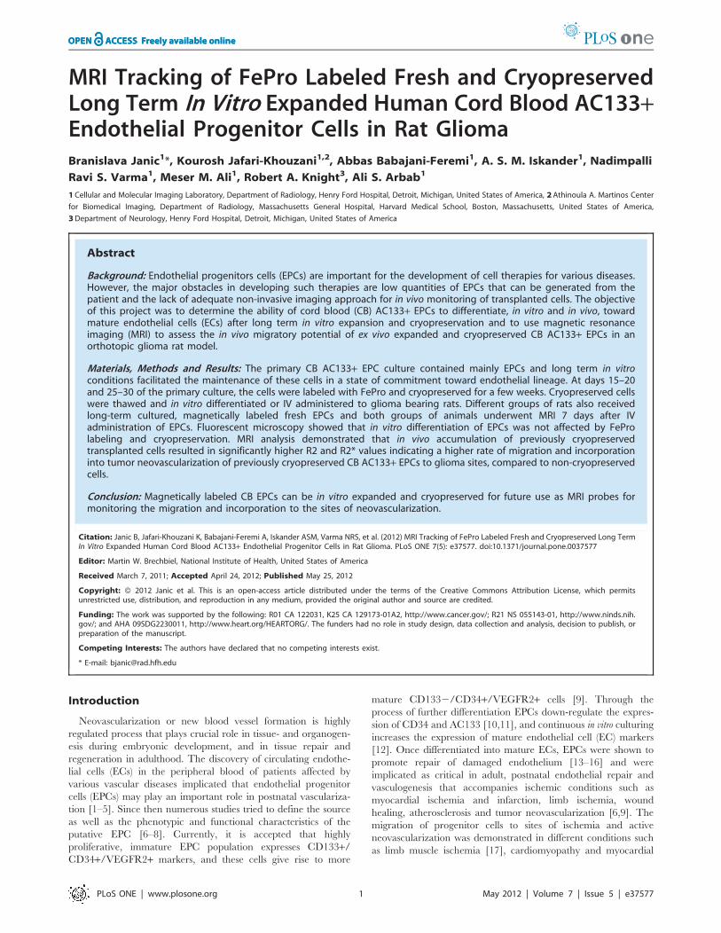
MRI Tracking of FePro Labeled Fresh and CryopreservedLong Term In Vitro Expanded Human Cord Blood AC133+Endothelial Progenitor Cells in Rat GliomaBranislava Janic1*, Kourosh Jafari-Khouzani1,2, Abbas Babajani-Feremi1, A. S. M. Iskander1, Nadimpalli
Ravi S. Varma1, Meser M. Ali1, Robert A. Knight3, Ali S. Arbab1
1Cellular and Molecular Imaging Laboratory, Department of Radiology, Henry Ford Hospital, Detroit, Michigan, United States of America, 2Athinoula A. Martinos Center
for Biomedical Imaging, Department of Radiology, Massachusetts General Hospital, Harvard Medical School, Boston, Massachusetts, United States of America,
3Department of Neurology, Henry Ford Hospital, Detroit, Michigan, United States of America
Abstract
Background: Endothelial progenitors cells (EPCs) are important for the development of cell therapies for various diseases.However, the major obstacles in developing such therapies are low quantities of EPCs that can be generated from thepatient and the lack of adequate non-invasive imaging approach for in vivo monitoring of transplanted cells. The objectiveof this project was to determine the ability of cord blood (CB) AC133+ EPCs to differentiate, in vitro and in vivo, towardmature endothelial cells (ECs) after long term in vitro expansion and cryopreservation and to use magnetic resonanceimaging (MRI) to assess the in vivo migratory potential of ex vivo expanded and cryopreserved CB AC133+ EPCs in anorthotopic glioma rat model.
Materials, Methods and Results: The primary CB AC133+ EPC culture contained mainly EPCs and long term in vitroconditions facilitated the maintenance of these cells in a state of commitment toward endothelial lineage. At days 15–20and 25–30 of the primary culture, the cells were labeled with FePro and cryopreserved for a few weeks. Cryopreserved cellswere thawed and in vitro differentiated or IV administered to glioma bearing rats. Different groups of rats also receivedlong-term cultured, magnetically labeled fresh EPCs and both groups of animals underwent MRI 7 days after IVadministration of EPCs. Fluorescent microscopy showed that in vitro differentiation of EPCs was not affected by FeProlabeling and cryopreservation. MRI analysis demonstrated that in vivo accumulation of previously cryopreservedtransplanted cells resulted in significantly higher R2 and R2* values indicating a higher rate of migration and incorporationinto tumor neovascularization of previously cryopreserved CB AC133+ EPCs to glioma sites, compared to non-cryopreservedcells.
Conclusion: Magnetically labeled CB EPCs can be in vitro expanded and cryopreserved for future use as MRI probes formonitoring the migration and incorporation to the sites of neovascularization.
Citation: Janic B, Jafari-Khouzani K, Babajani-Feremi A, Iskander ASM, Varma NRS, et al. (2012) MRI Tracking of FePro Labeled Fresh and Cryopreserved Long TermIn Vitro Expanded Human Cord Blood AC133+ Endothelial Progenitor Cells in Rat Glioma. PLoS ONE 7(5): e37577. doi:10.1371/journal.pone.0037577
Editor: Martin W. Brechbiel, National Institute of Health, United States of America
Received March 7, 2011; Accepted April 24, 2012; Published May 25, 2012
Copyright: � 2012 Janic et al. This is an open-access article distributed under the terms of the Creative Commons Attribution License, which permitsunrestricted use, distribution, and reproduction in any medium, provided the original author and source are credited.
Funding: The work was supported by the following: R01 CA 122031, K25 CA 129173-01A2, http://www.cancer.gov/; R21 NS 055143-01, http://www.ninds.nih.gov/; and AHA 09SDG2230011, http://www.heart.org/HEARTORG/. The funders had no role in study design, data collection and analysis, decision to publish, orpreparation of the manuscript.
Competing Interests: The authors have declared that no competing interests exist.
* E-mail: [email protected]
Introduction
Neovascularization or new blood vessel formation is highly
regulated process that plays crucial role in tissue- and organogen-
esis during embryonic development, and in tissue repair and
regeneration in adulthood. The discovery of circulating endothe-
lial cells (ECs) in the peripheral blood of patients affected by
various vascular diseases implicated that endothelial progenitor
cells (EPCs) may play an important role in postnatal vasculariza-
tion [1–5]. Since then numerous studies tried to define the source
as well as the phenotypic and functional characteristics of the
putative EPC [6–8]. Currently, it is accepted that highly
proliferative, immature EPC population expresses CD133+/CD34+/VEGFR2+ markers, and these cells give rise to more
mature CD1332/CD34+/VEGFR2+ cells [9]. Through the
process of further differentiation EPCs down-regulate the expres-
sion of CD34 and AC133 [10,11], and continuous in vitro culturing
increases the expression of mature endothelial cell (EC) markers
[12]. Once differentiated into mature ECs, EPCs were shown to
promote repair of damaged endothelium [13–16] and were
implicated as critical in adult, postnatal endothelial repair and
vasculogenesis that accompanies ischemic conditions such as
myocardial ischemia and infarction, limb ischemia, wound
healing, atherosclerosis and tumor neovascularization [6,9]. The
migration of progenitor cells to sites of ischemia and active
neovascularization was demonstrated in different conditions such
as limb muscle ischemia [17], cardiomyopathy and myocardial
PLoS ONE | www.plosone.org 1 May 2012 | Volume 7 | Issue 5 | e37577


Preparation of Ferumoxides-Protamine Sulfate (FePro)Complex and Labeling of CB AC133+ cellsAt days 10–15 and 25–30 of primary culture, AC133+ cells
were labeled according to our previously described method [29].
In brief, cells were suspended at the concentration of 46106 cell/
ml in serum free RPMI and commercially available ferumoxides
suspension (Fe) (Feridex IV; Bayer-Schering Pharma, Wayne, NJ,
USA) was added to the cells at the final concentration of 100 mg/ml. Immediately after, preservative-free protamine sulfate (Pro)
(American Pharmaceuticals Partners, Shaumburg, IL, USA) was
added in the same manner to the final concentration of 3 mg/ml.
Pro was supplied as 10 mg/ml of stock solution and was freshly
diluted to a concentration of 1 mg/ml in distilled water at the time
of use. Cells were plated in 24-well plate cell culture dish, 0.5 ml
per well and incubated in the presence of FePro complexes for
15 minutes at 37uC, 5% CO2, after which complete growth media
was added (0.5 ml per well) and the labeling procedure was further
continued for 4 h at 37uC, 5% CO2. Upon labeling, cells were
harvested, washed two times with 16 PBS and either cryopre-
served or intravenously administered to glioma bearing nude rats.
Cell labeling efficiency was determined by Prussian blue staining
and by determining the intracellular iron concentration according
to our published method [29]. Prussian blue staining was also
employed for confirming the presence of intracellular iron in cells
that were labeled with FePro and cryopreserved prior to
differentiation.
Cryopreservation of EPCsFollowing in vitro culturing, cells were harvested, washed twice
with PBS and resuspended at 106106cells/ml in freezing media
that contained 5% DMSO, 10% human serum albumin (HSA),
5% of 10% hydroxyethyl starch (Pentastarch)(Braun Inc, USA)
and 70% of serum free basal CellGroH SCGM media (CellGenix).
Cells were placed in cryogenic vials and ‘‘dump-frozen’’ from
room temperature to 285uC at a slow cooling rate of 1uC/min
after which vials were kept at the constant temperature of 285uCfor few weeks. Before IV administration, cells were thawed by
standard fast thawing method in a water bath at 37uC [30]. Cells
were then incubated in complete stem cell growth media for 1–2 h
at 37uC, 5% CO2, washed, suspended in 2 ml of sterile PBS and
induced to differentiate or IV administered to the animals.
Figure 1. CB AC133+ EPCs-expression of cell surface markers during long term in vitro culture. The data depicts CD133 and CD34 proteinexpression levels in cells cultured for 5, 11 and 25 days and the levels of CD117 in cells cultured for 25 days (A). Flow cytometric histograms from onerepresentative experiment are shown (n= 3). At least 10,000 live gated cells were analyzed for FITC, PE or PE-Cy5 expression. Isotype controls areshown as black histograms. Panel B shows cells induced to differentiate at day 25–30 of primary culture.doi:10.1371/journal.pone.0037577.g001
Effect of In Vitro Expansion and Freezing on EPCs
PLoS ONE | www.plosone.org 3 May 2012 | Volume 7 | Issue 5 | e37577

Human Glioma U251 CellsHuman glioma cells (U251, a generous gift from Dr. Steve
Brown, Henry Ford Hospital) were grown in Dulbecco’s modified
eagles medium (DMEM) supplemented with 10% fetal bovine
serum (FBS) in 5% CO2 at 37uC in a humidified incubator. For
implantation into rat brain, the cells were harvested and
resuspended in serum free media. A total of 46105 cells in 5 mlwas implanted into the rat brain.
Animal modelAll animal experiments and housing conditions were approved
by the Institutional Animal Care and Use Committee (IACUC) of
Henry Ford Health System. At day 0, athymic nude rats, 6–8
weeks of age and 150–170 g of weight (Charles River Laboratory,
Inc.) were anesthetized by intraperitoneal injection using ketamin/
xylazine (100 mg/kg ketamine, 15 mg/kg xylazine) and placed on
a small animal stereotactic device (Kopf, Cayunga, CA). The
surgical zone was shaved and swabbed with betadine solution, eyes
coated with Lacri-lube. After draping, a 1-cm incision was made
2 mm to the right of the midline, 1 mm retro-orbitally and the
skull was exposed with cotton-tip applicators. A HP-4 dental drill
bit was used with a micromanipulator to drill a hole 3 mm to the
right and 1 mm anterior to the bregma, with care not to penetrate
the dura. A #2701 10 mL Hamilton syringe with a #4 point, 26 s
gauge-needle containing 5 ml of 46105 of U251 human glioma
tumor cells was lowered to the depth of 3.5 mm, then raised to the
depth of 2.5 mm. The U251 cells were injected stepwise at a rate
of 0.5 mL/30 sec. Two to three minutes after completing the
injection, the syringe was withdrawn in a stepwise manner and the
surgical hole was sealed with bone wax. Finally, the skull was
swabbed with betadine before suturing the incision.
Administration of FePro labeled EPCsEleven days after U251 human glioma tumor cells implantation,
the animals (n = 10 per group) received an intravenous injection of
106106 of either fresh or previously cryopreserved FePro labeled
CB AC133+ EPCs that have been expanded for 10–15 or 25–30
days under in vitro conditions. For injection of previously
cryopreserved cells, cells were thawed, incubated in complete
stem cell growth media for 1–2 h at 37uC and 5% CO2. The cells
were then washed, suspended in 2 ml of sterile PBS and injected.
Figure 2. CB AC133+ EPCs expression of CD31, vWF and KDR and DiI-Ac-LDL uptake in differentiated progenitors – effect of FeProlabeling and cryopreservation. Expression of CD31, VEGFR2 and vWF in differentiated cells that were prior to differentiating (at days 25–30 of theprimary culture) labeled with FePro and cryopreserved for few weeks (D, E, F and G). Control cells were induced to differentiate at days 25–30 of theprimary culture without previous FePro labeling and cryopreservation (A, B and C). Positive signals for CD31, VEGFR2 and vWF were visualized witha FITC conjugated secondary antibody (green). Nuclei were visualized with DAPI (blue). VEGFR2 positive (middle panels B and E) cells also exhibitedthe uptake of DiI-Ac-LDL (red). Representative photomicrographs (406) of differentiated cells. Scale bar = 100 mm.doi:10.1371/journal.pone.0037577.g002
Effect of In Vitro Expansion and Freezing on EPCs
PLoS ONE | www.plosone.org 4 May 2012 | Volume 7 | Issue 5 | e37577

The control group of animals received intravenous injection of
2 ml of sterile PBS. On day 18 animals underwent in vivo MRI.
In vivo MR imaging and analysisImage acquisition. Rats were studied by MRI 18 days after
U 251 tumor implantation (7 days after IV administration of
EPCs). The animals were anesthetized with 2.0% isoflurane in
oxygen carrier gas and secured to a customized cradle. Core
temperature was maintained at 37.0uC. MRI was performed using
20-cm bore superconducting magnet 7T (Magnex Scientific,
Abingdon, UK) interfaced to Bruker console (Billerica, MA, USA).
After positioning using a triplanar FLASH sequence, T2- and
T2*- weighted images were acquired. Spin echo T2-weighted
images (T2WI) were obtained using a standard two-dimensional
Fourier transformation (2DFT) multi-slice (21slices) multiecho (4
echoes) MRI sequence (TE= 15, 30, 45, and 60 msec,
TR=2000 msec, 32 mm FOV, 1 mm slice thickness, 2566256
matrix, and NEX=2). The T2*-weighted images (T2*WI) were
obtained using a standard multislice (21 slices) multi gradient-echo
(4 echoes) MRI (TE= 11, 22, 33, and 44 msec, TR=5000 msec,
32 mm FOV, 1 mm slice thickness, 2566256 matrix, and
NEX=2).
Image Analysis. R2 (1/T2) and R2* (1/T2*) maps were
created from the T2WI and T2*WI image sets, respectively. The
R2 and R2* maps were created with a least square fit on a pixel-
by-pixel basis using an exponential model of the time series
extracted from the multi-echo T2-weighted spin-echo and
gradient-echo images, respectively using our custom made
software Eigentool (http://www.radiologyresearch.org/eigentool.
htm). Supplemental data show the representative multi-echo
T2*WI images and signal intensity changes obtained from animals
that received either cryopreserved or fresh magnetically labeled
EPCs (Figure S1). The R2 and R2* values were determined by
hand drawn irregular ROIs encircling the tumors for every section
that contained tumor. The tumor area R2 and R2* values were
normalized to the corresponding contra-lateral brain regions to
produce a ratio of tumor to normal brain.
Immunohistochemistry and Prussian blue stainingFor histological analysis of brain tissue, animals were euthanized
immediately after MRI imaging session. Animals were intrave-
nously or intraperitonealy administered 150–200 mg/kg of
Pentobarbital and then perfused with 100 mL of saline and
100 ml of 3% paraformaldehyde. The whole brain was collected
and fixed in 4% paraformaldehyde and 3% sucrose. The fixed
brain was then placed in 200–400 g coronal rat-brain matrix
(Activational Systems Inc., Warren, MI) and cut into 1-mm blocks
for processing and paraffin embedding. Some tissues were also
processed as frozen sections. The embedded blocks were serially
cut into 6–15 mm thick sections and analyzed by Prussian blue
staining for the presence of FePro labeled administered CB
AC133+ EPCs. Consecutive tissue sections were evaluated by
standard immunofluorescence staining techniques for the expres-
sion of von Willebrand factor (vWF) and CD 31 using rabbit anti-
human anti-vWF and mouse anti-human anti-CD31 antibodies
(both from DAKOCytomation). Endothelial lining was also
detected by FITC conjugated tomato lectin (Sigma). Prussian
blue staining was performed according to our previously reported
method [31].
Statistical AnalysisSample size was 10 animals per each group. Data are expressed
as mean 6 SD. Statistically significant difference was determined
with one way or multi ANOVA analysis followed by Fisher’s
PLSD post-hoc test. A p-value of ,0.05 was considered
significant.
Results
Long term expanded CB AC133+ EPCs actively proliferateand differentiate into endothelia-like cellsTo further explore the effect of long term ex vivo expansion,
FePro labeling and cryopreservation on the ability of EPCs to
differentiate in vitro towards ECs, CB AC133+ EPCs were
maintained in vitro in suspension culture for 30 days. Cells were
occasionally analyzed by flow cytometry to monitor expression of
CD133, CD34 and CD117 markers. Upon isolation, majority of
cells exhibited the expression of AC133 and CD 34 (.90%). As
expected, and in accordance with our previous experience [32],
over time cells down-regulated the expression of AC133 and
Figure 3. MRI detection of FePro labeled long-term culturedfrozen and fresh CB AC133+ EPCs in glioma. At days 10–15 (A, B)and 25–30 (E, F) of the primary culture, cells were labeled with FeProand cryopreserved for few weeks. On the day of IV administration, thecells were thawed, incubated for 1–2 hours in stem cell media, washedand IV injected. A control group of rats received freshly prepared FeProlabeled cells at 10–15 (C, D) and 25–30 (G, H) days of cultures. Sevendays after cell administration multi-echo gradient-echo MRI wereobtained using a 7 Tesla small animal MRI system. All animals receivingeither frozen or fresh FePro labeled cells exhibited low signal intensityareas around tumors (arrows). Corresponding DAB enhanced Prussianblue stained sections showed iron positive cells at the tumor margins.Both frozen and fresh FePro labeled cells migrated and accumulated intumor sites.doi:10.1371/journal.pone.0037577.g003
Effect of In Vitro Expansion and Freezing on EPCs
PLoS ONE | www.plosone.org 5 May 2012 | Volume 7 | Issue 5 | e37577

CD34. At day 5 in culture, expression of AC133 and CD34 was
detected on 51% and 30% of cells, respectively, while at day 11
only 16.9% of cells expressed AC 133 and 0.5% of cells expressed
CD34 (Figure 1). In comparison, the expression of CD117 (c-kit)
marker was still detected on more than 60% of cells at day 25,
indicating the presence of actively proliferating progenitors. The
endothelial differentiation potential of long term CB AC133+culture was characterized at day 10–15 and 25–30 of primary
culture and compared to the differentiation potential of thawed
cells that were previously labeled with FePro and cryopreserved at
day 10–15 and 25–30 of primary culture. Cells were differentiated
2 weeks, and analyzed for the expression of mature EC specific
markers. In addition, the uptake of DiI-Ac-LDL as an assessment
of differentiated ECs’ functional integrity [33,34] was also
analyzed. Fluorescent microscopy revealed that the majority of
cells exhibited expression of CD31, VEGFR2 and vWF when they
were induced to differentiate at days 10–15 (data not shown) and
25–30 (Figure 2A–C) of primary culture. As shown in Figure 2D–
E, expression of the same markers was not affected by previous
FePro labeling and cryopreservation. Both non-cryopreserved and
FePro labeled cryopreserved differentiated cells exhibited uptake
of DiI-Ac-LDL that appeared as uniform, perinuclear red
fluorescence. In addition, DAB enhanced Prussian blue staining
of FePro labeled cryopreserved differentiated cells demonstrated
intracellularly incorporated iron (Figure 2G). These results
indicate that long term in vitro culturing, FePro labeling and
cryopreservation did not impede the CB AC133+ cells’ potential
to differentiate towards ECs.
MRI and histological detection of migration andaccumulation of FePro labeled, long term culturedcryopreserved and fresh CB AC133+ EPCs in rat gliomamodelIn all animals, T2- and T2*-weighted images (T2WI and
T2*WI, respectively) detected growing glioma tumors within the
brains. In addition, MRI of all animals that received either frozen
or fresh FePro labeled cells showed low signal intensity areas
within and around the tumor tissue due to accumulation of the
administered cells. These hypointense regions were more pro-
nounced on T2*WI (Figure 3, panels A, C, E and G) and were
detected in all animals receiving FePro labeled cells, regardless of
the time of in vitro cells expansion (10–15 versus 20–25 days) or
fresh vs. cryopreserved. The presence of administered FePro
labeled cells was confirmed by DAB enhanced Prussian blue
staining of the tissue sections corresponding to the areas exhibiting
Figure 4. MRI relaxation parameters in tumors. Analyses of R2* values normalized to contralateral normal hemisphere (an indirect indicator ofiron positive cell accumulation ) showed significantly higher (p,0.05) accumulation of iron positive cells in animals that received previouslycryopreserved, FePro labeled CB AC133+EPCs that were in vitro expanded for 10–15 and 25–30 days. Bars: means 6 SD. * p,0.05 frozen day 10–15versus fresh day 25–30; 1 p,0.05 frozed day 25–30 versus fresh day 10–15 and fresh day 25–30.doi:10.1371/journal.pone.0037577.g004
Effect of In Vitro Expansion and Freezing on EPCs
PLoS ONE | www.plosone.org 6 May 2012 | Volume 7 | Issue 5 | e37577

hypointense voxels and it demonstrated multiple, iron positive cells
within the periphery and inside the tumors (Figure 3, panels B, D,
F and H).On the other hand, similar low signal intensity indicating
the presence of iron positive cells was not detected in animals that
received IV injection of non-labeled EPCs (Figure S2). Image
analysis demonstrated that, normalized R2 and R2* values
obtained from animals that received FePro labeled CB AC133+EPCs that were previously in vitro expanded for 25–30 days and
cryopreserved for couple of weeks, were significantly higher
(p,0.05) compared to that of animals that received non-
cryopreserved FePro labeled cells (Figure 4). Significant differ-ences were observed compared to both, fresh cells expanded for
10–15 and 25–30 days. On the other hand, R2 and R2* values
obtained from animals that received FePro labeled CB AC133+EPCs previously in vitro expanded for 10–15 days and cryopre-
served for couple of weeks, were significantly higher (p,0.05)
compared only to that of the animals that received non-
cryopreserved FePro labeled cells in vitro expanded for 25–30 days
(R2* data depicted in Figure 3). No significant differences in R2
and R2* values were detected when the same were compared to
non-cryopreserved FePro labeled cells in vitro expanded for 10–15
days (p = 0.065). To further determine whether the administered
cells associated and/or incorporated into the tumor neovascular
structures, tissue sections were analyzed for the expression of
neoangiogenic and endothelial differentiation markers by immu-
nohistochemistry. For these purposes tissue sections that were
consecutive to those that positively stained with DAB enhanced
Prussian blue (showing the presence of administered FePro labeled
cells) were analyzed. Additional staining with tomato lectin
revealed overlapping localization of iron positive cells with tumor
associated vasculature that was indicative of neovascular in-
corporation of administered cells (Figure 5). In addition, the same
iron positive cells co-localized with the strong immunoreactivity
detected by staining with anti-human vWF and anti-human CD31
antibodies, indicating in vivo differentiation of transplanted EPCs.
Similar results were observed between animals that received
cyropreserved FePro labeled cells expanded for both 10–15 and
Figure 5. Effect of cryopreservation on in vivo angiogenic properties of CB AC133+ EPCs-immunohistology. At days 10–15 and 25–30of the primary culture cells were labeled with FePro and cryopreserved for few weeks. Seven days after IV administration of thawed FePro labeledcells to glioma bearing rats, tissues were harvested and analyzed by DAB enhanced Prussian blue staining, FITC conjugated tomato lectin (green) andRho conjugated antibodies that recognized vWF and CD31 expression. Panels A–C depict tissue sections of animals receiving frozen labeled cells thatwere cultured for 10–15 days. Control animals received 10–15 days cultured non-cryopreserved FePro labeled cells (D–F). Same experiments weredone with cells expanded for 25–30 days. Tissue sections from the animals receiving frozen AC133+ are shown in panels G–I, while the section fromcontrol group receiving fresh cells are shown in J–L. Scale bar = 100 mm.doi:10.1371/journal.pone.0037577.g005
Effect of In Vitro Expansion and Freezing on EPCs
PLoS ONE | www.plosone.org 7 May 2012 | Volume 7 | Issue 5 | e37577

25–30 days, and animals that received fresh (non-cryopreserved)
FePro labeled cells expanded for both 10–15 and 25–30 days.
Discussion
Endothelial progenitor cells (EPCs) have been introduced as one
of the best candidates for developing cell based therapies for
various conditions that involve vasculogenesis. Yet, to be able to
realize such therapies, sufficient numbers of matching EPCs need
to be generated, cryopreserved, banked and readily be available
for treatment. This proved to be a challenging task due to very low
numbers of EPCs in vivo, and according to previous reports these
cells are exceptionally rare in bone marrow and peripheral blood
(,0.05% and #0.01% of mononuclear cells, respectively) [35,36].
In recent years however, human umbilical cord blood (CB) has
been established as a source of various types of stem/progenitor
cells [37–41] that can provide higher numbers of EPCs [35,42].
Although therapeutic potential has been ascribed to EPCs derived
from various sources, new evidence indicates that CB may provide
distinct advantages over other EPCs’ sources in terms of ontogeny
[43], higher telomerase activity and associated proliferation
potential [44,45] and lower risk of graft versus host disease
[46,47]. In addition, CB is the largest source of stem cells available
and the use of CB EPCs may be extremely advantageous in elderly
and sick whose endogenous adult stem cell supply and bone
marrow response may be inadequate, depleted and/or exhausted
[48,49]. In addition, cord blood is available without risk to mother
or infant [50] and the non-invasive nature of CB collection and
potential for easy and efficient characterization and banking, grant
CB derived stem cells a unique therapeutic prospect [51].
Currently available data also indicate that CB AC133+ cells
may be one of the best candidates for developing therapeutics for
vascular ischemic diseases, in particular. Although there is no
consensus on optimal numbers of cells needed to produce
successful in vivo therapeutic effects, in vitro cell amplification, as
well as long term cryopreservation appear to be necessary steps in
achieving optimal graft conditions.
Here we used our previously established method to isolate
AC133+ EPCs from CB, resulting in a cellular population of more
than 90% purity [32]. Upon isolation, CB AC133+ EPCs were
expanded under previously established optimal culture conditions
[32] for up to 30 days. At different time points in culture, cell
phenotype was characterized by flow cytometry. The expression of
AC133 and CD34 cell surface markers was down regulated over
time, with the complete loss of expression by day 25 of culture.
The observed down-regulation indicated the presence of more
mature endothelial progenitors and was in agreement with
previously reported EPCs’ phenotypic changes during in vitro
culturing [32,35,42,52–54]. At the same time, the majority of cells
still expressed CD117 (c-kit) marker that indicates the presence of
actively proliferating progenitors. In addition, during 30 days of in
vitro culturing CB AC133+ EPCs maintained the ability to give rise
to the functional progeny, i.e. mature endothelial cells as the
majority of cells expressed CD31, VEGF receptor 2 (KDR) and
vWF factor after 2 weeks of differentiation. These markers are
considered as the expression hallmarks of endothelial cell type [35]
and their expression was not affected by prior FePro labeling and
cryopreservation. In addition, long term expanded CB AC133+cultured under differentiation conditions, with or without previous
FePro labeling and cryopreservation, also exhibited uptake of DiI-
Ac-LDL. Altogether, these data demonstrated that FePro labeling
and cryopreservation did not affect the expected changes
associated with EC differentiation, i.e. properties necessary to
carry out in vivo angiogenic effects of mature endothelial cells.
Next, we hypothesized that cryopreservation of long term
expanded CB AC133+EPCs would not attenuate their in vivo
angiogenic properties and that magnetic cell labeling coupled with
MRI would be valuable for real time in vivo assessment of
migration and neovascular incorporation of these cells. Clinical
application of EPCs is considered to be most achievable in
conditions that are characterized by tissue ischemia where the
therapeutic approach may involve using these cells as a re-
generative tool for treating human vascular diseases or as a delivery
vehicle or target to restrict vascular growth in tumors [55–57]. The
common attribute in both, vascular and tumor pathologies, is that
tissue ischemia increases SDF-1 in situ expression, generating
a strong migratory signal for circulating EPCs [24,58]. The
capacity to migrate in response to such stimuli is crucial for the
EPCs angiogenic potential and involvement in neovascularization
processes [19,59,60]. Our previous studies on glioma tumor
animal models showed that both locally [22] and systemically [61]
administered FePro labeled CB AC133+ cells migrated and
incorporated into the tumor vasculatures and this in vivo CB
AC133+ cells’ migratory potential correlated with the increased
expression of SDF-1 within the glioma tumor tissue [22]. In
addition, the latest clinical studies demonstrate increased mobili-
zation of circulating EPCs in malignant glioma patients that
correlate with tumor angiogenic activity [62]. Therefore, we opted
to use a rat glioma model to assess the effect of cryopreservation on
the in vivo functional aspects of long term expanded CB AC133+EPCs, by employing MRI. MRI as a noninvasive imaging
technique has been shown efficient in in vivo monitoring of
temporal and spatial migration of stem and other cells labeled with
ferumoxides [31,63–66]. Magnetic labeling with FePro has also
been shown to not alter cell metabolism, proliferation, viability,
and differentiation capacity [29,31]. In the current study we
administered CB AC133+ EPCs that had previously been in vitro
expanded (10–15 and 25–30 days), FePro labeled and cryopre-
served to glioma bearing rats. In both, group of animals that
received either non-cryopreserved or cryopreserved cells, cell
migration and tissue incorporation at day 18 of tumor de-
velopment was detected on MRI as a hypointense region. This
hypointensity was mainly at the periphery of the tumor and was
due to the significant shortening of the T2 and T2* relaxation
times by the iron oxide incorporated within the endosomes of
FePro labeled, administered cells. The hypointense area observed
on MRI correlated with the area where iron positive cells were
detected by Prussian blue staining. In addition, iron positive cells
colocalized with ongoing tumor angiogenesis and expressed
human endothelial markers vWF and CD31. These findings
indicated in vivo differentiation of iron labeled EPCs towards
a more mature endothelial phenotype. Together, the patterns
observed by MRI and histopathology demonstrated infiltration
and incorporation of FePro labeled CB AC133+ EPCs into tumor
neovascularization that was not attenuated by previous cryopres-
ervation of the cells. Interestingly, MRI analysis revealed that the
animals which received FePro labeled cells that were cryopre-
served before IV administration exhibited significantly higher R2
and R2* values, that would suggest higher numbers of accumu-
lated cells within the tumors. The actual mechanism responsible
for this phenomenon is unknown, but may involve better in vivo
survival or proliferation of previously cryopreserved cells. Previous
reports on transplantation of stem/progenitor cells derived from
various sources have not established a significant in vivo advantage
when cryopreserved cells were used. However, it is interesting to
note that earlier work exploring the effects of cryopreservation on
transplanted human heart cells demonstrated that cryopreserva-
tion increased cell proliferation and reduced the immunogenicity
Effect of In Vitro Expansion and Freezing on EPCs
PLoS ONE | www.plosone.org 8 May 2012 | Volume 7 | Issue 5 | e37577
of transplants [67]. Cord blood derived stem cell transplants have
also been shown to exhibit a lower incidence of graft-versus –host
disease with allogeneic grafts than transplants generated from
bone marrow or peripheral blood, despite the HLA disparity
[46,47]. Nevertheless, if cryopreservation can be utilized to further
reduce or eliminate graft-versus –host disease incidence, it would
be an extremely significant step towards clinical use of CB
generated EPC transplants. In general, cryopreservation process is
a critical step in long term preservation of any type of stem cells;
however this step may be even more critical when it comes to CB
derived cells since the cells are harvested at the time of birth, with
intent of use at much later time point. Currently, the majority of
cryopreservation protocols for CB derived cells have largely been
adapted from methods originally designed for bone marrow or
peripheral blood hematopoietic cells (HSCs) and most commonly
involve freezing of minimally separated nucleated or mononuclear
cells without further, more specific cell separation [68]. While
cryopreservation of HSCs for clinical use is routinely performed
[69], cryogenic procedures for other progenitor cells are still being
defined at the laboratory level. In this study we focused on
cryopreservation of CB derived AC133+ EPCs. The ultimate goal
in optimizing cryogenic conditions is to develop protocols with
translational potential and in compliance with current Good
Manufacturing Practices (cGMP). Therefore, the majority of
studies focused on using xeno-free (i.e. FBS free) media with low
concentration of permeable cryoprotectant (CPA), such as DMSO
[70,71]. In addition, large molecular weight, non-permeable CPAs
such as hydroxyethyl starch also proved beneficial in reducing the
cell injury due to water crystallization and intracellular ice
formation [72]. The present study demonstrates a cryopreservation
method that utilizes media containing xeno-free components with
low concentrations of CPA reagents to produce favorable
cryopreservation conditions that enabled previously frozen cells
to preserve and in vivo exhibit proangiogenic properties such as
migration and vascular incorporation. In addition, the cryopres-
ervation method described does not require complex equipment or
procedures and all the components could be cGMP compatible
and clinically relevant.
In summary, the effects of cryopreservation on in vitro and in vivo
angiogenic properties of long term expanded CB AC133+ cells
were evaluated. The study demonstrated that under in vitro
conditions, the differentiation potential of long term expanded CB
AC133+EPCs was not affected by cryopreservation. On the other
hand, in vivo accumulation of previously cryopreserved trans-
planted cells resulted in significantly higher R2 and R2* values
indicating higher rate of migration and neovascular incorporation
of these cells. In addition, we show that FePro labeling of EPCs
and their in vivo tracking by MRI in an animal glioma model may
be valuable tool for exploring the role of EPCs in vasculo- and
angiogenic processes. However, to better understand the mechan-
isms behind the results presented here, further investigation is
warranted.
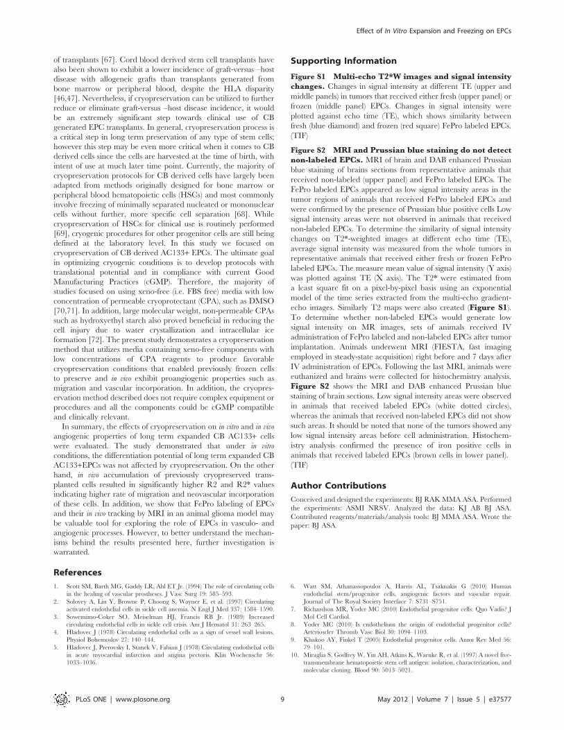
Supporting Information
Figure S1 Multi-echo T2*W images and signal intensitychanges. Changes in signal intensity at different TE (upper and
middle panels) in tumors that received either fresh (upper panel) or
frozen (middle panel) EPCs. Changes in signal intensity were
plotted against echo time (TE), which shows similarity between
fresh (blue diamond) and frozen (red square) FePro labeled EPCs.
(TIF)
Figure S2 MRI and Prussian blue staining do not detectnon-labeled EPCs. MRI of brain and DAB enhanced Prussian
blue staining of brains sections from representative animals that
received non-labeled (upper panel) and FePro labeled EPCs. The
FePro labeled EPCs appeared as low signal intensity areas in the
tumor regions of animals that received FePro labeled EPCs and
were confirmed by the presence of Prussian blue positive cells Low
signal intensity areas were not observed in animals that received
non-labeled EPCs. To determine the similarity of signal intensity
changes on T2*-weighted images at different echo time (TE),
average signal intensity was measured from the whole tumors in
representative animals that received either fresh or frozen FePro
labeled EPCs. The measure mean value of signal intensity (Y axis)
was plotted against TE (X axis). The T2* were estimated from
a least square fit on a pixel-by-pixel basis using an exponential
model of the time series extracted from the multi-echo gradient-
echo images. Similarly T2 maps were also created (Figure S1).To determine whether non-labeled EPCs would generate low
signal intensity on MR images, sets of animals received IV
administration of FePro labeled and non-labeled EPCs after tumor
implantation. Animals underwent MRI (FIESTA, fast imaging
employed in steady-state acquisition) right before and 7 days after
IV administration of EPCs. Following the last MRI, animals were
euthanized and brains were collected for histochemistry analysis.
Figure S2 shows the MRI and DAB enhanced Prussian blue
staining of brain sections. Low signal intensity areas were observed
in animals that received labeled EPCs (white dotted circles),
whereas the animals that received non-labeled EPCs did not show
such areas. It should be noted that none of the tumors showed any
low signal intensity areas before cell administration. Histochem-
istry analysis confirmed the presence of iron positive cells in
animals that received labeled EPCs (brown cells in lower panel).
(TIF)
Author Contributions
Conceived and designed the experiments: BJ RAK MMA ASA. Performed
the experiments: ASMI NRSV. Analyzed the data: KJ AB BJ ASA.
Contributed reagents/materials/analysis tools: BJ MMA ASA. Wrote the
paper: BJ ASA.
References
1. Scott SM, Barth MG, Gaddy LR, Ahl ET Jr. (1994) The role of circulating cells
in the healing of vascular prostheses. J Vasc Surg 19: 585–593.
2. Solovey A, Lin Y, Browne P, Choong S, Wayner E, et al. (1997) Circulating
activated endothelial cells in sickle cell anemia. N Engl J Med 337: 1584–1590.
3. Sowemimo-Coker SO, Meiselman HJ, Francis RB Jr. (1989) Increased
circulating endothelial cells in sickle cell crisis. Am J Hematol 31: 263–265.
4. Hladovec J (1978) Circulating endothelial cells as a sign of vessel wall lesions.
Physiol Bohemoslov 27: 140–144.
5. Hladovec J, Prerovsky I, Stanek V, Fabian J (1978) Circulating endothelial cells
in acute myocardial infarction and angina pectoris. Klin Wochenschr 56:
1033–1036.
6. Watt SM, Athanassopoulos A, Harris AL, Tsaknakis G (2010) Human
endothelial stem/progenitor cells, angiogenic factors and vascular repair.
Journal of The Royal Society Interface 7: S731–S751.
7. Richardson MR, Yoder MC (2010) Endothelial progenitor cells: Quo Vadis? J
Mol Cell Cardiol.
8. Yoder MC (2010) Is endothelium the origin of endothelial progenitor cells?
Arterioscler Thromb Vasc Biol 30: 1094–1103.
9. Khakoo AY, Finkel T (2005) Endothelial progenitor cells. Annu Rev Med 56:
79–101.
10. Miraglia S, Godfrey W, Yin AH, Atkins K, Warnke R, et al. (1997) A novel five-
transmembrane hematopoietic stem cell antigen: isolation, characterization, and
molecular cloning. Blood 90: 5013–5021.
Effect of In Vitro Expansion and Freezing on EPCs
PLoS ONE | www.plosone.org 9 May 2012 | Volume 7 | Issue 5 | e37577

11. Yin AH, Miraglia S, Zanjani ED, Almeida-Porada G, Ogawa M, et al. (1997)
AC133, a novel marker for human hematopoietic stem and progenitor cells.
Blood 90: 5002–5012.
12. Fujiyama S, Amano K, Uehira K, Yoshida M, Nishiwaki Y, et al. (2003) Bone
marrow monocyte lineage cells adhere on injured endothelium in a monocyte
chemoattractant protein-1-dependent manner and accelerate reendothelializa-
tion as endothelial progenitor cells. Circ Res 93: 980–989.
13. Bhattacharya V, McSweeney PA, Shi Q, Bruno B, Ishida A, et al. (2000)
Enhanced endothelialization and microvessel formation in polyester grafts
seeded with CD34 (+) bone marrow cells. Blood 95: 581–585.
14. Gehling UM, Ergun S, Schumacher U, Wagener C, Pantel K, et al. (2000) In
vitro differentiation of endothelial cells from AC133-positive progenitor cells.
Blood 95: 3106–3112.
15. Hill JM, Zalos G, Halcox JP, Schenke WH, Waclawiw MA, et al. (2003)
Circulating endothelial progenitor cells, vascular function, and cardiovascular
risk. N Engl J Med 348: 593–600.
16. Hu Y, Davison F, Zang Z, Xu Q (2003) Endothelial replacement and
angiogenesis in arteriosclerotic lesions of allografts are contributed by circulating
progenitor cells. Circulation 108: 3122–3127.
17. Hur J, Yoon CH, Lee CS, Kim TY, Oh IY, et al. (2007) Akt is a key modulator
of endothelial progenitor cell trafficking in ischemic muscle. Stem Cells 25:
1769–1778.
18. Askari AT, Unzek S, Popovic ZB, Goldman CK, Forudi F, et al. (2003) Effect of
stromal-cell-derived factor 1 on stem-cell homing and tissue regeneration in
ischaemic cardiomyopathy. Lancet 362: 697–703.
19. Kawamoto A, Gwon HC, Iwaguro H, Yamaguchi JI, Uchida S, et al. (2001)
Therapeutic potential of ex vivo expanded endothelial progenitor cells for
myocardial ischemia. Circulation 103: 634–637.
20. Navarro-Sobrino M, Rosell A, Hernandez-Guillamon M, Penalba A, Ribo M, et
al. (2010) Mobilization, endothelial differentiation and functional capacity of
endothelial progenitor cells after ischemic stroke. Microvascular Research In
Press, Corrected Proof.
21. Rad AM, Iskander AS, Janic B, Knight RA, Arbab AS, et al. (2009) AC133+progenitor cells as gene delivery vehicle and cellular probe in subcutaneous
tumor models: a preliminary study. BMC Biotechnol 9: 28.
22. Arbab AS, Janic B, Knight RA, Anderson SA, Pawelczyk E, et al. (2008)
Detection of migration of locally implanted AC133+ stem cells by cellular
magnetic resonance imaging with histological findings. FASEB J 22: 3234–3246.
23. Varma NR, Janic B, Iskander AS, Shankar A, Bhuiyan MP, et al. (2012)
Endothelial Progenitor Cells (EPCs) as Gene Carrier System for Rat Model of
Human Glioma. PLoS ONE 7: e30310.
24. Ceradini DJ, Kulkarni AR, Callaghan MJ, Tepper OM, Bastidas N, et al. (2004)
Progenitor cell trafficking is regulated by hypoxic gradients through HIF-1
induction of SDF-1. Nat Med 10: 858–864.
25. Isner JM, Asahara T (1999) Angiogenesis and vasculogenesis as therapeutic
strategies for postnatal neovascularization. J Clin Invest 103: 1231–1236.
26. Banai S, Shweiki D, Pinson A, Chandra M, Lazarovici G, et al. (1994)
Upregulation of vascular endothelial growth factor expression induced by
myocardial ischaemia: implications for coronary angiogenesis. Cardiovasc Res
28: 1176–1179.
27. Carmeliet P, Jain RK (2000) Angiogenesis in cancer and other diseases. Nature
407: 249–257.
28. Urbich C, Dimmeler S (2004) Endothelial progenitor cells: characterization and
role in vascular biology. Circ Res 95: 343–353.
29. Janic B, Rad AM, Jordan EK, Iskander AS, Ali MM, et al. (2009) Optimization
and validation of FePro cell labeling method. PLoS One 4: e5873.
30. Katayama Y, Yano T, Bessho A, Deguchi S, Sunami K, et al. (1997) The effects
of a simplified method for cryopreservation and thawing procedures on
peripheral blood stem cells. Bone Marrow Transplant 19: 283–287.
31. Arbab AS, Yocum GT, Kalish H, Jordan EK, Anderson SA, et al. (2004)
Efficient magnetic cell labeling with protamine sulfate complexed to ferumoxides
for cellular MRI. Blood 104: 1217–1223.
32. Janic B, Guo AM, Iskander ASM, Varma NRS, Scicli AG, et al. (2010) Human
Cord Blood-Derived AC133+ Progenitor Cells Preserve Endothelial Progenitor
Characteristics after Long Term In Vitro Expansion. PLoS ONE 5: e9173.
33. Ma G, Lin XX, Jin YB, Li W, Wang W, et al. (2008) [Isolation, culture and
characterization of endothelial cells in infantile hemangioma]. Zhonghua Zheng
Xing Wai Ke Za Zhi 24: 144–147.
34. Werner C, Bohm M, Friedrich EB (2008) Role of integrin-linked kinase for
functional capacity of endothelial progenitor cells in patients with stable
coronary artery disease. Biochem Biophys Res Commun 377: 331–336.
35. Eggermann J, Kliche S, Jarmy G, Hoffmann K, Mayr-Beyrle U, et al. (2003)
Endothelial progenitor cell culture and differentiation in vitro: a methodological
comparison using human umbilical cord blood. Cardiovasc Res 58: 478–486.
36. Gross P, Herbrig K (2004) Role of endothelial progenitor cells in cardiovascular
pathology. Rocz Akad Med Bialymst 49: 174–177.
37. Lee OK, Kuo TK, Chen WM, Lee KD, Hsieh SL, et al. (2004) Isolation of
multipotent mesenchymal stem cells from umbilical cord blood. Blood 103:
1669–1675.
38. Moise KJ Jr. (2005) Umbilical cord stem cells. Obstet Gynecol 106: 1393–1407.
39. Kogler G, Sensken S, Airey JA, Trapp T, Muschen M, et al. (2004) A new
human somatic stem cell from placental cord blood with intrinsic pluripotent
differentiation potential. J Exp Med 200: 123–135.
40. Fibbe WE, Noort WA, Schipper F, Willemze R (2001) Ex vivo expansion and
engraftment potential of cord blood-derived CD34+ cells in NOD/SCID mice.
Ann N Y Acad Sci 938: 9–17.
41. Hao QL, Shah AJ, Thiemann FT, Smogorzewska EM, Crooks GM (1995) A
functional comparison of CD34 + CD38- cells in cord blood and bone marrow.
Blood 86: 3745–3753.
42. Senegaglia AC, Brofman PR, Aita CA, Dallagiovanna B, Rebelatto CL, et al.
(2008) In vitro formation of capillary tubules from human umbilical cord blood
cells with perspectives for therapeutic application. Rev Bras Cir Cardiovasc 23:
467–473.
43. McGuckin CP, Forraz N (2008) Potential for access to embryionic-like cells from
human umbilical cord blood. Cell Prolif 41: 31–40.
44. Yao C-L, Feng Y-H, Lin X-Z, Chu I-M, Hsieh T-B, et al. (2006)
Characterization of Serum-Free Ex Vivo–Expanded Hematopoietic Stem Cells
Derived from Human Umbilical Cord Blood CD133+ Cells. Stem Cells and
Development 15: 70–78.
45. Gammaitoni L, Weisel KC, Gunetti M, Wu K-D, Bruno S, et al. (2004) Elevated
telomerase activity and minimal telomere loss in cord blood long-term cultures
with extensive stem cell replication. Blood 103: 4440–4448.
46. Yossi C, Arnon N (2004) Umbilical cord blood transplantation–how, when and
for whom? Blood reviews 18: 167–179.
47. Rocha V, Wagner JE, Sobocinski KA, Klein JP, Zhang M-J, et al. (2000) Graft-
Versus-Host Disease in Children Who Have Received a Cord-Blood or Bone
Marrow Transplant from an HLA-Identical Sibling. N Engl J Med 342:
1846–1854.
48. Scheubel RJ, Zorn H, Silber RE, Kuss O, Morawietz H, et al. (2003) Age-
dependent depression in circulating endothelial progenitor cells in patients
undergoing coronary artery bypass grafting. J Am Coll Cardiol 42: 2073–2080.
49. Goldberg JL, Laughlin MJ, Pompili VJ (2007) Umbilical cord blood stem cells:
implications for cardiovascular regenerative medicine. J Mol Cell Cardiol 42:
912–920.
50. Woods EJ, Pollok KE, Byers MA, Perry BC, Purtteman J, et al. (2007) Cord
Blood Stem Cell Cryopreservation. Transfusion Medicine and Hemotherapy 34:
276–285.
51. Suzanne MW, Marcela C (2005) Stem cell medicine: Umbilical cord blood and
its stem cell potential. Seminars in fetal & neonatal medicine 10: 209–220.
52. Yang C, Zhang ZH, Li ZJ, Yang RC, Qian GQ, et al. (2004) Enhancement of
neovascularization with cord blood CD133+ cell-derived endothelial progenitor
cell transplantation. Thromb Haemost 91: 1202–1212.
53. Bonanno G, Mariotti A, Procoli A, Corallo M, Rutella S, et al. (2007) Human
cord blood CD133+ cells immunoselected by a clinical-grade apparatus
differentiate in vitro into endothelial- and cardiomyocyte-like cells. Transfusion
47: 280–289.
54. Kuci S, Kuci Z, Schmid S, Seitz G, Muller I, et al. (2008) Efficient in vitro
generation of adult multipotent cells from mobilized peripheral blood CD133+cells. Cell Prolif 41: 12–27.
55. Timmermans F, Plum J, Yoder MC, Ingram DA, Vandekerckhove B, et al.
(2008) Endothelial progenitor cells: Identity defined? J Cell Mol Med.
56. Young PP, Vaughan DE, Hatzopoulos AK (2007) Biologic properties of
endothelial progenitor cells and their potential for cell therapy. Prog Cardiovasc
Dis 49: 421–429.
57. Dong C, Goldschmidt-Clermont PJ (2007) Endothelial progenitor cells:
a promising therapeutic alternative for cardiovascular disease. J Interv Cardiol
20: 93–99.
58. Fraisl P, Mazzone M, Schmidt T, Carmeliet P (2009) Regulation of angiogenesis
by oxygen and metabolism. Dev Cell 16: 167–179.
59. Kawamoto A, Tkebuchava T, Yamaguchi J, Nishimura H, Yoon YS, et al.
(2003) Intramyocardial transplantation of autologous endothelial progenitor cells
for therapeutic neovascularization of myocardial ischemia. Circulation 107:
461–468.
60. Terranova VP, DiFlorio R, Lyall RM, Hic S, Friesel R, et al. (1985) Human
endothelial cells are chemotactic to endothelial cell growth factor and heparin.
J Cell Biol 101: 2330–2334.
61. Arbab AS, Pandit SD, Anderson SA, Yocum GT, Bur M, et al. (2006) Magnetic
resonance imaging and confocal microscopy studies of magnetically labeled
endothelial progenitor cells trafficking to sites of tumor angiogenesis. Stem Cells
24: 671–678.
62. Rafat N, Beck G, Schulte J, Tuettenberg J, Vajkoczy P (2010) Circulating
endothelial progenitor cells in malignant gliomas. J Neurosurg 112: 43–49.
63. Anderson SA, Glod J, Arbab AS, Noel M, Ashari P, et al. (2005) Noninvasive
MR imaging of magnetically labeled stem cells to directly identify neovascu-
lature in a glioma model. Blood 105: 420–425.
64. Anderson SA, Shukaliak-Quandt J, Jordan EK, Arbab AS, Martin R, et al.
(2004) Magnetic resonance imaging of labeled T-cells in a mouse model of
multiple sclerosis. Ann Neurol 55: 654–659.
65. Arbab AS, Jordan EK, Wilson LB, Yocum GT, Lewis BK, et al. (2004) In vivo
trafficking and targeted delivery of magnetically labeled stem cells. Hum Gene
Ther 15: 351–360.
66. Hoehn M, Kustermann E, Blunk J, Wiedermann D, Trapp T, et al. (2002)
Monitoring of implanted stem cell migration in vivo: a highly resolved in vivo
magnetic resonance imaging investigation of experimental stroke in rat. Proc
Natl Acad Sci U S A 99: 16267–16272.
Effect of In Vitro Expansion and Freezing on EPCs
PLoS ONE | www.plosone.org 10 May 2012 | Volume 7 | Issue 5 | e37577

67. Yokomuro H, Shiono N, Ozawa T, Fujii T, Watanabe Y, et al. (2010) Effect of
Cryopreservation on Cell Proliferation and Immunogenicity of TransplantedHuman Heart Cells. Ann Thorac Cardiovasc Surg 16.
68. Broxmeyer HE (2008) Cord blood hematopoietic stem cell transplantation.
69. Alencar S, Garnica M, Luiz RR, Nogueira CM, Borojevic R, et al. (2010)Cryopreservation of peripheral blood stem cell: the influence of cell
concentration on cellular and hematopoietic recovery. Transfusion 50:2402–2412.
70. Zeisberger SM, Schulz JC, Mairhofer M, Ponsaerts P, Wouters G, et al. (2010)
Biological and physicochemical characterization of a serum- and xeno-free
chemically defined cryopreservation procedure for adult human progenitor cells.
Cell Transplant.71. Rubinstein P, Dobrila L, Rosenfield RE, Adamson JW, Migliaccio G, et al.
(1995) Processing and cryopreservation of placental/umbilical cord blood for
unrelated bone marrow reconstitution. Proc Natl Acad Sci U S A 92:10119–10122.
72. Sputtek A, Horn EP, Schulte Am Esch J, Kuhnl P (2001) [Cryopreservation ofred blood cells using hydroxyethyl starch (HES)-From laboratory investigations
to clinical application]. Anasthesiol Intensivmed Notfallmed Schmerzther 36
Suppl 2: S162–164.
Effect of In Vitro Expansion and Freezing on EPCs
PLoS ONE | www.plosone.org 11 May 2012 | Volume 7 | Issue 5 | e37577
Copyright © 2022 FDOKUMEN