Phase I single-dose study of intracavitary-administered Nimotuzumab labeled with 188Re in adult...
7
©2008 Landes Bioscience. Do not distribute. [Cancer Biology & Therapy 7:3, 1-6; March 2008]; ©2008 Landes Bioscience 1 Cancer Biology & Therapy 2008; Vol. 7 Issue 3 This manuscript has been published online, prior to printing.Once the issue is complete and page numbers have been assigned, the citation will change accordingly. Radioimmunotherapy (RIT) may improve the management of malignant gliomas. A Phase I clinical trial was performed to evaluate, for the first time, the toxicity and clinical effect of an intracavitary administration of a single dose of Nimotuzumab (h- R3) labeled wit 188 Re. Nimotuzumab is a humanized monoclonal antibody directed against epidermal growth factor receptors. Three patients with anaplastic astrocytoma (AA) and 8 with glioblastoma multiforme (GBM) were intended to be treated with 3 mg of mAb labelled with 10 or 15 mCi of 188 Re. In patients treated with 10 mCi (n = 6) transitory worsening of pre-existing neurological symptoms were observed. Two patients treated with 15 mCi (n = 4) developed early severe neurological symptoms and one also devel- oped late severe toxicity (radionecrosis). In the group treated with 10 mCi, 1 GBM patient died in progression 6 months after the treatment, 2 patients (1 GBM and 1 AA) developed stable disease during 3 months. One GBM patient had partial response for more than 1 year and 2 patients (1 GBM and 1 AA) were asymptomatic and in complete response after 3 years of treatment. Maximal tolerated dose of the radioimmunoconjugate 188 Re-Nimotuzumab was 3 mg of the h-R3 labelled with 10 mCi of 188 Re. The radio- immunoconjugate showed a high retention in the surgical created resection cavity and the brain adjacent tissues with a mean value of 85.5% of the injected dose one hour post-administration. This radioimmunoconjugate may be relatively safe and a promising therapeutic approach for treating high grade gliomas. Introduction The life expectancy of patients with high-grade gliomas, in partic- ular glioblastoma multiforme (GBM) is still very poor. Standard treatments including surgery, radiation and, if suitable, systemic chemotherapy, result in median survival times ranging from 1 year for GBM to 3 years for anaplastic astrocitomas (AA). 1,2 Standard treatment is not able to control tumor progression for a long-lasting period since in most cases; microscopic tumor cell clus- ters located in the peritumoral brain tissue are let and usually become the starting point for early tumor recurrence. 3,4 Complete surgical resection or eradication of the tumor by subse- quent radiation therapy is usually impossible. The median overall survival for patients with recurrent GBM is 25 weeks, and 47 weeks for those with recurrent AA. 5 These patients are appropriate candidates for clinical trials designed to improve local control by adding newer forms of treatments to the standard therapy. To overcome these limitations, more specific approaches for brain tumor treatment in the form of locoregional radioimmunotherapy (RIT) have been introduced. 6-10 The infusion of radiolabelled monoclonal antibodies directly into the postsurgical resection cavity has enabled the delivery of high radiation doses to the affected area without important harm to the surrounding normal brain tissue or distant organs. More than half of astrocytomas of high grade, especially of the novo type, overexpress the epidermal growth factor receptor (EGF-R). 11 EGF-R signal transduction pathways have been correlated with various processes that contribute to the development of malignancy, such as cell cycle progression, inhibition of apoptosis, tumor cell motility and metastasis. EGF-R overexpression has also been associ- ated with chemo and radioresistance. 12 Nimotuzumab (h-R3) is a humanized monoclonal antibody (MAb), IgG1 isotype, that recognizes an epitope located in the extracellular domain of the EGF-R. The antibody was obtained by transplanting the complementary determining regions (CDR) of the murine MAb ior egf/r3 (IgG2a) to a human framework, assisted by computer modelling. 13 Nimotuzumab has shown a synergic effect when combined with external bean radiotherapy and it has been well tolerated through the Clinical Study Phase I single-dose study of intracavitary-administered Nimotuzumab labeled with 188 Re in adult recurrent high-grade glioma Angel Casacó 1, * Gerardo López, 2 Iván García, 2 José Arsenio Rodríguez, 3 Ramsés Fernández, 2 Javier Figueredo, 3 Leonel Torres, 4 Alejandro Perera, 4 Juan Batista, 4 René Leyva, 5 Yamilé Peña, 4 Zaida Amador, 5 Addys González, 1 Barbara Estupiñan, 2 Marcos Coca, 4 Abel Hernández, 4 Miguel Puig, 2 Marbelia Iglesias, 3 Astrid Hernández, 6 Mayra Ramos, 1 Leyanis Rodríquez 7 and Niurelkis Suarez 8 1 Centro de Inmunología Molecular (CIM); 2 Centro Internacional de Restauración Neurológica (CIREN); 3 Centro de Investigaciones Médico Quirúrgicas (CIMEQ); 4 Centro de Investigaciones Clínicas (CIC); 5 Centro de Isótopos (CENTIS); 6 Centro Nacional de Ensayos Clínicos (CENCEC); 7 Instituto de Farmacia y Alimentos (IFAL); 8 Instituto Superior de Ciencias Médicas (ISCM); La Habana, Cuba Key words: gliomas, nimotuzumab, anti-EGF-R monoclonal antibody, h-R3, radioimmunotherapy, rhenium, brain tumor *Correspondence to: Angel Casacó Parada; Division of Clinical Trials; Center of Molecular Immunology; 216 Esq. a 15; Siboney; Playa; A. Postal 16040; Habana 11600; Cuba; Tel.: 537.217933; Fax: 537.335049 or 333509; Email: casaco@ cim.sld.cu Submitted: 10/08/07; Revised: 12/04/07; Accepted: 12/13/07 Previously published online as a Cancer Biology & Therapy E-publication: www.landesbioscience.com/journals/cbt/article/5414
Transcript of Phase I single-dose study of intracavitary-administered Nimotuzumab labeled with 188Re in adult...
©2008
Land
es B
ioscie
nce.
Do not
dist
ribut
e.
[Cancer Biology & Therapy 7:3, 1-6; March 2008]; ©2008 Landes Bioscience
1 Cancer Biology & Therapy 2008; Vol. 7 Issue 3
Thi
s man
uscr
ipt h
as b
een
publ
ished
onl
ine,
prio
r to
prin
ting.
Onc
e th
e iss
ue is
com
plet
e an
d pa
ge n
umbe
rs h
ave
been
ass
igne
d, th
e ci
tatio
n w
ill ch
ange
acc
ordi
ngly.
Radioimmunotherapy (RIT) may improve the management of malignant gliomas. A Phase I clinical trial was performed to evaluate, for the first time, the toxicity and clinical effect of an intracavitary administration of a single dose of Nimotuzumab (h-R3) labeled wit 188Re. Nimotuzumab is a humanized monoclonal antibody directed against epidermal growth factor receptors. Three patients with anaplastic astrocytoma (AA) and 8 with glioblastoma multiforme (GBM) were intended to be treated with 3 mg of mAb labelled with 10 or 15 mCi of 188Re. In patients treated with 10 mCi (n = 6) transitory worsening of pre-existing neurological symptoms were observed. Two patients treated with 15 mCi (n = 4) developed early severe neurological symptoms and one also devel-oped late severe toxicity (radionecrosis). In the group treated with 10 mCi, 1 GBM patient died in progression 6 months after the treatment, 2 patients (1 GBM and 1 AA) developed stable disease during 3 months. One GBM patient had partial response for more than 1 year and 2 patients (1 GBM and 1 AA) were asymptomatic and in complete response after 3 years of treatment. Maximal tolerated dose of the radioimmunoconjugate 188Re-Nimotuzumab was 3 mg of the h-R3 labelled with 10 mCi of 188Re. The radio-immunoconjugate showed a high retention in the surgical created resection cavity and the brain adjacent tissues with a mean value of 85.5% of the injected dose one hour post-administration. This radioimmunoconjugate may be relatively safe and a promising therapeutic approach for treating high grade gliomas.
IntroductionThe life expectancy of patients with high-grade gliomas, in partic-
ular glioblastoma multiforme (GBM) is still very poor. Standard
treatments including surgery, radiation and, if suitable, systemic chemotherapy, result in median survival times ranging from 1 year for GBM to 3 years for anaplastic astrocitomas (AA).1,2
Standard treatment is not able to control tumor progression for a long-lasting period since in most cases; microscopic tumor cell clus-ters located in the peritumoral brain tissue are let and usually become the starting point for early tumor recurrence.3,4
Complete surgical resection or eradication of the tumor by subse-quent radiation therapy is usually impossible.
The median overall survival for patients with recurrent GBM is 25 weeks, and 47 weeks for those with recurrent AA.5 These patients are appropriate candidates for clinical trials designed to improve local control by adding newer forms of treatments to the standard therapy.
To overcome these limitations, more specific approaches for brain tumor treatment in the form of locoregional radioimmunotherapy (RIT) have been introduced.6-10
The infusion of radiolabelled monoclonal antibodies directly into the postsurgical resection cavity has enabled the delivery of high radiation doses to the affected area without important harm to the surrounding normal brain tissue or distant organs.
More than half of astrocytomas of high grade, especially of the novo type, overexpress the epidermal growth factor receptor (EGF-R).11
EGF-R signal transduction pathways have been correlated with various processes that contribute to the development of malignancy, such as cell cycle progression, inhibition of apoptosis, tumor cell motility and metastasis. EGF-R overexpression has also been associ-ated with chemo and radioresistance.12
Nimotuzumab (h-R3) is a humanized monoclonal antibody (MAb), IgG1 isotype, that recognizes an epitope located in the extracellular domain of the EGF-R. The antibody was obtained by transplanting the complementary determining regions (CDR) of the murine MAb ior egf/r3 (IgG2a) to a human framework, assisted by computer modelling.13
Nimotuzumab has shown a synergic effect when combined with external bean radiotherapy and it has been well tolerated through the
Clinical Study
Phase I single-dose study of intracavitary-administered Nimotuzumab labeled with 188Re in adult recurrent high-grade gliomaAngel Casacó1,* Gerardo López,2 Iván García,2 José Arsenio Rodríguez,3 Ramsés Fernández,2 Javier Figueredo,3 Leonel Torres,4 Alejandro Perera,4 Juan Batista,4 René Leyva,5 Yamilé Peña,4 Zaida Amador,5 Addys González,1 Barbara Estupiñan,2 Marcos Coca,4 Abel Hernández,4 Miguel Puig,2 Marbelia Iglesias,3 Astrid Hernández,6 Mayra Ramos,1 Leyanis Rodríquez7 and Niurelkis Suarez8 1Centro de Inmunología Molecular (CIM); 2Centro Internacional de Restauración Neurológica (CIREN); 3Centro de Investigaciones Médico Quirúrgicas (CIMEQ); 4Centro de Investigaciones Clínicas (CIC); 5Centro de Isótopos (CENTIS); 6Centro Nacional de Ensayos Clínicos (CENCEC); 7Instituto de Farmacia y Alimentos (IFAL); 8Instituto Superior de Ciencias Médicas (ISCM); La Habana, Cuba
Key words: gliomas, nimotuzumab, anti-EGF-R monoclonal antibody, h-R3, radioimmunotherapy, rhenium, brain tumor
*Correspondence to: Angel Casacó Parada; Division of Clinical Trials; Center of Molecular Immunology; 216 Esq. a 15; Siboney; Playa; A. Postal 16040; Habana 11600; Cuba; Tel.: 537.217933; Fax: 537.335049 or 333509; Email: [email protected]
Submitted: 10/08/07; Revised: 12/04/07; Accepted: 12/13/07
Previously published online as a Cancer Biology & Therapy E-publication:www.landesbioscience.com/journals/cbt/article/5414
©2008
Land
es B
ioscie
nce.
Do not
dist
ribut
e.
Nimotuzumab labeled with 188Re in glioma patients
www.landesbioscience.com Cancer Biology & Therapy 2
intravenous route for treating the advanced epithelial-derived cancer patients including high-grade glioma patients.14-17
Recent data from a phase I/II clinical trial, with 200 mg/week of Nimotuzumab for 6 weeks, administered by IV infusion, combined with external radiotherapy in 24 patients with high grade astrocy-toma showed no grade 3/4 adverse events. Four patients achieved complete response, 5 a partial response and 11 stable disease, while the median survival time was 22.17 months for all subjects.17
Preclinical toxicological studies in relevant animal species have also demonstrated the low toxicity effect of Nimotuzumab when it was administered intravenously to monkeys or intracerebrally to rats.18-22
Rhenium 188 (Re-188) from a tungsten-188/rhenium-188 (188W/188Re)-radionuclide generator system, represents an attrac-tive alternative radionuclide. Rhenium-188 (t1/2 = 16.9 h) is produced after the beta decay of the tungsten-188 parent (t1/2 69.0 d). In addition to the emission of high-energy electrons (Eb = 2.118 MeV), 188Re also decays with emission of a gamma photon with an energy of 155 keV, 15% abundance. Besides the therapeutic useful-ness of 188Re in different diseases, the emission of 155 keV gamma photon is a second advantage since the biodistribution of 188Re-labeled antibodies can be evaluated in vivo with a gamma camera. In addition, since Re-188 and technetium 99 have similar chemical properties, both can be conjugated to antibodies using similar chem-istry methods.23-25
A recent intracavitary RIT study in a few number of patients bearing malignant gliomas and using a 188Re-labelled anti-tenascin antibody has shown a substantially prolonged survival.26
The aims of the present study were to evaluate the safety and the maximal tolerated dose (MTD) of 188Re-labeled antiepidermal growth factor receptor monoclonal antibody Nimotuzumab (h-R3) administered by the intracavitary route, in adult patients with recur-rent high grade glioma. The assessment of antitumor effect was a secondary end point of the study. A detailed biodistribution and internal dosimetry study was previously reported.27
ResultsEleven patients with recurrent high-grade glioma were enrolled
in the study, 8 had GBM, and 3 had AA. One GBM patient was excluded after surgery due to an important tumor progression 4
weeks after surgery, which occluded the indwelling catheter and precluded the radioimmunoconjugate admin-istration.
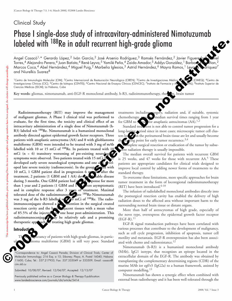
All, but one patient received one dose of the radioimmunoconjugate. Patient number 4 had a recurrent AA and was included in the first group (10 mCi), after treatment, the patient achieved a complete response lasting for more than 15 months but, 26 months after treatment the tumor recurred at the same place. Another surgery was carried out and the new pathological study revealed the presence of a GBM tumor. On a compassionate use basis, and after
the approval of the Cuban Regulatory Agency, the patient received a second dose of the radioimmunoconjugate. The patient died 1 year after the second treatment (Fig. 1).
Patient’s characteristics are listed in Table 1.Regarding safety, in patients treated with 10 mCi of 188Re (n
= 6), a transitory worsening of pre-existing neurological symp-toms was observed, even though they had no severe or very severe toxicity related to the study drug .
Two patients treated with 15 mCi (n = 4) developed severe or very severe neurological symptoms. Patient 8 developed a worsening of his pre-existing neurological symptoms with significant brain edema, patient 5 developed radionecrosis 6 months after RIT as a late treat-ment complication (Table 2).
In spite of the main objective of the study was to monitor side effects and to determine the maximal tolerated dose (MTD), 2 patients developed complete responses, 1, partial response and 2, stable diseases (Table 1).
After a median follow-up of 46 months (28.7–62.8 months), the mean and median overall survival was 16.76 and 6.07 months respectively.
For those patients that were treated with 10 mCi, the mean and median survival have corresponded to 25.14 and 18.7 months respectively.
No antiidiotypic IgG or IgM response was induced in any patient. Antibody radiolabeling and immunoreactivity assay. All radio-
labeling procedures were performed under aseptic conditions in a shielded laminar flow hood. All glassware, plastics and solutions were sterile and pyrogen free. The purified humanized MAb h-R3 was labeled with a specific activity range from 5.00–6.67 mCi/mg protein. A mean of 94.0 ± 2.3% of 188Re was bound to the IgG1 and 2.68 ± 0.62% to glucoheptonate as determined by paper chro-matography.
Instant paper chromatography of labeled MAb in acetone showed that about 2.41 ± 0.70% or less free perrhenate ran at the Rf = 1.0.
The radiocolloid determination was 1.50 ± 0.27%.The immuno-reactive fraction (IRF) of 188Re-h-R3 measured by flow cytometry analysis and by Lidmo method on the H-125 human lung adeno-carcinoma cell line was 0.78, with a correlation coefficient of r = 0.9984.
Biodistribution and dosimetric calculations. The biodistribution results showed that liver, kidneys and urinary bladder presented the
Figure 1. MRI, axial views from patient number 4. (A) T2 pre-RIT view showing post-surgical tumoral cavity of the tumoral nucleus with perilesional edema. (B) T1 post-gadolinium view, 15 months after RIT showing the post-surgical tumoral cavity without evidences of tumor or edema. (C) T1 post-gadolinium view, 26 months after RIT showing an increased gadolinium uptake zone and perilesional edema with mass effect at the same place of primary tumor. The patient died 1 year later.
©2008
Land
es B
ioscie
nce.
Do not
dist
ribut
e.
Nimotuzumab labeled with 188Re in glioma patients
3 Cancer Biology & Therapy 2008; Vol. 7 Issue 3
higher Nimotuzumab uptakes. Liver and kidneys reached maximum values of 5.6% the injected dose (ID) and 4.6% ID, respec-tively at 48 hours post injection, meanwhile the urinary bladder accumulated a maximum value of 3.3% ID, 24 hours after de radioimmunoconjugate administration. The 188Re-Nimotuzumab
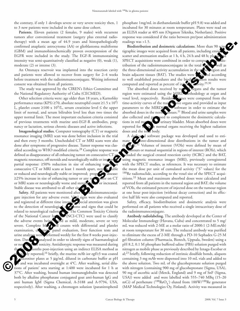
uptake in SCRC and BAT correspond to of 85.5 ± 10.3% ID, 1 hour post injection with a biological half life of approximately 22.7 ± 8.9 hours. About 6.2 ± 0.8% ID was excreted during the first 48 hours post-administration by the urinary pathway. Figure 2 shows planar views of the 188Re-Nimotuzumab distribution in the total body of patient number 6, one hour after the radioimmunoconju-gate administration.
The dosimetric studies showed that the average of the mean absorbed doses received in the tumour region were 24.1 ± 2.9 Gy and 31.1 ± 6.4 Gy, in groups I and II respectively, while the maximum doses in these VOIs were 58.9 Gy and 79.3 Gy. Dosimetric results in the tumour region showed a high variability among all patients; it seem to be due to the great differences found between the analyzed subjects regarding the cavities volumes and the 188Re kinetics in the VOIs; similar results have been reported by other authors.35
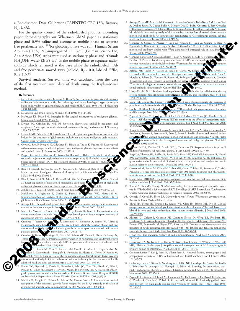
As expected, kidneys, liver and urinary bladder received the higher mean absorbed doses (0.754, 0.223 and 0.604 mGy/MBq, respec-tively), all these values were lower than the reported limits inducing radiotoxicity. Figure 3 shows the absorbed dose received by the main target organs.
These results showed that the use of locoregional RIT of high grade gliomas with 188Re-Nimotuzumab allow the delivery of a high tumoricidal dose to the tumour region without a significant irradia-tion of normal organs.
DiscussionLocal radioimmunotherapy for treating high-grade brain gliomas
using anti-EGF-R mAb is an attractive modality because this target expression is enhanced on malignant brain tissues as compared with the normal counterparts. On the other hand, high quantities of the radiolabeled mAb can penetrate into brain tissues and can kill those antigen-negative tumor cells, which have no specific radiolabeled antibody localized on their surface.36,37
It has been previously described that Nimotuzumab is a potent anti-cancer agent both, in vitro and in vivo by exerting a combined antiproliferative, antiangiogenic and proapoptotic activity in tumors overexpressing EGF-R.38
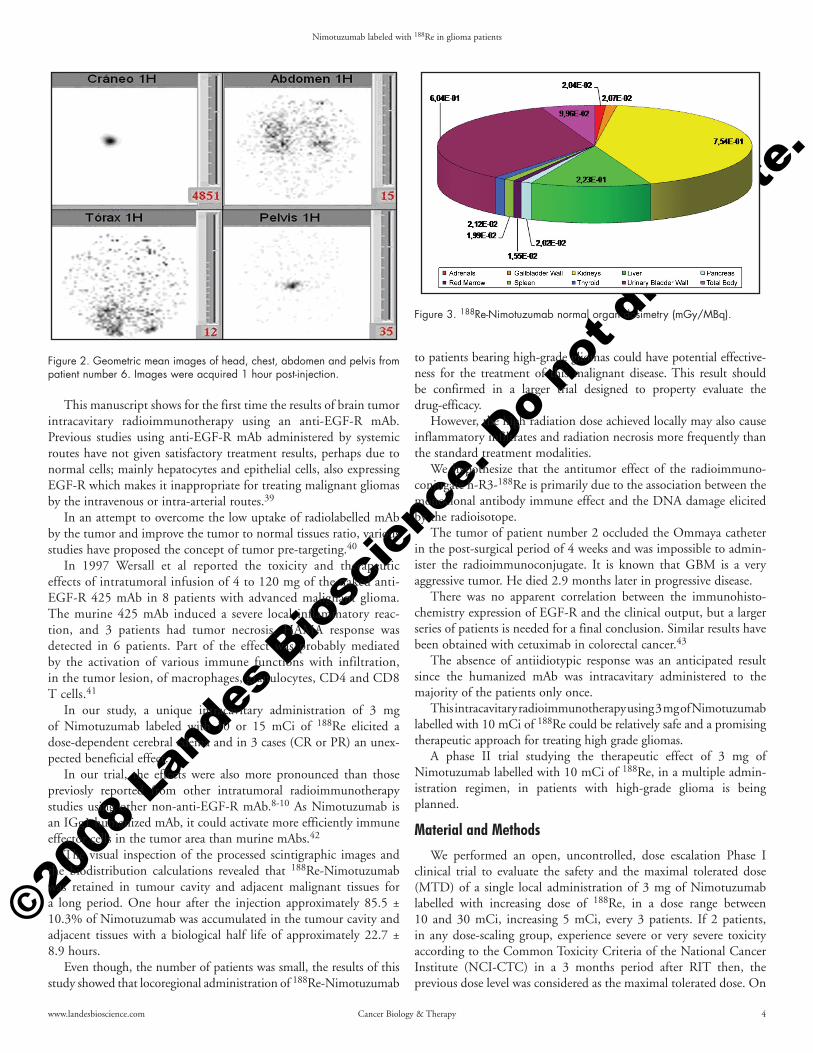
Table 2 Treatment related-toxicity
Inclusion Dose Adverse reactionnumber level (Grade)1 I Headache (moderate); confusion (moderate).2 - -3 I Increased level of SGPT (moderate); headache (moderate); seizures (moderate); objective muscle weakness (moderate).4 I Increased level of SGPT (mild); seizures (moderate).5 II Increased level of SGPT (mild); increased level of SGOT (mild); headache (moderate); seizures (moderate); confusion (moderate); aphasia (severe); radionecrosis (severe).6 II Increased level of SGOT (mild); aphasia (mild); headache (mild).7 II Seizures (moderate).8 II Aphasia (severe); confusion (very severe); brain edema (severe).9 I Increased level of SGPT (mild).10 I Increased level of SGPT (moderate); seizures (moderate).11 I Confusion (moderate); aphasia (moderate).
SGPT = Serum glutamic pyruvic transaminase. SGOT = Serum glutamic oxalacetic transaminase; Patient number 2 was not treated due to an Ommaya camera obstruction.
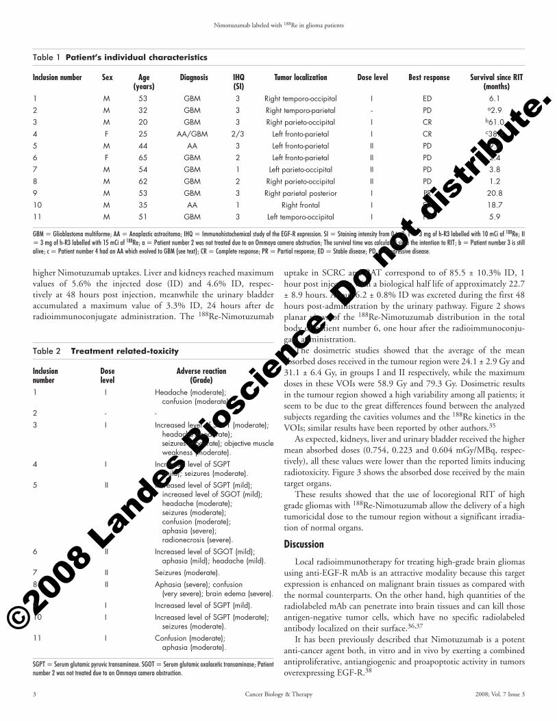
Table 1 Patient’s individual characteristics
Inclusion number Sex Age Diagnosis IHQ Tumor localization Dose level Best response Survival since RIT (years) (SI) (months)1 M 53 GBM 3 Right temporo-occipital I ED 6.12 M 32 GBM 3 Right temporo-parietal - PD a2.93 M 20 GBM 3 Right parieto-occipital I CR b61.04 F 25 AA/GBM 2/3 Left fronto-parietal I CR c38.45 M 44 AA 3 Left fronto-parietal II PD 9.46 F 65 GBM 2 Left fronto-parietal II PD 2.47 M 54 GBM 1 Left parieto-occipital II PD 3.8 8 M 62 GBM 2 Right parieto-occipital II PD 1.29 M 53 GBM 3 Right parietal posterior I PR 20.810 M 35 AA 1 Right frontal I ED 18.711 M 51 GBM 3 Left temporo-occipital I PD 5.9
GBM = Glioblastoma multiforme; AA = Anaplastic astrocitoma; IHQ = Immunohistochemical study of the EGF-R expression. SI = Staining intensity from 0 to 3; I = 3 mg of h-R3 labelled with 10 mCi of 188Re; II = 3 mg of h-R3 labelled with 15 mCi of 188Re; a = Patient number 2 was not treated due to an Ommaya camera obstruction; The survival time was calculated since the intention to RIT; b = Patient number 3 is still alive; c = Patient number 4 had an AA which evolved to GBM (see text); CR = Complete response; PR = Partial response; ED = Stable disease; PD = Progressive disease.
©2008
Land
es B
ioscie
nce.
Do not
dist
ribut
e.
Nimotuzumab labeled with 188Re in glioma patients
www.landesbioscience.com Cancer Biology & Therapy 4
This manuscript shows for the first time the results of brain tumor intracavitary radioimmunotherapy using an anti-EGF-R mAb. Previous studies using anti-EGF-R mAb administered by systemic routes have not given satisfactory treatment results, perhaps due to normal cells; mainly hepatocytes and epithelial cells, also expressing EGF-R which makes it inappropriate for treating malignant gliomas by the intravenous or intra-arterial routes.39
In an attempt to overcome the low uptake of radiolabelled mAb by the tumor and improve the tumor to normal tissues ratio, various studies have proposed the concept of tumor pre-targeting.40
In 1997 Wersall et al reported the toxicity and therapeutic effects of intratumoral infusion of 4 to 120 mg of the naked anti-EGF-R 425 mAb in 8 patients with advanced malignant glioma. The murine 425 mAb induced a severe local inflammatory reac-tion, and 3 patients had tumor necrosis. HAMA response was detected in 6 patients. Part of the effect was probably mediated by the activation of various immune functions with infiltration, in the tumor lesion, of macrophages, granulocytes, CD4 and CD8 T cells.41
In our study, a unique intracavitary administration of 3 mg of Nimotuzumab labeled with 10 or 15 mCi of 188Re elicited a dose-dependent cerebral edema and in 3 cases (CR or PR) an unex-pected beneficial effect.
In our trial, the effects were also more pronounced than those previosly reported from other intratumoral radioimmunotherapy studies using other non-anti-EGF-R mAb.8-10 As Nimotuzumab is an IGg1 humanized mAb, it could activate more efficiently immune effector cells in the tumor area than murine mAbs.42
The visual inspection of the processed scintigraphic images and the biodistribution calculations revealed that 188Re-Nimotuzumab was retained in tumour cavity and adjacent malignant tissues for a long period. One hour after the injection approximately 85.5 ± 10.3% of Nimotuzumab was accumulated in the tumour cavity and adjacent tissues with a biological half life of approximately 22.7 ± 8.9 hours.
Even though, the number of patients was small, the results of this study showed that locoregional administration of 188Re-Nimotuzumab
to patients bearing high-grade gliomas could have potential effective-ness for the treatment of this malignant disease. This result should be confirmed in a larger trial designed to property evaluate the drug-efficacy.
However, the high radiation dose achieved locally may also cause inflammatory infiltrates and radiation necrosis more frequently than the standard treatment modalities.
We hypothesize that the antitumor effect of the radioimmuno-conjugate h-R3-188Re is primarily due to the association between the monoclonal antibody immune effect and the DNA damage elicited by the radioisotope.
The tumor of patient number 2 occluded the Ommaya catheter in the post-surgical period of 4 weeks and was impossible to admin-ister the radioimmunoconjugate. It is known that GBM is a very aggressive tumor. He died 2.9 months later in progressive disease.
There was no apparent correlation between the immunohisto-chemistry expression of EGF-R and the clinical output, but a larger series of patients is needed for a final conclusion. Similar results have been obtained with cetuximab in colorectal cancer.43
The absence of antiidiotypic response was an anticipated result since the humanized mAb was intracavitary administered to the majority of the patients only once.
This intracavitary radioimmunotherapy using 3 mg of Nimotuzumab labelled with 10 mCi of 188Re could be relatively safe and a promising therapeutic approach for treating high grade gliomas.
A phase II trial studying the therapeutic effect of 3 mg of Nimotuzumab labelled with 10 mCi of 188Re, in a multiple admin-istration regimen, in patients with high-grade glioma is being planned.
Material and MethodsWe performed an open, uncontrolled, dose escalation Phase I
clinical trial to evaluate the safety and the maximal tolerated dose (MTD) of a single local administration of 3 mg of Nimotuzumab labelled with increasing dose of 188Re, in a dose range between 10 and 30 mCi, increasing 5 mCi, every 3 patients. If 2 patients, in any dose-scaling group, experience severe or very severe toxicity according to the Common Toxicity Criteria of the National Cancer Institute (NCI-CTC) in a 3 months period after RIT then, the previous dose level was considered as the maximal tolerated dose. On
Figure 2. Geometric mean images of head, chest, abdomen and pelvis from patient number 6. Images were acquired 1 hour post-injection.
Figure 3. 188Re-Nimotuzumab normal organ dosimetry (mGy/MBq).
©2008
Land
es B
ioscie
nce.
Do not
dist
ribut
e.
Nimotuzumab labeled with 188Re in glioma patients
5 Cancer Biology & Therapy 2008; Vol. 7 Issue 3
the contrary, if only 1 develops severe or very severe toxicity then, 1 to 3 new patients were included in the same dose cohort.
Patients. Eleven patients (2 females, 9 males) with recurrent tumors after conventional treatment (surgery plus external radio-therapy) with a mean age of 44.9 years and histopathologically confirmed anaplastic astrocytoma (AA) or glioblastoma multiforme (GBM) and immunohistochemically proven overexpression of the EGFR were included in the study. The EGF-R immunostaining intensity was semi-quantitatively classified as negative (0), weak (1), moderate (2) or intense (3).
An Ommaya reservoir was implanted into the resection cavity and patients were allowed to recover from surgery for 2–4 weeks before treatment with the radioimmunoconjugate. Writing informed consent was obtained from all patients.
The study was approved by the CIREN’s Ethics Committee and the National Regulatory Authority of Cuba (CECMED).
Other selection criteria were: age older than 18 years, a Karnofsky performance status (KPS) ≥70, absolute neutrophil count ≥1.5 x 10⁹/L, platelet count ≥100 x 109/L, serum creatinine level ≤ the upper limit of normal, and serum bilirubin level less than two times the upper normal limit. The most important exclusion criteria consisted of previous treatments with murine anti-EGF-R antibodies, preg-nancy or lactation, serious chronic diseases and active infections.
Imagenological studies. Computer tomography (CT) or magnetic resonance imaging (MRI) scan was done before inclusion in the trial and then every 3 months. Unscheduled CT or MRI scans were also done after symptoms of progressive disease. Tumor response was clas-sified according to WHO modified criteria.28 Complete response was defined as disappearance of all enhancing tumor on consecutive CT or magnetic resonance, off steroids and neurologically stable or improved; partial response: ≥50% reduction in size of enhancing tumor on consecutive CT or MRI scans at least 1 month apart, steroids stable or reduced and neurologically stable or improved; progressive disease: ≥25% increase in size of enhancing tumor or any new tumor on CT or MRI scans or neurologically worse and steroids stable or increased. Stable disease was attributed to all other situations.
Safety. All patients were monitored after the radioimmunoconju-gate injection for any adverse event. Vital signs were also evaluated and registered at different time intervals. Special attention was given to the detection of neurological symptoms and signs that could be related to neurological radiotoxicity. The Common Toxicity Criteria of the National Cancer Institute (NCI-CTC) were used to classify the adverse events being scaled as mild, moderate, severe or very severe. Complete blood cell counts with differential and platelet examination, chemistry panel evaluation, liver function tests and urine analyses were performed weekly for the first 8 weeks post-injec-tion. Data were analyzed in order to identify signs of haematological or normal tissue toxicity. Antiidiotypic response was measured during the first 6 months post-injection using an indirect ELISA method as previously reported;16 briefly, the murine mAb ior egf/r3 was coated on microtiter plates at 5 μg/mL diluted in carbonate buffer at pH 9.6 and was incubated overnight at 4°C. After washing, serial dilu-tions of patient’ sera starting at 1:400 were incubated for 1 h at 37°C. After washing, bound human immunoglobulin was detected both by alkaline phosphatase conjugated goat anti-human IgG and anti human IgM (Sigma Chemical, A-3188 and A-9794, USA, respectively). After washing, a chromogen solution (paranitrophenil
phosphate 1mg/mL in diethanolamide buffer pH 9.8) was added and incubated for 30 minutes at room temperature. Plates were read on an ELISA reader at 405 nm (Organon Teknika, Netherlans). Positive response was considered if the ratio between pre/post administration readings was >1.5.
Biodistribution and dosimetric calculations. More than 90 scin-tigraphic images were acquired from all patients, including emission, scatter and attenuation studies at 1 h, 4 h, 24 h and 48 h; planar and SPECT acquisitions were combined in order to compute the biodis-tribution of the radioimmunoconjugate in the total body, as well as the three-dimensional activity accumulation in the tumor region and brain adjacent tissues (BAT). The studies were processed according to well established procedures and the biodistribution results were computed and reported as percent of injected activity.29,30
The absorbed doses received by normal organs and the tumor region were estimated using the MIRD methodology at organ and voxel level, respectively. Residence times were computed from the time-activity curves of the main source organs and provided as input parameters to the MIRDOSE 3 software in order to estimate the absorbed doses in the normal tissues.31 Blood and urine samples were also collected and processed to complement the dosimetric calcula-tions in red marrow and urinary bladder. Mean absorbed doses were reported for the nine normal organs receiving the highest radiation doses and the total body.
A dedicated software package was developed and used to esti-mate the three-dimensional dose distributions in the tumor and the BAT.32 Volumes of interest (VOIs) were defined by mean of isocontours or manual sequential in regions of interest (ROIs), which included the surgical created resection cavity (SCRC) and the BAT, using magnetic resonance images (MRI), previously corregistered with the SPECT studies, as references. It was necessary to estimate the mean dose per unit of cumulated activity (“S” values) for the 188Re radionuclide, according to the voxel size of the SPECT acqui-sitions.33 Mean and maximum absorbed doses were calculated and reported from all patients in the tumoral region and BAT; the volume of VOIs, the estimated percent of injected dose on the tumour region at one hour post-injection (without decay correction) and its effec-tive half life were also computed and reported.
Safety, efficacy, biodistribution and dosimetric analysis were performed on all patients who received a single intracavitary dose of the radioimmunoconjugate.
Antibody radiolabeling. The antibody developed at the Center of Molecular Immunology (Havana, Cuba) and concentrated to 5 mg/mL was reduced with 2-ME at a molar ratio of 2000:1 (2-ME:mAb) at room temperature for 30 min. The reduced antibody was purified to eliminate the excess of 2-ME through a PD-10 Sephadex G-25 M gel filtration column (Pharmacia, Biotech, Uppsala, Sweden) using a pH 8.2, 0.1 M phosphate buffered saline (PBS) solution purged with nitrogen as mobile phase as previously described by Iznaga-Escobar et al;23 briefly, following reduction of intrinsic disulfide bonds, aliquots containing 3 mg mAb were dispensed into 10 mL vials and added to the above solution. Two mL of the glucoheptonate solution purged with nitrogen (containing 900 mg of glucoheptonate (Sigma, USA), 90 mg of ascorbic acid (Merck, England) and 9 mg of SnF (Sigma, USA)) were added and were labelled with 555–740 MBq (15–20 mCi) of perrhenate (188ReO4
-) eluted from 188W/188Re generator (MAP Medical Technologies Oy, Finland). Activity was measured in
©2008
Land
es B
ioscie
nce.
Do not
dist
ribut
e.
Nimotuzumab labeled with 188Re in glioma patients
www.landesbioscience.com Cancer Biology & Therapy 6
a Radioisotope Dose Calibrator (CAPINTEC CRC-15R, Ramsey, NJ, USA).
For the quality control of the radiolabeled product, ascending paper chromatography on Whatman 3MM paper as stationary phase and 0.9% saline and acetone as mobile phase to separate free perrhenate and 188Re-glucoheptonate was run. Human Serum Albumin (HSA, 1%)-impregnated ITLC-SG (Gelman Science Inc, Ann Arbor, USA) strips were used as stationary phase and ethanol: NH4OH: Water (2:1:5 v/v) as the mobile phase to separate radio-colloids which remained at the base while the radiolabeled mAb and free perrhenate moved away (colloid, Rf = 0.0, labeled 188Re, Rf = 1.0.34
Survival analysis. Survival time was calculated from the data of the first treatment until date of death using the Kaplan-Meier method.
References 1. Davis FG, Freels S, Grutsch J, Barlas S, Brem S. Survival rates in patients with primary
malignant brain tumors stratified by patient age and tumor histological type: an analysis based on surveillance, epidemiology, and end results (SEER) data, 1973-1991. J Neurosurg 1998; 88:1-10.
2. De Angelis LM. Brain tumors. N Engl J Med 2001; 344:114-23. 3. Harbaugh KS, Black PM. Strategies in the surgical management of malignant gliomas.
Semin Surg Oncol 1998; 14:26-33. 4. Devaux BC, OíFallon JR, Kelly PJ. Resection, biopsy, and survival in malignant glial
neoplasms. A retrospective study of clinical parameters, therapy, and outcome. J Neurosurg 1993; 78:767-75.
5. Halatsch ME, Schmidt U, Behnke-Mursch J, et al. Epidermal growth factor receptor inhi-bition for the treatment of glioblastoma multiforme and other malignant brain tumours. Cancer Treat Rev 2006; 32:74-89.
6. Goetz C, Riva P, Poepperl G, Gildehaus FJ, Hischa A, Tatsch K, Reulen HJ. Locoregional radioimmunotherapy in selected patients with malignant glioma: experiences, side effects and survival times. J Neurooncol 2003; 62:321-8.
7. Popperl G, Gotz C, Gildehaus FJ, Yousry TA, Reulen HJ, Hahn K, Tatsch K. Initial experi-ences with adjuvant locoregional radioimmunotherapy using 131I-labeled monoclonal anti-bodies against tenascin (BC-4) for treatment of glioma (WHO III and IV). Nuklearmedizin 2002; 41:120-8.
8. Riva P, Franceschi G, Riva N, Casi M, Santimaria M, Adamo M. Role of nuclear medicine in the treatment of malignant gliomas: the locoregional radioimmunotherapy approach. Eur J Nucl Med 2000; 27:601-9.
9. Riva P, Franceschi G, Arista A, Frattarelli M, Riva N, Cremonini AM, Giuliani G, Casi M. Local application of radiolabeled monoclonal antibodies in the treatment of high grade malignant gliomas: a six-year clinical experience. Cancer 1997; 80:2733-42.
10. Zalutsky MR. Targeted radiotherapy of brain tumours. Br J Cancer. 2004; 90:1469-73. 11. Nishikawa R, Sugiyama T, Narita Y, Furnari F, Cavenee WK, Matsutani M.
Immunohistochemical analysis of the mutant epidermal growth factor, deltaEGFR, in glioblastoma. Brain Tumor Pathol 2004; 21:53-6.
12. Arteaga CL. The epidermal growth factor receptor: from mutant oncogene in nonhuman cancers to therapeutic target in human neoplasia. Semin Oncol. 2002; 29:3-9.
13. Mateo C, Moreno E, Amour K, Lombardero J, Harris W, Perez R. Humanization of a mouse monoclonal antibody that blocks the epidermal growth factor receptor: recovery of antagonistic activity. Immunotechnology 1997; 3:71-81.
14. Crombet T, Torres O, Rodriguez V, Menendez A, Stevenson A, Ramos M, Torres F, Figueredo R, Veitia I, Iznaga N, Perez R, Lage A. Phase I clinical evaluation of a neutralizing monoclonal antibody against epidermal growth factor receptor in advanced brain tumor patients: preliminary study. Hybridoma 2001; 20:131-6.
15. Crombet T, Torres L, Neninger E, Catala M, Solano ME, Perera A, Torres O, Iznaga N, Torres F, Perez R, Lage A. Pharmacological evaluation of humanized anti-epidermal growth factor receptor, monoclonal antibody h-R3, in patients with advanced epithelial-derived cancer. J Immunother 2003; 26:139-48.
16. Crombet T, Osorio M, Cruz T, Roca C, del Castillo R, Mon R, Iznaga-Escobar N, Figueredo R, Koropatnick J, Renginfo E, Fernandez E, Alvarez D, Torres O, Ramos M, Leonard I, Perez R, Lage A. Use of the humanized anti-epidermal growth factor receptor monoclonal antibody h-R3 in combination with radiotherapy in the treatment of locally advanced head and neck cancer patients. J Clin Oncol 2004; 22:1646-54.
17. Ramos TC, Figueredo J, Catala M, Gonzalez S, Selva JC, Cruz TM, Toledo C, Silva S, Pestano Y, Ramos M, Leonard I, Torres O, Marinello P, Perez R, Lage A. Treatment of high-grade glioma patients with the humanized anti Epidermal Growth Factor Receptor (EGFR) antibody h-R3: Report from a Phase I/II trial. Cancer Biol Ther 2006; 5:375-9.
18. Maceira M, Rengifo E, Cedeno M, Merino N, Casaco Parada A. Immunohistochemical recognition of the epidermal growth factor receptor by the h-R3 antibody in the skin of experimental animals. App Immunohistochem Mol Morphol 2004; 12:360-3.
19. Arteaga-Perez ME, Maceira M, Casaco A, Hernandez-Sosa O, Bada-Barro AM, Leon-Goni A, Orphee-Suarez R, Cuevas-Fiallo A, Moreno-Diaz D, Padro-Gutierrez P, Baro-Gonzalez F, Rodriguez-Rodriguez V, Charoo-Ruiz L, Vazquez-Castro F, Ballester-Labrada A, Cedeno M. Multiple dose toxicity study of the humanized anti-epidermal growth factor receptor monoclonal antibody h-R3 intravenously administered to Cercopithecus aethiops sabaeus monkeys. Hum Exp Toxicol 2004; 23:219-27.
20. Gonzalez B, Casaco A, Alvarez P, Leon M, Arteaga M, Leon A, Santana E, Bada A, Figueredo R, Hernandez R, Iznaga-Escobar N, Gonzalez F, Perez R. Radiotoxicity of h-R3 monoclonal antibody labeled with 188Re administered intracerebrally in rats. Hum Exp Toxicol 2000; 19:684-92.
21. Gonzalez-Navarro B, Casaco A, Alvarez P, León A, Santana E, Bada A, Figueredo R, Iznaga-Escobar N, Perez R. Local and systemic toxicity of h-R3, an anti-epidermal growth factor receptor monoclonal antibody, labeled with 188osmiun after the intracerebral administration in rats. Exp. Toxicol Pathol 2005; 56:313-9.
22. Arteaga ME, Ledon N, Casaco A, Pardo B, García M, Boleda M, Viña L, Orphee R, Hernandez O, Gonzalez C, Fuentes D, Rodriguez V, Charro L, Baro F, Macias A, Perez A, Morales Y, Subiros N, Gonzalez B, Ramos M, Rodriquez L, Ballester-Labrada A, Crombet T. Systemic and Skin Toxicity in Cercopithecus aethiops sabaeus monkeys treated during 26 weeks with a high intravenous dose of the anti-epidermal growth factor receptor mono-clonal antibody nimotuzumab. Cancer Biol Ther 2007, 9:(in press).
23. Iznaga-Escobar N. 188Re-direct labelling of monoclonal antibodies for radioimmunotherapy of solid tumors: Biodistribution, normal organ dosimetry, and toxicology. Nucl Med Biol 1998; 25:441-7.
24. Jeong JM, Chung JK. Therapy with 188re-labeled radiopharmaceuticals. An overview of promising results from initial clinical trials. Cancer Biother Radiopharm 2003; 18:707-17.
25. Lambert B, Klerk J. Clinical applications of 188Re-labelled radiopharmaceuticals for radio-nuclide therapy. Nucl Med Commun 2006; 27:223-9.
26. Popperl G, Gotz C, Rachinger W, Schnell O, Gildehaus FJ, Tonn JC, Tatsch K. Serial O-(2-[(18)F]fluoroethyl)-L: -tyrosine PET for monitoring the effects of intracavitary radio-immunotherapy in patients with malignant glioma. Eur J Nucl Med Mol Imaging 2006; 33:792-800.
27. Torres L, Coca M, Batista J, Casaco A, Lopez G, Garcia I, Perera A, Peña Y, Hernandez A, Sanchez Y, Romero S, Fernandez R, Prats A, Leyva R. Biodistribution and internal dosim-etry of the 188Re labelled humanized monoclonal antibody anti-Epidemal Growth Factor receptor Nimotuzumab in the locoregional treatment of malignant gliomas. Nucl Med Commun 2008; 29:66-75.
28. Macdonald DR, Cascino TL, Schold SC Jr, Cairncross JG. Response criteria for phase II studies of supratentorial malignant glioma. J Clin Oncol 1990; 8:1277-80.
29. Siegel JA, Thomas SR, Stubbs JB, Stabin MG, Hays MT, Koral KF, Robertson JS, Howell RW, Wessels BW, Fisher DR, Weber DA, Brill AB. MIRD pamphlet no. 16: techniques for quantitative radiopharmaceutical biodistribution data acquisition and analysis for use in human radiation dose estimates. J Nucl Med 1999; 40:37-61.
30. Cremonesi M, Ferrari M, Chinol M, Stabin MG, Grana C, Prisco G, Robertson C, Tosi G, Paganelli G. Three-step radioimmunotherapy with 90Y-biotin: dosimetry and pharmacoki-netics in cancer patients. Eur J Nucl Med 1999; 26:110-20.
31. Stabin MG. MIRDOSE-the personal computer software for internal dose assessment in nuclear medicine. J Nucl Med 1996; 37:538-46.
32. Torres LA, Coca MA, Cornejo N. A Software package for tridimentional patient-specific dosim-etry in 188Re labelled h-R3 locoregional RIT. Procedings of IAEA International Conference on Quality Assurance and new techniques in radiation medicine 2006; 146:92-94.
33. Cornejo N, Coca MA, Torres LA. Cálculo de valores “S” para 188Re en una geometría voxel. Revista de Física Médica 2006; 7:101-6.
34. Thrall JH, Freitas JE, Swanson D, Rogers WL, Clare JM, Brown ML, Pitt B. Clinical comparison of cardiac blood pool visualization with technetium-99m red blood cells labeled in vivo and with technetium-99m human serum albumin. J Nucl Med 1978; 19:796-803.
35. Akabani G, Cokgor I, Coleman RE, Gonzalez Trotter D, Wong TZ, Friedman HS, Friedman AH, Garcia-Turner A, Herndon JE, DeLong D, McLendon RE, Zhao XG, Pegram CN, Provenzale JM, Bigner DD, Zalutsky MR. Dosimetry and dose-response rela-tionships in newly diagnosed patients treated with 131I-labelled anti-tenascin monoclonal antibody therapy. Int J Rad Oncol Biol Phys 2000; 46:947-58.
36. Dixon KL. The radiation biology of radioimmunotherapy. Nucl Med Commun 2003; 24:951-7.
37. Libermann TA, Nusbaum HR, Razon N, Kris R, Lax I, Soreq H, Whittle N, Waterfield MD, Ullrich A, Schlessinger J. Amplification and overexpression of EGF receptor gene in primary human glioblastomas. J Cell Sci Suppl 1985; 3:161-72.
38. Crombet-Ramos T, Rak J, Pérez R, Viloria-Petit A. Antiproliferative, antiangiogenic and proapoptotic activity of h-R3: A humanized anti-EGFR antibody. Int J Cancer 2002; 101:567-75.
39. Carlsson J, Ren ZP, Wester K, Sundberg AL, Heldin NE, Hesselager G, Persson M, Gedda L, Tolmachev V, Lundqvist H, Blomquist E, Nister M. Planning for intracavitary anti-EGFR radionuclide therapy of gliomas. Literature review and data on EGFR expresión. J Neuooncol 2006; 77:33-45.
40. Paganelli G, Grana C, Chinol M, Cremonesi M, De Cicco C, De Braud F, Robertson C, Zurrida S, Casadio C, Zoboli S, Siccardi AG, Veronesi U. Antibody-guided three-step therapy for high grade glioma with yttrium-90 biotin. Eur J Nucl Med 1999; 26:348-57
©2008
Land
es B
ioscie
nce.
Do not
dist
ribut
e.
Nimotuzumab labeled with 188Re in glioma patients
7 Cancer Biology & Therapy 2008; Vol. 7 Issue 3
41. Wersall P, Ohlsson I, Biberfeld P, Collins VP, von Krusenstjerna S, Larsson S, Mellstedt H, Boethius J. Intratumoral infusion of the monoclonal antibody, mAb 425, against the epider-mal-growth-factor receptor in patients with advanced malignant glioma. Cancer Immunol Immunother 1997; 44:157-64.
42. Masucci G, Ragnhammar P, Wersall P, Mellstedt H. Granulocyte-monocyte colony-stimu-lating-factor augments the inter-leukin-2 induced cytotoxic activity of human lymphocytes in the absence and presence of mouse or chimeric monoclonal antibodies (mAb17-1A). Cancer Immunol Immunother 1990; 31:231-5.
43. Chung KY, Shia J, Kemeny NE, Shah M, Schwartz GK, Tse A, Hamilton A, Pan D, Schrag D, Schwartz L, Klimstra DS, Fridman D, Kelsen DP, Saltz LB. Cetuximab shows activity in colorectal cancer patients with tumors that do not express the epidermal growth factor receptor by immunohistochemistry. J Clin Oncol 2005; 23:1803-10.