Tissue distribution and toxicity of intravenously administered titanium dioxide nanoparticles in...
7
Arch Toxicol (2008) 82:151–157 DOI 10.1007/s00204-007-0253-y 123 TOXICOKINETICS AND METABOLISM Tissue distribution and toxicity of intravenously administered titanium dioxide nanoparticles in rats Eric Fabian · Robert Landsiedel · Lan Ma-Hock · Karin Wiench · Wendel Wohlleben · Ben van Ravenzwaay Received: 6 August 2007 / Accepted: 15 October 2007 / Published online: 14 November 2007 © Springer-Verlag 2007 Abstract The tissue distribution and toxicity of intrave- nously administered nanoparticles of titanium dioxide (TiO 2 ) (>10 wt.% at <100 nm size) were investigated because of the fundamental importance to obtain informa- tion on the kinetics of this widely used nanoparticle in a sit- uation of 100% bioavailability. Male Wistar rats were treated with single intravenous injections of a suspension of TiO 2 in serum (5 mg/kg body weight), and the tissue con- tent of TiO 2 was determined 1, 14, and 28 days later. Bio- chemical parameters and antigens in serum were also assessed to determine potential pathological changes. The health and behavior of the animals were normal throughout the study. There were no detectable levels of TiO 2 in blood cells, plasma, brain, or lymph nodes. The TiO 2 levels were highest in the liver, followed in decreasing order by the lev- els in the spleen, lung, and kidney, and highest on day 1 in all organs. TiO 2 levels were retained in the liver for 28 days, there was a slight decrease in TiO 2 levels from day 1 to days 14 and 28 in the spleen, and a return to control levels by day 14 in the lung and kidney. There were no changes in the cytokines and enzymes measured in blood samples, indicating that there was no detectable inXamma- tory response or organ toxicity. Overall, rats exposed to TiO 2 nanoparticles by a route that allows immediate sys- temic availability showed expected tissue distribution, no obvious toxic health eVects, no immune response, and no change in organ function. Therefore, even with 100% bio- availability of the 5 mg/kg TiO 2 dose aVorded by the intra- venous route of administration, there were no remarkable toxic eVects evident in the experimental animals. These results indicate that TiO 2 nanoparticles could be used safely in low doses. Keywords Nano-TiO2 · Tissue distribution · Adverse eVects · Toxicokinetics · Cytokines Introduction The increased use of nano-sized materials in the past sev- eral years has compelled the scientiWc community to inves- tigate the potential hazards of these unique and useful materials. Indeed, the International Life Sciences Institute Research Foundation/Risk Science Institute Nanomaterial Toxicity Screening Work Group has recently discussed the importance of studying nanoparticles for their potential toxic eVects (Oberdörster et al. 2005). Moreover, a step- wise approach for human health hazard evaluation of nano- particles was already proposed by the European Center for Ecotoxicology and Toxicology of Chemicals (ECETOC) during a workshop held in November 2005. One of the most widely used nanoparticles is titanium dioxide (TiO 2 ). TiO 2 is a very versatile compound that has many uses, depending on its particle size. As early as 1939, TiO 2 was used in biological experiments to study symmetry of bone marrow in dog and rat extremities (Huggins 1939). This compound has also been used as a whitening agent and in sunscreen products to reXect ultraviolet (UV) sunrays. Most recently, TiO 2 is being tested in “smog-busting” Xagstones for sidewalks near Covent Garden in London, where 30,000 vehicles travel the road each day, and in similar programs E. Fabian · R. Landsiedel · L. Ma-Hock · K. Wiench · B. van Ravenzwaay (&) BASF Product Safety, BASF Aktiengesellschaft, Ludwigshafen, Germany e-mail: [email protected] W. Wohlleben BASF Polymer Physics, BASF Aktiengesellschaft, Ludwigshafen, Germany
Transcript of Tissue distribution and toxicity of intravenously administered titanium dioxide nanoparticles in...

Arch Toxicol (2008) 82:151–157
DOI 10.1007/s00204-007-0253-yTOXICOKINETICS AND METABOLISM
Tissue distribution and toxicity of intravenously administered titanium dioxide nanoparticles in rats
Eric Fabian · Robert Landsiedel · Lan Ma-Hock · Karin Wiench · Wendel Wohlleben · Ben van Ravenzwaay
Received: 6 August 2007 / Accepted: 15 October 2007 / Published online: 14 November 2007© Springer-Verlag 2007
Abstract The tissue distribution and toxicity of intrave-nously administered nanoparticles of titanium dioxide(TiO2) (>10 wt.% at <100 nm size) were investigatedbecause of the fundamental importance to obtain informa-tion on the kinetics of this widely used nanoparticle in a sit-uation of 100% bioavailability. Male Wistar rats weretreated with single intravenous injections of a suspension ofTiO2 in serum (5 mg/kg body weight), and the tissue con-tent of TiO2 was determined 1, 14, and 28 days later. Bio-chemical parameters and antigens in serum were alsoassessed to determine potential pathological changes. Thehealth and behavior of the animals were normal throughoutthe study. There were no detectable levels of TiO2 in bloodcells, plasma, brain, or lymph nodes. The TiO2 levels werehighest in the liver, followed in decreasing order by the lev-els in the spleen, lung, and kidney, and highest on day 1 inall organs. TiO2 levels were retained in the liver for28 days, there was a slight decrease in TiO2 levels from day1 to days 14 and 28 in the spleen, and a return to controllevels by day 14 in the lung and kidney. There were nochanges in the cytokines and enzymes measured in bloodsamples, indicating that there was no detectable inXamma-tory response or organ toxicity. Overall, rats exposed toTiO2 nanoparticles by a route that allows immediate sys-temic availability showed expected tissue distribution, noobvious toxic health eVects, no immune response, and no
change in organ function. Therefore, even with 100% bio-availability of the 5 mg/kg TiO2 dose aVorded by the intra-venous route of administration, there were no remarkabletoxic eVects evident in the experimental animals. Theseresults indicate that TiO2 nanoparticles could be used safelyin low doses.
Keywords Nano-TiO2 · Tissue distribution · Adverse eVects · Toxicokinetics · Cytokines
Introduction
The increased use of nano-sized materials in the past sev-eral years has compelled the scientiWc community to inves-tigate the potential hazards of these unique and usefulmaterials. Indeed, the International Life Sciences InstituteResearch Foundation/Risk Science Institute NanomaterialToxicity Screening Work Group has recently discussed theimportance of studying nanoparticles for their potentialtoxic eVects (Oberdörster et al. 2005). Moreover, a step-wise approach for human health hazard evaluation of nano-particles was already proposed by the European Center forEcotoxicology and Toxicology of Chemicals (ECETOC)during a workshop held in November 2005. One of themost widely used nanoparticles is titanium dioxide (TiO2).TiO2 is a very versatile compound that has many uses,depending on its particle size. As early as 1939, TiO2 wasused in biological experiments to study symmetry of bonemarrow in dog and rat extremities (Huggins 1939). Thiscompound has also been used as a whitening agent and insunscreen products to reXect ultraviolet (UV) sunrays. Mostrecently, TiO2 is being tested in “smog-busting” Xagstonesfor sidewalks near Covent Garden in London, where 30,000vehicles travel the road each day, and in similar programs
E. Fabian · R. Landsiedel · L. Ma-Hock · K. Wiench · B. van Ravenzwaay (&)BASF Product Safety, BASF Aktiengesellschaft, Ludwigshafen, Germanye-mail: [email protected]
W. WohllebenBASF Polymer Physics, BASF Aktiengesellschaft, Ludwigshafen, Germany
123
152 Arch Toxicol (2008) 82:151–157
in France, Italy, and Japan (Ross 2007). The TiO2 in a spe-cial top layer of the Xagstone is hoped to reduce pollutionwhen activated by sunlight by converting ambient nitrogendioxide gas into less toxic nitrates. The versatility of TiO2
is due in part to its useful properties, including being insol-uble in aqueous solutions, chemically unreactive in biologi-cal systems, non-radioactive, not radio-opaque, not costly,easy to prepare in stable suspensions in Wne particulateform, and easy to measure by simple chemical techniques.
Most of the research on nanoparticle size TiO2 hasemployed routes of administration like inhalation, intratra-cheal instillation, or oral gavage (Grassian et al. 2007;Wang et al. 2007; Warheit et al. 2007). Recent inhalationstudies with nano-sized TiO2 indicated a very strongagglomeration of the nanomaterial, with eVectively very lit-tle (<1% of total weight) of nano-sized particles remainingin the atmosphere (Ma-Hock et al. 2007). We undertook aninvestigative study to determine the toxicity and tissue dis-tribution of intravenously administered nanoparticles ofTiO2 because of the fundamental importance to obtaininformation on the kinetics of this widely used nanoparticlein a situation of 100% bioavailability. Information gainedfrom such a study will give insight into the basic pharmaco-kinetics of TiO2. In addition, studies on the biokinetics of asubstance provide relevant information for the evaluationof test results from other toxicological studies and for theextrapolation of data from animals to humans.
For this purpose, rats were treated intravenously withsingle injections of a suspension of TiO2 in serum. The con-tent of TiO2 was determined 1, 14, and 28 days after treat-ment in designated subgroups of male rats. Moreover, anextended set of biochemical parameters and antigens weremeasured in serum to gain information on potential patho-logical changes after exposure to TiO2.
Materials and methods
This study was conducted considering the following guide-lines: EC Commission Directive 87/302/EEC (EC Com-mission Directive 1988), OECD Guidelines for Testing ofChemicals (Method No. 417) (OECD Guidelines 1984),U.S. EPA, Health EVects Guidelines, OPPTS 870.7485(U.S. EPA 1998), and the Japan/MAFF: Guidelines on theCompiling of Test Results on Toxicity (Japan/MAFF2001).
Test substance application preparation and assessment
The test substance, titanium dioxide [TiO2; CAS no. 13463-67-7], consisted of both anatase and rutile forms (70/30),had no surface coating, and was stored at room tempera-ture. The TiO2 particles were in the size range 20–30 nm
and had a BET of 48.6 m2/g. The TiO2 application prepara-tion consisted of TiO2 at a concentration of 0.5% in apooled sample of rat serum. A total of two TiO2 applicationpreparations were prepared, one for each application day,using ultrasonication for at least 10 min and vigorous mix-ing with a blender (Ultra-Turax). Three representative 200-�l samples of the mixture were taken from each applicationpreparation before, during, and after dosing. The sampleswere examined to verify the accuracy of TiO2 concentrationand determine the stability and homogeneity of the test sub-stance in the application preparation. The TiO2 particle sizedistribution was measured in a separate sample followingthe same preparation protocol.
To determine the TiO2 concentration, the test substanceapplication preparation was heated in a beaker with ammo-nium sulfate and sulfuric acid. Next, a mixed acid solution(nitric acid : sulfuric acid : perchloric acid, 2:1:1, v/v/v)was added, and the solution was heated again. Followingoxidation, the mineral acids were evaporated, and the resi-due was taken up with hydrochloric acid. The TiO2 contentof the resultant solution was measured by inductively cou-pled plasma atomic emission spectrometry (ICP-AES)using a Varian “Vista pro” ICP-AES spectrometer (calibra-tion: external; nebulizer: Meinhard; plasma power:1,200 W; wavelength: Ti 337.800 nm).
The TiO2 particle size distribution was determined byanalytical ultracentrifugation (AUC) of »500 �l of a testsubstance application preparation that was made in parallelwith that used for the intravenous administration. At theacceleration of up to 300,000g used in AUC, solutes andnanoparticles sediment into fractions that are separatedaccording to their size in the range 0.5–10,000 nm.Agglomerates larger than »10 �m diameter sedimentbefore data acquisition starts and cannot be quantiWed.Simultaneous detection by synchronized optics quantiWesthe amount and the diameter of each fraction independently(Cölfen 2004). When there is a low concentration of nano-particles in a high-concentration protein medium, nanopar-ticles are easily discerned from sedimenting proteinsbecause of their much higher density and resulting fastersedimentation of many orders of magnitude, and because oftheir higher refractive index and high turbidity. We used aBeckman model XL ultracentrifuge that we modiWed forthe online recording of sedimentation with turbidity, inter-ference, and Schlieren or UV detection (Mächtle and Bör-ger 2006). This modiWcation allows the use of interferenceand turbidity optics with a ramped rpm-proWle to quantifythe amounts of ultraWne fraction (diameter <100 nm), Wnefraction (100–1,000 nm), and larger-scale material(>1 �m). The evaluation of the AUC raw data incorporatesthe fractal morphology of nanoparticle aggregates andapplies the fractional dimension of 2.1 together with thesedimentation relation as speciWed in Eq. (6) of the refer-
123

Arch Toxicol (2008) 82:151–157 153
ence (Lin et al. 1990). This value of the fractional dimen-sion has been shown to be universal for all reaction-limitedcolloid aggregates (Lin et al. 1990). If in the speciWc testsubstance application preparation the fractional dimensionof the aggregates were higher (lower), corresponding to amore compact (loose) structure of the aggregates, theretrieved particles sizes would shift to lower (higher)values.
Animals and treatment
Male Wistar rats [strain Crl : WI (Han); purchased fromCharles River Laboratories, Sulzfeld, Germany], were usedin this study. At the start of acclimatization and prior todosing, the rats were 7–12 weeks old and weighed 200–300 g. The animals were housed individually in type IIIMacrolon cages under conventional hygienic conditions, at20–24°C and 30–70% relative humidity, and with naturalday/night light rhythm. Animals were fed pelleted Kliba labdiet (Provimi Kliba, SA, Kaiseraugst, Switzerland), givenwooden gnawing blocks (Typ NGM E-022; Abedd® Laband Vet Service GmbH, Vienna, Austria), and allowedaccess to tap water ad libitum. The diet feed was assayedfor chemical and microbial contaminants by the supplier,and the purity of the drinking water was regularly deter-mined by the local municipal authorities. The health statusof the animals was determined daily by visual observation.
Experimental design and analyses
A total of 12 male Wistar rats, selected based on health sta-tus and to provide a narrow range of body weights, wererandomly assigned to three experimental groups, with onecontrol and three treated animals in each group. The controlanimals were injected intravenously (iv) via the tail veinwith »1 ml of sterile saline and the treated animals wereinjected intravenous via the tail vein with the test substanceapplication preparation which contained TiO2 at a dose of5 mg/kg body weight (»1 ml of test substance preparation/kg of rat body weight). This dose was chosen because itwas high enough to allow spectroscopic quantiWcation ofresidue levels in organs of interest (based on estimated cal-culations of residue levels with a distribution volume of 1and a known limit of quantitation (loq) of the atomicabsorption method used) and low enough to be acutely non-toxic (determined in a subgroup of animals by dosing with5 mg/kg and observing no visible symptoms of toxicity for1 day). The tails of lightly anesthetized animals werewarmed in a beaker of warm water, and blood Xow wasstimulated by wiping with alcohol pads before beinginjected with either saline (controls) or the TiO2 applicationpreparation (followed by »750 �l of saline to Xush theTiO2 application preparation into the vein). Following
treatment, the animals were returned to their individualcages. At 1, 14, and 28 days post-intravenous injection ofsaline or the TiO2 application preparation, animals ingroups 1, 2, and 3, respectively, were anesthetized with iso-Xurane and killed by cervical dislocation. Blood sampleswere taken, and tissues and organs were collected andweighed immediately after killing of the animals. Subse-quently, residue levels of TiO2 were determined in samplesof blood cells, plasma, kidney, spleen, brain, lymph nodes(mediastenal, mesenteric, and popliotenal), liver, and lung.
Samples of blood cells, plasma, lymph nodes, kidney,spleen, and brain were digested in an automated digestionapparatus. After addition of an ammonium sulfate tablet,sulfuric acid was added and the mixture was heated. Then,a mixture of nitric, sulfuric, and perchloric acids (2:1:1 v/v/v) was added to the mixture, which solution was heatedagain. Following oxidation, the mineral acids were evapo-rated, and the residue was taken up with hydrochloric acid.The TiO2 content in the obtained solution was measured byInductively Coupled Plasma Atomic Emission Spectrome-try (ICP-AES) with a Thermo Jarrell Ash “IRIS 1” spec-trometer (calibration: external; nebulizer: Meinhard orUltrasonic; plasma power: 1,150 W; Ti wavelength:338.376 nm).
Samples of liver and lung were treated with sulfuric acidand heat in a platinum dish. After digestion, the acid wasevaporated and the residue was ashed at 600°C in a muZefurnace. The ash was then mixed with a sodium/potassiumcarbonate–borax mixture and melted above a blowtorchXame. The resulting Xux was cooled down, dissolved inwater, and acidiWed with hydrochloric acid. The TiO2 con-tent in the obtained solution was measured by ICP-AES,with a VARIAN “Vista pro” (calibration: external; nebu-lizer: Meinhard; plasma power: 1,200 W; Ti wavelength:334.941 nm).
In addition to TiO2 tissue distribution, various cytokinesand enzymes (a total of 67 parameters) were measured inblood samples to assess potential inXammatory responsesand/or organ injury due to intravenousexposure to TiO2.These parameters were measured at Rules-Based Medicine,Inc. (Austin, TX, USA). The parameters measured to assessinXammatory responses were divided into four classes: (1)chemokines that are attractants for monocytes, Th lympho-cytes, eosinophils, basophils, and neutrophils; (2) indicatorsof cell proliferation or inhibition; (3) indicators of inXam-mation (e.g., cytokines); and (4) indicators of immunemodulation. Examples of parameters measured to assess theeVects of TiO2 on liver function include serum levels oftotal bilirubin (TBIL), alkaline phosphatase (ALP), alanineaminotransferase (ALT), and aspartate aminotransferase(AST). Similarly, kidney function and nephrotoxicity fol-lowing TiO2 exposure were assessed by measuring serumlevels of urea and creatinine (Cr).
123
154 Arch Toxicol (2008) 82:151–157
Results
Animal diet and health
Analysis of the diet feed and water showed no contami-nants that would inXuence the study results. The observedhealth status and behavior of the animals was normalthroughout the study, indicating that there were no toxiceVects on these parameters of exposure to TiO2 at 5 mg/kgbody weight.
TiO2 application preparations
The TiO2 used in the application preparations was a stable,homogenous, white solid. The TiO2 concentration in pooledserum in the two application preparations used for intrave-nous injection was very consistent; that is, 4,963, 5,011,4,951 as well as 4,959, 4,991, and 4,928 �g/g in preparation1 and 2, respectively.
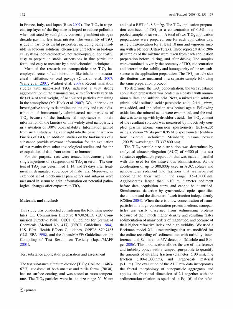
We used analytical ultracentrifuge (AUC) to measureboth the highly concentrated and very turbid samples usedfor the intravenous injections and a sample that was dilutedwith FBS gold. We found (see Fig. 1) that the particle sizeof the TiO2 used for intravenous injection was mostly in theWne fraction with components up to 1 �m. Approximately10 wt.% of the particles are found in the ultraWne- or nano-size range (<0.1 �m; i.e., <100 nm). To corroborate thisresult, we demonstrated agreement between size distribu-tions from interference or turbidity detection. The main rea-son for this reasonably good dispersion of the nanoparticlesin the serum is presumably the interface activity of albu-mines. Albumines show at least Wve diVerent binding sitesfor an entire variety of molecules (inorganic minerals, pro-teins, polar organic molecules, fats, and chiral centers), ren-dering albumines eVective as wetting and dispersing agents(Kragh-Hansen 1990).
Organ weights
Organ weights were consistent among all 12 animals and atall three experimental times (days 1, 14, and 21) of sacriWce(see Table 1).
TiO2 tissue distribution
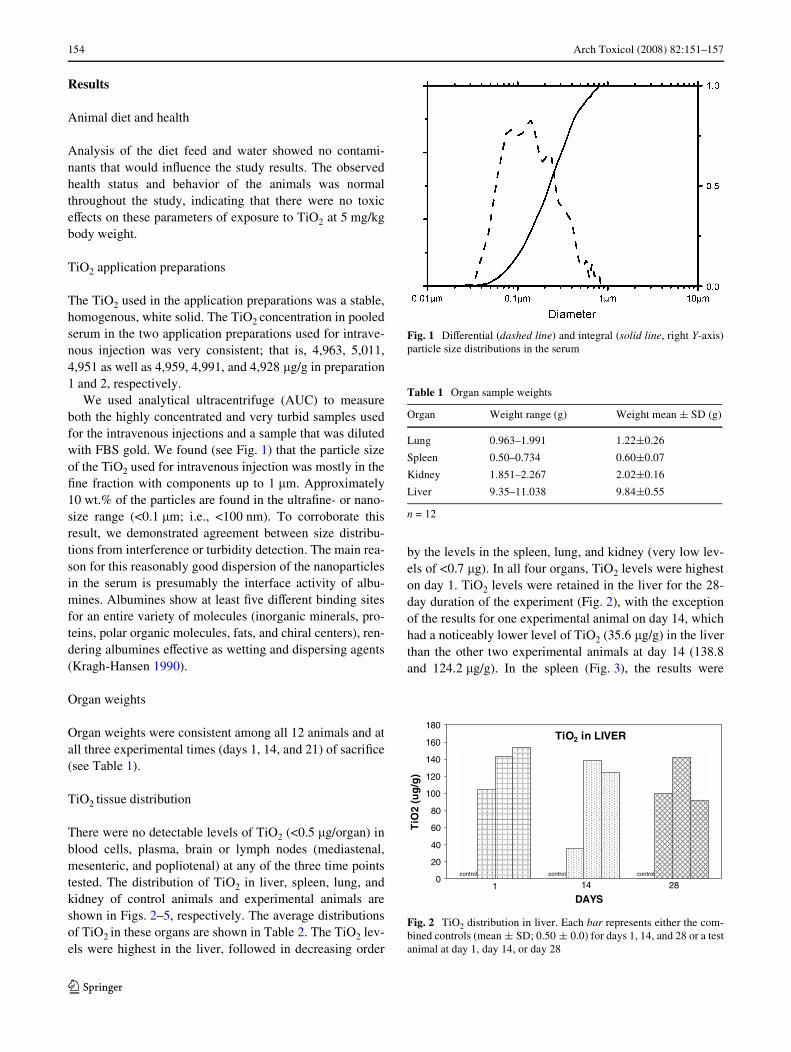
There were no detectable levels of TiO2 (<0.5 �g/organ) inblood cells, plasma, brain or lymph nodes (mediastenal,mesenteric, and popliotenal) at any of the three time pointstested. The distribution of TiO2 in liver, spleen, lung, andkidney of control animals and experimental animals areshown in Figs. 2–5, respectively. The average distributionsof TiO2 in these organs are shown in Table 2. The TiO2 lev-els were highest in the liver, followed in decreasing order
by the levels in the spleen, lung, and kidney (very low lev-els of <0.7 �g). In all four organs, TiO2 levels were higheston day 1. TiO2 levels were retained in the liver for the 28-day duration of the experiment (Fig. 2), with the exceptionof the results for one experimental animal on day 14, whichhad a noticeably lower level of TiO2 (35.6 �g/g) in the liverthan the other two experimental animals at day 14 (138.8and 124.2 �g/g). In the spleen (Fig. 3), the results were
Fig. 1 DiVerential (dashed line) and integral (solid line, right Y-axis)particle size distributions in the serum
Table 1 Organ sample weights
n = 12
Organ Weight range (g) Weight mean § SD (g)
Lung 0.963–1.991 1.22§0.26
Spleen 0.50–0.734 0.60§0.07
Kidney 1.851–2.267 2.02§0.16
Liver 9.35–11.038 9.84§0.55
Fig. 2 TiO2 distribution in liver. Each bar represents either the com-bined controls (mean § SD; 0.50 § 0.0) for days 1, 14, and 28 or a testanimal at day 1, day 14, or day 28
1 14 28
TiO2 in LIVER
0
20
40
60
80
100
120
140
160
180
DAYS
)g/
gu(
2Oi
T
controlcontrolcontrol
123

Arch Toxicol (2008) 82:151–157 155
more variable than in the liver, but showed a trend of aslight decrease in TiO2 levels from day 1 to days 14 and 28.An early distribution of TiO2 to the lung on day 1 is shownin Fig. 4, with a return to near control levels by day 14.Similarly, in the kidney (Fig. 5), there was an increase inTiO2 levels compared with control on day 1 and a return tocontrol levels by day 14.
EVects of TiO2 exposure on biochemical parameters in serum
Various cytokines and enzymes (a total of 67 parameters)were measured in blood samples to assess potential inXam-matory responses and/or organ injury due to intravenousexposure to TiO2. None of these parameters showed deWni-tive changes at days 1, 14, or 28 following TiO2 exposure,indicating that there was no detectable inXammatoryresponse or organ toxicity.
Discussion
The eVects of TiO2 nanoparticles have been studied in sev-eral animal models (e.g., rats, mice, and rabbits) followingexposure by multiple routes of administration, includingwhole body exposure, dermal exposure, gastric lavage, andinhalation (Bermudez et al. 2004; Grassian et al. 2007;Wang et al. 2007). The most discussed aspect at present isthe induction of reactive oxygen species being due to chem-ical properties of TiO2 and/or to inXammatory responsesdue to particle exposure of the lung. After inhalation expo-sure, nanoparticles accumulate in the lung, leading tophagocytosis and, depending on the atmospheric concentra-tion, perhaps particle overload.
Fig. 3 TiO2 distribution in spleen. Each bar represents either the com-bined controls (mean § SD; 0.80 § 1.0) for days 1, 14, and 28 or a testanimal at day 1, day 14, or day 28
)g/
gu(
2Oi
T
0
10
20
30
40
50
60
70
80
90
100
control controlcontrol
DAYS1 2814
TiO2 in SPLEEN
Fig. 4 TiO2 distribution in lung. Each bar represents either thecombined controls (mean § SD; 0.567 § 0.115) for days 1, 14, and 28or a test animal at day 1, day 14, or day 28
0
1
2
3
4
5
6
7
8
9
10
DAYS
)g/
gu(
2Oi
T
14 281
control control control
TiO2 in LUNG
Table 2 TiO2 distribution in organs
Organ TiO2 on day 1 TiO2 on day 14 TiO2 on day 28
Controla Treatedb Controla Treatedb Controla Treatedb
Liver 0.5 133.8 § 26.0 0.5 99.5 § 55.8 0.5 111.3 § 27.3
Spleen 0.8 78.7 § 19.3 0.9 48.8 § 27.8 0.7 33.3 § 28.8
Lung 1.5 8.8 § 0.6 1.7 2.8 § 1.4 1.5 2.3 § 1.0
Kidney 0.3 0.67 § 0.06 0.3 0.2 § 0 0.2 0.2 § 0
Values expressed as mean § SD (�g/g)a n = 1b n = 3
Fig. 5 TiO2 distribution in kidney. Each bar represents either thecombined controls (mean § SD; 0.267 § 0.058) for days 1, 14, and 28or a test animal at day 1, day 14, or day 28
TiO2 in KIDNEY
0
0,1
0,2
0,3
0,4
0,5
0,6
0,7
0,8
DAYS
)g/
gu(
2Oi
T
1 14 28
control control
control
123

156 Arch Toxicol (2008) 82:151–157
In this study, we determined the distribution of TiO2
nanoparticles following intravenous administration to gaininsight into the kinetics of this material. Moreover, weinvestigated the eVect of TiO2 on a great number of bio-chemical parameters. Particle eVects are not expected to bethe same after inhalation or after intravenous application.With the experimental setting of the current study, we donot aim to mimic inhalative exposure but to address the cur-rent concern, once nanoparticles enter the body, either viaingestion or after inhalation, they may become systemicavailable and cause toxic eVect in organs other than the portof entry (e.g., lung). The route of application (intravenous)produced a “worst case” scenario of 100% bioavailability.For this purpose, we made great eVort to prepare a applica-tion formulation containing high amount of nanoparticles.The test article preparation used in this study was con-Wrmed to be homogeneous, stable, of constant concentra-tion, and comprised of >10 wt.% nanoparticles of <100 nm.Accurate knowledge of the particle size is important as evi-denced by recent studies showing that the eVects of TiO2
can diVer depending on the exact size of the nanoparticle(Grassian et al. 2007; Warheit et al. 2007; Zhang et al.1999).
The overall results of this study indicate that ratsexposed to TiO2 nanoparticles by a route that allowedimmediate systemic availablity (i.e., intravenous adminis-tration) showed no obvious toxic or adverse health eVects atthe dose level of 5 mg/kg body weight. This lack of adverseeVects corroborates epidemiologic research that TiO2 haslow toxicity in humans (Chen and Fayerweather 1988;BoVetta et al. 2004) and is similar to that reported by Wanget al. (2007) who studied both nanoparticles (25 and 80 nm)and Wne-sized (155 nm) particles of TiO2 administered toadult mice by oral gavage in a much larger, single dose of5 g/kg body weight. As in our study, these researchersreported no obvious acute toxicity in male and female miceat 2 weeks after exposure to nanoparticles of TiO2. A recentreport comparing two routes of administration of largedoses of 4.7-nm particles of TiO2 in female mice alsorevealed that the test substance was well tolerated whengiven intravenously (560 mg/kg) (Umbreit et al. 2007).
Blood parameters that are known to indicate an immuneresponse or reXect changes in organ function wereunchanged in the animals exposed to intravenous TiO2 atthe dose of 5 mg/kg body weight used in this study. Therewere no trends or indications of change in enzymes reXec-tive of inXammatory response or organ (liver, kidney, andheart) toxicity. Therefore, even with 100% bioavailabilityof the TiO2 dose of 5 mg/kg body weight aVorded by theintravenous route of administration, there were no remark-able toxic eVects evident in the experimental animals. In asimilar study, female mice tolerated exposure to very largeintravenous doses (560 mg/kg) of 4.7-nm particles of TiO2
(Umbreit et al. 2007) with no clinical or hematologic signsof a systemic inXammatory response. Further analysis (inthis same study) of the immunological eVects in femalemice of very large intravenous doses (560 mg/kg) of 4.7-nm particles of TiO2 (Weaver et al. 2007) showed T-cellproliferation in lymph nodes that were cultured with Con-Afor 3 days. Cytokine analyses of these mitogen-stimulatedlymph node cells revealed increased production of TNF-alpha, IL-6, and gamma-interferon, indicating that theimmune system may be sensitive to the test nanoparticle.The diVerence between the inXammatory response resultsof our investigative study and the results of the intravenousstudy with mice described above most likely is related tothe much higher (100-fold) dose, although it cannot beexcluded that the sizes of nanoparticles or the diVerentexperimental animal species used could have also contrib-uted to the diVerence.
A similar lack of indication of organ injury to that foundin our study was recently reported in the intravenous studyby Umbreit and colleagues (Umbreit et al. 2007). Macro-scopically, all tissues examined (brain, liver, spleen, lymphnodes, and kidney) appeared normal. In contrast, TiO2
nanoparticles (80 nm) administered at a much higher dose(5 g/kg) to male and female mice by a single oral gavage(Wang et al. 2007) have been reported to cause an elevatedALT/AST enzyme ratio and elevated levels of serum LDH,both of which are indicative of hepatic injury, in femaleanimals only. In the same study, elevated serum BUN andCR levels in female mice indicated kidney dysfunction.The hepatic injury was conWrmed with observations ofincreased liver weight and hepatocyte necrosis, and the kid-ney dysfunction was evidenced in a swollen renal glomeru-lus and proteinic liquid accumulation in the renal tubules.The authors suggest that retention of TiO2 nanoparticles inthe liver and kidney, due to size and diYcult clearance ofTiO2 (Oberdörster et al. 2005), are the causes of the organinjury. Wang et al. (2007) also reported possible hypoxia ofcells in the heart as evidenced by elevated serum LDHcombined with elevated CK and alpha-HBDH. This dis-crepancy in results concerning biochemical parameters inserum between our study and that of Wang et al. (2007) ismost likely attributable to diVerences in TiO2 dose (1,000-fold).
The tissue distribution and elimination of the test sub-stance was as expected considering the intravenous route ofexposure and given the function of the liver and kidney asroutes of metabolism and elimination (of TiO2). As shownin Figs. 2–5 and Table 2, the TiO2 levels were highest onday 1 in all four organs. TiO2 levels were retained in theliver for the 28-day duration of the experiment (Fig. 2).This result is similar to a very early study by Huggins andFroehlich (1966) in which TiO2 particles (0.2–0.4 �m)injected intravenously to rats at a dose of 250 mg/kg
123
Arch Toxicol (2008) 82:151–157 157
accumulated in liver (69% at 5 min and 80% at 15 min). AsigniWcant accumulation of TiO2 80-nm nanoparticles com-pared with control has also been shown in the liver offemale mice at 2 weeks after a single 5-mg/kg gastric gav-age dose (Wang et al. 2007).
The distribution of TiO2 in the spleen (Fig. 3) was morevariable than that in the liver, but showed a trend of a slightdecrease in TiO2 levels from day 1 to days 14 and 28. Anearly distribution of TiO2 to the lung and kidneys on day 1 isshown in Figs. 4 and 5, respectively, with a return to near con-trol levels by days 14 in both organs. Retention of TiO2 nano-particles was reported to reach statistical signiWcancecompared with controls in the spleen, lung, and kidney offemale mice 2 weeks following exposure to the test substanceby gastric gavage (Wang et al. 2007). Using the same route ofadministration, yet a much larger dose of TiO2, Umbreit et al.(2007) reported rapid clearance of TiO2 from the circulationand accumulation in the lungs, liver, and spleen of exposedmice, as evidenced by the whitish color of these tissues. Therewere no detectable levels of TiO2 (<0.5 �g) in blood cells orplasma at any of the three times tested in our study, also sug-gesting a rapid clearance of the test substance from the bloodinto the lung, spleen, kidney, and liver.
This study is rather preliminary in terms of safety evalua-tion of TiO2. Even though no biochemical markers werealtered as compared to control animals, TiO2 has not beencleared from liver and spleen within the observation period,indicating that after continuous exposure TiO2 would be accu-mulating in these organs even at low exposure levels. A largerstudy with more animals would be needed to conWrm that atthe low dose used, nanoparticles of TiO2 do not cause adversehealth issues, inXammatory responses, or organ toxicity/dys-function in rats, whereupon, organs and tissues should beinvestigated by histological examinations. Considering theaccumulation eVect, the recovery period may be extended toat least 3 months. The data from such studies would provideaccurate pharmacokinetic and biokinetic descriptions of thishighly useful substance that would be relevant for the evalua-tion of test results from other toxicological studies and for theextrapolation of data from animals to humans.
References
Bermudez E, Mangum J, Wong B, Asgharian B, Hext P, Warheit D,Everitt JI (2004) Pulmonary responses of mice, rats, and Ham-sters to subchronic inhalation of ultraWne titanium dioxide parti-cles. Toxicol Sci 77:347–357
BoVetta P, Soutar A, Cherrie JW, Granath F, Anderson A, Anttila A,Blettner M, Gaborieau V, Klug SJ, Langard S, Luce D, Merletti F,Miller B, Mirabelli D, Pukkala E, Adami HO, Weiderpass E(2004) Mortality among workers employed in the titanium dioxideproduction industry in Europe. Cancer Causes Control 15:697–706
Chen JL, Fayerweather WE (1988) Epidemiologic study of workersexposed to titanium dioxide. J Occup Med 30:937–942
Cölfen H (2004) Analytical ultracentrifugation of nanoparticles. In:Nalwa HS (ed) Encyclopedia of Nanoscience and Nanotechnol-ogy. American ScientiWc Publishers, Valencia, CA, pp 67–88
EC Commission Directive 87/302/EEC of November 18, 1987; Part B:Methods for the determination of toxicity (1988). Toxicokinetics;OYcial Journal of the European Communities No. L 133:51-54
ECETOC (2006) Workshop on testing strategies to establish the safetyof Nanomaterials 7–8 November 2005, Barcelona, Workshop Re-port No. 7. ECETOC, Brussels, Belgium
Grassian VH, O’Shaughnessy PT, Adamcakova-Dodd A, PettiboneJM, Thorne PS (2007) Inhalation exposure study of titanium diox-ide nanoparticles with a primary size of 2 to 5 nm. Environ HealthPerspect 115(3):397–402
Huggins CB (1939) A quantitative study of the activity of the reticulo-endothelial structures in bone marrow in normal and ischemiclimbs as indicated by India ink and titanium dioxide. Anat Rec74:231
Huggins CB, Froehlich JP (1966) High concentration of injected titaniumdioxide in abdominal lymph nodes. J Exp Med 124(6):1099–1106
Japan/MAFF: guidelines on the Compiling of Test Results on Toxic-ity; Tests on In Vivo Fate In Animals (2001)
Kragh-Hansen U (1990) Dan Med Bull 37:57Lin MY, Lindsay HM, Weitz DA, Ball RC, Klein R, Meakin P (1990)
Universal reaction-limited colloid aggregation. Phys Rev A41:2005–2020
Mächtle W, Börger L (2006) Analytical Ultracentrifugation of poly-mers and Nanoparticles. Springer, Berlin
Ma-Hock L, Gamer AO, Landsiedel R, Leibold E, Frechen T, Sens B,Linsenbuehler M, van Ravenzwaay B (2007) Generation andcharacterization of test atmospheres with nanomaterials. InhalToxicol 19:833–848. doi:10.1080/08958370701479190
Oberdörster G, Maynard A, Donaldson K, Castranova V, Fitzpatrick J,Ausman K et al (2005) Principles for characterizing the potentialhuman health eVects from exposure to nanomaterials: elements ofa screening strategy. Particle Fibre Toxicol. doi:10.1186/1743-8977-2-8 [Online 6 October 2005]
OECD Guidelines for Testing of Chemicals (1984) Method No. 417Toxicokinetics, Version dated 4.4.1984
Ross R (2007) Clearing the air. Town Ctry Mag April: 68Umbreit T, Weaver JL, Miller TJ, Zhang J, Shah R, Stratmeyer ME,
Tomazic-Jezic V (2007) Toxicology of titanium dioxide (TiO2)nanoparticles: 1. Characterization and tissue distribution in sub-cutaneously and intravenously injected mice. Toxicologist96(1):287. Society of Toxicology 46th Annual Meeting and Tox-Expo. Abstract #1386. March 25–29, 2007, Charlotte, NC
U.S. EPA, Health EVects Guidelines, OPPTS 870.7485, Metabolismand Pharmacokinetics, August (1998). http://www.epa.gov/opptstrs/ publications/oppts_Harmonized/S70_Health_EVects_Test_Guidelines/Series/S70-7485.pdf
Wang J, Zhou G, Chen C, Yu H, Wang T, Ma Y, Jia G, Gao Y, Li B,Sun J, Li Y, Jiao F, Zhao Y, Chai Z (2007) Acute toxicity and bio-distribution of diVerent sized titanium dioxide particles in miceafter oral administration. Toxicol Lett 168(2):176–185
Warheit DB, Webba TR, Reeda KL, Frerichsb S, Sayesa CM (2007)Pulmonary toxicity study in rats with three forms of ultraWne-TiO2 particles: diVerential responses related to surface properties.Toxicology 230(1):90–104
Weaver J, Umbreit TH, Miller TJ, Zhang J, Stratmeyer ME, Tomazic-Jezic V (2007) Toxicology of titanium dioxide (TiO2) nanoparti-cles: 2. Immunological eVects in subcutaneously and intrave-nously injected mice. Toxicologist 96(1):288. Society ofToxicology 46th Annual Meeting and ToxExpo. Abstract #1391,March 25–29, 2007, Charlotte, NC
Zhang HZ, Penn RL, Hamers RJ, BanWeld JF (1999) Enhanced adsorp-tion of molecules on surfaces of nanocrystalline particles. J PhysChem B 103:4656–4662
123