Mouse Mast Cell Protease 4 Is the Major Chymase in Murine Airways and Has a Protective Role in...
8
Mouse Mast Cell Protease 4 Is the Major Chymase in Murine Airways and Has a Protective Role in Allergic Airway Inflammation 1 Ida Waern,* Sofia Jonasson, † Josephine Hjoberg, † Anders Bucht, ‡§ Magnus Åbrink, ¶ Gunnar Pejler,* and Sara Wernersson 2 * It is widely established that mast cells (MCs) have a harmful role in asthma, for example by secreting various proinflammatory substances stored within their secretory granule. However, in this study, we show that one of the substances stored within MC granule, chymase, in fact has a protective role in allergic airway inflammation, indicating that MCs may possess both harmful and protective activities in connection with this type of disease. Wild-type (WT) mice and mice lacking mouse MC protease 4 (mMCP- 4), a chymase that is functionally homologous to human chymase, were sensitized and challenged with OVA, followed by the assessment of airway physiology and inflammatory parameters. Our results show that the airway hyperresponsiveness was sig- nificantly higher in mMCP-4 / as compared with WT mice. Moreover, the degree of lung tissue inflammation was markedly higher in mice lacking mMCP-4 than in WT controls. Histological analysis revealed that OVA sensitization/challenge resulted in a marked increased in the thickness of the smooth muscle cell (SMC) layer and, notably, that the degree of SMC layer thickening was more pronounced in mMCP-4 / animals than in WT controls, thus indicating that chymase may have an effect on airway SMCs. In support of this, mMCP-4-positive MCs were located in the close vicinity of the SMC layer, mainly in the upper airways, and mMCP-4 was shown to be the major chymase expressed in these MCs. Taken together, our results indicate that chymase present in the upper airways protects against allergic airway responses, possibly by regulating SMCs. The Journal of Immu- nology, 2009, 183: 6369 – 6376. A llergic asthma is a complex disease characterized by re- versible airflow obstruction, airway hyperresponsive- ness (AHR), 3 and airway inflammation and remodeling. Mast cells (MCs) are key effector cells in allergic diseases, and they may contribute to asthma pathology in several ways (1). MCs can be activated by different types of stimuli, e.g., by allergen- induced cross-linking of the high-affinity IgE receptor, FcRI, and this may result in degranulation (2). Upon MC degranulation, a broad range of preformed bioactive substances is released, includ- ing histamine, cytokines, proteoglycans, and various proteases, with the latter group consisting of chymases, tryptases, and car- boxypeptidase A (CPA) (3). A role of MCs in asthma has been suggested for many years, supported by a multitude of findings (1, 4, 5). MC degranulation is often seen in asthmatic lungs, and various MC mediators can be found in bronchoalveolar lavage (BAL) fluid from asthmatic patients. Furthermore, a strong infiltration of MCs into the air- way smooth muscle cell (SMC) layer is a prominent feature of asthma, especially in allergic asthma (6), and is associated with AHR (7). Involvement of MCs in allergic asthma is also sup- ported by animal studies. Thus, mice lacking functional MCs are less responsive in models of asthma in which allergic airway inflammation is induced by administration of OVA without ex- ternal adjuvant (8 –11). Although a role for MCs in allergic asthma is now widely rec- ognized, it is still unclear by which mechanism MCs contribute to airway responses. One plausible scenario would be that the pre- formed proteases that are released upon MC degranulation con- tribute by provoking different types of airway responses. In line with this idea, MC tryptase has been shown to induce airway in- flammation and AHR, and tryptase inhibitors can reduce these types of responses (12–14). Also, chymase has been implicated in inflammatory responses, as shown by the ability of chymases to induce eosinophil and neutrophil tissue infiltration (15–17). More- over, a chymase polymorphism has been linked to allergic asthma (18), although it is not clear in which way this polymorphism affects chymase levels and/or activity. Most reports implicate MC proteases as proinflammatory agents, but it cannot be excluded that they, in addition, may have protective functions. In fact, it has been shown that the presence of chymase in small airways correlates with preserved airway function in severe asthmatics, thus suggest- ing a protective role of chymase (19). *Department of Anatomy, Physiology and Biochemistry, Swedish University of Ag- ricultural Sciences, Uppsala, Sweden; † Department of Medical Sciences, Uppsala University, Uppsala, Sweden; ‡ Department of Respiratory Medicine and Allergy, Umeå University, Umeå, Sweden; § Swedish Defense Research Agency, Umeå, Swe- den; and ¶ Department of Medical Biochemistry and Microbiology, Uppsala Univer- sity, Uppsala, Sweden Received for publication January 21, 2009. Accepted for publication September 12, 2009. The costs of publication of this article were defrayed in part by the payment of page charges. This article must therefore be hereby marked advertisement in accordance with 18 U.S.C. Section 1734 solely to indicate this fact. 1 This work was supported by grants from the Vårdal Foundation, the Swedish Society of Medicine, the Swedish Research Council, the Swedish Cancer Foun- dation, Gustaf V:s 80-year Anniversary Foundation, and the Swedish Heart-Lung Foundation. 2 Address correspondence and reprint requests to Dr. Sara Wernersson, Swedish Univer- sity of Agricultural Sciences, Department of Anatomy, Physiology and Biochemistry, BMC, Box 575, 75123 Uppsala, Sweden. E-mail address: [email protected] 3 Abbreviations used in this paper: AHR, airway hyperresponsiveness; BAL, bron- choalveolar lavage; CAE, chloroacetate esterase; C L , lung compliance; CPA, car- boxypeptidase A; FGF, fibroblast growth factor; i.n., intranasal; MC, mast cell; mMCP, mouse mast cell protease; PDGF, platelet-derived growth factor; R L , lung resistance; SMC, smooth muscle cell; WT, wild type. Copyright © 2009 by The American Association of Immunologists, Inc. 0022-1767/09/$2.00 The Journal of Immunology www.jimmunol.org/cgi/doi/10.4049/jimmunol.0900180
-
Upload
independent -
Category
Documents
-
view
3 -
download
0
Transcript of Mouse Mast Cell Protease 4 Is the Major Chymase in Murine Airways and Has a Protective Role in...

Mouse Mast Cell Protease 4 Is the Major Chymase in MurineAirways and Has a Protective Role in Allergic AirwayInflammation1
Ida Waern,* Sofia Jonasson,† Josephine Hjoberg,† Anders Bucht,‡§ Magnus Åbrink,¶
Gunnar Pejler,* and Sara Wernersson2*
It is widely established that mast cells (MCs) have a harmful role in asthma, for example by secreting various proinflammatorysubstances stored within their secretory granule. However, in this study, we show that one of the substances stored within MCgranule, chymase, in fact has a protective role in allergic airway inflammation, indicating that MCs may possess both harmful andprotective activities in connection with this type of disease. Wild-type (WT) mice and mice lacking mouse MC protease 4 (mMCP-4), a chymase that is functionally homologous to human chymase, were sensitized and challenged with OVA, followed by theassessment of airway physiology and inflammatory parameters. Our results show that the airway hyperresponsiveness was sig-nificantly higher in mMCP-4�/� as compared with WT mice. Moreover, the degree of lung tissue inflammation was markedlyhigher in mice lacking mMCP-4 than in WT controls. Histological analysis revealed that OVA sensitization/challenge resulted ina marked increased in the thickness of the smooth muscle cell (SMC) layer and, notably, that the degree of SMC layer thickeningwas more pronounced in mMCP-4�/� animals than in WT controls, thus indicating that chymase may have an effect on airwaySMCs. In support of this, mMCP-4-positive MCs were located in the close vicinity of the SMC layer, mainly in the upper airways,and mMCP-4 was shown to be the major chymase expressed in these MCs. Taken together, our results indicate that chymasepresent in the upper airways protects against allergic airway responses, possibly by regulating SMCs. The Journal of Immu-nology, 2009, 183: 6369–6376.
A llergic asthma is a complex disease characterized by re-versible airflow obstruction, airway hyperresponsive-ness (AHR),3 and airway inflammation and remodeling.
Mast cells (MCs) are key effector cells in allergic diseases, andthey may contribute to asthma pathology in several ways (1). MCscan be activated by different types of stimuli, e.g., by allergen-induced cross-linking of the high-affinity IgE receptor, Fc�RI, andthis may result in degranulation (2). Upon MC degranulation, abroad range of preformed bioactive substances is released, includ-ing histamine, cytokines, proteoglycans, and various proteases,
with the latter group consisting of chymases, tryptases, and car-boxypeptidase A (CPA) (3).
A role of MCs in asthma has been suggested for many years,supported by a multitude of findings (1, 4, 5). MC degranulationis often seen in asthmatic lungs, and various MC mediators canbe found in bronchoalveolar lavage (BAL) fluid from asthmaticpatients. Furthermore, a strong infiltration of MCs into the air-way smooth muscle cell (SMC) layer is a prominent feature ofasthma, especially in allergic asthma (6), and is associated withAHR (7). Involvement of MCs in allergic asthma is also sup-ported by animal studies. Thus, mice lacking functional MCsare less responsive in models of asthma in which allergic airwayinflammation is induced by administration of OVA without ex-ternal adjuvant (8 –11).
Although a role for MCs in allergic asthma is now widely rec-ognized, it is still unclear by which mechanism MCs contribute toairway responses. One plausible scenario would be that the pre-formed proteases that are released upon MC degranulation con-tribute by provoking different types of airway responses. In linewith this idea, MC tryptase has been shown to induce airway in-flammation and AHR, and tryptase inhibitors can reduce thesetypes of responses (12–14). Also, chymase has been implicated ininflammatory responses, as shown by the ability of chymases toinduce eosinophil and neutrophil tissue infiltration (15–17). More-over, a chymase polymorphism has been linked to allergic asthma(18), although it is not clear in which way this polymorphismaffects chymase levels and/or activity. Most reports implicate MCproteases as proinflammatory agents, but it cannot be excluded thatthey, in addition, may have protective functions. In fact, it has beenshown that the presence of chymase in small airways correlateswith preserved airway function in severe asthmatics, thus suggest-ing a protective role of chymase (19).
*Department of Anatomy, Physiology and Biochemistry, Swedish University of Ag-ricultural Sciences, Uppsala, Sweden; †Department of Medical Sciences, UppsalaUniversity, Uppsala, Sweden; ‡Department of Respiratory Medicine and Allergy,Umeå University, Umeå, Sweden; §Swedish Defense Research Agency, Umeå, Swe-den; and ¶Department of Medical Biochemistry and Microbiology, Uppsala Univer-sity, Uppsala, Sweden
Received for publication January 21, 2009. Accepted for publication September12, 2009.
The costs of publication of this article were defrayed in part by the payment of pagecharges. This article must therefore be hereby marked advertisement in accordancewith 18 U.S.C. Section 1734 solely to indicate this fact.1 This work was supported by grants from the Vårdal Foundation, the SwedishSociety of Medicine, the Swedish Research Council, the Swedish Cancer Foun-dation, Gustaf V:s 80-year Anniversary Foundation, and the Swedish Heart-LungFoundation.2 Address correspondence and reprint requests to Dr. Sara Wernersson, Swedish Univer-sity of Agricultural Sciences, Department of Anatomy, Physiology and Biochemistry,BMC, Box 575, 75123 Uppsala, Sweden. E-mail address: [email protected] Abbreviations used in this paper: AHR, airway hyperresponsiveness; BAL, bron-choalveolar lavage; CAE, chloroacetate esterase; CL, lung compliance; CPA, car-boxypeptidase A; FGF, fibroblast growth factor; i.n., intranasal; MC, mast cell;mMCP, mouse mast cell protease; PDGF, platelet-derived growth factor; RL, lungresistance; SMC, smooth muscle cell; WT, wild type.
Copyright © 2009 by The American Association of Immunologists, Inc. 0022-1767/09/$2.00
The Journal of Immunology
www.jimmunol.org/cgi/doi/10.4049/jimmunol.0900180

In this study, we addressed the role of MC chymase in allergicasthma by comparing the inflammatory responses and airway re-activity in wild-type (WT) mice with those in mice lacking mousemast cell protease 4 (mMCP-4). Of the several murine MC chy-mases (mMCP-1, -2, -4, -5, and -9), mMCP-4 has similar tissuedistribution, substrate specificity, and heparin-binding propertiesas human chymase, and is therefore likely to be its functionalcounterpart (20–22). We show that mice lacking mMCP-4 developmarkedly elevated AHR as compared with WT counterparts, in-dicating that chymase has a protective role in allergic airwayresponses.
Materials and MethodsMice and immunization protocol
Mice deficient in mMCP-4 (mMCP-4�/�) (20) were backcrossed for 13generations to the C57BL/6J genetic background. Experimental groupswere age and sex matched, and WT littermates (mMCP-4�/�) were used ascontrols. Animals were bred and maintained in the animal facility at Upp-sala Biomedical Centre. All experiments were approved by the local ethicalcommittee. Eight- to 12-wk-old mice were immunized i.p. with 10 �g ofOVA (Sigma-Aldrich) in 150 �l of PBS on days 1, 3, 6, 8, 10, 13, and 15.Control mice were nonimmunized. On days 31, 34, and 36, all mice werechallenged with 20 �g of OVA in 20 �l of PBS intranasally (i.n.). Separateexperiments included mice that were immunized i.p. with OVA, but notchallenged i.n. with OVA.
Airway reactivity and bronchoalveolar lavage
One or 2 days after the last Ag challenge, mice were anesthetized, trache-ostomized, and coupled to a FlexiVent ventilator (23, 24). Airway respon-siveness to incremental doses of i.v. methacholine was assessed. Resultsare presented as lung resistance (RL) and lung compliance (CL). Resultswere pooled from seven separate and matched experiments, and data wereobtained from a total of 10 mice per group. BAL fluid was collected byrinsing the lungs twice with 1 ml of HBSS, and the total number of cellswas determined. Cytospin slides were prepared and stained with May-Grunwald and Giemsa, followed by differential count of at least 200 cellsper slide.
ELISA
Analysis of OVA-specific IgE in sera was performed using Mouse IgEELISA Quantification Kit (Bethyl Laboratories). Serum samples were di-luted 1/3, and anti-OVA IgE Abs were quantified with biotinylated OVA(2 �g/ml), prepared as described (25), followed by incubation with 0.05 Uof streptavidin-peroxidase conjugate (Roche Diagnostics). For OVA-spe-cific IgG ELISA, serum samples diluted 1/1000 were added to OVA-coatedmicrotiter plates, and bound IgG was detected with horseradish-conjugatedanti-mouse IgG (dilution 1/1000; GE Healthcare). For enzyme detection,3,3�,5�5tetramethylbenzidine liquid substrate (Sigma-Aldrich) was used.
Histology
The left lung lobes were fixed in 4% paraformaldehyde, paraffin embedded,and cut into 6-�m sections. Samples were deparaffinized and stained withH&E, toluidine blue, or chloroacetate esterase, as described earlier (26).Trachea was embedded in OCT and kept in �70°C before cutting 6-�msections. Lung tissue inflammation was evaluated by blinded analyses ofnumbers of infiltrating cells per defined field of H&E-stained lung sectionsfrom five to seven animals per group. Cells were counted at �400 mag-nification, using an eyepiece fitted with a square graticule that covered afield of view measuring 0.06 mm2. Tissue inflammation in larger vs smallerairways was evaluated by separately counting the cells in regions aroundprimary and secondary airways, or the cells around terminal bronchioles,respiratory bronchioles, and alveoli, respectively. The numbers of cells inat least three randomly selected regions for each individual were deter-mined, and mean values were used for statistical calculations. The thick-ness of the smooth muscle layer was evaluated by blinded scoring from 0to 5 of regions around the upper bronchus, resulting in an average score foreach section. Number of MCs was evaluated by counting toluidine blue-stained cells in the connective tissue and smooth muscle layer underneaththe epithelia of the primary bronchi. Chymotryptic activity was detected byblinded scoring from 0 to 4 of the chloroacetate esterase-staining intensityof MCs around the primary bronchi.
Western blot
Tissue extracts from the upper (one-fourth) part of the left lung lobe werehomogenized in PBS/1 mM EDTA/1% Triton X-100 and centrifuged at13,400 rpm at 4°C for 20 min, and supernatants were collected for analysesof fibronectin, smooth muscle �-actin, and �-actin. The resulting pelletswere extracted with PBS/1 mM EDTA/1% Triton X-100 containing 2.0 MNaCl, and supernatants were used for analyses of mMCP-1, mMCP-2,mMCP-4, mMCP-5, mMCP-6, CPA, and �-actin. Samples were separatedunder reducing conditions on 12% SDS-PAGE gels (or 8% SDS-PAGEgels for fibronectin). Western blots were performed using Abs to smoothmuscle �-actin (Sigma-Aldrich; 1/750 dilution), �-actin (Santa Cruz Bio-technology), antisera to fibronectin (a gift of S. Johansson, UppsalaUniversity, Uppsala, Sweden; 1/250 dilution), or antisera to mMCP-1,mMCP-2, mMCP-4, mMCP-5, mMCP-6, and CPA (26, 27) (1/500 dilu-tion). Purified mMCP-4 (28), mucosal-type bone marrow-derived MCs(26), or peritoneal cell-derived MCs (29) were used as positive controls formMCP-4, mMCP-2, and mMCP-5, respectively. Western blot signals forsmooth muscle �-actin were visualized and quantified using the ImageReader V 1.7E software. Data were measured as pixels/mm2, and resultswere obtained by dividing the values for smooth muscle �-actin samples bythe values of corresponding �-actin control.
In vitro cleavage
Cleavage of platelet-derived growth factor (PDGF)-BB (provided by C.-H.Heldin, Uppsala University, Uppsala, Sweden) and fibroblast growth factor(FGF)-2 (PeproTech) was performed using 10 �g of substrate and 0.5 �gof purified mMCP-4 (28) in PBS/0.05% Triton X-100. Reactions (37°C)were interrupted at 20 min, 2 h, or 6 h by addition of SDS-PAGE samplebuffer, followed by boiling for 5 min. Samples were run under reducingconditions on a 12% SDS-PAGE gel. Results were obtained by AgNO3
staining.
Statistical analysis
Statistical differences were determined with Student’s t test for differentialcount and ELISA, with Mann-Whitney U test for histology and densitometry,and with two-way ANOVA for airway reactivity measurements, using Graph-Pad Prism 4.0 (GraphPad). All values are displayed as means � SEM.
ResultsEnhanced allergic airway inflammation in mMCP-4�/� mice
To investigate the role of mMCP-4 in allergic airway inflamma-tion, we compared the OVA-induced accumulation of cells in BALand lung tissue of WT and mMCP-4�/� mice. Sensitization andchallenge with OVA resulted in a prominent increase of total BALcells in both mMCP-4�/� and WT mice, mainly due to accumu-lation of eosinophils (Fig. 1A). However, the numbers of OVA-induced BAL eosinophils, neutrophils, macrophages, and lympho-cytes did not differ significantly between mMCP-4�/� and WTmice (Fig. 1A). Lung histology demonstrated a perivascular andperibroncheal infiltration of leukocytes, mainly eosinophils, in an-imals that had been both sensitized and challenged with OVA,whereas control animals (challenged only) showed no substantialsigns of inflammation (Fig. 1B). The tissue around larger airways(primary and secondary bronchi) displayed a higher density of in-flammatory cells compared with that around smaller airways (Fig1, C and D). Although OVA-induced tissue inflammation was ev-ident in both WT and mMCP-4�/� mice, a significant increase inthe number of infiltrating cells around both larger and smaller air-ways was seen in the absence of mMCP-4 (Fig. 1, B–D), thussuggesting a role for chymase as a negative regulator of allergen-induced airway inflammation.
AHR in the absence of mMCP-4
To analyze whether the increase in tissue inflammation in the ab-sence of mMCP-4 was accompanied by an increased AHR, inva-sive lung mechanics measurements were performed 1 or 2 daysafter the last OVA challenge. OVA-sensitized and OVA-chal-lenged mMCP-4�/� mice exhibited significantly enhanced RL ascompared with OVA-sensitized/challenged mMCP-4�/� mice
6370 PROTECTIVE ROLE OF CHYMASE IN ASTHMA

( p � 0.01), as well as compared with nonsensitized controls ( p �0.001; Fig. 2A). Thus, induction of AHR was clearly seen in OVA-sensitized/challenged mMCP-4�/� mice, but was undetectable inthe corresponding WT mice. These results strongly suggest that thepresence of mMCP-4 in WT mice protects from AHR in this
model of allergic airway inflammation. In line with the increasedRL, OVA-sensitized/challenged mMCP-4�/� mice displayed asignificant decrease in CL as compared with nonimmunizedmMCP-4�/� controls at the dose of 1 mg/kg methacholine ( p �0.05; Fig. 2B).
Normal allergic sensitization in mMCP-4�/� mice
The increased airway inflammation and AHR seen in mMCP-4�/�
mice could potentially be explained by a deficient control of thehumoral immune response during OVA sensitization. To studythe effects of mMCP-4 on Ab production, we measured thesystemic OVA-specific IgE and IgG levels in all groups ofmice. Mice presensitized with OVA had significantly increasedserum levels of OVA-specific IgE (Fig. 3A) and IgG (Fig. 3B)compared with nonsensitized controls. However, no significantdifferences in the levels of OVA-specific IgE or IgG were ob-served when comparing WT and mMCP-4�/� animals (Fig. 3).This finding does not support a major role of mMCP-4 in thesensitization process.
Localization of MCs in upper airways
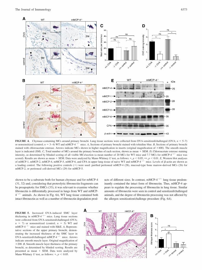
We evaluated the presence of MCs in the airways by toluidine bluestaining of lung tissue sections. In agreement with previous ob-servations (30), MCs were only rarely seen in the tissue aroundsmaller airways, either in control or sensitized/challenged animals(data not shown). In contrast, MCs were more abundant in thetissue around the larger upper airways, in particular in regionsclose to the primary bronchus (Fig. 4A) and trachea (supplementaldata).4 MC numbers and distribution were similar in WT andmMCP-4�/� animals, and there was no obvious difference in thedegree of MC degranulation when comparing OVA-sensitized/challenged mice and control mice (data not shown). Furthermore,no difference in numbers of MCs around the primary bronchus ortrachea was observed between sensitized/challenged animals vscontrols (Fig. 4C, and data not shown). Notably, MCs were par-ticularly abundant in the tissue below the epithelium and thesmooth muscle layer of the primary bronchus, and MCs in thisregion were frequently seen in close vicinity to the SMC layer(Fig. 4, A and B).
mMCP-4 is the major source of chymotryptic activity in lungMCs
To detect chymotrypsin-like activity in the upper airways, we usedthe chloroacetate esterase assay, resulting in red stain where activechymase is present. As shown in Fig. 4, B and D, chymase activitywas readily detected in WT MCs, whereas mMCP-4�/� MCs ex-hibited a severely reduced chymase activity, as demonstrated bythe faint staining. This indicates that mMCP-4 accounts for a majorpart of the chymase activity stored within lung MCs, i.e., otherchymases such as mMCP-1, mMCP-2, mMCP-5, or mMCP-9 con-tribute to a lesser extent to the total chymotrypsin-like activity inMCs of the upper airways. Moreover, OVA sensitization/challengedid not significantly affect the chymase activity of WT or mMCP-4�/� lung MCs, thus arguing against the possibility of a compen-satory up-regulation of other chymases during allergic airway in-flammation in mMCP-4�/� mice (Fig. 4D).
The presence of mMCP-4 in the larger airways of WT mice wasalso confirmed by Western blot analyses of the upper (one-fourth)part of the left lung lobe or of the trachea (Fig. 4E and supple-mental data). In contrast, mMCP-4 protein was below the level of
4 The online version of this article contains supplemental material.
FIGURE 1. Allergic airway inflammation in BAL and lung tissue ofmMCP-4�/� and WT mice. Mice were sensitized with OVA or were non-sensitized (controls). All mice received three i.n. instillations with 20 �g ofOVA. A, Differential counts of leukocytes in BAL fluid (n � 17–23, datapooled from 8 independent experiments). Data are presented as mean num-ber of cells � SEM. Differences between groups were analyzed by Stu-dent’s t test, as follows: �, p � 0.05; ��, p � 0.01; or ���, p � 0.001. B,Lung histology of representative sections from OVA-immunized mice (up-per panels) and controls (lower panels). Arrows indicate leukocyte infil-trates. Original magnifications were �100. Lung tissue inflammationaround larger (C) or smaller (D) airways was evaluated by blinded count-ing of number of infiltrating cells per field, as described in Materials andMethods (n � 5–7, data pooled from 3 independent experiments). Data arepresented as mean number of infiltrating cells/field � SEM. Data wereanalyzed by Mann-Whitney U test, and significant differences are indi-cated, as follows: �, p � 0.05 or ��, p � 0.01.
6371The Journal of Immunology

detection in lower parts of the lungs (data not shown). As ex-pected, mMCP-4 protein was not detected in tissue from mMCP-4�/� animals (Fig. 4E and supplemental data). In a previous study,it was demonstrated that peritoneal MCs from mMCP-4�/� micedisplay normal morphology, normal IgE-induced MC degranula-tion, and normal levels of other granule-stored proteases, i.e.,mMCP-5, mMCP-6, and CPA (20). To confirm that mMCP-4�/�
airway MCs exhibit an unaltered phenotype, we in this studyanalyzed for the presence of mMCP-1, mMCP-2, mMCP-5,mMCP-6, and CPA in upper airways. No substantial differencebetween mMCP-4�/� mice and WT controls could be detected,although a minor tendency of higher levels of mMCP-1 andmMCP-6, and lower levels of mMCP-2, was seen in the absence ofmMCP-4 (Fig. 4E). These findings demonstrate that there are nosubstantial compensatory alterations of the major MC proteases inthe absence of mMCP-4.
Increased OVA-induced smooth muscle thickening in primarybronchi of mMCP-4�/� mice
The localization of chymase-containing MCs mainly around theupper airways prompted us to further analyze the local tissue re-sponse at this site. Considering the profound effect of mMCP-4deficiency on AHR (Fig. 2), it was of particular interest to evaluateOVA-induced alterations in the SMC layer. Histological analysesrevealed a substantial OVA-induced thickening of the SMC layer
of the primary bronchi of mMCP-4�/� mice, whereas only a mod-est, nonsignificant thickening of the SMC layer was seen in theWT mice (Fig. 5). The increased smooth muscle thickness inmMCP-4�/� mice occurs only in response to the complete asthmaprotocol (OVA sensitization and challenge), because neither sen-sitization alone nor i.n. challenge alone had any affect on thesmooth muscle thickness of the primary bronchi (Fig. 5, and sup-plemental data). This is in line with a role for mMCP-4 only duringlocal allergic airway inflammation, i.e., when mMCP-4 is releasedupon MC degranulation during the effector phase of the disease.Moreover, the SMC layer thickness of OVA-sensitized/challengedmMCP-4�/� mice was significantly increased as compared withthat in OVA-sensitized/challenged WT mice (Fig. 5B). To furtheraddress the effect of mMCP-4 in tissue remodeling, we analyzedtotal levels of SMC �-actin in the upper (one-fourth) part of thelungs by Western blot analysis. As shown in Fig. 6, A and B, therewas a significantly lower level of total SMC �-actin in the (one-fourth) upper part of lung tissue from mMCP-4�/� control micecompared with that in WT controls, despite no obvious histologicaldifferences in the airway smooth muscle compartment (Fig. 5, anddata not shown). We cannot exclude that there is an altered lungtissue homeostasis and/or a generally reduced SMC �-actin ex-pression in the absence of mMCP-4 that is not dependent onallergic inflammation. Notably, there was a tendency of an aller-gen-induced increase in levels of total SMC �-actin in mMCP-4�/� mice, whereas the opposite tendency was seen for WT mice(Fig. 6B). Although these tendencies are compatible with an in-creased SMC response to allergen sensitization/challenge in theabsence of mMCP-4, the differences between groups did not reachstatistical significance. Thus, despite a clear effect of mMCP-4 onthe smooth muscle thickening of the primary bronchus (Fig. 5A),there was no substantial effect of mMCP-4 on the OVA-inducedincrease of total SMC �-actin levels of the upper (one-fourth) partof the lungs (including SMC around both small and large airwaysand blood vessels). Taken together, these observations suggest arole for mMCP-4 in the local control of allergen-induced tissueremodeling, i.e., in the control of SMC hyperplasia and/or hyper-trophy of primary airways.
Degradation of SMC mitogens and fibronectin by mMCP-4
To investigate possible mechanisms by which mMCP-4 could reg-ulate SMCs, we examined whether mMCP-4 can cleave purifiedSMC mitogens. Indeed, purified mMCP-4 cleaved both PDGF-BBand FGF-2 (Fig. 6, C and D). Another potential substrate formMCP-4 could be fibronectin. Fibronectin has previously been
FIGURE 2. AHR to methacholine in WT and mMCP-4�/� mice. Lung mechanics measurements were performed 24 or 48 h after last i.n. challenge ofOVA-sensitized (OVA) or nonsensitized (control) mice. A, RL was significantly enhanced in OVA-immunized mMCP-4�/� mice both vs controls (p �0.001) and vs OVA-immunized WT mice (p � 0.01), as determined by two-way ANOVA. B, CL. Statistical significances for individual doses are shown,as follows: �, p � 0.05 or ��, p � 0.01 of OVA-immunized mMCP-4�/� mice vs OVA-immunized WT mice; †, �0.05; ††, �0.01; or †††, �0.001 ofOVA-immunized groups vs controls; not significant, p � 0.05 (data not shown). Data were obtained from 7 independent experiments with a total of 10mice per group.
FIGURE 3. Normal OVA-specific Ab production in mMCP-4�/� mice.Sera from OVA-sensitized/challenged (OVA) or nonsensitized (control)WT or mMCP-4�/� mice were collected after allergen challenge. Sampleswere analyzed by ELISA for OVA-specific IgE (A) and OVA-specific IgG(B). Results are displayed as OD values � SEM. ���, p � 0.001 of OVA-immunized groups vs controls. Data were pooled from eight independentexperiments (n � 17–23).
6372 PROTECTIVE ROLE OF CHYMASE IN ASTHMA
shown to be a substrate both for human chymase and for mMCP-4(31, 32) and, considering that proteolytic fibronectin fragments canbe proapoptotic for SMCs (33), it was relevant to examine whetherfibronectin is differentially processed in lungs from WT and mMCP-4�/� animals. As shown in Fig. 6A, WT lung tissue contained bothintact fibronectin as well as a number of fibronectin degradation prod-
ucts of different sizes. In contrast, mMCP-4�/� lung tissue predom-inantly contained the intact form of fibronectin. Thus, mMCP-4 ap-pears to regulate the processing of fibronectin in lung tissue. Similaramounts of fibronectin were seen in control and sensitized/challengedanimals, and the degree of fibronectin processing was not affected bythe allergen sensitization/challenge procedure (Fig. 6A).
FIGURE 4. Chymase-containing MCs around primary bronchi. Lung tissue sections were collected from OVA-sensitized/challenged (OVA; n � 5–7)or nonsensitized (control; n � 3–4) WT and mMCP-4�/� mice. A, Sections of primary bronchi stained with toluidine blue. B, Sections of primary bronchistained with chloroacetate esterase. Arrows indicate MCs shown in higher magnification in inserts (original magnification of �400). The smooth musclelayer is indicated (SM). C, Total number of MCs around the primary bronchus of each section, shown as mean � SEM. D, Chloroacetate esterase stainingintensity, as determined by blinded scoring of all visible MCs/section (a mean number of 20 MCs for WT mice and 7.7 MCs for mMCP-4�/� mice wasscored). Results are shown as mean � SEM. Data were analyzed by Mann-Whitney U test, as follows: �, p � 0.05; ��, p � 0.01. E, Western blot analysesof mMCP-1, mMCP-2, mMCP-4, mMCP-5, mMCP-6, and CPA in upper lung tissue of naive WT and mMCP-4�/� mice. Levels of �-actin are shown asa loading control. The following positive controls (�) were used: purified peritoneal mMCP-4 (28), mucosal-type bone marrow-derived MCs (26) formMCP-2, or peritoneal cell-derived MCs (29) for mMCP-5.
FIGURE 5. Increased OVA-induced SMC layerthickening in mMCP-4�/� mice. Lung tissue sectionswere collected from OVA-sensitized/challenged (OVA;n � 7) or nonsensitized (control; n � 5) WT andmMCP-4�/� mice and stained with H&E. A, Represen-tative sections of the upper primary bronchi, demon-strating the increased thickness of the SMC layer inOVA-sensitized/challenged mMCP-4�/� mice. Arrowsindicate smooth muscle layer. Original magnification of�200. B, Smooth muscle layer thickness of the primarybronchi, as determined by blinded scoring. Results arepresented as mean � SEM. Data were analyzed byMann-Whitney U test, as follows: �, p � 0.05.
6373The Journal of Immunology

DiscussionThis is the first study in which the role of MC chymase in allergicairway responses is addressed by using a chymase-deficient mousestrain. We show that in response to OVA immunization and chal-lenge, mMCP-4�/� mice develop a more pronounced tissue in-flammation, AHR, and SMC remodeling than WT mice, indicatingthat chymase is a negative regulator of these features. Conse-quently, chymase does not seem to contribute to the overall det-rimental effects of MCs in allergic airway inflammation (11). In-stead, chymase could have a crucial role in counteracting theharmful reactions induced by other bioactive substances releasedby MCs or other inflammatory cells.
At first glance, the dampening effect of endogenous mMCP-4 onthe OVA-induced tissue inflammation could appear to be in dis-crepancy with the reported proinflammatory properties of chy-mases (15–17). However, studies demonstrating proinflammatoryeffects were frequently conducted by administration of purifiedchymases. Obviously, the context of an administered chymase,e.g., dose, timing, and localization, is radically different from thatof the endogenous counterpart. Furthermore, the physiologicalform of both human chymase and mMCP-4 is in complex withserglycin proteoglycans, and this association affects chymase func-tion (3). In other approaches, chymase functions have been in-ferred based on studies using various chymase inhibitors. How-ever, most chymase inhibitors have been developed to act againsthuman chymase, with the activity and selectivity toward the rele-vant animal chymase being less characterized. Therefore, the useof genetically targeted mice selectively lacking functional chy-mase may be a more relevant approach for identifying physiolog-ical effects of endogenous chymase in different inflammatory set-tings. The mechanism by which mMCP-4 dampens OVA-inducedlung tissue inflammation is not known, but could involve degra-dation of proinflammatory cytokines and chemokines (34). In ad-dition, because mMCP-4 is needed for proper processing of con-nective tissue components, as shown previously (20) and in Fig. 6,this may facilitate the transfer of leukocytes from the tissue intoBAL. In support of the latter notion, the numbers of OVA-inducedBAL eosinophils were similar in WT and mMCP-4�/� mice, de-spite a significantly lower degree of tissue inflammation in the WT
mice. Although it needs to be firmly established, a possible ex-planation for the lower ratio of BAL vs tissue inflammation inmMCP-4�/� mice could be that the presence of unprocessed con-nective tissue components in the absence of mMCP-4 hinders themigration of cells over the lung epithelium. Alternatively, it ispossible that impairment of cytokine degradation could lead to anincreased cell activation in the mMCP-4�/� mice that renders thecells more adhesive to the tissue.
A major finding in this study was the development of AHR, onlyin mMCP-4�/� mice, indicating that the presence of mMCP-4 inWT mice protects against AHR in this model. Notably, AHR wastotally absent in OVA-sensitized and -challenged WT C57BL/6mice. Although C57BL/6 mice are known to be low responders inMC-independent protocols involving alum (35, 36), they havebeen reported to develop a significant AHR in MC-dependent pro-tocols in which external adjuvant is omitted during OVA sensiti-zation (37, 38). We used a protocol that is almost identical with aMC-dependent protocol described by Williams and Galli (9). Theabsence of AHR in WT C57BL/6 mice in the present study mightbe explained by the relatively low dose of OVA during i.n. chal-lenge (20 �g of OVA as in Ref. 9, instead of 200 �g of OVA asin Refs. 37 and 38). Differences in the assessments of lung me-chanics (e.g., dissimilar ventilators and routes of methacholine de-livery) might also explain the discrepancies in the levels of AHRbetween this and other studies.
Strong OVA-specific IgE and IgG responses were seen in bothWT and mMCP-4�/� mice, and there were no significant differ-ences in the levels of produced Abs between the two genotypes.This indicates that the OVA sensitization was efficient, and sug-gests that mMCP-4 does not have a major impact on the generationof adaptive immune responses, when using this immunization pro-tocol. Thus, the increased airway responsiveness of mMCP-4�/�
mice most likely depends on mMCP-4-regulated events during theacute phase of this model, rather than during the sensitizationphase.
An obvious and important question concerns the mechanism bywhich mMCP-4 affects AHR. Considering the location of mMCP-4-containing MCs in close association of the SMC layer of theupper bronchi, a direct effect of chymase on SMC remodeling is an
FIGURE 6. Altered tissue remodeling and cleavageof PDGF-BB and FGF-2 by mMCP-4. A, Lung homog-enates from OVA-sensitized/challenged (OVA) or non-sensitized (control) WT or mMCP-4�/� mice were sub-jected to Western blot analysis for fibronectin and SMC�-actin. �-actin was used as loading control. B, Densi-tometry of SMC �-actin vs �-actin immunoblot. Resultsare presented as arbitrary units (a.u.), in which intensityfor �-actin was set to 1. Data were analyzed by Mann-Whitney U test, as follows: �, p � 0.05. C, Cleavage ofPDGF-BB by purified mMCP-4. D, Cleavage of FGF-2by purified mMCP-4. Cleavage was allowed for 20 min,2 h, and 6 h, followed by SDS-PAGE of products underreducing conditions.
6374 PROTECTIVE ROLE OF CHYMASE IN ASTHMA

attractive hypothesis. In support of this, we found a higher SMCresponsiveness in the absence of mMCP-4, as demonstrated by theOVA-induced thickening of the SMC layer and increase of SMC�-actin only in the lungs of mMCP-4�/� mice. Because the SMClayer most likely has a major impact on airway responses, i.e.,bronchoconstriction, an effect of mMCP-4 on SMC responsivenessis well in line with the observed effects on the AHR. It is alsointeresting to note that infiltration of the SMC layer with MCsappears to be a hallmark of allergic asthma, and that most of theMCs infiltrating the SMC layer are chymase positive (6, 7). Hence,chymase is abundant in the SMC layer of allergic asthmatics andmay thus have the potential to influence SMC function in a similarfashion, as was seen in this study.
The exact mechanism by which chymase could affect SMCs isintriguing. One possibility would be that mMCP-4 degrades SMCgrowth factors, thus counteracting the SMC hyperplasia typicallyseen in allergic airway inflammation. In line with such a scenario,we demonstrate that two major SMC growth factors, PDGF-BBand FGF-2, indeed are substrates for mMCP-4. Furthermore,Lazaar et al. (39) previously showed that chymase reduces epider-mal growth factor-induced proliferation of SMCs. An alternativeexplanation for how mMCP-4 may control SMC responses wouldbe that the fibronectin fragments generated in the presence ofmMCP-4 could be proapoptotic for SMCs (33), thus possibly pre-venting an exaggerated SMC expansion in response to OVA treat-ment. However, numerous other potential targets for chymase havebeen described, many of which could directly or indirectly affectSMCs and AHR, e.g., substance P, vasoactive intestinal peptide,endothelin-1, angiotensin I, IL-13, and C3a (3).
Notably, several data presented in this study demonstrate aneffect of mMCP-4 in control mice that had not been sensitized withOVA. For instance, we found a defect in fibronectin degradation inmMCP-4�/� nonsensitized mice as compared with WT counter-parts, indicating a role for mMCP-4 in regulating normal lunghomeostasis. Moreover, a minor difference in CL was observedbetween nonsensitized mMCP-4�/� and WT mice, possibly re-flecting an altered lung tissue composition in mMCP-4�/� mice.
We identify in this study mMCP-4 as the major source of chy-motryptic activity in the lungs of mice, as demonstrated by theseverely reduced chloroacetate esterase (CAE)-staining intensityof mMCP-4�/� lung MCs. The absolute fraction of remainingchymotryptic activity in the absence of mMCP-4 is difficult toevaluate, and is most likely overestimated in Fig. 4D, due to therisk of unintentionally neglecting mMCP-4�/� MCs that are to-tally CAE negative. Nonetheless, a faint, but evident CAE stainingwas seen in several mMCP-4�/� MCs. This is in contrast to thetotal absence of CAE staining observed in mice lacking all chy-mase activity due to the deletion of the enzyme dipeptidyl pepti-dase I that activates mMCP-4 as well as other chymases (40).Therefore, the remaining CAE activity in mMCP-4�/� MCsstrongly suggests the presence of low amounts of one or severalother dipeptidyl peptidase I-dependent chymases in airway MCs.Indeed, we found detectable, but low amounts of mMCP-1,mMCP-2, and mMCP-5 in the lungs that could contribute to CAEstaining in the absence of mMCP-4 (Fig. 4E). Moreover, we can-not exclude that airway MCs also express low amounts of cathep-sin G, a chymotryptic neutrophil protease that has been reported tobe expressed also by MCs (41).
The protective role of chymase in AHR suggested by this studyis well in line with two reports demonstrating positive correlationsbetween lung function and the presence of chymase-containingMCs, either in the small airways of severe asthmatics (19) or in theupper airways of mild to moderate asthmatics (42). However, astriking difference between humans and mice is that MCs are
widely distributed within both the upper and lower airways in hu-mans, whereas MCs are mainly present in the upper airways ofmice (30, 43). It could therefore be speculated that the impact ofchymase in human airway responses could be even more pro-nounced than in mouse. Furthermore, it is important to stress thatwhereas only one chymase is expressed by human MCs, mMCP-4is only one of several chymase genes that are expressed by murineMCs. Thus, it is possible that other murine chymases could havefunctions that overlap with those of mMCP-4. In contrast, the se-verely reduced chymotryptic activity detected in the lung MCs ofmMCP-4�/� mice, even during allergic inflammation, arguesagainst a major role of other murine chymases in airway responses.Moreover, there is strong evidence suggesting that mMCP-4 con-stitutes the relevant functional counterpart of human MC chymase(see above). It is therefore likely that effects of mMCP-4 mimicthose of human chymase.
The results presented in this study, combined with previousfindings demonstrating a harmful role of MCs in similar models ofallergic asthma (8–10), indicate that MCs can possess both pro-tective and deleterious activities during a given disorder. Possibly,harmful and protective effects of MCs could occur during differenttime frames with, for example, initial activities that promote theairway response being followed by the mounting of protective ac-tivities that down-regulate the initial proinflammatory activities. Itshould also be emphasized that individual MC mediators may haveseparate roles in different types of inflammation. For example, itwas recently reported that mMCP-4 has a harmful role in collagen-induced arthritis, by contributing both to the Ab production andinflammation (44). Apparently, the precise role of MC chymase inan inflammatory setting may thus vary extensively, most likelydepending on the repertoire and availability of suitable chymasesubstrates.
Taken together, the results presented in this study suggest thatchymase has a role in counteracting the harmful effects of MCsduring allergic airway responses, possibly via limiting tissue in-flammation and SMC remodeling.
AcknowledgmentsWe thank Ricardo Feinstein (the Swedish National Veterinary Institute) forconsultations regarding histology.
DisclosuresThe authors have no financial conflict of interest.
References1. Bradding, P., A. F. Walls, and S. T. Holgate. 2006. The role of the mast cell in
the pathophysiology of asthma. J. Allergy Clin. Immunol. 117: 1277–1284.2. Galli, S. J., S. Nakae, and M. Tsai. 2005. Mast cells in the development of
adaptive immune responses. Nat. Immunol. 6: 135–142.3. Pejler, G., M. Åbrink, M. Ringvall, and S. Wernersson. 2007. Mast cell proteases.
Adv. Immunol. 95: 167–255.4. Boyce, J. A. 2003. The role of mast cells in asthma. Prostaglandins Leukotrienes
Essent. Fatty Acids 69: 195–205.5. Okayama, Y., C. Ra, and H. Saito. 2007. Role of mast cells in airway remodeling.
Curr. Opin. Immunol. 19: 687–693.6. Amin, K., C. Janson, G. Boman, and P. Venge. 2005. The extracellular deposition
of mast cell products is increased in hypertrophic airways smooth muscles inallergic asthma but not in nonallergic asthma. Allergy 60: 1241–1247.
7. Brightling, C. E., P. Bradding, F. A. Symon, S. T. Holgate, A. J. Wardlaw, andI. D. Pavord. 2002. Mast-cell infiltration of airway smooth muscle in asthma.N. Engl. J. Med. 346: 1699–1705.
8. Kobayashi, T., T. Miura, T. Haba, M. Sato, I. Serizawa, H. Nagai, andK. Ishizaka. 2000. An essential role of mast cells in the development of airwayhyperresponsiveness in a murine asthma model. J. Immunol. 164: 3855–3861.
9. Williams, C. M., and S. J. Galli. 2000. Mast cells can amplify airway reactivityand features of chronic inflammation in an asthma model in mice. J. Exp. Med.192: 455–462.
10. Taube, C., X. Wei, C. H. Swasey, A. Joetham, S. Zarini, T. Lively, K. Takeda,J. Loader, N. Miyahara, T. Kodama, et al. 2004. Mast cells, Fc�RI, and IL-13 arerequired for development of airway hyperresponsiveness after aerosolized aller-gen exposure in the absence of adjuvant. J. Immunol. 172: 6398–6406.
6375The Journal of Immunology

11. Yu, M., M. Tsai, S. Y. Tam, C. Jones, J. Zehnder, and S. J. Galli. 2006. Mast cellscan promote the development of multiple features of chronic asthma in mice.J. Clin. Invest. 116: 1633–1641.
12. Clark, J. M., W. M. Abraham, C. E. Fishman, R. Forteza, A. Ahmed, A. Cortes,R. L. Warne, W. R. Moore, and R. D. Tanaka. 1995. Tryptase inhibitors blockallergen-induced airway and inflammatory responses in allergic sheep.Am. J. Respir. Crit. Care Med. 152: 2076–2083.
13. Molinari, J. F., M. Scuri, W. R. Moore, J. Clark, R. Tanaka, and W. M. Abraham.1996. Inhaled tryptase causes bronchoconstriction in sheep via histamine release.Am. J. Respir. Crit. Care Med. 154: 649–653.
14. Wong, G. W., P. S. Foster, S. Yasuda, J. C. Qi, S. Mahalingam, E. A. Mellor,G. Katsoulotos, L. Li, J. A. Boyce, S. A. Krilis, and R. L. Stevens. 2002. Bio-chemical and functional characterization of human transmembrane tryptase(TMT)/tryptase �: TMT is an exocytosed mast cell protease that induces airwayhyperresponsiveness in vivo via an interleukin-13/interleukin-4 receptor �/signaltransducer and activator of transcription (STAT) 6-dependent pathway. J. Biol.Chem. 277: 41906–41915.
15. He, S., and A. F. Walls. 1998. Human mast cell chymase induces the accumu-lation of neutrophils, eosinophils and other inflammatory cells in vivo.Br. J. Pharmacol. 125: 1491–1500.
16. Watanabe, N., K. Miura, and Y. Fukuda. 2002. Chymase inhibitor ameliorateseosinophilia in mice infected with Nippostrongylus brasiliensis. Int. Arch. Al-lergy Immunol. 128: 235–239.
17. Terakawa, M., Y. Tomimori, M. Goto, and Y. Fukuda. 2006. Mast cell chymaseinduces expression of chemokines for neutrophils in eosinophilic EoL-1 cells andmouse peritonitis eosinophils. Eur. J. Pharmacol. 538: 175–181.
18. Sharma, S., U. M. Rajan, A. Kumar, A. Soni, and B. Ghosh. 2005. A novel(TG)n(GA)m repeat polymorphism 254 bp downstream of the mast cell chymase(CMA1) gene is associated with atopic asthma and total serum IgE levels.J. Hum. Genet. 50: 276–282.
19. Balzar, S., H. W. Chu, M. Strand, and S. Wenzel. 2005. Relationship of smallairway chymase-positive mast cells and lung function in severe asthma.Am. J. Respir. Crit. Care Med. 171: 431–439.
20. Tchougounova, E., G. Pejler, and M. Åbrink. 2003. The chymase, mouse mastcell protease 4, constitutes the major chymotrypsin-like activity in peritoneumand ear tissue: a role for mouse mast cell protease 4 in thrombin regulation andfibronectin turnover. J. Exp. Med. 198: 423–431.
21. Andersson, M. K., U. Karlson, and L. Hellman. 2008. The extended cleavagespecificity of the rodent �-chymases rMCP-1 and mMCP-4 reveal major func-tional similarities to the human mast cell chymase. Mol. Immunol. 45: 766–775.
22. Reynolds, D. S., R. L. Stevens, W. S. Lane, M. H. Carr, K. F. Austen, andW. E. Serafin. 1990. Different mouse mast cell populations express various com-binations of at least six distinct mast cell serine proteases. Proc. Natl. Acad. Sci.USA 87: 3230–3234.
23. Jonasson, S., L. Swedin, M. Lundqvist, G. Hedenstierna, S. E. Dahlen, andJ. Hjoberg. 2008. Different effects of deep inspirations on central and peripheralairways in healthy and allergen-challenged mice. Respir. Res. 9: 23.
24. Jonasson, S., G. Hedenstierna, H. Hedenstrom, and J. Hjoberg. 2009. Compari-sons of effects of intravenous and inhaled methacholine on airway physiology ina murine asthma model. Respir. Physiol. Neurobiol. 165: 229–236.
25. Svensson, L., B. Lilliehook, R. Larsson, and A. Bucht. 2003. �� T cells contributeto the systemic immunoglobulin E response and local B-cell reactivity in allergiceosinophilic airway inflammation. Immunology 108: 98–108.
26. Braga, T., M. Grujic, A. Lukinius, L. Hellman, M. Åbrink, and G. Pejler. 2007.Serglycin proteoglycan is required for secretory granule integrity in mucosal mastcells. Biochem. J. 403: 49–57.
27. Forsberg, E., G. Pejler, M. Ringvall, C. Lunderius, B. Tomasini-Johansson,M. Kusche-Gullberg, I. Eriksson, J. Ledin, L. Hellman, and L. Kjellen. 1999.
Abnormal mast cells in mice deficient in a heparin-synthesizing enzyme. Nature400: 773–776.
28. Pejler, G., and A. Karlstrom. 1993. Thrombin is inactivated by mast cell secretorygranule chymase. J. Biol. Chem. 268: 11817–11822.
29. Malbec, O., K. Roget, C. Schiffer, B. Iannascoli, A. R. Dumas, M. Arock, andM. Daeron. 2007. Peritoneal cell-derived mast cells: an in vitro model of matureserosal-type mouse mast cells. J. Immunol. 178: 6465–6475.
30. Miller, H. R., and A. D. Pemberton. 2002. Tissue-specific expression of mast cellgranule serine proteinases and their role in inflammation in the lung and gut.Immunology 105: 375–390.
31. Vartio, T., H. Seppa, and A. Vaheri. 1981. Susceptibility of soluble and matrixfibronectins to degradation by tissue proteinases, mast cell chymase and cathepsinG. J. Biol. Chem. 256: 471–477.
32. Tchougounova, E., E. Forsberg, G. Angelborg, L. Kjellen, and G. Pejler. 2001.Altered processing of fibronectin in mice lacking heparin: a role for heparin-dependent mast cell chymase in fibronectin degradation. J. Biol. Chem. 276:3772–3777.
33. Leskinen, M. J., K. A. Lindstedt, Y. Wang, and P. T. Kovanen. 2003. Mast cellchymase induces smooth muscle cell apoptosis by a mechanism involving fi-bronectin degradation and disruption of focal adhesions. Arterioscler. Thromb.Vasc. Biol. 23: 238–243.
34. Zhao, W., C. A. Oskeritzian, A. L. Pozez, and L. B. Schwartz. 2005. Cytokineproduction by skin-derived mast cells: endogenous proteases are responsible fordegradation of cytokines. J. Immunol. 175: 2635–2642.
35. Takeda, K., E. Hamelmann, A. Joetham, L. D. Shultz, G. L. Larsen, C. G. Irvin,and E. W. Gelfand. 1997. Development of eosinophilic airway inflammation andairway hyperresponsiveness in mast cell-deficient mice. J. Exp. Med. 186:449–454.
36. Takeda, K., A. Haczku, J. J. Lee, C. G. Irvin, and E. W. Gelfand. 2001. Straindependence of airway hyperresponsiveness reflects differences in eosinophil lo-calization in the lung. Am. J. Physiol. 281: L394–L402.
37. Nakae, S., H. Suto, G. J. Berry, and S. J. Galli. 2007. Mast cell-derived TNF canpromote Th17 cell-dependent neutrophil recruitment in ovalbumin-challengedOTII mice. Blood 109: 3640–3648.
38. Nakae, S., C. Lunderius, L. H. Ho, B. Schafer, M. Tsai, and S. J. Galli. 2007. TNFcan contribute to multiple features of ovalbumin-induced allergic inflammation ofthe airways in mice. J. Allergy Clin. Immunol. 119: 680–686.
39. Lazaar, A. L., M. I. Plotnick, U. Kucich, I. Crichton, S. Lotfi, S. K. Das, S. Kane,J. Rosenbloom, R. A. Panettieri, Jr., N. M. Schechter, and E. Pure. 2002. Mastcell chymase modifies cell-matrix interactions and inhibits mitogen-induced pro-liferation of human airway smooth muscle cells. J. Immunol. 169: 1014–1020.
40. Wolters, P. J., C. T. Pham, D. J. Muilenburg, T. J. Ley, and G. H. Caughey. 2001.Dipeptidyl peptidase I is essential for activation of mast cell chymases, but nottryptases, in mice. J. Biol. Chem. 276: 18551–18556.
41. Schechter, N. M., A. M. Irani, J. L. Sprows, J. Abernethy, B. Wintroub, andL. B. Schwartz. 1990. Identification of a cathepsin G-like proteinase in the MCTCtype of human mast cell. J. Immunol. 145: 2652–2661.
42. Zanini, A., A. Chetta, M. Saetta, S. Baraldo, R. D’Ippolito, A. Castagnaro,M. Neri, and D. Olivieri. 2007. Chymase-positive mast cells play a role in thevascular component of airway remodeling in asthma. J. Allergy Clin. Immunol.120: 329–333.
43. Boyce, J. A., and K. F. Austen. 2005. No audible wheezing: nuggets and conun-drums from mouse asthma models. J. Exp. Med. 201: 1869–1873.
44. Magnusson, S. E., G. Pejler, S. Kleinau, and M. Abrink. 2009. Mast cell chymasecontributes to the antibody response and the severity of autoimmune arthritis.FASEB J. 23: 875–882.
6376 PROTECTIVE ROLE OF CHYMASE IN ASTHMA