Motif Decomposition of the Phosphotyrosine Proteome Reveals a New N-terminal Binding Motif for SHIP2
12
Motif Decomposition of the Phosphotyrosine Proteome Reveals a New N-terminal Binding Motif for SHIP2* □ S Martin Lee Miller‡, Stefan Hanke§, Anders Mørkeberg Hinsby‡, Carsten Friis‡, Søren Brunak‡, Matthias Mann§, and Nikolaj Blom‡¶ Advances in mass spectrometry-based proteomics have yielded a substantial mapping of the tyrosine phospho- proteome and thus provided an important step toward a systematic analysis of intracellular signaling networks in higher eukaryotes. In this study we decomposed an un- characterized proteomics data set of 481 unique phos- photyrosine (Tyr(P)) peptides by sequence similarity to known ligands of the Src homology 2 (SH2) and the phos- photyrosine binding (PTB) domains. From 20 clusters we extracted 16 known and four new interaction motifs. Us- ing quantitative mass spectrometry we pulled down Tyr(P)-specific binding partners for peptides correspond- ing to the extracted motifs. We confirmed numerous pre- viously known interaction motifs and found 15 new inter- actions mediated by phosphosites not previously known to bind SH2 or PTB. Remarkably, a novel hydrophobic N-terminal motif ((L/V/I)(L/V/I)pY) was identified and val- idated as a binding motif for the SH2 domain-containing inositol phosphatase SHIP2. Our decomposition of the in vivo Tyr(P) proteome furthermore suggests that two- thirds of the Tyr(P) sites mediate interaction, whereas the remaining third govern processes such as enzyme activation and nucleic acid binding. Molecular & Cel- lular Proteomics 7:181–192, 2008. Phosphorylation-dependent protein-protein interaction is one of the key organizing principles in intracellular signaling events. The phosphotyrosine binding (PTB) 1 domain and the Src homology 2 (SH2) domain are modular domains that typically bind phosphotyrosine (Tyr(P))-containing peptides (1, 2). “Linear motifs” (unstructured sequence recognition patches with conserved residues at specific positions (3)) that direct Tyr(P)-dependent interaction have traditionally been studied using degenerate oriented peptide libraries. Such studies revealed that PTB and SH2 domains have preference for specific amino acids N- and C-terminal to the Tyr(P) resi- due, respectively (4, 5). Recent methodological developments in MS-based proteom- ics have made it possible to identify hundreds to thousands of protein phosphorylation sites in a single project (6 –14). Exten- sive mapping of the phosphoproteome is an important step toward analyzing the regulatory components of the cell. Be- cause the majority of newly identified phosphopeptides are uncharacterized with respect to signaling context, there is now a unique opportunity to mine the phosphoproteome for novel phosphorylation motifs. Methods have been developed that successfully mine for overrepresented motifs from large protein data sets in general (15–17) and more recently also from phos- phoproteomics data sets (18). However, these methods do not partition the data set into smaller subsets with high sequence similarity prior to motif extraction. Sequence patches flanking the motif also govern phosphorylation-dependent recognition (19); consequently there is a risk of extracting false positive motifs from functionally unrelated peptides. Furthermore the above mentioned methods are in silico approaches and do not combine prediction with experimental validation. To overcome such limitations in the area of Tyr(P) motif discovery and classification, one may partition the data set into smaller subsets e.g. by sequence similarity with known kinase or binding substrates prior to motif extraction. Thus, the risk of retrieving false positive motifs is minimized because overrepresented motifs are extracted from peptides closely related in sequence and function. Besides Tyr(P) recognition motifs for kinases and interaction domains, there may also potentially exist Tyr(P) motifs that mediate other processes than binding such as e.g. enzyme activation and nucleic acid binding. Thus, it is essential to validate the extracted motifs both by experimental and bioinformatical means to obtain a functional classification. With this in mind we developed a motif extraction and validation methodology and classified Tyr(P) motifs on a pro- From the ‡Center for Biological Sequence Analysis, Technical Uni- versity of Denmark, Kemitorvet, Building 208, DK-2800 Lyngby, Denmark and §Department of Proteomics and Signal Transduction, Max Planck Institute for Biochemistry, Am Klopferspitz 18, 82152 Martinsried, Germany Received, May 24, 2007, and in revised form, October 5, 2007 Published, MCP Papers in Press, October 15, 2007, DOI 10.1074/ mcp.M700241-MCP200 1 The abbreviations used are: PTB, phosphotyrosine binding; PAM, partitioning around medoids; ITIM, immunoreceptor tyrosine-based inhibition motif; SH2, Src homology 2; GO, Gene Ontology; IPI, Inter- national Protein Index; zf-C 2 H 2 , Cys 2 -His 2 zinc finger protein; N- WASP, neural Wiskott-Aldrich syndrome protein; PI3K, phosphatidyl- inositol 3-kinase; GAP, GTPase-activating protein; STAT, signal transducers and activators of transcription; PIP 3 , phosphatidylinositol 3,4,5-trisphosphate. Research © 2008 by The American Society for Biochemistry and Molecular Biology, Inc. Molecular & Cellular Proteomics 7.1 181 This paper is available on line at http://www.mcponline.org
-
Upload
independent -
Category
Documents
-
view
4 -
download
0
Transcript of Motif Decomposition of the Phosphotyrosine Proteome Reveals a New N-terminal Binding Motif for SHIP2
Motif Decomposition of the PhosphotyrosineProteome Reveals a New N-terminal BindingMotif for SHIP2*□S
Martin Lee Miller‡, Stefan Hanke§, Anders Mørkeberg Hinsby‡, Carsten Friis‡,Søren Brunak‡, Matthias Mann§, and Nikolaj Blom‡¶
Advances in mass spectrometry-based proteomics haveyielded a substantial mapping of the tyrosine phospho-proteome and thus provided an important step toward asystematic analysis of intracellular signaling networks inhigher eukaryotes. In this study we decomposed an un-characterized proteomics data set of 481 unique phos-photyrosine (Tyr(P)) peptides by sequence similarity toknown ligands of the Src homology 2 (SH2) and the phos-photyrosine binding (PTB) domains. From 20 clusters weextracted 16 known and four new interaction motifs. Us-ing quantitative mass spectrometry we pulled downTyr(P)-specific binding partners for peptides correspond-ing to the extracted motifs. We confirmed numerous pre-viously known interaction motifs and found 15 new inter-actions mediated by phosphosites not previously knownto bind SH2 or PTB. Remarkably, a novel hydrophobicN-terminal motif ((L/V/I)(L/V/I)pY) was identified and val-idated as a binding motif for the SH2 domain-containinginositol phosphatase SHIP2. Our decomposition of thein vivo Tyr(P) proteome furthermore suggests that two-thirds of the Tyr(P) sites mediate interaction, whereasthe remaining third govern processes such as enzymeactivation and nucleic acid binding. Molecular & Cel-lular Proteomics 7:181–192, 2008.
Phosphorylation-dependent protein-protein interaction isone of the key organizing principles in intracellular signalingevents. The phosphotyrosine binding (PTB)1 domain and theSrc homology 2 (SH2) domain are modular domains that
typically bind phosphotyrosine (Tyr(P))-containing peptides (1,2). “Linear motifs” (unstructured sequence recognitionpatches with conserved residues at specific positions (3)) thatdirect Tyr(P)-dependent interaction have traditionally beenstudied using degenerate oriented peptide libraries. Suchstudies revealed that PTB and SH2 domains have preferencefor specific amino acids N- and C-terminal to the Tyr(P) resi-due, respectively (4, 5).
Recent methodological developments in MS-based proteom-ics have made it possible to identify hundreds to thousands ofprotein phosphorylation sites in a single project (6–14). Exten-sive mapping of the phosphoproteome is an important steptoward analyzing the regulatory components of the cell. Be-cause the majority of newly identified phosphopeptides areuncharacterized with respect to signaling context, there is nowa unique opportunity to mine the phosphoproteome for novelphosphorylation motifs. Methods have been developed thatsuccessfully mine for overrepresented motifs from large proteindata sets in general (15–17) and more recently also from phos-phoproteomics data sets (18). However, these methods do notpartition the data set into smaller subsets with high sequencesimilarity prior to motif extraction. Sequence patches flankingthe motif also govern phosphorylation-dependent recognition(19); consequently there is a risk of extracting false positivemotifs from functionally unrelated peptides. Furthermore theabove mentioned methods are in silico approaches and do notcombine prediction with experimental validation.
To overcome such limitations in the area of Tyr(P) motifdiscovery and classification, one may partition the data setinto smaller subsets e.g. by sequence similarity with knownkinase or binding substrates prior to motif extraction. Thus,the risk of retrieving false positive motifs is minimized becauseoverrepresented motifs are extracted from peptides closelyrelated in sequence and function. Besides Tyr(P) recognitionmotifs for kinases and interaction domains, there may alsopotentially exist Tyr(P) motifs that mediate other processesthan binding such as e.g. enzyme activation and nucleic acidbinding. Thus, it is essential to validate the extracted motifsboth by experimental and bioinformatical means to obtain afunctional classification.
With this in mind we developed a motif extraction andvalidation methodology and classified Tyr(P) motifs on a pro-
From the ‡Center for Biological Sequence Analysis, Technical Uni-versity of Denmark, Kemitorvet, Building 208, DK-2800 Lyngby,Denmark and §Department of Proteomics and Signal Transduction,Max Planck Institute for Biochemistry, Am Klopferspitz 18, 82152Martinsried, Germany
Received, May 24, 2007, and in revised form, October 5, 2007Published, MCP Papers in Press, October 15, 2007, DOI 10.1074/
mcp.M700241-MCP2001 The abbreviations used are: PTB, phosphotyrosine binding; PAM,
partitioning around medoids; ITIM, immunoreceptor tyrosine-basedinhibition motif; SH2, Src homology 2; GO, Gene Ontology; IPI, Inter-national Protein Index; zf-C2H2, Cys2-His2 zinc finger protein; N-WASP, neural Wiskott-Aldrich syndrome protein; PI3K, phosphatidyl-inositol 3-kinase; GAP, GTPase-activating protein; STAT, signaltransducers and activators of transcription; PIP3, phosphatidylinositol3,4,5-trisphosphate.
Research
© 2008 by The American Society for Biochemistry and Molecular Biology, Inc. Molecular & Cellular Proteomics 7.1 181This paper is available on line at http://www.mcponline.org

teome level. Operating in sequence space, we stretched theMS-mapped Tyr(P) peptides over a backbone of ligands al-ready known to be involved in Tyr(P)-dependent interaction.Using experimentally verified Tyr(P) ligands of the PTB andSH2 domains as both a clustering backbone and as a controlfor the partitioning, we split a literature-extracted data set ofmammalian Tyr(P) peptides into 20 different clusters. We ob-tained a meaningful clustering because the controls parti-tioned correctly into separate clusters.
From the 20 clusters we extracted both known and un-known phosphorylation motifs, and peptides matching thesemotifs were synthesized and assayed for phosphorylation-specific interaction partners using a peptide pulldown assaybased on quantitative proteomics (20). In contrast to theoriented peptide library approach that uses artificial degener-ate peptides, we used naturally occurring peptides as baits topull down binding partners from the cell lysate. Moreoverbecause the interaction partners are in competition for bind-ing, mimicking the in vivo binding situation, the risk of findingkinetically unfavorable interaction motifs is minimized. Finallythis technique can potentially identify new types of domainswith modification-specific binding capability.
Using the pulldown assay we identified the expected bind-ing partners for numerous known C-terminally directed SH2domain motifs. We also found 15 new phosphorylation-de-pendent interactions mediated by phosphosites not previ-ously shown to direct interaction. Surprisingly we identified anew N-terminal hydrophobic motif ((L/V/I)(L/V/I)pY where pYis phosphotyrosine) for the SH2 domain-containing inositolphosphatase SHIP2. The specificity of the motif was con-firmed by mutational analysis. Surprisingly this motif is N-terminally directed, which is in contrast to previous observa-tions showing that binding of prototypical SH2 domains aredirected by C-terminal recognition (21). Until now the onlyother known SH2 domain binding motif that is partly directedby N-terminal recognition is the immunoreceptor tyrosine-based inhibition motif (ITIM) (I/L/V)XpYXX(I/L/V) (22, 23).
On a proteome level we analyzed which Gene Ontology(GO) categories were overrepresented in proteins matchingthe extracted motifs. We found that motifs that mediate inter-action in the pulldown assay are typically found in proteinsinvolved in signal transduction, whereas non-binding motifsare found in enzymes and ion- and nucleic acid-binding pro-teins. Thus, we estimate that one-third of the in vivo Tyr(P)sites are not directly involved in interaction via domains suchas SH2 and PTB but rather are sites that could alter thecatalytic activity of enzymes or modulate the DNA bindingaffinity of e.g. transcription factors.
EXPERIMENTAL PROCEDURES
Data Set Preparation—Large scale data sets of tyrosine phospho-rylation sites mapped in MS/MS experiments with mammalian celllines were collected from the literature (8, 10, 12–14, 24, 25) yieldinga total of 847 tyrosine phosphorylation sites. To filter out phos-phopeptides from closely related homologs and orthologs only
unique 13-mer peptide sequences with the Tyr(P) centrally positionedwere considered. This reduced the MS-based data set to 481 phos-phopeptides distributed in 380 proteins. Furthermore 162 experimen-tally verified Tyr(P) peptide ligands of one PTB domain and 10 differ-ent SH2 domains were extracted from the Phospho.ELM database(26). The 162 peptides were included in the data set as positivecontrols, resulting in a data set of 643 Tyr(P) peptides (see supple-mental Table 3). The criteria for selecting the positive controls werethe existence of a consensus binding motif and that a suitable amountof examples could be obtained.
Generation of Weight Matrices—13-mers of the 162 phosphopep-tide ligands of the 11 respective PTB and SH2 domains (see Table I)were used to create 11 weight matrices using the weight matrix modeof EasyGibbs 1.0 (27). Default settings were used except motif lengthwas set to 13 fixed around the central Tyr(P) residue. Subsequently allphosphopeptides in the MS-based data set (481) and the positivecontrol data set (162) were scored by each of the 11 weight matrices,and thus each phosphopeptide could be represented as a vector ofthe 11 weight matrix scores.
Clustering Using Partitioning around Medoids (PAM)—A matrixconsisting of the 11 weight matrix scores and the 643 phosphopep-tides was generated and subsequently clustered by the PAM method(28) using the cluster package in R. The PAM algorithm is a robustversion of k-means, and it searches for a specified number of me-doids (representatives), k, around which clusters are constructed. Theclusters are generated by minimizing the sum of the dissimilarities ofall observations and assigning them to their closest medoid. Using ahypergeometrical test (see “Statistics”) the optimal number of clusters(k � 20) was inferred because this resulted in the best partitioning ofthe positive controls. We use z-scores, i.e. multiples of standarddeviations from the mean, to account for the different numeric rangesof the measured parameters.
The choice of an appropriate clustering algorithm is a complex onebecause no given algorithm is universally superior (29, 30). Rather thebest choice will depend on the data set and in particular on whatconstitutes a good distance measure for it. Another relevant concernis the desired outcome and whether a hierarchical or partitional resultis preferable. Many sophisticated methods exist that are capable ofautomatically determining the number of “natural clusters” in the datalike the popular density-based clustering algorithms that can describevery complex non-circular relations in the data (31). It is, however, notclear whether the ability to recognize non-circular structures in thedata is beneficial in this case. Proteins that share the same featuresare likely to be related and will form a circular relation in feature space.On the other hand, an elongated cluster in feature space will containproteins that share only some features but not others, and the bio-logical implications thereof can be quite diverse. Other than beingcomputationally effective and easy to implement, the PAM algorithmwas selected because it satisfies the need for a robust clusteringalgorithm and because its reliance on an Euclidean distance measureensures that the result can be easily interpreted. The primary weak-ness of PAM is the need to arbitrarily select a number of clusters forthe data, which in this case is overcome by the mentioned applicationof the hypergeometric test.
Dendrogram and Sequence Logo Plots—Weight matrices of thepeptides in the 20 clusters were made using positional weighting ofthe three residues flanking the central Tyr(P) residue (27) and used tocalculate distance matrices as described previously (32). The dis-tance matrices were used as input to the program neighbor fromversion 3.5 of PHYLIP (Phylogeny Inference Package). To estimatethe significance of the neighbor-joining clustering we used the boot-strap method and estimated the consensus tree by bootstrapping for1000 repetitions as described earlier (32).
The frequencies of amino acids at particular positions in each
Motif Decomposition of the Tyr(P) Proteome
182 Molecular & Cellular Proteomics 7.1
cluster were calculated, and subsequently sequence logo plots wereused for graphic visualization (33). Each position in the aligned se-quences corresponds to a column in the logo plot. The height of thecolumn represents the degree of conservation at that position,whereas the height of the individual letters is proportional to therelative frequency of this amino acid residue. The maximal height ofthe column for the 20-amino acid alphabet is log220 � 4.32 bits.
Extraction of Motifs and Selection of Peptides to Synthesize—Theidentified phosphomotifs in each of the 20 clusters were found usingthe publicly available TEIRESIAS pattern discovery tool from IBMBioinformatics (17). Parameters were set so the extracted motifs werewithin a window of 13 residues centered on the phosphoresidue. Theminimal number of literals in the motif was set to 4, and the aminoacids were grouped according to their chemical nature (Ala/Gly, Asp/Glu, Phe/Tyr, Lys/Arg, Ile/Leu/Met/Val, Gln/Asn, Ser/Thr, Pro, Trp,His, and Cys (17)). For each of the 20 clusters the most abundantmotif was selected, and subsequently one peptide matching the motifwas chosen from the respective cluster. Because multiple peptides ineach cluster matched the extracted motif, peptides from mouse andpeptides not previously known to be involved in phosphorylation-de-pendent interaction were preferred. In the few cases (three) wheremouse sequences could not be obtained, peptides from humans withhigh homology in mouse were chosen.
Gene Ontology Analysis—Gene Ontology categories were obtainedfrom Gene Ontology Annotation mouse database version 29.0. Theextracted motifs were matched to proteins in the International ProteinIndex (IPI) mouse proteome version 3.20. Using a hypergeometricaltest (see “Statistics”) with the total proteome as background we foundthe 10 most overrepresented GO terms in retrieved proteins. The hitswere inspected manually, and the consensus GO term was assessedfor each motif. For the purpose of the hypergeometrical test, eachannotated GO category was taken to include all of its ancestral termsto avoid problems with diverging levels of annotation.
Statistics—To determine whether the positive controls were signif-icantly overrepresented in specific clusters compared with the wholedata set, hypergeometric sampling without replacement (34) wasperformed. The hypergeometric test is a statistical test used to de-scribe the arbitrariness of a sampling without replacement from abackground of true or false examples. The probability (p) to observea given or more extreme situation by a pure coincidence is given bythe hypergeometric distribution,
P�X � x�N,M,K��M
x ��N � MK � x �
�NK�
(Eq. 1)
where N is the total number of peptides, M is the number of peptidesin the given set, K is the number of peptides in a particular cluster, andx is the number of K that belongs to M. A Bonferroni correction wasperformed to correct for multiple comparisons. In the case of GOanalysis, we performed the test once for each GO category present inthe data and evaluated the probability of sampling the set of retrievedproteins from the background of the total proteome by mere chance,considering a protein ‘true‘ or ‘false‘ depending on whether it hadbeen assigned the category in question. The end result of this testwas one p value for each GO category, describing the degree ofoverrepresentation of that particular assignment in the retrieved setagainst the background of the entire proteome.
Cell Culture—Mouse C2C12 muscle cells were grown in arginine-and lysine-deficient Dulbecco’s modified Eagle’s medium with 10%dialyzed fetal bovine serum for at least five passages and thenswitched to 2% dialyzed fetal bovine serum to differentiate the cellsfor 8 days. In accordance with the stable isotope labeling by aminoacids in cell culture (SILAC) procedure, one cell population was
supplemented with normal isotopic abundance L-arginine (Sigma)and L-lysine, and the other was supplemented with �99% isotopicabundance [13C6,15N4]arginine and [13C8]lysine (Aldrich) as describedpreviously (35). Thereby full labeling of all proteins was achieved.
Peptide Synthesis and Pulldowns—Desthiobiotinylated peptideswere synthesized on a solid-phase peptide synthesizer using amideresin (Intavis, Koeln, Germany). All peptides were designed as15-mers with the Tyr(P) residue placed centrally at position 7 or 8expect for one peptide from cluster 1 (see Table I) that was 20amino acids long. The peptides were synthesized with an N-termi-nal biotin and an SG dipeptide linker. Peptides were synthesized aspairs in phosphorylated and a non-phosphorylated “control” form.The identity and purity of the synthesized peptides was confirmedby mass spectrometric analysis. For pulldowns, 1.5 nmol of immo-bilized peptide was added to an average of 1.5 mg of cell lysate.Dynabeads MyOne Streptavidin were saturated with biotinylatedpeptide prior to incubation with cell lysates. Cells were lysed asdescribed previously (36), and equal amounts of protein were incu-bated overnight with the respective immobilized peptides at 4 °C.After three rounds of washing with lysis buffer, beads of pulldownpairs with the phosphorylated form and control were combined (20),and bound proteins were eluted using 16 mM biotin. Eluted proteinswere precipitated and subsequently digested with trypsin for LC-MS/MS analysis.
LC/MS/MS, Database Searching, and Quantitation—After reduc-tion in 1 �g of DTT and alkylation with 5 �g of iodoacetamide theeluted proteins were in-solution digested with 1 �g of endoproteinaseLys-C (Wako) for 3 h at room temperature. Subsequently sampleswere diluted with 4 volumes of 50 mM NH4HCO3 and further digestedwith 1 �g of trypsin (Promega) overnight at room temperature. Pep-tide mixtures were desalted on stop and go extraction tips (37) andloaded onto reversed phase analytical columns for liquid chromatog-raphy (38). Peptides were eluted from the analytical column by amultistep linear gradient running from 2 to 40% acetonitrile in 100 minand sprayed directly into the orifice of an LTQ-FT or an LTQ-Orbitrapmass spectrometer (Thermo Electron, Bremen, Germany). Proteinswere identified by MS/MS by information-dependent acquisition offragmentation spectra of multiply charged peptides. The peak list wasgenerated using in-house software, raw2msm version 1.2, with de-fault settings. The identified proteins were then searched against themouse IPI database using the Mascot (version 2.1.0) algorithm (39).The MS/MS ion search parameters were set as follows: enzymespecificity for trypsin, trypsin/Pro � AspPro; maximum number ofmissed cleavages, 2; fixed modification, carbamidomethylcysteine;variable modifications, oxidation (Met), N-acetyl (protein), deamida-tion (NQ), [13C6,15N4]arginine, [13C8]lysine, and pyro (N-terminal QC);mass tolerance for precursor ions, 5 ppm; fragment mass tolerance,0.5 Da; database version, IPI_mouse mouse_v314 with 68,655 en-tries. Common contaminants like human keratins were manuallyadded. No species-specific restrictions were used. MSQuant(SourceForge) was used for quantitation and spectra validation. MS-Quant uses peak area and extracted ion chromatogram forquantitation.
Determination of Significant Binding Partners—Intensity ratios oflabeled to unlabeled forms of each validated tryptic peptide and theassociated average ratio for the whole protein were obtained byMSQuant. We used ‘crossover‘ experiments in which the specificinteraction partners were required to have inverse ratios comparedwith the ‘normal‘ experiment (20). A significant binder was defined asa protein with a log ratio at least three standard deviations over the logaverage ratio (�3 log(�) � log(�)) of all the proteins identified in apulldown experiment. Furthermore the binder had to be confirmed inat least two pulldown experiments (normal and crossover experi-ment), and we only report specific binders for the phosphopeptide
Motif Decomposition of the Tyr(P) Proteome
Molecular & Cellular Proteomics 7.1 183
and not the non-phosphorylated peptides. Finally at least one peptidehad to have a score above 30, corresponding to p � 0.05. In the 64pulldowns performed we identified a few sequence-unspecific bind-ers with high affinity to either the phosphorylated or non-phosphoryl-ated peptides (staphylococcal nuclease domain-containing protein,eukaryotic translation initiation factor, peptidylprolyl isomerase B,RNA-binding protein SiahBP, and RIKEN cDNA 2410104I19). Theseproteins were excluded because we consider them as false positivebinders, i.e. they bind in a sequence-unspecific manner and occa-sionally bind most strongly to the non-phosphorylated peptide. In allthe pulldown experiments an average of 140 � 41 proteins wasquantified with an average ratio of 1.217 � 0.529.
RESULTS
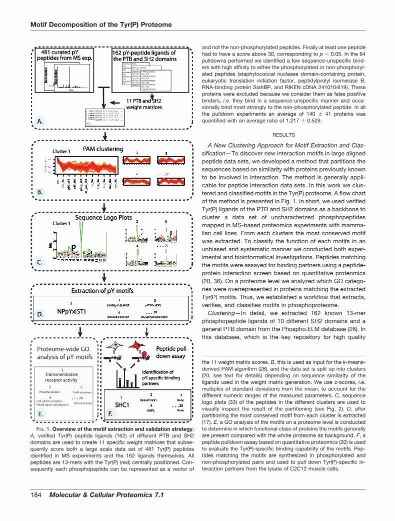
A New Clustering Approach for Motif Extraction and Clas-sification—To discover new interaction motifs in large alignedpeptide data sets, we developed a method that partitions thesequences based on similarity with proteins previously knownto be involved in interaction. The method is generally appli-cable for peptide interaction data sets. In this work we clus-tered and classified motifs in the Tyr(P) proteome. A flow chartof the method is presented in Fig. 1. In short, we used verifiedTyr(P) ligands of the PTB and SH2 domains as a backbone tocluster a data set of uncharacterized phosphopeptidesmapped in MS-based proteomics experiments with mamma-lian cell lines. From each clusters the most conserved motifwas extracted. To classify the function of each motifs in anunbiased and systematic manner we conducted both exper-imental and bioinformatical investigations. Peptides matchingthe motifs were assayed for binding partners using a peptide-protein interaction screen based on quantitative proteomics(20, 36). On a proteome level we analyzed which GO catego-ries were overrepresented in proteins matching the extractedTyr(P) motifs. Thus, we established a workflow that extracts,verifies, and classifies motifs in phosphoproteome.
Clustering—In detail, we extracted 162 known 13-merphosphopeptide ligands of 10 different SH2 domains and ageneral PTB domain from the Phospho.ELM database (26). Inthis database, which is the key repository for high quality
481 curated pY
11 PTB and SH2 weight matrices
Extraction of pY-motifs
Peptide pull-down assay
2 3
stat
_sh
2
ras_
gap
_sh
2
grb
2_sh
2
plc
-g_s
h2
p85
_sh
2
Cluster 1
10 R
DHLSVP
11 T
WRKPGYDFLSQNE
12 I
RQPAGEDS
13 H
ALK
IYPGRED||
1
DQRCTNVLYAHESKFPG
2
HRLPQNFDGKEATS
3
PI
CANQVLSTRE
4
NITYGKAVLSQED
5
KVQNTFHAEDGSP
6
SPCKYEVFGTI
L
7
Y0
1
2
3
bits
|
1
EGNQ
IKRHLCYVTAS
2
DRKMPFHAYTI
VLS
3
WYPCTGDSAEQVN
4
VRPDKSLCEQTN
5
EITGKHQNDARSP
6
SGYFLDQCVATE
I
7
Y
8
QSDKHRNYGFLE
9
VMENKPLGTFAQS
10 T
CI
EHRYQWDVPASLF
11 E
GH
ILQYNFPRVAKTS
12 T
CKHRAI
PNVSDLFGY
13 E
CI
YTLWRGKPANVS
|Cluster 1
PAM clustering
NPpYx[ST][ILMV]pYx[ILMV]P
|
1
HMNRGYSPALDTE
2
RGNFYLVHESKDT
3
TCGNA
IQRKPSLDE
4
SAR
IHVNEKD
5
GNFYI
PESVTADL
6
FHCASNVEKP
ILY
2
4
pYESPxx[DE]
[DE]xx[ILV][ILV]pY
2 3
4
SHC1RasGAP, Nck1 None
SHIP2
4
Relati
ve Abu
ndance
Relativ
e Abun
dance
peptides from MS exp.162 pY-peptide ligands ofthe PTB and SH2 domains
1
A.
B.
C.
D.
F.
YVNTTLYEKFTYA PTBIAGNPAYRSFSNS PTBASVNPEYFSAADV PTBSVQNPVYHNQPLN PTBSLDNPDYQQDFFP PTB
YNSVVLYSTPPIA Crk_SH2LSEETPYSYPTGN Crk_SH2GEEEHVYSFPNKQ Crk_SH2GALSPLYGVPETN Crk_SH2SDTDSSYCIPTAG
AVKMSQYRDNFLS Grb_SH2AIKMGQYKESFAN Grb_SH2PKSPGEYVNIEFG Grb_SH2SSDDVRYVNAFKF Grb_SH2ISKAQEYFNIKSR
PNEHQRGTASLV
11 I
SHPTLRYFNGAEQK
12 V
EWTSDQGRPK
13 D
SNVTQMAFEYGLKR
|
. . . [AG]xpYxx[ILMV]x[KR]
20
None
20. . .
Identification of pY-specific binding partners
EKFTYA 27.2 0.08 4.21 ...RSFSNS 31.5 -0.22 10.2 ...FSAADV 24.2 4.18 12.8 ...HNQPLN 33.6 4.97 15.3 ...
5 SLDNPDYQQDFFP 35.5 -3.35 12.5 ......643...
Sequence Logo Plots
Proteome-wide GO analysis of pY-motifs
Transmembrane receptor activity
Phosphorylation T-cell activation
Cell surface receptor linked signal transduction
2 3
4
1
Kinase activity
20. . .
E.
FIG. 1. Overview of the motif extraction and validation strategy.A, verified Tyr(P) peptide ligands (162) of different PTB and SH2domains are used to create 11 specific weight matrices that subse-quently score both a large scale data set of 481 Tyr(P) peptidesidentified in MS experiments and the 162 ligands themselves. Allpeptides are 13-mers with the Tyr(P) (red) centrally positioned. Con-sequently each phosphopeptide can be represented as a vector of
the 11 weight matrix scores. B, this is used as input for the k-means-derived PAM algorithm (28), and the data set is split up into clusters(20, see text for details) depending on sequence similarity of theligands used in the weight matrix generation. We use z-scores, i.e.multiples of standard deviations from the mean, to account for thedifferent numeric ranges of the measured parameters. C, sequencelogo plots (33) of the peptides in the different clusters are used tovisually inspect the result of the partitioning (see Fig. 2). D, afterpartitioning the most conserved motif from each cluster is extracted(17). E, a GO analysis of the motifs on a proteome level is conductedto determine in which functional class of proteins the motifs generallyare present compared with the whole proteome as background. F, apeptide pulldown assay based on quantitative proteomics (20) is usedto evaluate the Tyr(P)-specific binding capability of the motifs. Pep-tides matching the motifs are synthesized in phosphorylated andnon-phosphorylated pairs and used to pull down Tyr(P)-specific in-teraction partners from the lysate of C2C12 muscle cells.
Motif Decomposition of the Tyr(P) Proteome
184 Molecular & Cellular Proteomics 7.1

annotated phosphorylation sites, these 11 domains had thehighest number of annotated ligands (see Table I for details).Thus, the 162 substrates of the 11 domains represent thebroadest available in vivo data set of Tyr(P) ligands. Theseligands were aligned with the Tyr(P) centrally positioned and
used to generate a position-specific weight matrix for each ofthe 11 domains. The 481 phosphopeptides in the MS-baseddata set and the 162 ligands of the PTB and SH2 domains(“positive controls”) were then scored by each of the 11weight matrices. Consequently each phosphopeptide was
TABLE IClustering and motif extraction of the Tyr(P) proteome
The PAM algorithm was used to partition the data set by sequence similarity with the known ligands of the Tyr(P) binding domains (positivecontrols). The first and second columns show the cluster number and the size of the clusters, respectively. The ability of the algorithm tosignificantly partition (p � 0.05) the positive controls into different clusters is shown in the third column. For example, eight out of a total of10 ligands of the Crk SH2 domain are grouped in cluster 2, corresponding to a significant overrepresentation in a Bonferonni correctedhypergeometric sampling test (p � 3.67e�08). The most conserved motif in each cluster was extracted and is stated in the fourth column. TheTyr(P) residue is indicated in bold, and an X represents any amino acid. Also indicated is the number of occurrences of the motif in therespective cluster and in the total data set. The identified motifs were matched to a library of known Tyr(P) binding motifs, and the expectedbinding partner is indicated (fifth column). 15–20-mer peptides matching the motifs were synthesized in pairs, one with a Tyr(P) as indicatedin bold. Furthermore the parent protein of the peptide is given by Swiss-Prot entry name, and the position of the Tyr(P) is stated. TheTyr(P)-dependent interaction partner(s) identified in a quantitative proteomics peptide-protein screen is shown in the last column (see alsoSupplemental Table 1). PLC, phospholipase C.
Cluster SizePositivecontrols
Extracted motifsMatched motifs,expected partner
PubMedID
Peptides synthesizedIdentifiedpartners
1 41 PTB10 of 10
NPXpYX(S/T)7 of 7
SHC PTB(NPXpY)
75427447541030
KEVCDGWSLPNPEpYYTLRYAELMO2_MOUSE, 48
SHC
2 36 Crk SH28 of 10
(I/L/M/V)pYX(I/L/M/V)P8 of 14
Crk/RasGAP SH2(pYXXP)
923379811607838
KPSTDPLpYDTPDTRGRIN1_MOUSE, 35
RasGAPNck1
3 29 Vav SH24 of 4
pYESPXX(D/E)5 of 5
Vav SH2(pYESP)
9151714 TETKTITpYESPQIDGE41L2_MOUSE, 889
None
4 28 (D/E)XXX(I/L/V)(I/L/V)pY4 of 6
New motif RETSKVIpYDFIEKTGWASL_MOUSE, 253
SHIP2
5 20 (D/E)(D/E)XXXpYXN4 of 6
Grb2 SH2(pYXN)
11994738 VYDEDSPpYQNIKILHSPSY_MOUSE, 147
Grb2RasGAP
6 41 Grb2 SH214 of 31
pYXN(I/L/M/V)XXL5 of 7
Grb2 SH2(pYXN)
11994738 ELFDDPSpYVNIQNLDSHC1_MOUSE, 423
Grb2
7 21 Grb2 SH26 of 31
(D/E)pYXN(I/L/M/V)4 of 11
Grb2 SH2(pYXN)
11994738 QPASVTDpYQNVSFSNITSN2_HUMAN, 858
Grb2
8 45 pY(I/L/M/V)XMXP4 of 10
p85-PI3K SH2(pYXXM)
751121011994738
PQRVDPNGpYMMMSPSIRS1_MOUSE, 658
p85-PI3Kap85-PI3K�
9 40 pY(D/E)X(I/L/M/V)X(I/L/M/V)5 of 22
Fps/Fes SH2(pY(E/D)X(I/V))
7511210 AGKQKLQpYEGIFIKDSF3A1_MOUSE, 757
None
10 32 (D/E)XXpY(D/E)X(I/L/M/V)7 of 27
Fps/Fes SH2(pY(E/D)X(I/V))
7511210 DGGSDQNpYDIVTIGAINP4A_HUMAN, 355
None
11 35 PI3K SH216 of 24
pY(I/L/M/V)PMXP6 of 7
p85 PI3K SH2(pYXXM)
751121011994738
NLHTDDGpYMPMSPGVIRS1_MOUSE, 608
p85-PI3K�
12 23 DpY(I/L/M/V)X(I/L/M/V)7 of 18
SHP2 SH2(pY(I/V/L)X(I/V/L))
7680959 DLINRMDpYVEINIDHVIGLN_MOUSE, 437
SHP2
13 28 SHP2 SH27 of 12
(I/L/M/V)XpY(I/L/M/V)X(I/L/M/V)D6 of 7
SHP2 SH2(pY(I/V/L)X(I/V/L))
7680959 DIKEKLCpYVALDFEQACTB_MOUSE, 218
SHP2
14 32 PLC� SH25 of 16
(I/L/M/V)pYXX(I/L/M/V)(I/L/M/V)5 of 11
General/SHP2 SH2(pY(I/V/L)X(I/V/L))
75112107680959
GKSKQPLpYSSIVTVEO88185_MOUSE, 948
SHP2SHIP2
15 29 RasGAP SH27 of 8
(A/G)(I/L/M/V)pYXXP6 of 10
Crk/RasGAP SH2(pYXXP)
923379811607838
GVVDSGVpYAVPPPAEBCAR1_HUMAN, 410
RasGAP
16 37 SHC SH29 of 13
PXEpYXXXXX(I/L/M/V)3 of 3
New motif TTEAPGEYFFSDGVRIMDH1_MOUSE, 400
None
17 25 Src SH27 of 14
pY(D/E)X(I/L/M/V)H4 of 6
Fps/Fes SH2(pY(E/D)X(I/V))
7511210 ELTAEFLpYDEVHPKQTWF2_MOUSE, 309
RasGAP
18 32 STAT SH215 of 19
pY(I/L/M/V)PQ4 of 4
STAT SH2(pYXXQ)
14966128 SGENFVPpYMPQFQTCLEPR_MOUSE, 1138
None
19 33 H(S/T)GXKPpYXCXXCG10 of 10
New motif RIHTGEKPpYECVQCGKZNF24_MOUSE, 335
None
20 36 (A/G)XpYXX(I/L/M/V)X(K/R)8 of 15
New motif KKNRIAIpYELLFKEGRS10_MOUSE, 12
None
Motif Decomposition of the Tyr(P) Proteome
Molecular & Cellular Proteomics 7.1 185
represented as a vector of the 11 weight matrix scores (Fig.1A). Based on this profile of vectors we clustered the total of643 peptides using the k-means-derived PAM algorithm (40).The PAM algorithm searches for a predefined number ofmedoids (representatives) around which clusters are con-structed. We tried various cluster sizes, and by using a hy-pergeometrical distribution test the number of clusters wasset to 20 because this gave the statistically best partition ofthe positive controls into different clusters (Fig. 1, B and C).
We were able to obtain a convenient fit of the model be-cause the positive controls were distributed such that all werestatistically overrepresented in separate clusters (p � 0.05)(see Table I). For example, 10 of a total of 10 ligands of thePTB domains were grouped in cluster 1 (p � 7.34e�12). Onlythe 31 ligands for the Grb2 SH2 domain were split up in twosignificant groups (clusters 6 and 7). Furthermore eight of the20 clusters did not contain a significant overrepresentation ofthe known ligands used.
Motif Extraction—From each of the 20 clusters the mostconserved motif was extracted with the TEIRESIAS patterndiscovery tool from IBM Bioinformatics (Fig. 1D) (17) as de-scribed under ‘Experimental Procedures.‘ The 20 identifiedmotifs are presented in Table I followed by the number ofmatches to peptides in the particular cluster and in the totaldata set. For example, a unique motif, NPXpYX(S/T), wasextracted from cluster 1 because all peptides (seven) thatmatched this motif were in this cluster. To compare the 20identified motifs with already characterized interaction motifs,we matched each motif to a comprehensive library of Tyr(P)interaction motifs in the Human Protein Reference Database(HPRD) (41). Using the same example again, the motif ex-tracted from cluster 1 could be matched to the NPXpY motifdescribed for the SHC and IRS-1 PTB domains (1), althoughour extended motif contains a Ser or Thr residue at position�2 from the Tyr(P) residue. Considering that the ligands (thepositive controls) of the PTB domain were grouped in cluster1, it is not surprising that we extracted the NPXpYX(S/T) motifin this cluster; however, only three of the seven matchingpeptides in cluster 1 were from these PTB ligands (data notshown). Because this was a general trend it shows the abilityof the clustering method to gather previously uncharacterizedpeptides with high sequence similarity to known ligands of thedifferent Tyr(P) binding domains. In total, 16 of the 20 identi-fied motifs could be matched to the motif library, showing anoverall consistency between the positive controls and theextracted and matched motifs. In four clusters new Tyr(P)motifs were identified.
Gene Ontology Analysis—We used a GO analysis of the 20extracted motifs on a proteome level to determine in whichtype of proteins the extracted motifs generally are present(Fig. 1E). We retrieved all proteins in the mouse proteome thatmatched the motifs and used a hypergeometrical test with thetotal proteome as background and thereby found the 10 mostoverrepresented GO terms in retrieved proteins (see “Exper-
imental Procedures” for details). Using the same exampleagain, the NPXYX(S/T) motif from cluster 1 was overrepre-sented in proteins involved in processes like ‘receptor activity‘and ‘intrinsic to membrane‘ with the consensus parent GOterm assessed to be ‘transmembrane receptor activity‘ (seesupplemental Table 2). Thus, analyzing the motif on a pro-teome level indicates that proteins containing the motif areinvolved in early signaling transduction. This makes sense inthat this is a motif for the PTB domain, which is found in proteinsthat function as molecular scaffolds and adaptors in signalingpathways (1).
Tyr(P)-specific Interaction Partners—To experimentally ver-ify the 20 extracted motifs we used a phosphorylation-specificpeptide-protein interaction screen (Fig. 1F) (20, 36). This as-say is based on differential labeling of proteins using stableisotope labeling by amino acids in cell culture (SILAC) makingit possible to distinguish specific binders from backgroundbinders by their isotope ratios determined by quantitativemass spectrometry (35, 42). The peptides are synthesized in aphosphorylated form and a non-phosphorylated form andused as baits to pull down competing binding partners fromcell lysate, thus mimicking the in vivo binding situation.
We synthesized peptide pairs matching the 20 extractedmotifs. If there were multiple matches we chose peptides withTyr(P) sites not previously known to mediate interaction. Us-ing this experimental approach we could test our clusteringand motif extraction method and investigate the relevance ofknown motifs in a near in vivo situation, i.e. endogenousproteins competing for binding, and potentially discover bind-ing partners for novel motifs. Again using cluster 1 as anexample, we synthesized a peptide pair from the engulfmentand cell motility protein 2 in which the Tyr-48 residue waseither phosphorylated or non-phosphorylated. This is an un-characterized phosphosite identified in a large scale phos-phoproteomics study (12), and it has not previously beenshown to direct Tyr(P)-dependent interaction. Using this pep-tide pair as bait we retrieved one specific binder with a ratiomore than three standard deviations over the log mean of atotal of 162 background binders. This protein, SHC-trans-forming protein 1 (SHC), which contains both a PTB and anSH2 domain, had a total Mascot score of 1654 with 15 iden-tified peptides of which nine were quantifiable (see supple-mental Table 1). Theoretically it could be either the SH2 or thePTB domain that binds the bait phosphopeptide; however,because the peptide matches the consensus NPXpY motifknown to direct PTB domain binding, it is most likely that SHCbinds to the peptide through its PTB domain.
Assaying the Tyr(P) Sequence Space for Interaction Part-ners—The identified phosphospecific binding partners of therepresentative peptides from each cluster can be seen inTable I. Besides the aforementioned SHC protein, these pro-teins all contain SH2 domains, making it very likely that thisdomain governed the phosphospecific interaction. The major-ity (13 of 20) of the peptides retrieved one or more interaction
Motif Decomposition of the Tyr(P) Proteome
186 Molecular & Cellular Proteomics 7.1

proteins. Of these proteins seven were unique because someproteins were identified several times. This is not surprisingbecause some of the clusters were close to each other insequence space resulting in extraction of similar motifs andultimately retrieving the same interaction partners.
To get an overview of the results of the pulldown experi-ments, the GO analysis, and the sequence similarity betweenthe different clusters, we generated weight matrices of thepeptides in the individual clusters and constructed a dendro-gram based on an alignment of these matrices (32). The treecan be seen in Fig. 2 together with sequence logo plots wherethe height of each position represents the degree of conser-vation (33). The logo plots visually illustrate a successful par-titioning because each cluster has a distinct pattern whereparticular amino acids are highly abundant at specific posi-tions flanking the central Tyr(P) residue. The tree is coloredaccording to the identified interaction partners retrieved bypeptides matching the motifs in the different clusters. Forexample, clusters 8 and 11 are close in sequence space withan overrepresentation of hydrophobic residues, especiallymethionine, at position �3 from the central tyrosine residue.Rather than being distinct clusters, these are more likely to besubsets of the same motif. Thus, from these two clusters the
motifs pY(I/L/M/V)XMXP and pY(I/L/M/V)PMXP were ex-tracted and matched to the consensus pYXXM in the library ofmotifs. Accordingly the two peptide pairs synthesized fromthese clusters both retrieve the PI3K-p85� protein. In thesame manner the majority of the expected interaction part-ners were identified using peptides matching to the well char-acterized C-terminally directed Tyr(P) motifs, such as pYXN,pYXXP, and pY(I/V/L)X(I/V/L), that retrieved Grb2, RasGAP,and SHP2, respectively. This illustrates the clear consistencybetween the sequence similarity of the clusters, the con-served residues in the motifs, and the interaction partnersidentified.
There were four clusters (clusters 3, 9, 10, and 18) where wedid not identify the partners (Vav, Fps/Fes, and STAT) asexpected from the signature of motifs alone (see Table I). Forinstance, we did not pull down the SH2 domain protein Fps/Fes when using peptides matching a pY(E/D)X(I/V/L) motif(clusters 9 and 10), which has previously been shown to directbinding (4). This motif was defined using in vitro orientedpeptide library experiments, which have the inherent risk ofdefining motifs that are not relevant in vivo. Whether this is thecase, the Fps/Fes protein was not present in the cell lysate, orbecause of technical limitations remains unclear. In total, here
3
5
76
2
1510
17
20
4
19
1
14
8
11 18
16
9
12
13
0.1
0
1
2
3
bits
23_1
2
|
1
IWMTFVNLQAS
2
NDE
ILTRKQ
3
VNRQSTI
ALGDK
4
RCFVDKANELGS
5
AMQNESTDGKL
6
PAEND
7
Y
8
GPDTKQEF
IALV
9
VCPWFNDTKQESALG
10 V
HTKQMRCF
IL
11 F
QK
IESNVPADH
12 D
MSRHVAL
IF
13 T
RGA
IVSPED|
0
1
2
3
bits
28_1
3
|
1
PTNDVER
IAGSLK
2
NPSLTHRYQDEAK
3
GFMRKPDLAE
4
RFE
IDAQLKS
5
GESD
IQTFNKALV
6
AHNPYKQ
ICEGDT
7
Y
8
NPDVSLTA
9
QI
DTYLSVGA
10 K
SGAR
IVL
11 G
IPESVND
12 P
MTLQ
INDCRSKF
13 G
HRKDSNEQ|
SHP2pYxx[ILV]
0
1
2
3
bits
35_1
1
|
1
RPFYLQDKHATESG
2
VTPLHFGKNSCRAED
3
IRETLDFYGPQS
4
FRLNSQDEGY
5
HVKNLDQGSE
6
EHKLA
IVGPND
7
Y
8
PKTDYI
CASMEV
9
NDVYGMSTHALEP
10 V
IWEQKTSLRAM
11 L
QVTKEDRGNS
12 I
LEFKGNASP
13 L
IQSKNDPAFCTG|
0
1
2
3
bits
45_8
|
1
DSTVHLAYI
ENKRQP
2
ENFRCVTSDGLP
I
3
EKNYVLRDTQ
ISGP
4
VHGLARFTYPQNDSE
5
LKCTSNPDQGEA
6
QAYERPGLSVND
IT
7
Y
8
NQALTM
IEGSV
9
YVCDNTHMQAGELRSKP
10 Q
FNEGTSHRC
IKYVLM
11 EV
MWHFGNYI
QTKLSPR
12 E
GVMDQFWAKTLRSNP
13 N
RCYAKSVLFTG
I |p85-PI3K
pYxxM
0
1
2
3
bits
20_5
|
1
VPYI
NQRDASGE
2
FARWI
QKMHDSE
3
GPVTSYDE
4
YNTEAGD
5
VATLPEYQNDGKS
6
SQ
IPLVED
7
Y
8
GMVAQSYE
9
ARMTKYLGSEN
10 N
YKML
ISRV
11 T
EI
WSQDNKG
12 E
SND
ILGKR
13 N
TPRVELKD|
0
1
2
3
bits
21_7
|
1
ITDMNKWPVLES
2
KQDRPGWESA
3
RYNVKDSE
4
RTLVKDCNAG
5
KRQYNFVAPETH
6
RAQLNVFTD
IE
7
Y
8
THKFDAQEGYSV
9
LTYASWGQRN
10 L
QAEKGSTI
V
11 R
IQMSLFPAVHNETGK
12 R
QAWYLKSFE
13 R
LYMDFKEHS|
0
1
2
3
bits
41_6
|
1
CHDES
IVYAKPTLR
2
TNLHAWMFVYRKG
ISP
3
NGAFVHTQKEPYDS
4
RITLHASMQEPGD
5
QVYLHARSKDNEGP
6
TAP
IDLVYSEQ
7
Y
8
NLSHATQKDRYVGE
9
FWHQMAPSRGKDEN
10 F
QCLDNYTKSRVG
IA
11 M
KWTGLAVRSEYQFP
12 A
YETLKDFSNPG
13 I
RYHNCMKFSAVLG|
Grb2pYxN
0
1
2
3
bits
29_3
|
1
NVRDLTI
APSGE
2
TNKDHLSQ
IMGVFE
3
MKSFAPRQGTYED
4
CNTAEDG
5
RKYI
PTGENDS
6
TQYACLSRPVDG
IE
7
Y
8
TI
ALQMDGSE
9
RKVALNTQ
IDES
10 R
DHLSVP
11 T
WRKPGYDFLSQNE
12 I
RQPAGEDS
13 H
ALK
IYPGRED|
0
1
2
3
bits
28_1
5
|
1
GEAL
ITNDYPSVR
2
QATDKN
IFRGSVP
3
NLFAHKGEDQP
4
QAKRMYTLVDESP
5
NHKTQAEDGS
6
WNGYDTEPVI
L
7
Y8
QYI
MNGEDSA
9
WQ
IPARDEVL
10 E
FTI
DSP
11 D
QTEYFRLP
12 A
TQNGSEPD
13 Q
NLETFRSAYP| 0
1
2
3
bits
36_2
|1
DQRCTNVLYAHESKFPG
2
HRLPQNFDGKEATS
3
PI
CANQVLSTRE
4
NITYGKAVLSQED
5
KVQNTFHAEDGSP
6
SPCKYEVFGTI
L
7
Y8
LCEMTAGHQSD
9
AQKYENFLTI
V
10 E
TYASP
11 I
QKGSADNHTERP
12 I
QLMREKHPSTVDAG
13 A
YI
PEQGFSLRN|
RasGAPpYxxP
0
1
2
3
bits
25_1
7
|
1
QACPSLGTD
2
LRVDK
ISATPG
3
TLQMCKWHAGRP
4
SMLFTPHAKRDE
5
SI
PKDLQFEYG
6
QGCAYVTI
ELDH
7
Y
8
GTAQVYNSED
9
GKFALPSYNDE
10 L
GAKNPSTI
V
11 A
TMDSVI
QEGH
12 S
RN
IQCTVKMFLPG
13 I
SRNADEQVTLPGK
|0
1
2
3
bits
32_1
0
|
1
TRCNMYVQKSDLEG
2
LHYNKVARSDGET
3
AHQ
IDPRTEVLS
4
CSQNVI
KRTADE
5
AHQVNEPRYSDG
6
SGYREC
IATVNL
7
Y
8
CQMAYVLNPTDSE
9
SCWFTMN
IRGYAVL
10 Y
TKWGEVC
ISRL
11 N
VQDGPERTAKS
12 R
YEPVI
GKLA
13 H
TPN
ILVADRKG|
0
1
2
3
bits
36_2
0
|
1
LNHYEVGSRDPK
2
AHRYQVL
ITESNPDG
3
IMTFNRQYKSEAGP
4
SVWMLHG
IPDARE
5
STRHFVNQPDEAG
6
KDPFQYTGVA
IL
7
Y
8
NTYCRKVDSEGA
9
HMQWYR
IGSAVEL
10 P
NEH
IQRGTASLV
11 I
SHPTLRYFNGAEQK
12 V
EWTSDQGRPK
13 D
SNVTQMAFEYGLKR
|
0
1
2
3
bits
28_4
|
1
HMNRGYSPALDTE
2
RGNFYLVHESKDT
3
TCGNA
IQRKPSLDE
4
SAR
IHVNEKD
5
GNFYI
PESVTADL
6
FHCASNVEKP
IL
7
Y
8
GI
CKYMPTDLSA
9
CGYALTI
FV
10 F
KDHRCPYETS
IV
11 K
RFNMAC
IQPSGVE
12 T
WHRAYE
IVDPFKL
13 T
NI
MYRQKGASFP|
0
1
2
3
bits
33_1
9
|
1
QDPMERLSH
2
VDMYRNELKSGT
3
LNATYSDVEMKQG
4
IRVGHSYWTDKE
5
TCSNYGDK
6
ITKVGAQSP
7
Y8
FCQRKYA
ITGSVE
9
NI
DSRAGVLTC
10 R
QI
AHSPGDNFELVT
11 L
TSDNGVQRPAKE
12 R
YEPGAVSC
13 D
SQ
IRVKALG|
New motif[ILV][ILV]pY
SHIP2
0
1
2
3
bits
32_1
4
|
1
QITPFKESGAVLND
2
RPHKAENLQDVST
3
NVHR
IETPLDSFKYGA
4
LCVRGSFDQKTPEN
5
CAHGEDKQSTNP
6
CRTGFAEDNPS
ILV
7
Y
8
VFYQETCDG
ISL
9
NGFPYITSVRADL
10 C
GSTAWFD
ILV
11 I
FQTGAVYSL
12 N
MRPQAETLK
IYG
13 T
RYLGN
IKVDP|
0
1
2
3
bits
41_1
|1
EGNQ
IKRHLCYVTAS
2 DRKMPFHAYTI
VLS
3 WYPCTGDSAEQVN
4
VRPDKSLCEQTN
5
EITGKHQNDARSP
6
SGYFLDQCVATE
I
7
Y8
QSDKHRNYGFLE
9
VMENKPLGTFAQS
10 T
CI
EHRYQWDVPASLF
11 E
GH
ILQYNFPRVAKTS
12 T
CKHRAI
PNVSDLFGY
13 E
CIYTLWRGKPANVS
|SHC
NPxpY
SHP2pYxx[ILV]
0
1
2
3
bits
32_1
8
|
1
IHNYGDQRPEKFLATS
2
AEYRVTSGPL
3
QRE
ILATDMPSNVG
4
RVAFDHPGTQES
5
VMEPRKTQNCADLGS
6
LTI
VDSQPAG
7
Y
8
NI
HWDAERMTFVQKL
9
EQGSHDRACKP
10 I
YGPSKELFTQ
11 M
GSEKTRDFHVNQAP
12 N
HREVPF
IAYQCLSD
13 W
ND
IVFTRSAL|
0
1
2
3
bits
37_1
6
|
1
INDVQLFKSTAP
2
CN
IEFWYSDPGTAL
3
IKHRFYELPAVGD
4
GDAVI
YSTQLPN
5
NYRVTH
ISGPEQL
6
AI
NKGTHYVQLDEP
7
Y
8
GKTHFYR
IQWASLV
9
VISWDKFQEHAYLGP
10 D
CGEPAMLSTYQVI
11 M
VITSFLYKGDRPHAQ
12 S
GMNKEYWQ
IPLRHDV
13 R
CMSYQF
ILTAEVGP
|0
1
2
3
bits
40_9
|
1
MHVI
DKREPQATSLG
2
NREMYPSHQGKDTA
3
MIAEHQKDYCTFPNVRLSG
4
CFRPHQTVD
ILGKAENS
5
HGSEQTNFYD
IKRALV
6
GI
AHRCSVKLDTEPQ
7
Y
8
FKGTEYLDACVIS
9
SVLEPHFIRDKMACQYTGN
10 R
GFHQTS
ILV
11 I
FCETHANKGRSDQV
12 H
QGTMNCKRYI
VPFL
13 LM
QVFNDYKPTEGS
|
GO: nucleic acid bindingH[ST]GxKPpYxCxxCG
GO: ion bindingpY[ILMV]PQ
GO: ion bindingPxEpYxxxxx[ILMV]
GO: catalytic activitypY[DE]x[ILMV]x[ILMV]
GO: catlytic activity[DE]xxpY[DE]x[ILMV]
GO: kinase activity[AG]xpYxx[ILMV]x[KR]
RasGAPpYxx[ILV]
GO: T-cell activationpYESPxx[DE]
GO: signal transducer activity, insulin receptor binding
GO: G-protein coupled receptor activity
GO: transmembrane receptor activity
GO: cell surface receptor linked signal transduction
GO: cell surface receptor linked signal transduction, phosphorylation
GO: Ras GTPase activator activity, regulation of transcription, insulin
receptor binding
GO: immune response, insulin receptor binding
GO: regulation of metabolism
FIG. 2. Dendrogram representing thein vivo Tyr(P) sequence space, motifs,and binding partners. Peptides in the 20different clusters are used to generateweight matrices that subsequently areused as input in a phylogenetic alignment.The tree is a consensus tree of 1000 boot-strap trees (32). The tree represents thedistance between the clusters in se-quence space, which is also visually illus-trated by the sequence logo plots (33) ofthe peptides in each cluster. The color ofthe branches is based on the Tyr(P)-de-pendent interaction partners that wereexperimentally identified using a pep-tide-protein interaction screen. Bothnovel and previously known consensusmotifs (see Table I, fifth column) thatgovern these specific interactions are in-dicated in the same color. Branches andextracted motifs are gray if no interactionpartners were retrieved via the motifs.Furthermore overrepresented GO termsfrom proteins in the whole mouse pro-teome containing the motifs are stated(see text for details). Note that the motifsthat do not retrieve a specific bindingpartner (gray) are typically found in pro-teins mediating processes such as ionbinding, ‘catalytic activity,‘ and nucleicacid binding.
Motif Decomposition of the Tyr(P) Proteome
Molecular & Cellular Proteomics 7.1 187
we report 15 phosphorylation-dependent interactions medi-ated by phosphosites not previously known to direct proteininteraction (see supplemental Table 1).
Non-binding Tyr(P) Motifs—We observed that motifs thatdo not mediate interaction in the pulldown assay are typicallyfound in proteins involved in processes other than signaltransduction (see Fig. 2). It is particularly interesting that weextracted a highly conserved H(S/T)GXKPpYXCXXXCG motiffrom a number of closely related peptides from Cys2-His2 zincfinger proteins (zf-C2H2) concentrated in cluster 19 that didnot pull down any phosphorylation-specific interaction part-ner. The phosphosite of the first tyrosine residue in the zf-C2H2 domain was also described recently in a study thatmined for novel motifs in the phosphoproteome (18), althoughthe identified motif in this work (EXXpY) was different from ourtop scoring motif (H(S/T)GXKPYXCXXXCG), which does notcontain an acidic residue in position �3. However, our secondbest motif in cluster 19 (HXGEXXpY) closely resembles thatreported by Schwartz and Gygi (18).
The H(S/T)GXKPYXCXXXCG motif is extremely specific forproteins containing the zf-C2H2 domain: of 33,758 proteinswe retrieved 656 matches, all of which had the GO term‘nucleic acid binding‘ (GO:0003676) (see supplemental Table2), whereas 647 of the 656 matches had the term ‘zinc ionbinding‘ (GO:0008270) (p � 1e�100).
Recently a role for phosphorylation of zf-C2H2 domains ininhibition of transcription has been suggested (43, 44), sup-posedly as a consequence of the negatively chargedphosphomoiety that reduces DNA affinity (45). Indirectly ourresults support this; because we did not retrieve any interac-tion partner for the synthesized phosphopeptide matching thezinc finger motif, it is unlikely that this motif directs protein-protein interaction, but rather phosphorylation of this motifmodulates nucleic acid binding.
Similarly the novel motifs from clusters 16 and 20 couldmediate mechanisms other than protein-protein interactions,for example, a kinase motif that mediates enzyme activation,nucleic acid binding, protein folding, etc. Supporting this, wefound these motifs to be present in proteins overrepresentedin proteins with GO terms ‘ion binding‘ and ‘kinase activity,‘respectively. Likewise the motifs from clusters where we didnot find the expected partners (clusters 3, 9, 10 and 18) are allexcept motif 3 overrepresented in proteins involved in enzy-matic processes and ion binding (see Fig. 2 or supplementalTable 2).
This indicates that the motifs not mediating protein bindingcould govern processes such as phosphorylation-dependentenzyme activation and nucleic acid binding. Taken togetherwith the results from the pulldown experiment where 13 of 20motifs mediated interaction, we estimate that one-third of theTyr(P) motifs in the proteome mediate processes other thaninteraction through prototypic SH2 and PTB domains.
Identification of a New N-terminal Hydrophobic Tyr(P) Bind-ing Motif for SHIP2—From cluster 4 we extracted a new
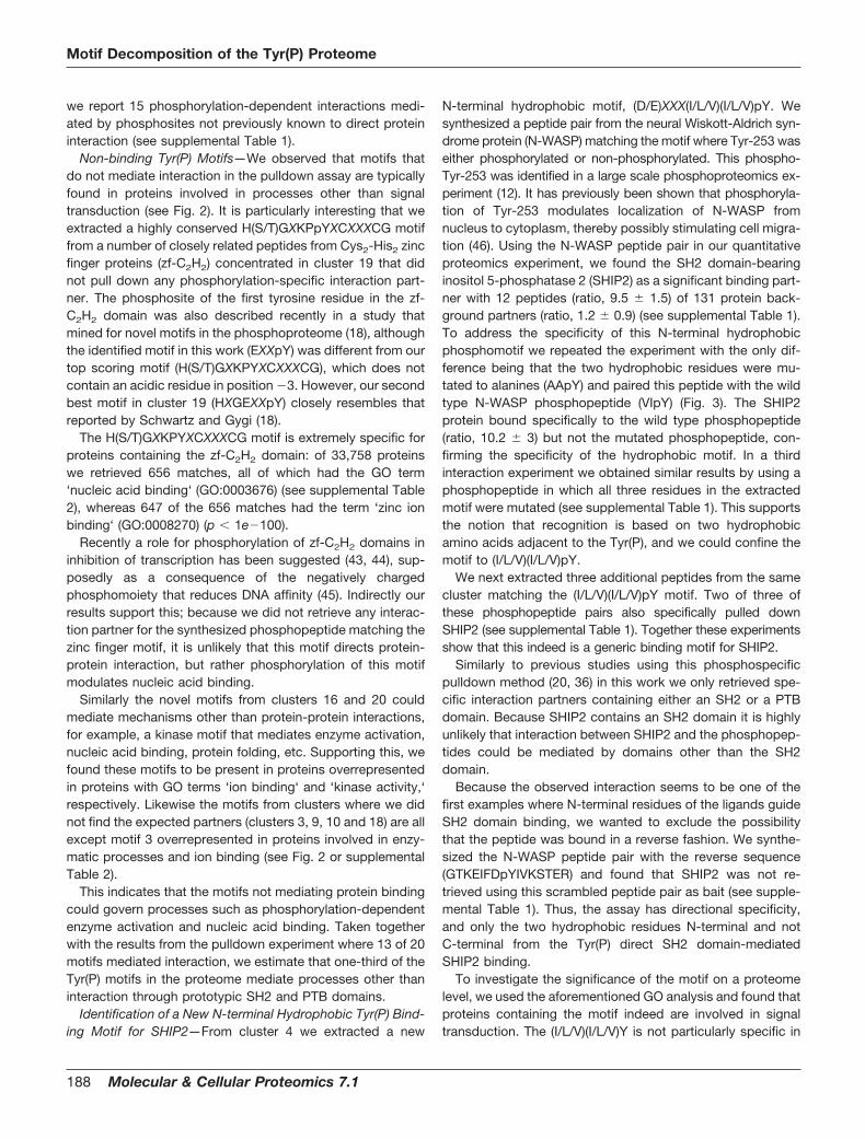
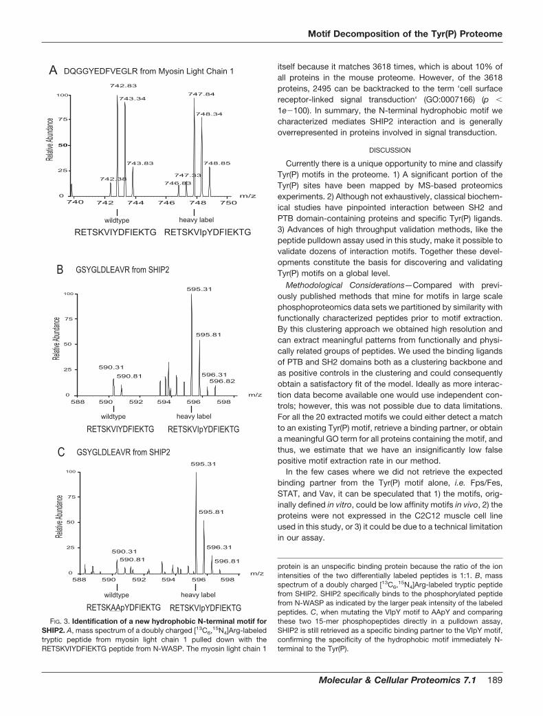
N-terminal hydrophobic motif, (D/E)XXX(I/L/V)(I/L/V)pY. Wesynthesized a peptide pair from the neural Wiskott-Aldrich syn-drome protein (N-WASP) matching the motif where Tyr-253 waseither phosphorylated or non-phosphorylated. This phospho-Tyr-253 was identified in a large scale phosphoproteomics ex-periment (12). It has previously been shown that phosphoryla-tion of Tyr-253 modulates localization of N-WASP fromnucleus to cytoplasm, thereby possibly stimulating cell migra-tion (46). Using the N-WASP peptide pair in our quantitativeproteomics experiment, we found the SH2 domain-bearinginositol 5-phosphatase 2 (SHIP2) as a significant binding part-ner with 12 peptides (ratio, 9.5 � 1.5) of 131 protein back-ground partners (ratio, 1.2 � 0.9) (see supplemental Table 1).To address the specificity of this N-terminal hydrophobicphosphomotif we repeated the experiment with the only dif-ference being that the two hydrophobic residues were mu-tated to alanines (AApY) and paired this peptide with the wildtype N-WASP phosphopeptide (VIpY) (Fig. 3). The SHIP2protein bound specifically to the wild type phosphopeptide(ratio, 10.2 � 3) but not the mutated phosphopeptide, con-firming the specificity of the hydrophobic motif. In a thirdinteraction experiment we obtained similar results by using aphosphopeptide in which all three residues in the extractedmotif were mutated (see supplemental Table 1). This supportsthe notion that recognition is based on two hydrophobicamino acids adjacent to the Tyr(P), and we could confine themotif to (I/L/V)(I/L/V)pY.
We next extracted three additional peptides from the samecluster matching the (I/L/V)(I/L/V)pY motif. Two of three ofthese phosphopeptide pairs also specifically pulled downSHIP2 (see supplemental Table 1). Together these experimentsshow that this indeed is a generic binding motif for SHIP2.
Similarly to previous studies using this phosphospecificpulldown method (20, 36) in this work we only retrieved spe-cific interaction partners containing either an SH2 or a PTBdomain. Because SHIP2 contains an SH2 domain it is highlyunlikely that interaction between SHIP2 and the phosphopep-tides could be mediated by domains other than the SH2domain.
Because the observed interaction seems to be one of thefirst examples where N-terminal residues of the ligands guideSH2 domain binding, we wanted to exclude the possibilitythat the peptide was bound in a reverse fashion. We synthe-sized the N-WASP peptide pair with the reverse sequence(GTKEIFDpYIVKSTER) and found that SHIP2 was not re-trieved using this scrambled peptide pair as bait (see supple-mental Table 1). Thus, the assay has directional specificity,and only the two hydrophobic residues N-terminal and notC-terminal from the Tyr(P) direct SH2 domain-mediatedSHIP2 binding.
To investigate the significance of the motif on a proteomelevel, we used the aforementioned GO analysis and found thatproteins containing the motif indeed are involved in signaltransduction. The (I/L/V)(I/L/V)Y is not particularly specific in
Motif Decomposition of the Tyr(P) Proteome
188 Molecular & Cellular Proteomics 7.1
itself because it matches 3618 times, which is about 10% ofall proteins in the mouse proteome. However, of the 3618proteins, 2495 can be backtracked to the term ‘cell surfacereceptor-linked signal transduction‘ (GO:0007166) (p �
1e�100). In summary, the N-terminal hydrophobic motif wecharacterized mediates SHIP2 interaction and is generallyoverrepresented in proteins involved in signal transduction.
DISCUSSION
Currently there is a unique opportunity to mine and classifyTyr(P) motifs in the proteome. 1) A significant portion of theTyr(P) sites have been mapped by MS-based proteomicsexperiments. 2) Although not exhaustively, classical biochem-ical studies have pinpointed interaction between SH2 andPTB domain-containing proteins and specific Tyr(P) ligands.3) Advances of high throughput validation methods, like thepeptide pulldown assay used in this study, make it possible tovalidate dozens of interaction motifs. Together these devel-opments constitute the basis for discovering and validatingTyr(P) motifs on a global level.
Methodological Considerations—Compared with previ-ously published methods that mine for motifs in large scalephosphoproteomics data sets we partitioned by similarity withfunctionally characterized peptides prior to motif extraction.By this clustering approach we obtained high resolution andcan extract meaningful patterns from functionally and physi-cally related groups of peptides. We used the binding ligandsof PTB and SH2 domains both as a clustering backbone andas positive controls in the clustering and could consequentlyobtain a satisfactory fit of the model. Ideally as more interac-tion data become available one would use independent con-trols; however, this was not possible due to data limitations.For all the 20 extracted motifs we could either detect a matchto an existing Tyr(P) motif, retrieve a binding partner, or obtaina meaningful GO term for all proteins containing the motif, andthus, we estimate that we have an insignificantly low falsepositive motif extraction rate in our method.
In the few cases where we did not retrieve the expectedbinding partner from the Tyr(P) motif alone, i.e. Fps/Fes,STAT, and Vav, it can be speculated that 1) the motifs, orig-inally defined in vitro, could be low affinity motifs in vivo, 2) theproteins were not expressed in the C2C12 muscle cell lineused in this study, or 3) it could be due to a technical limitationin our assay.
protein is an unspecific binding protein because the ratio of the ionintensities of the two differentially labeled peptides is 1:1. B, massspectrum of a doubly charged [13C6,15N4]Arg-labeled tryptic peptidefrom SHIP2. SHIP2 specifically binds to the phosphorylated peptidefrom N-WASP as indicated by the larger peak intensity of the labeledpeptides. C, when mutating the VIpY motif to AApY and comparingthese two 15-mer phosphopeptides directly in a pulldown assay,SHIP2 is still retrieved as a specific binding partner to the VIpY motif,confirming the specificity of the hydrophobic motif immediately N-terminal to the Tyr(P).
595.31
595.81
596.31590.31
596.81
588 590 592 594 596 598m/z
595.31
595.81
590.31590.81
596.82596.31
GSYGLDLEAVR from SHIP2
RETSKVIYDFIEKTG
740 742 744 746 748 750m/z0
25
50
75
100
ecnadnubA evitaleR
742.83
747.84743.34
748.34
743.83 748.85
747.33742.38
746.83
DQGGYEDFVEGLR from Myosin Light Chain 1
588 590 592 594 596 598m/z
590.81
C
B
A
RETSKVIpYDFIEKTG
RETSKAApYDFIEKTG RETSKVIpYDFIEKTG
RETSKVIYDFIEKTG RETSKVIpYDFIEKTG
0
25
50
75
100
ecnadnubA evitaleR
0
25
50
75
100
ecnadnubA evita leR
GSYGLDLEAVR from SHIP2
wildtype heavy label
wildtype heavy label
wildtype heavy label
FIG. 3. Identification of a new hydrophobic N-terminal motif forSHIP2. A, mass spectrum of a doubly charged [13C6,15N4]Arg-labeledtryptic peptide from myosin light chain 1 pulled down with theRETSKVIYDFIEKTG peptide from N-WASP. The myosin light chain 1
Motif Decomposition of the Tyr(P) Proteome
Molecular & Cellular Proteomics 7.1 189
Implications for a New Motif for SHIP2—One of our findingswas that the SH2 domain-containing inositol 5-phosphataseSHIP2 binds to a novel N-terminal hydrophobic motif, (I/L/V)(I/L/V)pY. The specificity of this motif was confirmed by muta-tional analysis. A scrambled peptide pair with the reversesequence and thus with the motif C-terminal of the Tyr(P) didnot retrieve SHIP2, confirming that the recognition lies on theN-terminal side of the Tyr(P). SHIP2 is also retrieved by twoother peptide pairs matching the motifs, indicating this is ageneric motif for SHIP2 binding.
In general, the C-terminal residues Tyr(P) �1 and �3 areconsidered as the most important for the binding specificity ofprototypic SH2 domains (1, 2). Interestingly the SH2 bindingmotif of SHIP2 that we describe is N-terminal, indicating thatthe peptide binding groove of some SH2 domains also ac-commodates residues N-terminal of the Tyr(P). In agreementwith this, the binding properties of the tandem SH2 domainsof the protein-tyrosine phosphatase SHP-2 are governed byresidues Tyr(P) �2 to �5 (47). SHP-1 and SHP-2 both bindthe (I/L/V)XpYXX(I/L/V) ITIM motif in the cytoplasmic part of Fcreceptors, and the Tyr(P) �2 hydrophobic residues have spe-cifically been shown to mediate binding (23, 48, 49). In con-trast to the prototypic SH2 domains that have an Arg in the�A2 binding pocket groove, the tandem SH2 domains of theSHP-1 and SHP-2 phosphatases instead have Gly (50). Sup-posedly this creates a gap that is filled by the side chain of theTyr(P) �2 residue of the bound peptide. Supporting this hy-pothesis, it has been shown that a single point mutation in�A2 Gly3 Arg disrupts the Tyr(P) �2-mediated binding spec-ificity of SHP-2 (48). Furthermore it is known that signalinglymphocytic activation molecule-associated protein (SAP)and Eat2 SH2 domains in part are directed by N-terminalbinding to ITIM motifs (51).
The binding motif of the SH2 domain of SHIP2 has notpreviously been investigated using degenerate peptide bind-ing experiments; however, the ITIM motif has also been re-ported as a docking point for the SH2 domain of SHIP2 (52,53). Combined with our observations, this indicates that thebinding specificity of SHIP2 may be conferred by hydrophobicresidues immediately upstream of the Tyr(P) with contribu-tions from downstream hydrophobic patches. To our knowl-edge, other than that of SHP, this is the only other reportedN-terminally directed Tyr(P) binding motif.
It could be speculated that the SHIP inositol phosphatasescould have a binding mechanism similar to that of SHP pro-tein-tyrosine phosphatases. However, in contrast to SHP theSH2 domains of the SHIP phosphatases resemble the proto-typic SH2 domain because they have the highly conserved�A2 Arg (50), indicating that the N-terminally directed bindingmechanisms differ between SHIP and SHP phosphatases.Because the crystal structure of the SHIP2 phosphatase witha bound ligand has not been resolved, the specific bindingmechanisms have yet to be described.
SHIP2 is involved in membrane signaling by dephospho-
rylating the 5-phosphate group of the key secondary messen-ger phosphatidylinositol 3,4,5-trisphosphate (PIP3). Throughthis action SHIP2 inhibits PI3K-mediated receptor tyrosinekinase signaling because PIP3 is generated by PI3K (54). Thenew motif we describe for the SH2 domain of SHIP2 fits intothis overall function because a GO term analysis of all proteinscontaining the (I/L/V)(I/L/V)pY motif revealed that these pro-teins are involved in cell surface receptor-linked signal trans-duction. Presumably SHIP2 could bind to a number of yetunidentified membrane signaling proteins through its SH2 do-main and in this way be translocated to the membrane or actcooperatively with these proteins. Negative regulators of PI3Kand PIP3 are attractive as antiobesity and diabetes drug targetsbecause PI3K is the main effector of insulin signaling. Recentlya role for SHIP2 as a candidate for such therapeutic interventionhas been proposed by studies of SHIP2 knock-out mice (55,56). Thus, the new motif described in this report may not only beinvolved in regulation of SHIP2-mediated signal transductionbut could also be relevant for medical use.
Conclusions—This work presents the first system-wide ap-proach to mine the proteome for Tyr(P) interaction motifsusing both bioinformatics methods and experimental valida-tion. Strikingly 16 of the 20 motifs extracted could be matchedto previously described interaction motifs. Our experimentalvalidation shows that the majority of the Tyr(P) interactionmotifs that previously have been defined in vitro are also ableto pull down interaction partners from complex lysate mix-tures. The GO analysis revealed that motifs that mediateinteraction in the pulldown assay are found in proteins in-volved in signal transduction, whereas remaining non-bindingmotifs are found in enzymes and ion- and nucleic acid-bindingproteins. This raises the intriguing possibility that about one-third of the in vivo Tyr(P) sites are not directly involved ininteraction via domains such as SH2 and PTB but rather aresites that could alter the catalytic activity of enzymes or mod-ulate the DNA binding affinity of e.g. transcription factors.
Perspectives—The developed clustering method is applicableto other types of complex large scale data sets of post-transla-tional modification where a substantial amount of peptide-pro-tein interactions have been identified. As MS-based methodsmap more modifications such as acetylation and methylationsites, such interactomes could be mined for conserved motifsand assayed for binding partners in a similar manner. Combin-ing proteomics and bioinformatics enables one to do large scalescreens in an unbiased way and thus allows one to reconfirmprevious knowledge and discover new mechanisms at the sametime. As mass spectrometric streamlining and automation ad-vances, entire post-translational modification proteomes can bemapped, binding motifs can identified, and thus ultimately thespecificity of signaling networks can be unraveled.
Acknowledgments—We thank members of the Department of Pro-teomics and Signal Transduction and the Center for Biological Se-quence Analysis for comments on the manuscript.
Motif Decomposition of the Tyr(P) Proteome
190 Molecular & Cellular Proteomics 7.1
* This work was supported by Interaction Proteome, FP6, ContractLSHG-CT-2003-505520, a grant from the European Commission inthe 6th framework program; by the Danish Platform for IntegrativeBiology, a grant from the Danish National Research Foundation (Dan-marks Grundforskningsfond); and by the Danish Research Agency(Forskningsstyrelsen). The costs of publication of this article weredefrayed in part by the payment of page charges. This article musttherefore be hereby marked “advertisement” in accordance with 18U.S.C. Section 1734 solely to indicate this fact.
□S The on-line version of this article (available at http://www.mcponline.org) contains supplemental material.
¶ To whom correspondence should be addressed. Tel.: 45-4525-2477; Fax: 45-4593-1585; E-mail: [email protected] or [email protected].
REFERENCES
1. Yaffe, M. B. (2002) Phosphotyrosine-binding domains in signal transduc-tion. Nat. Rev. Mol. Cell Biol. 3, 177–186
2. Pawson, T. (2004) Specificity in signal transduction: from phosphotyrosine-SH2 domain interactions to complex cellular systems. Cell 116, 191–203
3. Bork, P., and Koonin, E. V. (1996) Protein sequence motifs. Curr. Opin.Struct. Biol. 6, 366–376
4. Songyang, Z., Shoelson, S. E., McGlade, J., Olivier, P., Pawson, T., Bustelo,X. R., Barbacid, M., Sabe, H., Hanafusa, H., and Yi, T. (1994) Specificmotifs recognized by the SH2 domains of Csk, 3BP2, fps/fes, GRB-2,HCP, SHC, Syk, and Vav. Mol. Cell. Biol. 14, 2777–2785
5. Songyang, Z., Margolis, B., Chaudhuri, M., Shoelson, S. E., and Cantley,L. C. (1995) The phosphotyrosine interaction domain of SHC recognizestyrosine-phosphorylated NPXY motif. J. Biol. Chem. 270, 14863–14866
6. Ballif, B. A., Villen, J., Beausoleil, S. A., Schwartz, D., and Gygi, S. P. (2004)Phosphoproteomic analysis of the developing mouse brain. Mol. Cell.Proteomics 3, 1093–1101
7. Beausoleil, S. A., Jedrychowski, M., Schwartz, D., Elias, J. E., Villen, J., Li,J., Cohn, M. A., Cantley, L. C., and Gygi, S. P. (2004) Large-scalecharacterization of HeLa cell nuclear phosphoproteins. Proc. Natl. Acad.Sci. U. S. A. 101, 12130–12135
8. Brill, L. M., Salomon, A. R., Ficarro, S. B., Mukherji, M., Stettler-Gill, M., andPeters, E. C. (2004) Robust phosphoproteomic profiling of tyrosine phos-phorylation sites from human T cells using immobilized metal affinitychromatography and tandem mass spectrometry. Anal. Chem. 76,2763–2772
9. Ficarro, S., Chertihin, O., Westbrook, V. A., White, F., Jayes, F., Kalab, P.,Marto, J. A., Shabanowitz, J., Herr, J. C., Hunt, D. F., and Visconti, P. E.(2003) Phosphoproteome analysis of capacitated human sperm. Evi-dence of tyrosine phosphorylation of a kinase-anchoring protein 3 andvalosin-containing protein/p97 during capacitation. J. Biol. Chem. 278,11579–11589
10. Ficarro, S. B., Salomon, A. R., Brill, L. M., Mason, D. E., Stettler-Gill, M.,Brock, A., and Peters, E. C. (2005) Automated immobilized metal affinitychromatography/nano-liquid chromatography/electrospray ionizationmass spectrometry platform for profiling protein phosphorylation sites.Rapid Commun. Mass Spectrom. 19, 57–71
11. Olsen, J. V., Blagoev, B., Gnad, F., Macek, B., Kumar, C., Mortensen, P.,and Mann, M. (2006) Global, in vivo, and site-specific phosphorylationdynamics in signaling networks. Cell 127, 635–648
12. Rush, J., Moritz, A., Lee, K. A., Guo, A., Goss, V. L., Spek, E. J., Zhang, H.,Zha, X. M., Polakiewicz, R. D., and Comb, M. J. (2005) Immunoaffinityprofiling of tyrosine phosphorylation in cancer cells. Nat. Biotechnol. 23,94–101
13. Salomon, A. R., Ficarro, S. B., Brill, L. M., Brinker, A., Phung, Q. T., Ericson,C., Sauer, K., Brock, A., Horn, D. M., Schultz, P. G., and Peters, E. C.(2003) Profiling of tyrosine phosphorylation pathways in human cellsusing mass spectrometry. Proc. Natl. Acad. Sci. U. S. A. 100, 443–448
14. Zheng, H., Hu, P., Quinn, D. F., and Wang, Y. K. (2005) Phosphotyrosineproteomic study of interferon � signaling pathway using a combination ofimmunoprecipitation and immobilized metal affinity chromatography.Mol. Cell. Proteomics 4, 721–730
15. Jonassen, I., Collins, J. F., and Higgins, D. G. (1995) Finding flexiblepatterns in unaligned protein sequences. Protein Sci. 4, 1587–1595
16. Nevill-Manning, C. G., Wu, T. D., and Brutlag, D. L. (1998) Highly specific
protein sequence motifs for genome analysis. Proc. Natl. Acad. Sci.U. S. A. 95, 5865–5871
17. Rigoutsos, I., and Floratos, A. (1998) Combinatorial pattern discovery inbiological sequences: the TEIRESIAS algorithm. Bioinformatics 14,55–67
18. Schwartz, D., and Gygi, S. P. (2005) An iterative statistical approach to theidentification of protein phosphorylation motifs from large-scale datasets. Nat. Biotechnol. 23, 1391–1398
19. Forman-Kay, J. D., and Pawson, T. (1999) Diversity in protein recognition byPTB domains. Curr. Opin. Struct. Biol. 9, 690–695
20. Schulze, W. X., and Mann, M. (2004) A novel proteomic screen for peptide-protein interactions. J. Biol. Chem. 279, 10756–10764
21. Songyang, Z., and Cantley, L. C. (1995) Recognition and specificity inprotein tyrosine kinase-mediated signalling. Trends Biochem. Sci. 20,470–475
22. Vely, F., and Vivier, E. (1997) Conservation of structural features reveals theexistence of a large family of inhibitory cell surface receptors and non-inhibitory/activatory counterparts. J. Immunol. 159, 2075–2077
23. Burshtyn, D. N., Yang, W., Yi, T., and Long, E. O. (1997) A novel phospho-tyrosine motif with a critical amino acid at position �2 for the SH2domain-mediated activation of the tyrosine phosphatase SHP-1. J. Biol.Chem. 272, 13066–13072
24. Hinsby, A. M., Olsen, J. V., Bennett, K. L., and Mann, M. (2003) Signalinginitiated by overexpression of the fibroblast growth factor receptor-1investigated by mass spectrometry. Mol. Cell. Proteomics 2, 29–36
25. Hinsby, A. M., Olsen, J. V., and Mann, M. (2004) Tyrosine phosphopro-teomics of fibroblast growth factor signaling: a role for insulin receptorsubstrate-4. J. Biol. Chem. 279, 46438–46447
26. Diella, F., Cameron, S., Gemund, C., Linding, R., Via, A., Kuster, B., Sicher-itz-Ponten, T., Blom, N., and Gibson, T. J. (2004) Phospho.ELM: adatabase of experimentally verified phosphorylation sites in eukaryoticproteins. BMC Bioinformatics 5, 79
27. Nielsen, M., Lundegaard, C., Worning, P., Hvid, C. S., Lamberth, K., Buus,S., Brunak, S., and Lund, O. (2004) Improved prediction of MHC class Iand class II epitopes using a novel Gibbs sampling approach. Bioinfor-matics 20, 1388–1397
28. Kaufman, L., and Rousseeuw, P. J. (1990) Finding Groups in Data: anIntroduction to Cluster Analysis, John Wiley, New York
29. Fraley, C., and Raftery, A. E. (1998) How many clusters? Which clusteringmethod? Answers via model-based cluster analysis. Comput. J. 41,578–588
30. Jain, A. K., Murth, M. N., and Flynn, P. J. (1999) Data clustering: a review.ACM Comput. Surv. 31, 264–323
31. Ester, M., Kriegel, H. P., Sander, S., and Xu, X. (1996) A density-basedalgorithm for discovering clusters in large spatial databases with noise, inProceedings of 2nd International Conference on Knowledge Discoveryand Data Mining, Portland, August 2–4, 1996 (Simoudis, E., Han, J., andFayyad, U., eds) Vol. 1, pp. 226–231, Association for the Advancementof Artificial Intelligence (AAAI) Press, Menlo Park, CA
32. Lund, O., Nielsen, M., Kesmir, C., Petersen, A. G., Lundegaard, C., Worn-ing, P., Sylvester-Hvid, C., Lamberth, K., Roder, G., Justesen, S., andBuus, S., and Brunak, S. (2004) Definition of supertypes for HLA mole-cules using clustering of specificity matrices. Immunogenetics 55,797–810
33. Schneider, T. D., and Stephens, R. M. (1990) Sequence logos: a new wayto display consensus sequences. Nucleic Acids Res. 18, 6097–6100
34. Johnson, L. N., Kotz, S., and Kemp, A. W. (1992) Univariate DiscreteDistributions, 2nd Ed., Wiley-Interscience, New York
35. Ong, S. E., Kratchmarova, I., and Mann, M. (2003) Properties of 13C-substituted arginine in stable isotope labeling by amino acids in cellculture (SILAC). J. Proteome Res. 2, 173–181
36. Schulze, W. X., Deng, L., and Mann, M. (2005) Phosphotyrosine interac-tome of the ErbB-receptor kinase family. Mol. Syst. Biol. 1, 2005 0008
37. Rappsilber, J., Ishihama, Y., and Mann, M. (2003) Stop and go extractiontips for matrix-assisted laser desorption/ionization, nanoelectrospray,and LC/MS sample pretreatment in proteomics. Anal. Chem. 75,663–670
38. Ishihama, Y., Rappsilber, J., Andersen, J. S., and Mann, M. (2002) Micro-columns with self-assembled particle frits for proteomics. J. Chromatogr.A 979, 233–239
39. Perkins, D. N., Pappin, D. J., Creasy, D. M., and Cottrell, J. S. (1999)
Motif Decomposition of the Tyr(P) Proteome
Molecular & Cellular Proteomics 7.1 191
Probability-based protein identification by searching sequence data-bases using mass spectrometry data. Electrophoresis 20, 3551–3567
40. Bateman, A., Birney, E., Cerruti, L., Durbin, R., Etwiller, L., Eddy, S. R.,Griffiths-Jones, S., Howe, K. L., Marshall, M., and Sonnhammer, E. L.(2002) The Pfam protein families database. Nucleic Acids Res. 30,276–280
41. Peri, S., Navarro, J. D., Amanchy, R., Kristiansen, T. Z., Jonnalagadda,C. K., Surendranath, V., Niranjan, V., Muthusamy, B., Gandhi, T. K.,Gronborg, M., Ibarrola, N., Deshpande, N., Shanker, K., Shivashankar,H. N., Rashmi, B. P., Ramya, M. A., Zhao, Z., Chandrika, K. N., Padma,N., Harsha, H. C., Yatish, A. J., Kavitha, M. P., Menezes, M., Choudhury,D. R., Suresh, S., Ghosh, N., Saravana, R., Chandran, S., Krishna, S.,Joy, M., Anand, S. K., Madavan, V., Joseph, A., Wong, G. W., Schi-emann, W. P., Constantinescu, S. N., Huang, L., Khosravi-Far, R., Steen,H., Tewari, M., Ghaffari, S., Blobe, G. C., Dang, C. V., Garcia, J. G.,Pevsner, J., Jensen, O. N., Roepstorff, P., Deshpande, K. S., Chinnaiyan,A. M., Hamosh, A., Chakravarti, A., and Pandey, A. (2003) Developmentof human protein reference database as an initial platform for approach-ing systems biology in humans. Genome Res. 13, 2363–2371
42. Ong, S. E., Blagoev, B., Kratchmarova, I., Kristensen, D. B., Steen, H.,Pandey, A., and Mann, M. (2002) Stable isotope labeling by amino acidsin cell culture, SILAC, as a simple and accurate approach to expressionproteomics. Mol. Cell. Proteomics 1, 376–386
43. Dovat, S., Ronni, T., Russell, D., Ferrini, R., Cobb, B. S., and Smale, S. T.(2002) A common mechanism for mitotic inactivation of C2H2 zinc fingerDNA-binding domains. Genes Dev. 16, 2985–2990
44. Saydam, N., Adams, T. K., Steiner, F., Schaffner, W., and Freedman, J. H.(2002) Regulation of metallothionein transcription by the metal-respon-sive transcription factor MTF-1: identification of signal transduction cas-cades that control metal-inducible transcription. J. Biol. Chem. 277,20438–20445
45. Jantz, D., and Berg, J. M. (2004) Reduction in DNA-binding affinity ofCys2His2 zinc finger proteins by linker phosphorylation. Proc. Natl.Acad. Sci. U. S. A. 101, 7589–7593
46. Wu, X., Suetsugu, S., Cooper, L. A., Takenawa, T., and Guan, J. L. (2004)Focal adhesion kinase regulation of N-WASP subcellular localization andfunction. J. Biol. Chem. 279, 9565–9576
47. Eck, M. J., Pluskey, S., Trub, T., Harrison, S. C., and Shoelson, S. E. (1996)Spatial constraints on the recognition of phosphoproteins by the tandem
SH2 domains of the phosphatase SH-PTP2. Nature 379, 277–28048. Huyer, G., and Ramachandran, C. (1998) The specificity of the N-terminal
SH2 domain of SHP-2 is modified by a single point mutation. Biochem-istry 37, 2741–2747
49. Beebe, K. D., Wang, P., Arabaci, G., and Pei, D. (2000) Determination of thebinding specificity of the SH2 domains of protein tyrosine phosphataseSHP-1 through the screening of a combinatorial phosphotyrosyl peptidelibrary. Biochemistry 39, 13251–13260
50. Liu, B. A., Jablonowski, K., Raina, M., Arce, M., Pawson, T., and Nash, P. D.(2006) The human and mouse complement of SH2 domain proteins-establishing the boundaries of phosphotyrosine signaling. Mol. Cell 22,851–868
51. Poy, F., Yaffe, M. B., Sayos, J., Saxena, K., Morra, M., Sumegi, J., Cantley,L. C., Terhorst, C., and Eck, M. J. (1999) Crystal structures of the XLPprotein SAP reveal a class of SH2 domains with extended, phosphoty-rosine-independent sequence recognition. Mol. Cell 4, 555–561
52. Muraille, E., Bruhns, P., Pesesse, X., Daeron, M., and Erneux, C. (2000) TheSH2 domain containing inositol 5-phosphatase SHIP2 associates to theimmunoreceptor tyrosine-based inhibition motif of Fc �RIIB in B cellsunder negative signaling. Immunol. Lett. 72, 7–15
53. Bruhns, P., Vely, F., Malbec, O., Fridman, W. H., Vivier, E., and Daeron, M.(2000) Molecular basis of the recruitment of the SH2 domain-containinginositol 5-phosphatases SHIP1 and SHIP2 by fc� RIIB. J. Biol. Chem.275, 37357–37364
54. Damen, J. E., Liu, L., Rosten, P., Humphries, R. K., Jefferson, A. B.,Majerus, P. W., and Krystal, G. (1996) The 145-kDa protein induced toassociate with Shc by multiple cytokines is an inositol tetraphosphateand phosphatidylinositol 3,4,5-triphosphate 5-phosphatase. Proc. Natl.Acad. Sci. U. S. A. 93, 1689–1693
55. Marion, E., Kaisaki, P. J., Pouillon, V., Gueydan, C., Levy, J. C., Bodson, A.,Krzentowski, G., Daubresse, J. C., Mockel, J., Behrends, J., Servais, G.,Szpirer, C., Kruys, V., Gauguier, D., and Schurmans, S. (2002) The geneINPPL1, encoding the lipid phosphatase SHIP2, is a candidate for type 2diabetes in rat and man. Diabetes 51, 2012–2017
56. Sleeman, M. W., Wortley, K. E., Lai, K. M., Gowen, L. C., Kintner, J., Kline,W. O., Garcia, K., Stitt, T. N., Yancopoulos, G. D., Wiegand, S. J., andGlass, D. J. (2005) Absence of the lipid phosphatase SHIP2 confersresistance to dietary obesity. Nat. Med. 11, 199–205
Motif Decomposition of the Tyr(P) Proteome
192 Molecular & Cellular Proteomics 7.1