Iranian Critical ELT: A Belated but Growing Intellectual Shift in Iranian ELT Community
Upload
khangminh22Category
view
1download
0
Folia Morphol. Vol. 76, No. 4, pp. 695–701
DOI: 10.5603/FM.a2017.0032 Copyright © 2017 Via Medica
ISSN 0015–5659 www.fm.viamedica.pl
O R I G I N A L A R T I C L E
695
Morphological variations of the vermiform appendix in Iranian cadavers: a study from developing countriesS. Mohammadi1, A. Hedjazi2, M. Sajjadian3, M. Rahmani4, M. Mohammadi5, M.D. Moghadam6 1Neurogenic Inflammation Research Centre, Faculty of Medicine, Mashhad University of Medical Sciences, Mashhad, Iran 2Legal Medicine Research Centre, Legal Medicine Organisation, Tehran, Iran 3Mashhad Legal Medicine Organisation, Mashhad, Iran 4Departmnt of Midwifery, Faculty of Medicine, Gonabad University of Medical Sciences, Gonabad, Iran 5Department of Public Health, School of Health, Shahid Beheshti University of Medical Sciences, Tehran, Iran 6Department of Community Medicine, School of Medicine, Mashhad University of Medical Sciences, Mashhad, Iran
[Received: 25 January 2017; Accepted: 16 March 2017]
Background: The vermiform appendix is a worm-like tube containing a large amount of lymphoid follicles. In our knowledge, there is a little standard data about the vermiform appendix in Iranian population. Therefore, the objective of this study was to investigate the normal appendix size in Iranian cadavers.Materials and methods: A cross-sectional study was undertaken between June 2014 and July 2015, in the autopsy laboratory, Legal Medicine Organisation, Razavi Khorasan province, Iran. A total of 693 cadavers with the mean age of 40.46 ± 20.99 years were divided into 10 groups. After writing down position of the appendix, the length, diameter and weight of the appendix were measured. Statistical analysis was performed using SPSS software.Results: The mean values of the demographic characteristics included — age: 40.46 ± 20.99 years; weight: 63.47 ± 17.84 kg; height: 159.95 ± 28.23 cm. The mean values of the appendix length, diameter, weight and index in the cadavers were 8.52 ± 2.99 cm, 12.17 ± 4.53 mm, 6.43 ± 3.26 g and 0.013 ± 0.01, respectively. The most common position of the appendix was retrocaecal in 71.7% of cases. Significant correlations were evident between the value of demographic data and appendix size (p < 0.05). The diameter (p = 0.002) and index of the appendix (p = 0.003) showed significant difference between males and females. Conclusions: Having standard data on the vermiform appendix is useful for cli-nicians as well as anthropologists. The findings of the present study can provide information about morphologic variations of the appendix in Iranian population. (Folia Morphol 2017; 76, 4: 695–701)
Key words: vermiform appendix, cadaver, anatomic variation
Address for correspondence: Dr. A. Hedjazi, Legal Medicine Research Centre, Legal Medicine Organisation, Tehran, Iran, tel: +98 513 8425946, fax: +98 513 8454403, e-mail: [email protected]
696
Folia Morphol., 2017, Vol. 76, No. 4
INTRODUCTIONThe vermiform appendix is a worm-shaped di-
verticulum that extends from the posteromedial surface of the caecum [23]. It contains abundant lymphoid nodules in its wall [23]. The base of ap-pendix is fairly constant whereas its apex can be found in the following situations: retrocaecal, pel-vic, subcaecal, preileal, postileal, and paracolic [28]. The appendix is suspended by a short triangular mesoappendix containing the appendicular nerves and vessels [23].
Mc Burney’s point is the surface landmark for the base of the appendix that it is situated in the middle third of the line joining the umbilicus to the right anterior superior iliac spine. The vermiform appen-dix appears at about the sixth week of gestation as a small diverticulum of the distal limb of the primitive midgut loop [21]. The primary intestinal loop ro-tates 270° counterclockwise around the axis of the superior mesenteric artery [21]. Since the vermiform appendix forms during descent of the caecum, the most common position of the appendix is retrocae-cal [21]. There are some different findings about appendix size in Asian population. The length of the appendix in the Indian cadavers ranges between 5.9 and 10.21, while the range of the appendix thick- ness was from 0.46 cm to 0.7 cm [2, 3, 8, 22]. The mean length of appendix was reported 6.03 cm in Thai-land [5]. This value obtained for weight ranges from 6.33 cm to 8.57 cm in Iranian population [9, 10, 24]. The common position of the appendix was retrocaecal in India [3, 15, 22, 25], retroileal in Thailand and pelvic in Iran [9, 10, 24].
Appendicitis has been identified as “the most common acute abdominal surgical emergency”. Knowledge of anatomical positions of the vermiform appendix is essential for appropriate treatment and management of appendicitis. Its variable positions may cause surgeons to make a wrong diagnosis. In addition, variation in anatomical location of the ap-pendix causes different clinical presentation which may mimic other diseases such as torsion of ovarian cyst, biliary colic, colitis, pelvic inflammatory diseases etc. There is a little data regarding the anthropo-metric values of the vermiform appendix in Iranian populations. Hence, the aim of this study was to determine standard size of the vermiform appendix among Iranian population and to compare them with the available literature.
MATERIALS AND METHODS A cross-sectional study was carried out in the
autopsy laboratory of the Forensic Medicine Organi-sation, Razavi Khorasan province, from June 2014 to July 2015. The protocol of the research was approved by the Ethics Research Committee of Mashhad Legal Medicine Organisation.
Sixty hundred and ninety-three cadavers (541 ma- les/152 females) with the mean age of 40.46 ± ± 20.99 years were divided into 10 different age groups: Group A (0–9 years), Group B (10–19 years), Group C (20–29 years), Group D (30–39 years), Group E (40–49 years), Group F (50–59 years), Group G (60–69 years), Group H (70–79 years), Group I (80–89 years) and Group J (90–99 years).
Fresh cadavers with no gross evidence of ab-dominal trauma, adhesions, peritonitis, fibrosis, and history of poisoning were included in the study. Non-Iranian cadavers with any pathologic abnor-mality or injury to the colon, caecum and appen-dix were excluded from the study. Demographic data, including age, sex, body weight and height were collected from cadaver’s file. Body mass in-dex (BMI) was calculated as body weight in kilo-grams divided by height in meters squared (kg/m2). The index of appendix was also calculated as appendix weight/body weight.
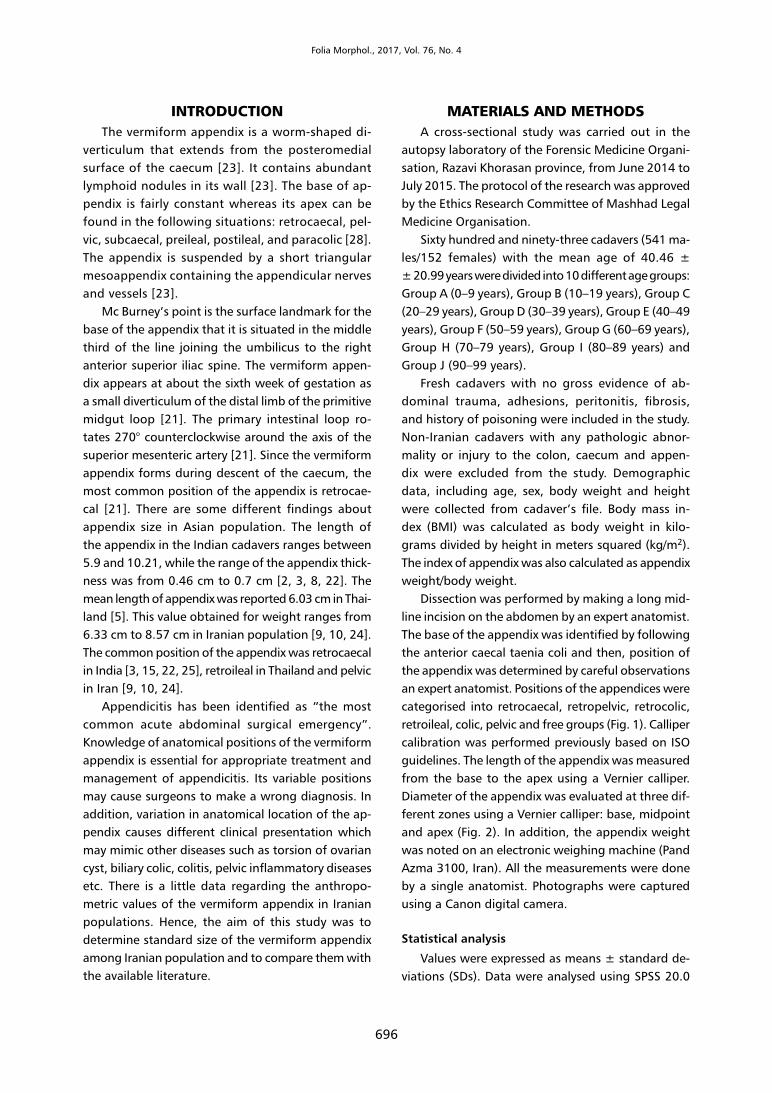
Dissection was performed by making a long mid-line incision on the abdomen by an expert anatomist. The base of the appendix was identified by following the anterior caecal taenia coli and then, position of the appendix was determined by careful observations an expert anatomist. Positions of the appendices were categorised into retrocaecal, retropelvic, retrocolic, retroileal, colic, pelvic and free groups (Fig. 1). Calliper calibration was performed previously based on ISO guidelines. The length of the appendix was measured from the base to the apex using a Vernier calliper. Diameter of the appendix was evaluated at three dif-ferent zones using a Vernier calliper: base, midpoint and apex (Fig. 2). In addition, the appendix weight was noted on an electronic weighing machine (Pand Azma 3100, Iran). All the measurements were done by a single anatomist. Photographs were captured using a Canon digital camera.
Statistical analysis
Values were expressed as means ± standard de-viations (SDs). Data were analysed using SPSS 20.0

697
S. Mohammadi et al., Morphological variations of the vermiform appendix in Iranian cadavers
software. P values less than 0.05 were considered significant. The normality of data was assessed us-ing the Kolmogorov-Smirnov test. The correlation between anthropometric parameters and variances such as the appendix length and the appendix thick-ness was evaluated using the Spearman correlation. Comparisons between groups were carried out on independent sample t-tests (for two groups) and analysis of variance (for more than two groups).
RESULTSDemographic data are summarised in Table 1.
A total of 693 Iranian cadavers (152 females/541 males) with a mean age of 40.46 ± 20.99 years were included in the study. The height of the cadav-ers ranges between 31 and 190, with an average of 159.95 cm. The values obtained for weight ranged from 1 to 120, with an average of 63.47 g. The mean BMI was 25.20 ± 26.41 kg/m2.
Figure 1. Histogram showing the incidence of anatomical positions of the appendix in Iranian cadavers (n = 693).
Figure 2. Vermiform appendix in Iranian cadavers. Arrows show appendix.

698
Folia Morphol., 2017, Vol. 76, No. 4
Table 2. Length, weight, index and external diameter of the appendix of Iranian cadavers in different age groups
Age groups Length [cm] External diameter of the appendix [mm] Weight [g] Index [%]
At the apex At the midpoint At the base
< 10 (n = 47) 2.54 ± 2.21* 1.65 ± 1.39* 1.57 ± 1.48* 1.76 ± 1.62* 2.65 ± 2.32* 0.05 ± 0.04*
10–19 (n = 57) 8.81 ± 2.38 4.24 ± 1.69 4.16 ± 1.46 4.01 ± 1.92 6.27 ± 2.86 0.01 ± 0.008
20–29 (n = 120) 9.40 ± 2.41 4.29 ± 1.68 4.33 ± 1.59 4.50 ± 2.05 6.43 ± 2.78 0.009 ± 0.004
30–39 (n = 119) 9.16 ± 2.51 4.70 ± 1.88 4.39 ± 1.63 4.40 ± 2.14 7.04 ± 3.21 0.01 ± 0.004
40–49 (n = 137) 8.95 ± 2.52 4.49 ± 2.00 4.11 ± 1.53 4.17 ± 1.84 7.20 ± 3.28 0.01 ± 0.005
50–59 (n = 79) 9.07 ± 2.48 4.58 ± 1.89 4.49 ± 1.25 4.15 ± 1.90 6.95 ± 3.25 0.01 ± 0.005
60–69 (n = 60) 8.81 ± 1.85 4.11 ± 2.06 4.14 ± 1.43 4.02 ± 1.66 6.81 ± 2.78 0. 01 ± 0.004
70–79 (n = 42) 9.10 ± 2.12 4.20 ± 1.70 3.74 ± 1.42 3.91 ± 1.62 6.59 ± 3.61 0.01 ± 0.005
80–89 (n = 24) 8.36 ± 3.24 3.42 ± 1.55 4.09 ± 1.33 3.82 ± 1.74 5.82 ± 3.18 0.009 ± 0.005
90–99 (n = 8) 8.70 ± 1.71 4.90 ± 2.48 3.80 ± 1.68 3.40 ± 1.63 7.60 ± 4.39 0.01 ± 0.008
Values are expressed as mean± standard deviation. Comparison between groups was made using ANOVA and Tukey test. *P = 0.000 compared to group B (10–19 years old)
Table 1. Demographic data of Iranian cadavers (n = 693) in Razavi Khorasan province, Iran
Age groups Age [years] Gender (female/male) Height [m] Weight [kg]
< 10 1.80 ± 2.70 21/26 75 ± 38.97 9.80 ± 15.79
10–19 15.85 ± 2.66 10/47 160.82 ± 16.42 60.47 ± 16.49
20–29 24.65 ± 2.69 32/88 168.03 ± 7.60 67.84 ± 8.04
30–39 34.33 ± 2.68 24/95 166.59 ± 16.83 69.08 ± 9.35
40–49 44.51 ± 2.55 19/118 166.67 ± 11.20 68.59 ± 9.38
50–59 53.78 ± 2.87 13/66 166.38 ± 17.01 68.97 ± 6.98
60–69 63.45 ± 2.68 12/48 165.63 ± 12.37 68.10 ± 5.85
70–79 74.07 ± 2.56 11/31 167.48 ± 7.69 65.64 ± 11.83
80–89 83.75 ± 3.35 9/15 162.50 ± 16.48 63.88 ± 6.99
90–99 91.25 ± 2.18 1/7 166.75 ± 6.73 61.88 ± 14.28
Values are presented as mean ± standard deviation or number.
Tables 2 and 3 show the anthropometric param-eters of the vermiform appendix in different gender and age groups. The mean length of the appendix was 8.52 cm (range, 0.5–16 cm). The average diam-eter of the tip, midpoint and base of the appendix measured 4.16 mm, 4.01 mm and 4.01 mm, respec-tively. The minimum weight of the appendix was 1 g and its maximum weight was 18 g. The index of the appendix varied from 0.001 to 0.18 with a mean value of 0.013. The longest appendix was observed in Group B, while the shortest was in Group A. The lowest diameter of the appendix was seen in cadavers 0–9 years old, while the highest diameter of the vermiform appendix was found in cadavers 40–49 years old. Weight of appendix was the largest in 9th decade, and it was the least
in 1th decade of life. Index of the appendix was the largest in Group A, and the least index was observed in Group I.
The length and weight of the appendix were high-er in males than females, although this difference did not reach statistical significance (p = 0.11). There was a significant difference in diameter (p = 0.002) and index of appendix (p = 0.003) between females and males. A significant difference was found in the ap-pendix size between Group A and Group B (p < 0.05).
The positions of the vermiform appendix were as follows: retrocaecal (71.7%), pelvic (14.7%), retroileal (6.5%), retropelvic (3.5%), colic (1.2%) and subcaecal (1.2%). The most common location of the vermiform appendix in all age groups was retrocaecal except Group A (Fig. 1). The retroileal

699
S. Mohammadi et al., Morphological variations of the vermiform appendix in Iranian cadavers
position was predominant in Group A. The most common position of the appendix was retrocaecal for both genders. Appendectomy was performed in 128 (18.5%) cadavers.
Table 4 shows correlation between the anthropo-metric parameters of the appendix and demographic values. Demographic data were strongly correlates with appendix size (p < 0.05).
DISCUSSIONThe main finding of the present study was that
the appendix length and width were 8.52 cm and 1.21 cm, respectively. The weight of the appendix ranges between 1 and 18, with an average of 6.43 g. The retrocaecal position of the appendix was high-est (71.7%) followed by pelvic (14.7%) and retroileal (6.5%). Rate of appendectomy was 18.5%, with a higher rate in males than females.
‘Gray’s Anatomy’ has been mentioned the length of the appendix ranges between 2 and 20, with an average of 9 cm [4]. Our findings are similar to the textbook’s data, as mean length of the appendix ranged from 0.5 to 16; with a mean of 8.52 cm. The mean length of appendix ranges from 5.3 to 6.9 cm in western countries that is less than that seen in our study [13, 20]. In a study in Germany, the mean length of the appendix was 6.3 cm in females and 7.5 cm in males [20]. In African studies the appendix length varies from 7.65 cm in the Kenian population to 11.7 cm in Zambian people [14, 17, 18]. In Senegal,
the mean length of appendix was 10.64 cm and its diameter was 67.7 cm [18].
There are various reports on the appendix size in Asian population. In a laparoscopic study by Gupta et al. [11] in New Delhi, the mean length and width of the appendix were reported 6.8 cm and 0.87 cm in children, while these values were 5.25 cm and 0.72 cm in adult. The length of the Indian cadavers ranges between 5.9 and 10.21 and the values obtai- ned for thickness ranges from 0.46 cm to 0.7 cm [2, 3, 8, 22]. In the present study, the appendix length and width were longer than those in Indian population (12.17 mm and 0.46 mm, respectively).
The mean length of the appendix was 6.03 cm in Thailand [5]. The average weight of the appendix was 6.33 g in males and 6.46 g in females. This was less than values seen in another Asian study [19]. The average length of appendix was 6.61 cm in males and 6.06 cm in females in Gorgan [10]. Tofighi in Tehran [24] and Ghorbani et al. [9] in Zanjan have reported that the mean length of appendix was 9.12 cm in males and 8.03 cm in females. In addition, significant correlation was found between appendix weight and age, which was consistent with our finding [11]. Our sample size was more than this study (693 vs. 200) and similar to other studies by Raschka et al. [20] and Tofighi et al. [24], the vermiform appendix was shorter in females than males. In addition, similar to finding of Raschka et al. [20] appendix length significantly correlates with height, weight, and BMI. However, our
Table 4. Correlation (r) between morphological parameters of appendix and demographic characterizes
Length [cm] External diameter of appendix [mm] Weight [g] Index [%]
At the apex At the midpoint At the base
Age 0.175* 0.144** 0.159* 0.118&* 0.193* –0.184*
Height 0.160* 0.295* 0.269* 0.298* 0.060 –0.380*
Body weight 0.199* 0.235* 0.282* 0.298* 0.108# –0.443*
Body mass index 0.220* 0.126*** 0.185* 0.183* 0.207* –0.273*
Correlations were assessed using Spearman correlation coefficients; *p = 0.000, **p = 0.001, ***p = 0.003, &p = 0.005, #p = 0.01
Table 3. Length, weight, index and external diameter of the appendix of Iranian cadavers of different genders
Gender Length [cm] External diameter of the appendix [mm] Weight [g] Index [%]
At the apex At the midpoint At the base
Female 8.09 ± 3.59 3.65 ± 1.82* 3.72 ± 1.86** 3.67 ± 2.20 6.33 ± 3.40 0.019 ± 0.02***
Male 8.64 ± 2.79 4.30 ± 1.99 4.10 ± 1.60 4.10 ± 1.94 6.46 ± 3.22 0.012 ± 0.01
Values are presented as mean ± standard deviation. The t-test for independent samples was used to compare; *p = 0.001, **p = 0.041; ***p = 0.003 between gender in cadavers

700
Folia Morphol., 2017, Vol. 76, No. 4
finding was inconsistent with the results of Bakar et al. [2], who found no correlation between age and appendix thickness.
In the textbook of ‘Gray’s Anatomy’, retrocaecal has been mentioned as the most common posi-tion of the vermiform appendix [4]. Similarly, in our study retrocaecal was the predominant position with frequency of 71.7%. The result of the present study is similar to finding in the United States that the incidence of retrocaecal position of appendix in adult group was the highest, while the highest position was retroileal in children group [13]. The various positions of the appendix were found among European population. Pelvic position has been re-ported as common position in Bosnia 57.7% [7] and United Kingdom 38% [1]. But in another study retrocaecal site was the most common location of the appendix [26].
Pelvic position was the most common site in Zam-bia (43.6%) and in Nigeria [14, 16]. In another study in Kenyan population the incidence of appendix posi-tion was noted retrocaecal in males and subileal in females [17]. Our findings were similar to African study in Ghana in which retrocaecal position was a predominant site of the appendix [6]. There was remarkable variation of appendix position in different regions of Asia. The most frequent position of the ap-pendix was found retrocaecal in India [3, 15, 22, 25] ranging from 55.5% to 68%. The most common location of the vermiform appendix was retroileal in Thai population [5], while pelvic was a predominant position in Iranian cadavers [9, 10, 24], which was inconsistent with our finding. Geographical changes, life style, genetic, race and dietary factors are known to play important roles in determining the position of the appendix [1, 2, 5, 7, 9, 10, 17].
Clinical presentations of appendicitis are related to the anatomical location of the vermiform appendix. Hence, the knowledge of its position is important. In retrocaecal appendicitis is difficult to elicit abdomen tenderness on palpation in the right inguinal region. Retrocaecal appendicitis causes less pain in the right inferior region of the abdomen [2]. Besides, it has also been believed to have more complication than other anatomic positions [12, 14, 27]. Since the vermiform appendix buds during descent of the caecum, retro-caecal site is the predominant anatomical position of the appendix [21].
The strength of our study was that the meas-urement of the anthropometric parameters of the
vermiform appendix in cadavers of Razavi Khorasan province was done for the first time. The limitation of this study was that the number of females was fewer than males in our samples, which may result in some sampling bias. The histology of appendix and distance between the appendix and spinoumbilical line were not examined in this study, and we recom-mend it in future studies. As a blind-ended pouch, ingested material can gain access to the lumen of the appendix and initiate inflammation and an infection (appendicitis). Of course, it also is a lymphatic organ and the lymphatic nodules tend to decrease with age. The size also may decrease with age, although that is not shown in this study. Clinically, it would be nice to see if the lumen was larger in the younger population and if that might correlate with the incidence of ap-pendicitis in the younger population. Unfortunately, this was not done in the current study.
CONCLUSIONS Having standard data on the vermiform appendix
is useful for clinicians as well as anthropologists. The findings of the present study can provide informa-tion about morphologic variations of the appendix in Iranian population. However, further studies with a larger sample size are required to make better decision.
Acknowledgements
This study was supported by a research grant (number 93032) from Research Council of Mashhad Legal Medicine Organisation. The authors would like to thank the Mashhad Legal Medicine Organisation for their help and financial support.
REFERENCES1. Ahmed I, Asgeirsson KS, Beckingham IJ, et al. The position
of the vermiform appendix at laparoscopy. Surg Radiol Anat. 2007; 29(2): 165–168, doi: 10.1007/s00276-007-0182-8, indexed in Pubmed: 17318285.
2. Bakar SM, Shamim M, Alam GM, et al. Negative cor-relation between age of subjects and length of the appendix in Bangladeshi males. Arch Med Sci. 2013; 9(1): 55–67, doi: 10.5114/aoms.2013.33349, indexed in Pubmed: 23515519.
3. Banerjee A, Kumar IA, Tapadar A, et al. Morphological Variations in the Anatomy of Caecum and Appendix — A Cadaveric Study. NJCA. 2012; 1: 30–35.
4. Borley NR. Gray’s anatomy; the anatomical basis of clinical practice. 39th ed. Churchill Livingstone, Edinburgh 2005: 1095–1395.
5. Chaisiwamongkol K, Chantaupalee T, Techataweewan N, et al. Position Variation of Vermiform Appendix in

701
S. Mohammadi et al., Morphological variations of the vermiform appendix in Iranian cadavers
Northeast Thai Cadavers. Srinagarind Med J. 2010; 25: 250–255.
6. Clegg-Lamptey JNA, Armah H, Naaeder SB, et al. Position and susceptibility to inflammation of vermiform appendix in Accra, Ghana. East Afr Med J. 2006; 83(12): 670–673, indexed in Pubmed: 17685212.
7. Denjalić A, Delić J, Delić-Custendil S, et al. [Variations in position and place of formation of appendix vermiformis found in the course of open appendectomy]. Med Arh. 2009; 63(2): 100–101, indexed in Pubmed: 19537667.
8. Geethanjali HT, Subhash LPA. study of variations in the position of vermiform appendix. Anatomica Karnataka. 2011; 5: 17–23.
9. Ghorbani A, Forouzesh M, Kazemifar A. Variation in ana-tomical position of vermiform appendix among iranian population: an old issue which has not lost its importance. Anat Res Int. 2014; 2014: 1–4, doi: 10.1155/2014/313575.
10. Golalipour MJ, Arya B, Azarhoosh R, et al. Anatomical Variations Of Vermiform Appendix In South-East Caspian Sea (Gorgan-IRAN). J Anat Soc India. 2003; 52: 141–143.
11. Gupta G, Srivastava SK, Mathur SK, et al. Histomorpho-metric characteristics of human vermiform appendix with special reference to lymphoid tissue. J Morphol Sci. 2012; 29: 135–139.
12. Herscu G, Kong A, Russell D, et al. Retrocecal appendix location and perforation at presentation. Am Surg. 2006; 72(10): 890–893, indexed in Pubmed:17058728.
13. Jorge A, Ferreira JR, Pacheco YG. Development of the vermiform appendix in children from different age ranges. Braz J Morphol Sci. 2009; 26: 68–76.
14. Katzarski M, Gopal Rao UK, Brady K. Blood supply and position of the vermiform appendix in Zambians. Med J Zambia. 1979; 13(2): 32–34, indexed in Pubmed: 263369.
15. Manisha C, Divyesh K, Sanjay D, et al. A study of mor-phology of vermifrom appendix in 200 cases. Int J Med Res Health Sci. 2013; 2(4): 780, doi: 10.5958/j.2319-5886.2.4.125.
16. Monks G, Blake JB. The normal appendix: its length, its mesentery, and its position or direction, as ob-
served in six hundred and fifty-six autopsies. Boston Med Surg J. 1902; 147(22): 581–583, doi: 10.1056/nejm190211271472201.
17. Mwachaka P, El-Busaidy H, Sinkeet S, et al. Variations in the position and length of the vermiform appen-dix in a black kenyan population. ISRN Anat. 2014; 2014: 871048, doi: 10.1155/2014/871048, indexed in Pubmed: 25938112.
18. Ndoye JMN, Ndiaye As, Ndiaye Ab, et al. [Cadaveric topography and morphometry of the vermiform ap-pendix]. Morphologie. 2005; 89(285): 59–63, indexed in Pubmed: 16110740.
19. Rahman M, Khalil M, Khalil M, et al. Mass of the Vermiform Appendix in Bangladeshi People. J Bangladesh Soc Physiol. 2009; 3(0), doi: 10.3329/jbsp.v3i0.1787.
20. Raschka S, Raschka C. [On the relationship between body dimensions and appendix length]. Anthropol Anz. 2008; 66(1): 67–72, indexed in Pubmed:18435206.
21. Sadler TW, Langman J. Medical embryology. 10th ed. Lip-pincott Williams & Wilkins, Philadelphia 2012: 224–226.
22. Salwe NA, Kulkarni PG, Sinha RS. Study of Morphological Variations of Vermiform Appendix and Caecum in Cadavers of Western Maharashtra Region. IJAPAS. 2014; 2: 31–41.
23. Snell R. Clinical Anatomy by Regions. 9th ed. Lippincott Williams & Wilkins, Philadelphia 2012: 182.
24. Tofighi H, Taghadosi-Nejad F, Abbaspour A, et al. The anatomical position of appendix in Iranian cadavers. Int J Med Toxicol Forensic Med. 2013; 3: 126–130.
25. Uttam KP, Humaira N, Tahmina B, et al. Position of vermi-form appendix: a postmortem study. Bangladesh J Anat. 2009; 7: 34–36.
26. Wakeley CP. The position of the vermiform appendix as ascertained by an analysis of 10,000 cases. J Anat. 1993; 67: 277–283, indexed in Pubmed:17104423.
27. Wani I. K-Sign in retrocaecal appendicitis: a case series. Cases J. 2009; 2(1): 157, doi: 10.1186/1757-1626-2-157.
28. Williams PL, Bannister LH, Berry MM, et al. Gray’s anato-my. In: Alimentary System. 39th ed. Churchill Livingstone, New York 2005: 1775–1776.
Copyright © 2022 FDOKUMEN