Morphological Quantitative Changes in the Number of Lymphocytes, Macrophages and Plasma Cells in the...
10
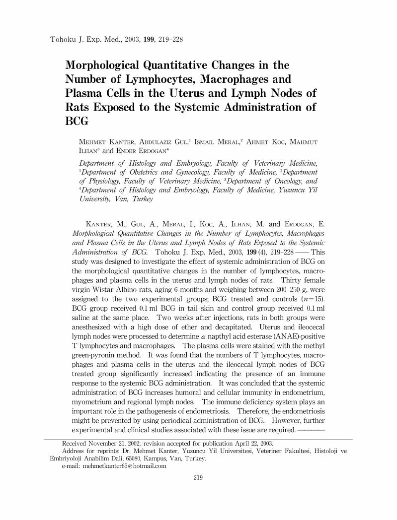
Morphological Quantitative Changes inthe Number of Lymphocytes,Macrophages and Plasma Cells inthe Uterus and LymphNodes of Rats Exposed to the Systemic Administrationof BCG MEHMET K ANTER ,A BDULAZIZ G UL ,I SMAIL MERAL ,A HMET K OC ,MAHMUT I LHAN andE NDER E RDOGAN Depar tment of Histology and Embr yology, Faculty of Veter inar y Medicine, Depar tment of Obstetr ics and Gynecology, Faculty of Medicine, Depar tment of Physiology, Faculty of Veter inar y Medicine, Depar tment of Oncology, and Depar tment of Histology and Embr yology, Faculty of Medicine, Yuzuncu Yil Univer sity, Van, Tur key K ANTER ,M.,G UL ,A.,MERAL ,I.,K OC ,A.,I LHAN ,M.and E RDOGAN ,E. Mor phological Quantitative Changes in the Numberof Lymphocytes, Macr ophages and Plasma Cells in the Uter us and Lymph Nodes of Rats Exposed to the Systemic Administr ation of BCG. TohokuJ.Exp.Med.,2003, 199 (4),219-228―― This study was designed to investigate the effect of systemic administration of BCGon themorphological quantitativechanges inthenumber of lymphocytes,macro- phages andplasmacells inthe uterus andlymphnodes of rats.Thirtyfemale virgin Wistar Albino rats,aging 6 months and weighing between 200-250 g,were assigned to thetwo experimental groups;BCG treated and controls( n=15). BCG groupreceived0.1 ml BCG intail skinandcontrol groupreceived0.1 ml saline at the same place.Twoweeks after injections,rats inbothgroups were anesthesizedwithahighdoseof ether anddecapitated.Uterus andileocecal lymph nodes were processed to determineαnapthyl acid esterase(ANAE)-positive Tlymphocytes and macrophages. The plasma cells were stained with the methyl green-pyronin method. It was found that the numbers of T lymphocytes,macro- phages andplasmacells intheuterusandtheileocecal lymphnodesof BCG treated group significantly increased indicating the presence ofan immune response to the systemic BCGadministration.It was concluded that the systemic administration of BCGincreases humoral and cellular immunity in endometrium, myometrium and regional lymph nodes. The immune deficiency system plays an important role in the pathogenesis of endometriosis.Therefore,the endometriosis might be prevented by using periodical administration of BCG. However,further experimental and clinical studies associated with these issue are required.―――― TohokuJ.Exp.Med.,2003, 199 ,219-228 ReceivedNovember 21,2002;revisionacceptedfor publicationApril 22,2003. Addressforreprints:Dr.Mehmet Kanter,YuzuncuYil Universitesi,VeterinerFakultesi,Histoloji ve Embriyoloji Anabilim Dali,65080,Kampus,Van,Turkey. e-mail:mehmetkanter65@hotmail.com 219
Transcript of Morphological Quantitative Changes in the Number of Lymphocytes, Macrophages and Plasma Cells in the...
Morphological Quantitative Changes in the
Number of Lymphocytes,Macrophages and
Plasma Cells in the Uterus and Lymph Nodes of
Rats Exposed to the Systemic Administration of
BCG MEHMET KANTER,ABDULAZIZ GUL,ISMAIL MERAL,AHMET KOC,MAHMUT
ILHAN and ENDER ERDOGAN
Department of Histology and Embryology, Faculty of Veterinary Medicine,
Department of Obstetrics and Gynecology, Faculty of Medicine, Department
of Physiology, Faculty of Veterinary Medicine, Department of Oncology, and
Department of Histology and Embryology, Faculty of Medicine, Yuzuncu Yil
University, Van, Turkey
KANTER,M.,GUL,A.,MERAL,I.,KOC,A.,ILHAN,M.and ERDOGAN,E.Morphological Quantitative Changes in the Number of Lymphocytes, Macrophages
and Plasma Cells in the Uterus and Lymph Nodes of Rats Exposed to the Systemic
Administration of BCG. Tohoku J.Exp.Med.,2003,199(4),219-228――This
study was designed to investigate the effect of systemic administration of BCG on
the morphological quantitative changes in the number of lymphocytes,macro-phages and plasma cells in the uterus and lymph nodes of rats.Thirty female
virgin Wistar Albino rats,aging 6 months and weighing between 200-250 g,were
assigned to the two experimental groups;BCG treated and controls(n=15).BCG group received 0.1 ml BCG in tail skin and control group received 0.1 ml
saline at the same place.Two weeks after injections,rats in both groups were
anesthesized with a high dose of ether and decapitated.Uterus and ileocecal
lymph nodes were processed to determineαnapthyl acid esterase(ANAE)-positive
T lymphocytes and macrophages.The plasma cells were stained with the methyl
green-pyronin method.It was found that the numbers of T lymphocytes,macro-phages and plasma cells in the uterus and the ileocecal lymph nodes of BCG
treated group significantly increased indicating the presence of an immune
response to the systemic BCG administration.It was concluded that the systemic
administration of BCG increases humoral and cellular immunity in endometrium,myometrium and regional lymph nodes.The immune deficiency system plays an
important role in the pathogenesis of endometriosis.Therefore,the endometriosis
might be prevented by using periodical administration of BCG.However,further
experimental and clinical studies associated with these issue are required.――――
Tohoku J.Exp.Med.,2003,199,219-228
Received November 21,2002;revision accepted for publication April 22,2003.Address for reprints:Dr.Mehmet Kanter,Yuzuncu Yil Universitesi,Veteriner Fakultesi,Histoloji ve
Embriyoloji Anabilim Dali,65080,Kampus,Van,Turkey.e-mail:mehmetkanter65@hotmail.com
219
BCG;endometriosis;uterus;lymph nodes;ratⒸ2003 Tohoku University Medical Press
The use of Bacillus Calmette-Guerin (BCG)in cancer has been encouraged by animal studies
indicating that it can prevent growth of tumor
transports and eliminate established tumors(Bast et al.1974).BCG was originally isolated
by Calmette and Guerin(Guerin 1957;Inooka et
al.1962)through progressive attenuation of a
virulent strain of Mycobacterium bovis.Hun-dreds of millions of vaccinations have been
performed with negligible mortality and mor-bidity(Lemonde et al.1971;Bansal and Sjogren
1973;Schwartz 1973).BCG is currently one of
the most effective agents for the prophylaxis
and cure of superficial bladder cancer(Haaff et
al.1985;Debruijine et al.1989).Although the
mechanisms of its antitumor activity are un-known,they are generally believed to act by the
immune system,since various components of
that system are known to be activated(Mitchell
and Murahata 1979;De Jong et al.1987).By
using immunohistochemical staining,De Jong et
al.(1987)have showed that the numbers of T
cells increased in the bladder wall after intra
vesical BCG administration in guinea pigs.Widely scattered B cells and macrophages were
also present,but were much fewer in number
than T cells(De Jong et al.1987).BCG is also a potent and persistent inducer
of lymphocyte trapping,particularly in regional
lymph nodes,where it also markedly enhances
the trapping response to a subsequent antigenic
challenge(Zats 1976).It has been suggested
that BCG activates both macrophages and T
cells in vitro(Mokyr and Mitchell 1975),and
that its effects are mediated in vivo by T cell
systems(Ariyan and gershon 1973;Mackaness
et al.1973;Lagrange and Mackaness 1975).It
has been also reported that B lymphocytes
increase in lymphoid organs following the
intraperitonal administration of BCG(Meyer et
al.1979).
Although many women have reflux seeding
of menstrual debris into the peritoneal cavity
but do not all develop endometriosis,there may
be genetic or immunologic factors that influence
susceptibility(Speroff et al.1999).Dmowski
and coworkers(1981)demonstrated that mon-keys with endometriosis had decreased cellular
immunity to endometrial tissue and women
with endometriosis demonstrate a decrease in
various measurements of immune response(Oosterlynck et al.1992;Vigano et al.1992).However,the presence of abnormal immunity in
the“precursor-stage”and the inverse correla-tion between these abnormalities and the stag
favor the altered immune system preceding the
endometriosis(Dmowski et al.1989;Gleicher
and Pratt 1993;Laçin et al.1998).This study was designed to investigate the
effect of systemic administration of BCG on the
morphological quantitative changes in the num-ber of lymphocytes,macrophages and plasma
cells in the uterus and lymph nodes of rats and
whether systemic prophylaxis with BCG can be
used for prevention of endometriosis.
MATERIALS AND METHODS
In this study 30 female 6 months old virgin
Wistar Albino rats weighing between 200-250 g
were used.Rats were assigned to two groups,BCG and controls(n=15),and fed with stan-dard feed and water ad libitum.In order to
maintain their biological rhythms stable 12
hours artificial and 12 hours dark require was
applied.All rats received humane care accord-ing to the criteria outlined in the“Guide for the
Care and Use of Laboratory Animals”prepared
by the National Academy of Sciences and publi-shed by the National Institutes of Health.BCG group received 0.1 ml BCG in tail skin
(Gul et al.2001)and control group received 0.1
M.Kanter et al.220
ml saline at the same place.Two weeks after
injections rats in both groups were anesthesized
with high dose ether and decapitated.Uterus
and ileocecal lymph nodes were processed to
determine a naphthyl acid esterase(ANAE)-positive T lymphocytes and macrophages.Briefly,the uterine and ileocecal lymph nodes
were removed and fixed in previously cooled
formalin-sucrose solution at+4℃ for 22 hours
and then they were kept+4℃ in Holt solution
for 22 hours(Mueller et al.1975).Tenμthin slides obtained using cryostat
(Microm,Germany)were transferred into the
formalin-gelatin covered glass.The materials
were left to dry at room temperature in order to
determine ANAE positive lymphocytes(Mueller
et al.1975)and macrophages(Zicca et al.1981).The slides were stained in an incubation solu-tion described by Mueller et al.(1975)at pH 7.2 for 5 minutes.After ANAE staining,the
slides were washed and processed for nucleus
staining for five minutes with 1% methyl green
dissolved in acetate buffer(pH 4.2).Materials’water was removed by preceding with alcohol
solutions at different concentrations and mate-rials were shinned with xylol and covered with
entellane.In order to plasma cells,same of the tissue
samples were fixed in formalin-alcohol fixation
solution for 24 hours.The routine histological
tissue processing protocol was applied to the
tissue samples.Methyl green-pyronine staining
was applied to 6μm-thick slides(Bancroft and
Cook 1984)obtained using rotary microtome
(Leica 2135,Leica Instruments GmbH,Nussloch,Germany).Materials were examined through
a research microscope (Nikon Optiphot 2,Tokyo).In order to determine the numerical distri-
bution of cell in the tissue samples,20 areas in
the endometrium-myometrium of the uterus and
the cortex-medulla of the lymph nodes were
chosen randomly and the cells were counted in
each area.The cell(100 square mm)densities
in each area were calculated and recorded as
cell number/mm.The data were expressed as
means with standard deviations(S.D.).Student’s
t-test was used to compare BCG treated vs.control rats.
RESULTS
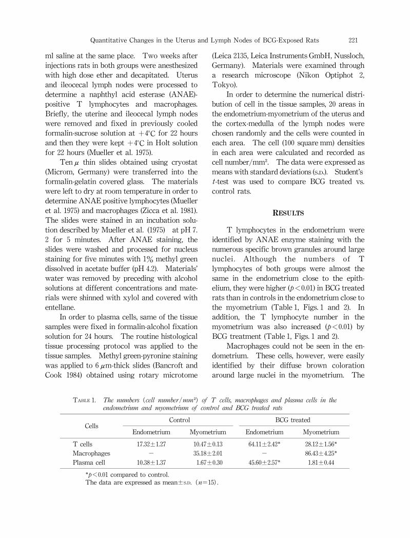
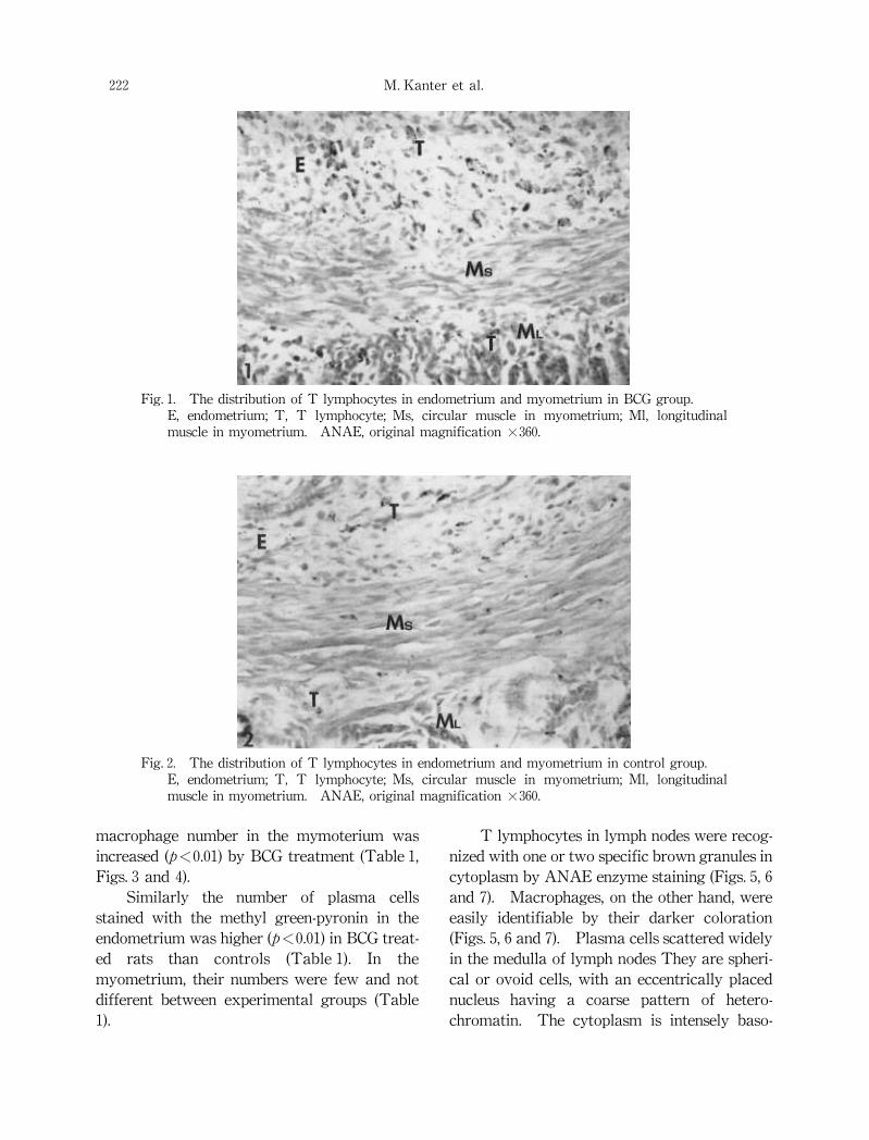
T lymphocytes in the endometrium were
identified by ANAE enzyme staining with the
numerous specific brown granules around large
nuclei. Although the numbers of T
lymphocytes of both groups were almost the
same in the endometrium close to the epith-elium,they were higher(p<0.01)in BCG treated
rats than in controls in the endometrium close to
the myometrium (Table 1,Figs.1 and 2).In
addition,the T lymphocyte number in the
myometrium was also increased(p<0.01)by
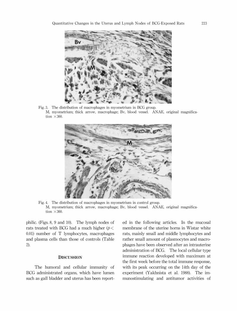
BCG treatment(Table 1,Figs.1 and 2).Macrophages could not be seen in the en-
dometrium.These cells,however,were easily
identified by their diffuse brown coloration
around large nuclei in the myometrium.The
Quantitative Changes in the Uterus and Lymph Nodes of BCG-Exposed Rats
TABLE 1. The numbers (cell number/mm )of T cells, macrophages and plasma cells in the
endometrium and myometrium of control and BCG treated rats
Cells Control
Endometrium Myometrium
BCG treated
Endometrium Myometrium
T cells 17.32±1.27 10.47±0.13 64.11±2.42 28.12±1.56
Macrophages - 35.18±2.01 - 86.43±4.25
Plasma cell 10.38±1.37 1.67±0.30 45.60±2.57 1.81±0.44
p<0.01 compared to control.The data are expressed as mean±S.D.(n=15).
221
macrophage number in the mymoterium was
increased(p<0.01)by BCG treatment(Table 1,Figs.3 and 4).Similarly the number of plasma cells
stained with the methyl green-pyronin in the
endometrium was higher(p<0.01)in BCG treat-ed rats than controls (Table 1).In the
myometrium,their numbers were few and not
different between experimental groups(Table
1).
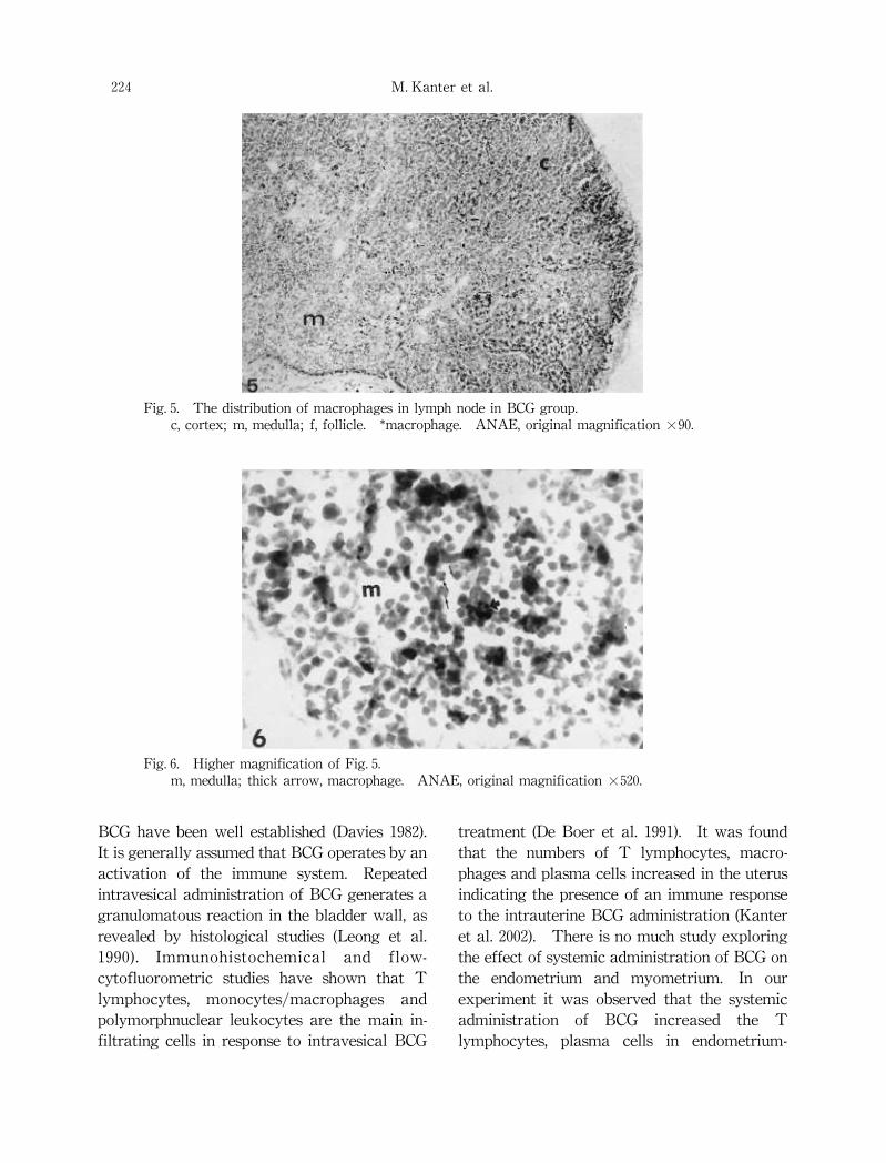
T lymphocytes in lymph nodes were recog-nized with one or two specific brown granules in
cytoplasm by ANAE enzyme staining(Figs.5,6
and 7).Macrophages,on the other hand,were
easily identifiable by their darker coloration(Figs.5,6 and 7).Plasma cells scattered widely
in the medulla of lymph nodes They are spheri-cal or ovoid cells,with an eccentrically placed
nucleus having a coarse pattern of hetero-chromatin.The cytoplasm is intensely baso-
Fig.1.The distribution of T lymphocytes in endometrium and myometrium in BCG group.E,endometrium;T,T lymphocyte;Ms,circular muscle in myometrium;Ml,longitudinal
muscle in myometrium.ANAE,original magnification×360.
Fig.2.The distribution of T lymphocytes in endometrium and myometrium in control group.E,endometrium;T,T lymphocyte;Ms,circular muscle in myometrium;Ml,longitudinal
muscle in myometrium.ANAE,original magnification×360.
M.Kanter et al.222
philic.(Figs.8,9 and 10).The lymph nodes of
rats treated with BCG had a much higher(p<0.01)number of T lymphocytes,macrophages
and plasma cells than those of controls(Table
2).
DISCUSSION
The humoral and cellular immunity of
BCG administrated organs,which have lumen
such as gall bladder and uterus has been report-
ed in the following articles.In the mucosal
membrane of the uterine horns in Wistar white
rats,mainly small and middle lymphocytes and
rather small amount of plasmocytes and macro-phages have been observed after an intrauterine
administration of BCG.The local cellular type
immune reaction developed with maximum at
the first week before the total immune response,with its peak occurring on the 14th day of the
experiment(Yakhnista et al.1988).The im-munostimulating and antitumor activities of
Fig.4.The distribution of macrophages in myometrium in control group.M,myometrium;thick arrow,macrophage;Bv,blood vessel.ANAE,original magnifica-tion×360.
Fig.3.The distribution of macrophages in myometrium in BCG group.M,myometrium;thick arrow,macrophage;Bv,blood vessel.ANAE,original magnifica-tion×360.
223 Quantitative Changes in the Uterus and Lymph Nodes of BCG-Exposed Rats
BCG have been well established(Davies 1982).It is generally assumed that BCG operates by an
activation of the immune system.Repeated
intravesical administration of BCG generates a
granulomatous reaction in the bladder wall,as
revealed by histological studies(Leong et al.1990). Immunohistochemical and flow-cytofluorometric studies have shown that T
lymphocytes,monocytes/macrophages and
polymorphnuclear leukocytes are the main in-filtrating cells in response to intravesical BCG
treatment(De Boer et al.1991).It was found
that the numbers of T lymphocytes,macro-phages and plasma cells increased in the uterus
indicating the presence of an immune response
to the intrauterine BCG administration(Kanter
et al.2002).There is no much study exploring
the effect of systemic administration of BCG on
the endometrium and myometrium.In our
experiment it was observed that the systemic
administration of BCG increased the T
lymphocytes,plasma cells in endometrium-
Fig.5.The distribution of macrophages in lymph node in BCG group.c,cortex;m,medulla;f,follicle. macrophage.ANAE,original magnification×90.
Fig.6.Higher magnification of Fig.5.m,medulla;thick arrow,macrophage.ANAE,original magnification×520.
M.Kanter et al.224

myometrium and macrophages in myometrium.The systemic administration of BCG caused the
similar humoral and cellular immunity as intra-uterine administration of BCG in the uterus and
lymph nodes.Because,the intrauterine admin-istration of BCG is needed laparatomy or
opened cervix in estrous cycle,systemic admin-istration of BCG might be preferred.The administration of BCG evokes a T
cell-mediated immune response in mouse
lymphoid organs(Ten Dam et al.1976).The
BCG induced activation of macrophages has
been adequately documented(Kelly 1976).It
has been also suggested that B lymphocytes
increases in lymphoid organs following the
intraperitoneal administration of BCG(Meyer
et al.1979).The enlargement of the lymph node T cell
areas,a systemic effect of BCG,may be due in
part to increased influx or trapping of recir-culating T cells as demonstrated by Zatz(1976)in peripheral lymph nodes after intravenous
Fig.7.The distribution of macrophages in lymph node in control group.c,cortex;m,medulla;f,follicle. macrophage.ANAE,original magnification×80.
Fig.8.The distribution of plasma cells in lymph node in BCG group.c,cortex;m,medulla;f,follicle. plasma cells.Methyl green-pyronine,original magnifica-tion×80.
225 Quantitative Changes in the Uterus and Lymph Nodes of BCG-Exposed Rats

Fig.9.The distribution of plasma cells in lymph node in control group.c,cortex;m,medulla;f,follicle;thick arrow,plasma cell.Methyl green-pyronine,original
magnification×80.
Fig.10.Higher magnification of Fig.9.m,medulla;thick arrow,plasma cell.Methyl green-pyronine,original magnification×520.
TABLE 2. The numbers (cell/mm )of T cells, macrophages and plasma cells in the cortex-medulla of lymph nodes of control and BCG treated rats
Cells Control
Cortex Medulla
BCG treated
Cortex Medulla
T cells 420.22±39.16 60.19±3.42 496.19±39.41 172.92±12.98
Macrophages 16.10±0.82 29.16±1.49 68.32±3.47 72.12±4.09
Plasma cell 13.17±0.55 51.53±3.97 17.08±0.60 536.38±46.90
p<0.01 compared to control.The data are expressed as mean±S.D.(n=15).
M.Kanter et al.226
injection of BCG.The increase of germinal
center areas also suggests a BCG-induced prolif-eration of B cells(Meyer et al.1979).In addi-tion,the number of T lymphocytes,macro-phages and plasma cells also increased in the
ileocecal lymph nodes of rats to the intrauterine
administration of BCG (Kanter et al.2002).Systemic administration of BCG was also found
to increase the T lymphocytes,macrophages
and plasma cells in lymphoid organs.Hence
the present data support previous investigation.There is ample evidence suggesting that the
immune system plays an important role in the
pathogenesis of endometriosis,and an altered
immune responsiveness has been proposed to
explain why some women develop en-dometriosis,whereas others do not(Braun and
Dmowski 1998).Because we did not try to
produce an endometriosis experimentally after
the systemic BCG administration,we do not
know whether systemic administration of BCG
could prevent the implantation of endometriosis.However,Gul et al.(2001)suggested that sys-temic prophylaxis with BCG prevented the im-plantation of endometriosis in rats.In our
study,since systemic BCG administration in-creased the immune responsiveness,we thought
it might also prevent the implantation of en-dometriosis.However,more studies are need-ed.In conclusion,systemic administration of
BCG increases humoral and cellular immunity
in endometrium,myometrium and regional
lymph nodes.The immune deficiency system
plays an important role in the pathogenesis of
endometriosis.Therefore,the endometriosis
might be prevented by using periodical adminis-tration of BCG.However,further experimen-tal and clinical studies associated with these
issue are required.
References
Ariyan,S.&Gershon,K.R.(1973)Augmentation of
the adoptive transfer of specific tumor immu-nity by non-specifically immunized macro-
phages. J. Natl. Cancer Inst.,51,1145-1148.Bancroft,J.D.& Cook,H.C.(1984) Manual of
histological techniques.Churchill Livingstone,New York.
Bansal,S.C.&Sjogren,H.O.(1973)Effects of BCG
on various facets of the immune response
against polyoma tumors in rats. Int. J. Can-cer.,11,162-171.
Bast,R.C.,Zbar,B.,Borsos,T.&Rapp,H.J.(1974)BCG and cancer.New Engl. J. Med.,20,1413-1419.
Braun, D.P. & Dmowski, W.P.(1998) En-dometriosis:abnormal endometrium and
dysfunctional immune response. Curr. Op.Obstet. Gynecol.,10,365-369.
Davies,M.(1982)Bacillus Calmette-Guerin as an
antitumor agent.The interaction with cells
of the mammalian immune system. Biochim.Biophys. Acta,651,143-147.
De Boer,E.C.,de Jong,W.H,van der Meijen,A.P.M.,Streenberg,P.A.,Witjes,F.,Vegt,P.D.J.,De-bruyne,F.M.J.&Ruitenberg,E.J.(1991)Leu-kocytes in the urine after intravesical BCG
treatment for superficial bladder cancer.A
flow citofluorometric analysis.Urol. Res.,19,45-50.
Debruijine,F.M.J.,Denis,L.& Van Der Meijden,A.P.M.(1989)BCG in superficial bladder
cancer.EORTC Genitourinary Group Mono-graph 6. Prog. Clin. Biol. Res.,310A.Liss,New York.
De Jong,W.H.,Steerenberg,P.A.&Ruitenberg,E.J.(1987)Bacillus Calmette-Guerin(BCG)and
its use for cancer immunotherapy.Res.Monogr. Immunol.,11,283-307.
Dmowski,W.P.,Steele,R.W.& Baker,G.F.(1981)Deficient cellular immunity in endometriosis.Am. J. Obstet. Gynecol.,141,377-380.
Dmowski,W.P.,Gebl,H.M.& Rawlins,R.G.(1989)Immunologic aspects of endometriosis.Ob-stet. Gynecol. Clin. North. Am.,16,93-103.
Gleicher,N.& Pratt,D.(1993)Abnormal(auto)immunity endometriosis Int. J. Gynecol. Ob-stet.,40(Suppl.),21-27.
Guerin,C.(1957)In:The History of BCG, BCG
Vaccination Against Tuberculosis,edited by
S.R.Rosenthal,Boston Little,Brown and
Company,pp.48-53.Gul,A.,Yasar,T.&Ugras,S.(2001)BCG vaccina-
tion to prevent implantation of endometriosis:an experimental study in rats. Eur. J. Obstet.Gynecol. Reprod. Biol.,98,209-212.
227 Quantitative Changes in the Uterus and Lymph Nodes of BCG-Exposed Rats

Haaff,E.O.,Dresner,S.M.,Kelley,D.R.,Ratliff,T.L.,
Shapiro,A.&Catalona,W.J.(1985)Role of
immunotherapy in the prevention of recur-rence and invasion of urothelial bladder
tumors:a review. Worl. J. Urol.,3,76-85.
Inooka,S.,Ebina,T.&Tekase,Y.(1962)Influence
of BCG vaccination on Ehrlich ascites tumor
in mice.Kekkak,37,503-505.Kanter,M.,Yoruk,M.,Koc,A.,Meral,I.&
Timurkan,H.H.(2002)A rat model for the
immune response to the intrauterine adminis-tration of BCG. Scand. J. Lab. Anim. Sci.,1,34-39.
Kelly,M.T.(1976)Activation of guinea pig macro-phages by cell walls of Mycobacterium bovis,strain BCG. Cell. Immunol.,26,254-263.
Laçin,S.,Sungurtekin,U.& Çapanogvlu,R.(1998)The dilemma of endometriosis:is the solution
immunomodulation. Jinekolji Obstetrik.,8,6-8.
Lagrange,P.H.&Mackaness,G.B.(1975)A stable
form of delayed type hypersensitivity. J. Exp.Med.,141,82-96.
Lemonde,P.,Dubreuil,R.& Guindon,A.(1971)Stimulating influence of Bacillus Calmette-Guerin on immunity to polyoma tumors and
spontaneous leukemia. J. Natl. Cancer Inst.,47,1013-1024.
Leong,A.S.Y.,Wannakrairot,P.,Jose,J.&Milios,J.(1990) Bacillus Calmette Guerin-treated
superficial bladder cancer:correlation of
morphology with immunophenotyping. J.Pathol. (Lond.),162,35-41.
Mackaness,G.B.,Auclair,D.J.& Lagrange,P.H.(1973)Immunopotentiation with BCG.I.Immune response to different strains and
preparations. J. Natl. Cancer Inst.,51,1655-1667.
Meyer,E.M.,Wilmsmann,K.,Schalke,W.&Grundmann,E.(1979) Quantification of
BCG-induced reactions of T and B areas in
peripheral lymphoid organs of young adult
BALB/c mice.A histometrical and autor-adiographical study. Path. Res. Pract.,164,127-140.
Mitchell,M.S.&Murahata,R.I.(1979)Modulation
of immunity by Bacillus Calmette-Guerin(BCG). Pharmacol. Ther.,4,329-353.
Mokyr,M.B.&Mitchell,M.S.(1975)Activation of
lymphoid cells by BCG in vitro. Cell. Im-munol.,15,264.
Mueller,J.,Del Re,G.B.,Buerki,H.,Keller,H.U.,Hess,M.W.&Cottier,H.(1975)Nonspecific
acid esterase activity:A criterion for differen-tiation of the T and B lymphocytes in mouse
lymph nodes. Eur. J. Immun.,5,270-274.
Oosterlynck,D.J.,Meuleman,C.,Waer,M.,Vande-putte,M.&Koninckx,P.R.(1992)The natu-ral killer activity of peritoneal fluid
lymphocytes is decreased in women with en-dometriosis. Fertil. Steril.,8,292-295.
Schwartz,E.(1973)Effect of mycobacterium bovis
BCG Praha on the growth of transplanted
Daels sarcoma in guinea-pigs.Neoplasma,20,375-386.
Speroff,L.,Glass,R.H.& Kase,N.G.(1999)En-dometriosis.In:Clinical Gynecologic Endo-crinology and Infertility,6th ed,Baltimore
Mayland,pp.1057-1073.
Ten Dam,H.G.,Toman,K.,Hitze,K.L.&Guld,J.(1976)Present knowledge of immunization
against tuberculosis. Bull. WHO,54,255-269.
Vigano,P.,Vercillini,P.,Di Blasio,A.M.,Colombo,A.,Candiani,G.B.&Vignal,M.(1992)Defi-cient antiendometrium lymphocyte-mediated
cytotoxicity in patients with endometriosis.Fertil. Steril.,58,292-296.
Yakhnista,A.G.,Stefanov,S.B.& Artyukh,E.V.(1988)Reaction of lymphocytes in en-dometrium to the intrauterine administration
of antigens.Arch. Anat. Hist. Embr.,64,74-77.
Zatz,M.M.(1976)Effects of BCG on lymphocyte
trapping. J. Immunol.,116,1587-1691.Zicca,A.,Leprini,A.,Cadoni,A.,Franzi,A.T.,Fer-
rarini M.& Grossi,C.E.(1981) Ultra-structural localization of alpha naphtyl acid
esterase in human T lymphocytes.Am. J.Pathol.,105,40-46.
M.Kanter et al.228