
1.1 Uterus and Its Functions
192
-
Upload
khangminh22 -
Category
Documents
-
view
2 -
download
0
Transcript of 1.1 Uterus and Its Functions

CHAPTER Ι
INTRODUCTION
1.1 Uterus and Its Functions
The uterus or womb is a major female reproductive organ of most mammals,
including humans. One end, the cervix, opens into the vagina; the other is connected
on both sides to the fallopian tubes. In mammals, the four main forms in which it is
found are: bipartite, as in cows; bicornuate, as in pigs; simplex, as with the pear-
shaped one found in humans and horses; and duplex, found in rodents.
The uterus is located in the pelvis immediately dorsal to the urinary bladder
and ventral to the rectum. It is held in place by several ligaments. Outside of
pregnancy, its size is several centimeters in diameter.
The uterus mostly consists of muscle, known as myometrium. The lining of
the uterine cavity is called the endometrium. In most mammals, including humans, the
endometrium builds a lining periodically which, if no pregnancy occurs, is shed or
reabsorbed. Shedding of the endometrial lining in humans is responsible for monthly
menstrual bleeding, known colloquially as woman's "period", throughout the fertile
years of a female. In other mammals there may be cycles set as widely apart as six
months or as frequently as a few days.
Although, there are morphological differences in the uteri among species,
they have a unique function. The main function of the uterus is to accept fertilized
ovum which (becomes) implanted into the endometrium, and derives nourishment
2
from blood vessels which develop exclusively for this purpose. The fertilized ovum
becomes an embryo, which then develops into fetus and gestates until childbirth. In
addition, the uterine changes during menstrual cycle are caused by changes in the
plasma concentration levels of estrogen and progesterone. During the proliferative
phase, an increasing plasma estrogen level stimulates growth of both the endomertium
and the underlying uterine smooth muscle (myometrium) for their receptors. Then,
following ovulation and formation of the corpus luteum (during the secretory phase),
progesterone acts upon this estrogen primed endometrium to convert it to an actively
secreting tissue. The changes are essential to make the endometrium a hospitable
environment for implantation and nourishment of the developing embryo. Uterine
quiescence is maintained by progesterone throughout pregnancy and is essential to
prevent premature delivery.
1.1.1 Anatomy of the Uterus
The uterine wall is thick and composed of three layers. The endometrium is
the inner mucosal layer lining the uterine cavity. It is covered with columnar
epithelium and contains abundant tubular glands. The myometrium, a very thick,
muscular layer, largely consists of bundles of smooth muscle fibers in longitudinal,
circular, and spiral patterns and is interlaced with connective tissues. During the
monthly female reproductive cycles and during pregnancy, the endometrium and
myometrium extensively change. The perimetrium consists of an outer serosal layer,
which covers the body of the uterus and part of the cervix (Shier et al., 2002).
3
1.1.2 Contractile Proteins
The structural and functional filaments of two major proteins in muscle cells
are thick and thin (Broderick and Broderick, 1990) filaments. The former comprises
thick myosin-containing filaments with thin actin-containing filaments. Their
interaction governs the extent of smooth muscle contraction (Taggart and Morgan,
2007). It has been suggested that this contraction arises via Ca2+-independent isoforms
of PKC, possibly via uninhibited thin filaments (Horowitz et al., 1996).
The Thick Filaments
Myosin (thick filaments) consist of two heavy chains (MHC) that form a
coiled rod-like structure together with a globular head domain, two regulatory light
chains (MLC20) and two essential light chains (MLC17). One of each type of the light
chain is associated with the head domain of MHC. Regulation of each myosin
subunit, whether that be in terms of expression or post translational modification, may
well participate in myometrial contractile adaptations with gestation. An elevation of
Ca2+ results in the co-operative binding to the calcium binding canmodulin protein
(CaM) and subsequent activation by Ca2+-(CaM)4 of the intracellular enzyme myosin
light chain kinase (MLCK). Ca2+-(CaM)4-MLCK, in turn, acts to increase the
serine/threonine phosphorylation of the regulatory light chains of myosin (MLC20). A
matching of the elevation of [Ca2+]i to phosphorylation of MLC20 has been reported in
uterine smooth muscle of many species and in response to diverse contractile stimuli
(Taggart et al., 1997; Word et al., 1993; Word et al., 1994; Shojo and Kaneko, 2001)
4
The Thin Filaments
Contractile thin filaments consist mainly of an alpha helical coil of actin and
associated proteins caldesmon and calponin (Marston and Redwood, 1991). Smooth
muscle actin however, has been suggested to exist as part of both a contractile domain
directly involved in force-generation events and a cytoskeletal domain important for
structural integrity (Small and Gimona, 1991).
Calmodulin
Calmodulin was previously discussed in relation to the function of Ca2+ as a
second messenger in hormone action. The calmodulin-Ca2+ complex thus formed
combines with and activates MLCK, an enzyme that catalyzes the phosphorylation
(addition of phosphate groups) of MLC, a component of the myosin cross bridges. In
smooth muscle (unlike striated muscle), the phosphorylation of myosin cross bridges
is the regulatory event that permits them to bind to actin and thereby produce a
contraction. Calmodulin can bind four Ca2+ ions but may have two already bound at
the c-terminal binding sites under resting conditions ie, low [Ca2+]i (Bavley et al.,
1996; Iohnson et al., 1996). Indeed, recently suggested a novel scheme where by a
portion of calmodulin is tightly bound to the myofilaments and the Ca2+ (Keirse,
1995; Larcombe-McDouall et al., 1999) for contraction diffuses to this site. The Ca2+-
calmodulin (CaM) interaction introduces significant delay between [Ca2+]i increase
and ensuring an increase of force. The activation of MLCK after Ca2+-calmodulin
binding is also relatively slow and could be one of the rate-limiting steps in
5
contraction along with recruitment and diffusion of calmodulin (Somlyo and Somlyo,
1990).
Myosin Light Chain Kinase
Myosin light chain kinase (MLCK) of smooth muscle consists of an actin-
binding domain at the N-terminal, the catalytic domain in the central portion, and the
myosin-binding domain at C-terminal. The kinase activity is mediated by the catalytic
domain that phosphorylates the myosin light-chain of 20 kDa (MLC20), activating
smooth muscle myosin to interact with actin (Nakamura et al., 2008).
Since MLCK is activated by Ca2+-calmodulin complex, it is somewhat
difficult to estimate the relative contribution of thick and thin filaments in the
activation process of smooth muscle contraction. However, the affinity of caldesmon
(CD) or calponin (CP) for Ca2+-CaM complex is two to three orders of magnitude
lower than that of Ca2+-CaM complex for MLCK (Walsh, 1994). This finding
suggests that Ca2+-CaM activated MLCK is the main mechanism of the contraction
that CD and CP act as second regulatory mechanisms (Savineau and Marthan, 1997).
MLCK can be phosphorylated in vitro by several kinases including PKA, PKC and
CaM kinase II (Nishikawa et al., 1978; Adelstein et al., 1978; Hashimoto and
Hodering, 1990; Ikebe and Reardon, 1990; Stull et al., 1993). It has been presented
that stimulation by agonists elevate the intracellular Ca2+ concentration of smooth
muscle, causing Ca2+ to bind with calmodulin (CaM). CaM in conjunction with Ca2+
(Ca-CaM) activates MLCK. Myosin, thus phosphorylated at MLC20 by MLCK, is in
an active form and interacts with actin to induce contraction (Bárány, 1979).
Myosin Light Chain Phosphatase
6
Myosin light chain (MLC) Phosphatase is physiologically responsible for the
dephosphorylation of the MLC20. The kinase is found to be bound tightly with myosin
and is not dissociated from myosin under physiological ionic conditions, suggesting
that under physiological condition the kinase is targeted for its substrate (Horowitz et
al., 1996).
Force regulation in smooth muscle is dependent on the activities of MLC
kinase and MLC phosphatase (Hartshorne et al., 1998; Gong et al., 1992). The activity
of MLC kinase is regulated by Ca2+-CaM (Hartshorne et al., 1998), whereas MLC
phosphatase was originally thought to be constitutively active and unregulated
(Hartshorne et al.,1998). However, there is abundant evidence that the activity of
MLC phosphatase can be both inhibited to produce Ca2+ sensitization (Hartshorne et
al., 1998; Somlyo and Somlyo, 1994; 1999) or an increase in force at a constant
[Ca2+]. Nitric oxide (NO) is the classical agent to produce Ca2+desensitization by
activating the soluble pool of guanylate cyclase, which in turn produces cGMP and
leads to the activation of type I cGMP-dependent protein kinase (PKGI). PKGI
mediates smooth muscle cell relaxation by several mechanisms. It has been
demonstrated that PKGI acts on the K+ channel to produce hyperpolarization of the
smooth muscle, decreases Ca2+ flux, and also activates MLC phosphatase (Surks et
al., 1999; Etter et al., 2001) to decrease the level of MLC20 phosphorylation and to
produce smooth muscle relaxation.
1.1.3 Uterine Contractile Activity
Excitation-Contraction Coupling
7
Excitation-contraction (EC) coupling in smooth muscles is triggered by a
sharp rise in the Ca2+ concentration within the cytoplasm of the muscle cells (Fox,
2004). The EC coupling starts with a depolarization of the plasma membrane that is
the activation threshold of voltage-activated dihydropyridine-sensitive L-type Ca2+
channels and causes them to open. The opened Ca2+ channels allow the influx of Ca2+
that not only contributes to the further explosive depolarization of the plasmalemma,
but also binds to the Ca2+ binding protein calmodulin (Matthew et al., 2004). Which is
structurally similar to troponin in striated muscles, calmodulin was previously
discussed in relation to the function of Ca2+ as a second messenger in hormone action.
The Ca2+-calmodulin complex thus formed combines with and activates MLCK, an
enzyme that catalyzes the phosphorylation of MLC, a component of the myosin cross
bridges (Fox, 2004) and thereby produce a contraction.
Uterine Smooth Muscle Contraction
Muscle contraction is turned on when sufficient amounts of Ca2+ bind to
troponin protein. This occurs when the concentration of Ca2+ of the sarcoplasm rises
above 10-6 molar (Fox, 2004). During normal contraction, when the cross bridges
attach to actin, they undergo power strokes and cause muscle contraction. The
contractile state of smooth muscle is determined predominantly by the level of
phosphorylated myosin, achieved largely via MLCK whose activity is regulated by
calmodulin (Somlyo and Somlyo, 1994; Walsh et al., 1996). In order for muscle to
relax, therefore, the attachment of myosin cross bridges to actin must be prevented.
The regulation of cross-bridge attachment to actin is a function of two proteins that
are associated with actin in the thin filaments.
8
Uterine Smooth Muscle Relaxation
The muscle relaxation is produced by the active transport of Ca2+ out of the
sarcoplasmic reticulum (SR). The SR is a modified endoplasmic reticulum, consisting
of interconnected sacs and tubes that surround each myofibril within the muscle cell.
Relaxation of the smooth muscle follows the closing of the Ca2+ channels and
lowering of the cytoplasmic Ca2+ concentration by the action of Ca2+-ATPase active
transport pumps (Fox, 2004). Under those conditions, calmodulin dissociates from the
myosin light-chain kinase, thereby inactivating this enzyme. The phosphate groups
that were added to the myosin are then removed by a different enzyme, a myosin
phosphatase. Dephosphorylation inhibits the cross bridge from binding to actin and
producing another power stroke.
Although the uterus is myogenic, myometrial activity can be regulated by both
adrenergic and cholinergic nerves. Adrenergic stimulation caused both contraction
and relaxation of the uterine smooth muscle through smooth muscle α-(excitatory)
and β- (inhibitory) adrenoreceptors (Marshall, 1970; O’Donnell et al., 1978; Digges,
1982). On the other hand chlorinergic stimulation has been shown to cause the
contraction of the myometrial through muscarinic receptors present on the smooth
muscle cells (Nakanishi and Wood, 1971; Hollingsworth, 1975; Morizaki et al.,
1989). Neuronal modulation, especially the β-adrenergic agonists (β-mimertics) are
the most commonly used tocolytic agents for the prevention of pre-term delivery
(Monga and Creasy, 1995). The rationale for using these compounds is based on their
9
ability to increase adenosine 3, 5 -cyclic monophosphate (cAMP) or guanosine 3, 5 -
cyclic monophosphate (cGMP) level in smooth muscle of the uterus through binding
to specific receptors linked to the stimulatory guanine neucleotide-dependent
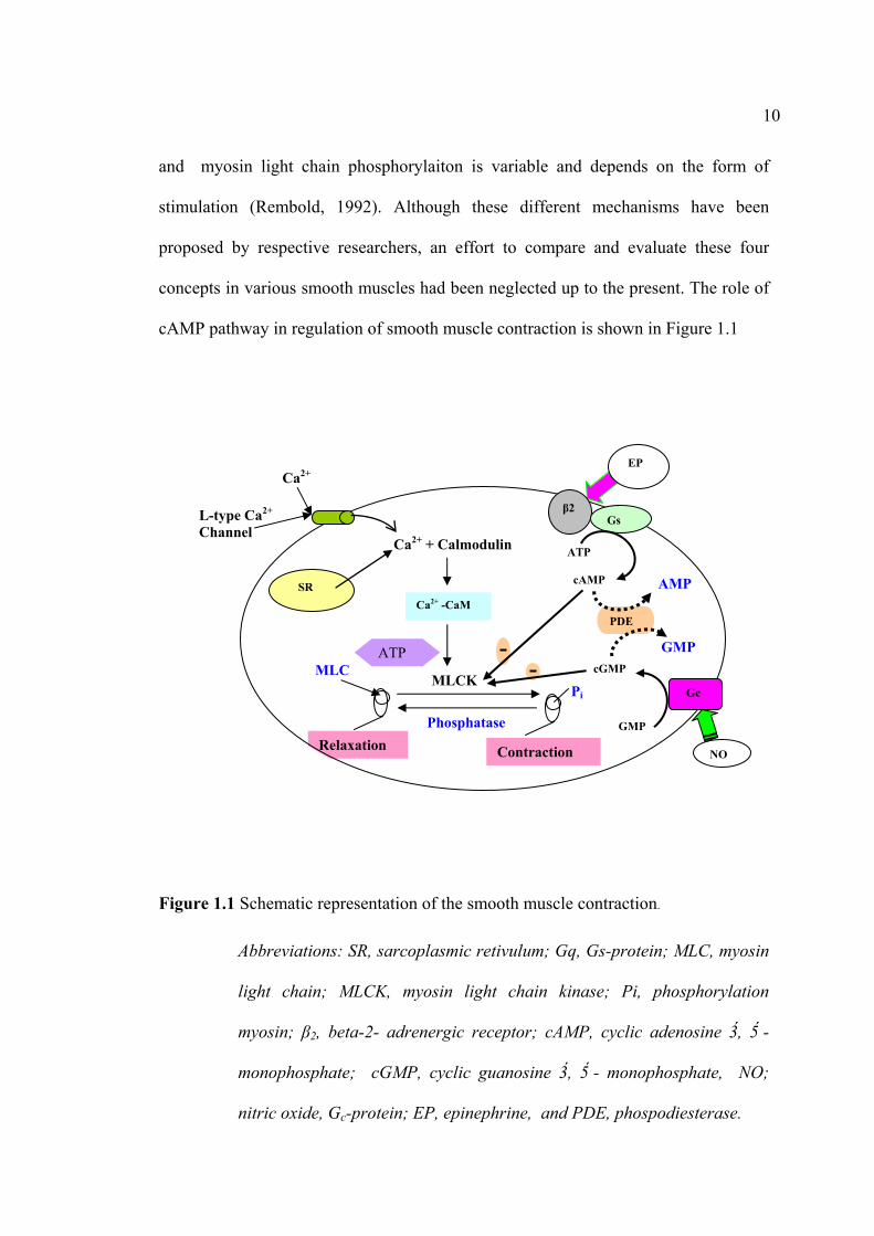
regulatory protein (Gi(s)), which, in turn, leading to uterine relaxation. cAMP and
cGMP are important second messengers that control many physiological processes,
including smooth muscle relaxation (Diamond, 1978). Adenylyl cyclase and guanylyl
cyclase synthesize cAMP and cGMP, respectively and phosphodiesterases (PDE)
enzyme degrades them (Fig. 1.1). The proposed mechanism for action of cAMP and
cGMP in smooth muscle relaxation can be described (see below).
Cyclic Nucleotide-Induced Relaxation
cAMP is an important intracellular second messenger in many tissues and
mediates the effect of multiple drugs and hormones. It is known that cAMP produces
relaxation of smooth muscle by activation of cAMP dependent protein kinase (PKA)
which interferes with several processes involved in smooth muscle contraction (Wray,
1993). cAMP causes relaxation by lowering in intracellular Ca2+ concentration
([Ca2+]i) by: 1) increasing extrusion of Ca2+ due to stimulation of both Ca2+ transport
and Ca2+ ATPase by the plasma membrane; 2) stimulating the Na+-K+ pump, thereby
lowering [Na]i which in turn enhance Ca2+ efflux on Na+- Ca2+ exchange; 3) causing
internal sequestration of Ca2+ (Casteels and Raeymaekers, 1979; Mueller and van
Breemen, 1979); and 4) inhibiting Ca2+ influx, although this has not been
demonstrated directly in the uterus (Scheid et al., 1979; Bullbring and den Hertog,
1980; Wray, 1993). However, recently data suggest that the relation between [Ca2+]i
10
and myosin light chain phosphorylaiton is variable and depends on the form of
stimulation (Rembold, 1992). Although these different mechanisms have been
proposed by respective researchers, an effort to compare and evaluate these four
concepts in various smooth muscles had been neglected up to the present. The role of
cAMP pathway in regulation of smooth muscle contraction is shown in Figure 1.1
Figure 1.1 Schematic representation of the smooth muscle contraction.
Abbreviations: SR, sarcoplasmic retivulum; Gq, Gs-protein; MLC, myosin
light chain; MLCK, myosin light chain kinase; Pi, phosphorylation
myosin; β2, beta-2- adrenergic receptor; cAMP, cyclic adenosine 3, 5 -
monophosphate; cGMP, cyclic guanosine 3, 5 - monophosphate, NO;
nitric oxide, Gc-protein; EP, epinephrine, and PDE, phospodiesterase.
Pi
Ca2+
L-type Ca2+ Channel
Relaxation
PDE
EP
NO
GMP
cGMP
Gc
AMP
GMP
β2Gs
ATP
cAMP SR
Ca2+ + Calmodulin
Ca2+ -CaM
Contraction
MLC
Phosphatase
MLCK
ATP

11
During the last decade, numerous studies have demonstrated the modulation of
inflammatory cell activations by selective PDE4 inhibitors. It is now established that
an elevation of cAMP is able to inhibit some of inflammatory processes (Lagente et
al., 2005). The increase of intracellular cAMP can be achieved through receptor
activation or inhibition of cAMP breakdown (Conti et al., 1995). PDEs are
responsible for the breakdown of intracellular cyclic nucleotides, from which PDE4
are the major cAMP metabolizing isoenzymes found in inflammatory and immune
cells (Lagente et al., 2005). The first generation of PDE4 inhibitors, although potent
anti-inflammatory agents, failed as pharmaceuticals owning to their emetic and gastric
side-effects (Mackenzie, 2004). It should be noted that the inhibitory effect of cAMP
was demonstrated under special conditions using high concentrations of cAMP (≥ 10-4
M) in the presence of fluoride or theophylline or by the use of protein kinase. In
contrast, the study reported that 3 × 10⁻⁶ M cAMP had no effect on 10-5 M Ca2+-
induced contraction of saponin-treated skinned smooth muscle in the presence of
exogenous protein kinase (Itoh et al., 1982). According to Ruegg et al. (1983), the
inhibitory effect of cAMP was best observed when a low concentration of Ca2+ was
used for the contraction. Nevertheless, the present observation shows that 10-5 M
cAMP had no effect on the contraction induced with 10-6 M Ca2+ or 10-5 M Ca2+ in the
saponin-treated skinned preparation. The reason for this report is still unclear for the
other smooth muscle. Since this problem is very important for understanding the Ca2+
regulation in smooth muscle, further studies are required.
12
It has been suggested that another nucleotide, cGMP, also act as an
intracellular mediator for relaxation in some type of smooth muscles. Through
phosphorylation of PKG, cGMP is thought to cause a decrease in cytosolic Ca2+ and
Ca2+ sensitivity of contractile proteins. Another mechanism by which cGMP may
promote relaxation of smooth muscle cells is membrane hyperpolarisation as a
consequence of K+ channel activation (Zhou et al., 2000). Thus, the role of cyclic
nucleotide both cAMP and cGMP in uterine relaxation is supported by several lines of
evidence. In addition, it has been proposed that the maintenance of uterine quiescence
during pregnancy is stimulated by cAMP and cGMP (Lopez Bernal et al., 1995; Telfe
et al., 2001).
1.2 Calcium Signaling and Uterine Contraction
1.2.1 L-type Ca2+ Channels
The properties of myometrial ion channels and their regulation by voltage and
agonist-occupied receptors have been reviewed recently (Sanborn, 1995). Ca2+
channels are expressed in the myometrium, including L-type voltage sensitive
channels. Of course, inward current by producing depolarization will also lead to the
opening of L-type Ca2+currents; hence there will be a synergy in their effects. T-type
Ca2+ channels are also voltage sensitive but open at more negative potentials than L-
type Ca2+ channels (-60 mV compared with -40 mV, respectively), have a smaller
conductance, and have peak currents at around -30 mV compared +10 mV for L-type
channels in the uterine cell (Triggle, 1998). The L-type Ca2+ channel opening is the
main source of the Ca2+ which activates the myofilaments, but as mentioned above,
IP3 induced Ca2+ release may make a contribution via release of Ca2+ to the deep
13
cytoplasm. It is also the case that the relation between force and Ca2+ may be altered,
a process known as Ca2+ (de) sensitization. One of the main modulatory pathways is
agonists, via Rho associated kinase (Wray et al., 2003; Somlyo and Somlyo, 2003;
Gerthoffer, 2005) and other agents, altering the activity of the MLCP. Recent data has
shown that the Rho kinase pathway can be activated in the absence of agonist, e.g. by
the action potential or high-K+ depolarization (Shabir et al., 2004; Ratz et al., 2005).
1.2.2 Calcium from the SR
Reticular pattern of the SR of the uterine myocytes is an interconnecting
membrane system of tubules and cisternae found throughout the cytoplasm. The SR is
able to take up Ca2+agonist the electrochemical gradient due to ATP-dependent Ca2+
pump in the SR membrane (Shmygol and Wray, 2004). The role of the SR is to
feedback and limit contractility by contribution of Ca2+ induced Ca2+ release (CICR)
through ryanodine (RyR) gated calcium channels producing force (Taggart and Wray,
1998). This may act to limit contractions and act as a calcium sink, rather than to
amplify contractility (Kupittayanant et al., 2002). As mentioned above, a rise in
intracellular [Ca2+] is associated with contraction. The rise of [Ca2+]i for myometrial
contraction may come from two sources, an extraclellular Ca2+ entry and Ca2+-
released from the SR store. There are two types of Ca2+ release channels in the SR
membrane: those gated by Ca2+ and known as ryanodine receptors (RyR) and those
gated by IP3, IP3 recepter (IP3R). Their expression is species dependent (Byrdyga et
al., 1995). The Ca2+-released from the store can occur through inositol trisphosphate
(IP3) gated channels giving rise to IICR via IP3R on the SR membrane, while the RyR
channels are also physiologically activated by Ca2+ itself, giving rise to CICR. The IP3
14
is generated when agonists such as prostaglandins (PGs) bind to their receptors on the
membrane, causing IP3-induced Ca2+ release (IICR) from the SR (Wray, 1993;
Luckas et al., 1999).
Recent work has clearly shown that Ca2+ signaling and contractility are
increased in myometrial preparation if SR Ca2+ release is inhibited (Taggart and
Wray, 1998; Kupittayanant et al., 2002). However, the SR of smooth muscles is less
developed than that of skeletal muscles, and Ca2+ release from this organelle may
account for only the initial phase of smooth muscle contraction. In the mouse, rat and
human myometrium pharmacological inhibition of the SR Ca2+ pump, e.g. by
cyclopiazonic acid (CPA), causes depletion of Ca2+ from the SR and increase
cytosolic [Ca2+] and contraction (Taggart and Wray, 1998; Tribe et al., 2000).
1.2.3 Calcium Sensitization
An increase in cytoplasmic [Ca2+] is the key event in excitation-contraction
coupling in smooth muscle and the relationship linking the [Ca2+]i value to force of
contraction represents the Ca2+ sensitivity of the contractile apparatus (Savineau and
Marthan, 1997). Recently, it has become evident that agonist-mediated Ca2+-
sensitisation has been observed in permeabilized myometrial preparations-where
myofilament activating Ca2+ can be clamped at sub-maximal levels-of rat, guinea-pig
and human (Izumi et al., 1994; Izumi et al., 1996; Somlyo and Somlyo, 1999; Lee et
al., 2001; Williams et al., 2005). In recent years, the focus of molecular mechanisms
mediating Ca2+-sensitisation of smooth muscle contractility has centered around
signaling pathways that impair myosin phosphatase activity, thereby elevating MLC20
15
phosphorylation and force. Additionally, the similarity in the effects of inhibiting
MLCK in human and rat uterus are in agreement with previous data, suggesting that
the basic mechanism of contraction is the same in both species (Wray, 1993).
1.3 Pathophysiological Problems of the Uterus
Several pathological conditions of the uterus has been reported. These include
dysmenorrhea or painful menstruation.
Dysmenorrhea
Dysmenorrhea is one of the most common gynecological conditions, and is a
leading cause of absenteeism by women from work, school and other activities. It is
estimated to affect almost half of all women at some time during their childbearing
years, usually appearing during adolescence and tending to decrease with age and
following pregnancy (Owen, 1984). It is characterized by pain occurring on the first
day of menses, usually coinciding with the onset of flow, but may not be present until
the second day. The term dysmenorrhea is derived from the Greek words dys,
meaning difficult/painful/abnormal, meno, meaning month, and rrhea, meaning flow
(Gerbie, 1987).
The symptom may vary among women. Lower abdominal cramping and pain
that may radiate to the thighs and lower back is the most prevalent symptom (Cahill,
1986). Headache, nausea, constipation or diarrhea, and urinary frequency are often
present, and vomiting may also occur (Owen, 1984; Gerbie, 1987). The symptoms
tend to peak after 24 hours and usually subside after 2 days. While many women
suffer mild discomfort during menstruation, dysmenorrhea is present if pain prevents
16
normal activity and requires over-the-counter or prescription medication (Gerbie,
1987).
There are three types of dysmenorrhea: primary, secondary, and membranous.
Primary dysmenorrhea is characterized by the absence of an organic etiology, while
secondary dysmenorrhea is associated with specific diseases or disorders.
Membranous dysmenorrhea (uterine cast) is rare and causes intense cramping pain as
a result of the passage of the intact endometrial cast through an undilated cervix
(Gerbie, 1987). A majority of women suffering from dysmenorrhea are diagnosed
with primary dysmenorrhea.
Primary dysmenorrhea is due to the production of PGs. PGs are hormone-like
compounds that function as mediators of a variety of physiological responses such as
inflammation, muscle contraction, vascular dilation, and platelet aggregation. They
are modified forms of unsaturated fatty acids, via the cyclooxygenase pathway, that
are synthesized in virtually all cells of the body (Lavin, 1986). Studies have
demonstrated that varying PG levels in the female reproductive tract affect the cyclic
regression of the corpus luteum and the shedding of the endometrium. PGs may also
mediate the effect of luteinizing hormone on ovulation (Budoff, 1983). The
association between the symptoms of dysmenorrheal and intrauterine production of
PGs goes back 49 years to the report of Pickles (Pickles, 1957), who first identified a
substance in menstrual fluid which stimulated contractions of human uterine smooth
muscle strips. This menstrual stimulant was subsequently found to contain PGF2α and
PGE2, with the PGF/PGE ratio higher in the endometrium and menstrual fluid of
women with primary dysmenorrheal (Pickles, 1957). PGF2α and PGE2 have opposing
vascular effects causing vasoconstriction and vasodilation, respectively (Rees et al.,
17
1984). While PGF2α administration stimulates uterine contractility during all phases of
the menstrual cycle, PGE2 may inhibit myometrial contractility during menstruation
and stimulate it during the proliferative and luteal phases (Rees et al., 1984). Since
they are both formed from a common precursor, arachidonic acid (AA), the increase
in PGF2α/PGE2 ratio indicates that synthesis can be directed preferentially towards the
PGF compounds (Downie et al., 1974). Several studies suggest that women with
primary dysmenorrhea have elevated concentrations of PGF2α and/or its metabolites
in the endometrium, menstrual fluid, and peripheral circulation (Rees et al., 1984;
Willman et al., 1976; Lundstrom and Green, 1978). These findings have led to the
hypothesis that painful menstruation may be due to hypertonicity of the myometrium
with accompanying uterine ischemia caused by the local release of excessive amounts
of PGs (Lundstrom et al., 1978). Furthermore, escape of PGs from the uterus into the
systemic circulation could be responsible for other symptoms of dysmenorrhea such
as gastrointestinal disturbances, faintness, dizziness, and headaches. This theory is
supported by several research findings: 1) higher PG levels (especially PGF2α) during
the secretory phase than in the proliferative phase of the menstrual cycle (Downie et
al., 1974; Willman et al., 1976; Singh et al., 1975; Levitt et al., 1975); 2) high PG
levels and high PGF2α/PGE2 ratio found in the endometrium and menstrual fluid of
women with dysmenorrheal (Budoff, 1983; Rees et al., 1984; Willman et al., 1976;
Lundstrom and Green, 1978); 3) administration which are produces symptoms similar
to dysmenorrheal (Roth-Brandel et al., 1970); and 4) PG inhibitors successfully
relieve symptoms of dysmenorrheal (Lunstrom and Green, 1978). The association
between the symptoms of dysmenorrheal and intrauterine production of PGs is
depicted in Fig. 1.2

18
Uterus
Endometrium
PGs
Cyclooxygenase
Uterine Contraction
Cramp/Painful
Dysmenorrhea
Treatment methods of primary dysmenorrhea include medications for pain and
oral contraceptive pills to regulate the menstrual cycle. Nutritional and lifestyle
medications play an important role, as well. In addition, herbal therapies have a long
history of use in the management of dysmenorrheal.
Figure 1.2 Schematic representation of the association between the symptoms of
dysmenorrheal and intrauterine production of PGs.
PG synthetase inhibitors (non-steroidal anti-inflammatory drugs), such as
ibuprofen, mefanamic acid, naproxen, and indomethacin, have been used as analgesic
treatment for dysmenorrhea since the early 1970s (Owen, 1984). Prior to their
discovery, women who had dysmenorrhea were dependent largely on narcotics or oral
19
contraceptives for pain relief (Budoff, 1983). PG inhibitors block PG synthesis early
in the inflammatory reaction by inhibiting the cyclooxygenase pathway. Once pain
has become severe, relief is unlikely. However, these drugs should not be used prior
to the onset of menses because of their teratogenic potential (Gerbie, 1987). In a
comprehensive review of clinical trials of PG inhibitors in the treatment of primary
dysmenorrhea, it was found that significant pain relief was reported for each of the
PG inhibitors for the majority of women (Owen, 1984). However, the authors
concluded that 9% to 22% of dysmenorrheic women will not benefit from PG
inhibitor treatment, possibly because some of these women may have secondary
dysmenorrhea. While PG inhibitors are generally recognized as effective against pain,
there are drawbacks. These drugs are not selective in their inhibition of PGs,
translating to a reduction of all PGs, good or bad. In addition, possible side effects
include dizziness, headache, nausea, vomiting, heartburn, and diarrhea, as well as
gastrointestinal tract damage with protracted use (Cahill, 1986; Bjarnason et al.,
1986).
Cyclic administration of oral contraceptives, usually in the lowest dosage but
occasionally with increased estrogen, is also used to alleviate pain. The mechanism of
pain relief may be related to absence of ovulation or to altered endometrium resulting
in decreased prostaglandin production during the luteal phase (Gerbie, 1987; Budoff,
1983). Surgery is a rare form of intervention used in women who do not respond to
medication.
1.4 Herbal Medication
20
Botanical medicines have been used to treat the symptoms of dysmenorrheal
for centuries throughout the world (Shils et al., 1994; Bensky and Gamble, 1993).
Herbs with a long history of use in treating women’s problems include cramp bark
(Viburnum opulus) and blue cohosh (Caulophyllum thalictroides). These plants relax
the uterine muscle by acting as antispasmodics and are used to relieve cramping,
along with pain in the lower back and thighs (Brinker, 1997; Jarboe et al., 1966;
Mabey, 1988); ginger root (Zingiber officinale), an inhibitor of prostaglandin
synthesis, has been used for thousands of years for its anti-inflammatory properties
(Srivastava and Mustafa, 1989; Taymor et al., 1964); wild lettuce leaf (Lactuca
elongata) has been used since ancient times for its pain-relieving and calmative
effects (Weiner and Weiner, 1994), and black cohosh (Cimicifuga racemosa) has
antispasmodic and analgesic properties, easing cramping and muscle tension (Mabey,
1988). Dong quai (Angelica sinensis) demonstrates uterine tonic activity, causing an
initial increase in uterine contraction followed by relaxation (Ozaki and Ma, 1990).
Growing evidence indicates that essential oils are useful for alleviation of
dysmenorrheal sequalee (Ostad et al., 2001; Ostad et al., 2004).
As stated above, there are still far too may women who encounter
dysmenorrheal difficulties. The cost of these, in terms of the socio-economic impact
of dysmenorrhea, such as absenteeism from work and school, as well as disruption of
social and athletic activities, are high, and fuel both clinical and scientific endeavors
directed towards prevention and treatment. It is a need therefore to understand the
underlying physiological mechanisms, and ultimately to improve prevention and
treatment. The thesis proposal is directed to the study of hyperactivity of the uterus
during PGF2α-and PGE2-induced contraction (as a model of dysmenorrhea) and the
21
effects of selected herbs on it. I had been screening the physiological effects of plant
oils extracted from some Thai medicinal plants on rat uterine relaxation. In the
screening test, oils obtained from five plants including Curcuma longa Linn.,
Curcuma zedoaria Rose., Boesenbergia pandurata Roxb., Ocinum bacilicum Linn.,
Ocinum sanctum Linn., Cymbopogon citraus Stapf., Zingiber cassumunar Roxb., and
Zingiber officinale Roscoe were used. Preliminary data showed that ginger oil
extracted from Zingiber officinale Roscoe showed a relaxation effect of the uterus.
Ginger oil is, therefore, will be used in this thesis.
Ginger Rhizomes (Zingiber officinale Roscoe)
Ginger rhizomes contain both volatile oils and nonvolatile pungent
compounds which can be extracted with solvents such as acetone or alcohol including
steam- and hydro-distillation. The hydro-distillation study showed that the volatile oil
compounds of fresh ginger rhizomes consist mainly of monoterpenes, such as β-
phellandrene 10.9%, camphene 11.85%, linalool 12.17%, geranial 2.2%, zingiberene
17.44%, β-sesquiphellandrene 5.01%, neral 4.17%, α-bisabolene 7.96%, α-curcumene
4.34%, α-farnesene 12.68% and α-muurolene 5.36% (Zhou et al., 2006). According to
these investigations, major constituents in the essential oil of the ginger rhizomes,
main constituents in the essential oil of the various kinds of the Japanese ginger
rhizomes, including the young shoots, are monoterpenes (Sakamura and Hayashi,
1978; Sakamura, 1987). Previous studies indicated that the essential oil of the green
ginger from Fiji has a high content of monoterpene aldehydes, such as neral and
geranial (Smith and Rhobinson, 1981). Moreover, the steam distillation of Australian
ginger rhizomes has been characterized by very high citral levels (51-71%) and
22
relatively low levels of the sesquiterpene hydrocarbons typical of ginger oil
(Wohlmuth et al., 2006). The main flavour components of ginger rhizome are the
monoterpene aldehydes geranial and neral, which impart a lemon-like quality to the
essential oil, and pungent phenolic derivatives, the most important of which is 6-
gingerol (Sakamula, 1987).
1.5 Aims
There are two main aims to the program of this work, which are
interconnected: 1) to investigate the effects of ginger oil and some of its active
compounds on prostaglandins-induced contraction, in particular their effects on
uterine relaxation; and 2) to increase our understandings of the physiological
mechanisms where by ginger oil inhibits uterine contractility arising either
spontaneously or PGF2α and PGE2 stimulation. Due to some difficulties to obtain
human myometrial tissues, rat myometrial tissues will be used in the study. However,
it has been suggested that the mechanisms of uterine smooth muscle contraction found
in rats are most likely the same as in humans (Wray et al., 2001).
1.6 References
Adelstein, R. S., Conti, M. A., Hathaway, D. R. and Klee, C. B. (1978).
Phosphorylation of smooth muscle myosin light chain kinase by the catalytic
subunit of adenosine 3,5 -monophosphate-depedent protein kinase. Journal of
Biology Chemistry. 253 : 8347 – 8350.
23
Bavley, P. M., Findlay, W. A. and Martin, S. R. (1996). Target recognition by
calmodulin: Dissecting the kinetics and affinity of interaction using short
peptide sequences. Protein Science. 5 : 1215 – 1228.
Bárány, M. (1979). Biochemistry of smooth muscle contraction. San Diego:
Academic Press.
Bensky, D. and Gamble, A. (1993). Chinese herbal medicine: Materia medica. pp
331 – 332. Washington: Eastland Press.
Bjarnason, I., et al. (1986). Effect of non-steroidal anti-inflammatory drugs and
prostaglandins on the permeability of the human small intestine. Gut. 27 :
1292 – 1297.
Brinker, F. A. (1997). Comparative review of eclectic female regulators. Journal of
Naturopathic Medicine. 7 : 11 – 25.
Broderick, R. and Broderick, K. A. (1990). Ultrastructure and calcium stores in the
myometrium. Quated in M. E. Carsten, and J. D. Miller. Uterine function and
Molecular Cellular Aspects. pp. 1 – 70. New York : Plenum Press.
Budoff, P. W. (1983). The use of prostaglandin inhibitors for the premenstrual
syndrome. Journal of Reproductive Medicine. 28 : 469 – 478.
Bullbring, E. and den Hertog, A. (1980). The action of isoprenaline on the smooth
muscle of the guinea-pig taenia coil. Journal of Physiology. 304 : 277 – 296.
Byrdyga, T. V., Taggart, M. J. and Wray, S. (1995). Major difference between rat and
guinea-pig ureter in the ability of agonists and caffeine to release Ca2+ and
influence force. Journal of Physiology (London). 489 : 327 – 335.
Cahill, M. (ed.). (1986). Signs and symptoms. Springhouse, PA: Springhouse.
24
Casteels, R. and Raeymaekers, L. (1979). The action of acetylcholine and
catecholamines on an intracellular calcium store in the smooth muscle of the
guinea-pig taenia coli. Journal of Physiology. 294 : 5 – 68.
Conti, M. Nemoz, G. Sette, C. and Vicini, E. (1995). Recent progress in
understanding the hormonal regulation of phosphodiesterase. Endocrine
Review. 16 : 370 – 389.
Diamond, J. (1978). Role of cyclic nucleotides in control of smooth muscle
contraction. pp. 327 – 340. Quated in W. J. George and L. J. Ignarro. advance
in cyclic nucleotide research. 9. New York : Raven Press.
Digges, K. C. (1982). Adrenoceptors in uterus. Journal of Autonomic
Pharmacology. 2 : 53 – 67.
Downie, J., Poyser, N. L. and Wunderlich, M. (1974). Levels of prostaglandins in
human endometrium during the normal menstrual cycle. Journal of
Physiology. 236 : 465 – 472.
Etter, E. F., et al., (2001). Activation of myosin light chain phosphatase in intact
arterial smooth muscle during nitric oxide-induce relaxation. Journal of
Biological Chemistry. 276 : 34681 – 34685.
Fox, S. I. (2004). Human physiology. 8thed. pp 326 – 358. New York: The McGraw-
Hill Companies.
Gerbie, M. D. (1987). Complications of menstruation: Abnormal uterine bleeding. In:
Pernoil, M. L. and Benson, R.C. (eds.). Current Obstetric & Gynecologic
Diagnosis and Treatment. (6th ed.). pp 612 – 617. Norwalk, CN: Appleton &
Lange.
25
Gerthoffer, W. T. (2005). Signal-transduction pathways that regulate visceral smooth
muscle function. ІІІ. Coupling of muscarinic receptors to signaling kinases and
effector proteins in gastrointestinal smooth muscles, American Journal of
Gastrointestinal of Liver Physiology. 288 : G849 – 853.
Gong, M. C., et al. (1992). Arachidonic acid inhibits myosin light chain Phosphatase
and sensitizes smooth muscle to Ca2+. Journal of Biology Chemistry. 267 :
21492 – 21498.
Hartshorne, D. J., Ito M. and Erdodi, F. (1998). Myosin light chain phosphatase:
Subunit composition, interactions and regulation. Journal of Muscular
Research Cell Motility. 19(4) : 325 – 341.
Hashimoto, Y. and Hodering, T. R. (1990). Phosphorylation of smooth muscle
myosin light chain kinase by Ca2+-calmodulin-dependent protein kinase II.
comparative study of the phosphorylation sites. Archives of Biochemistry
and Biophysics. 278 : 41 – 45.
Hollingsworth, M. (1975). Mechanical responses of rat isolated uterine horns to
transmural stimulation. British Journal of Pharmacology. 55 : 41 – 46.
Horowitz, A., Clemént-Chomienne, O., Walsh, M. P. and Morgan, K. G. (1996). Ε-
Isoenzyme of protein kinase C induces a Ca2+-independent contraction in
vascular smooth muscle. American Journal of Physiology. 271 : C589 – 594.
Ikebe, M. and Reardon, S. (1990). Phosphorylaiton of smooth muscle myosin light
chain kinase by smooth muscle Ca2+ /calmodulin-dependent multi-functional
protein kinase. Journal of Biological Chemistry. 265 : 8975 – 8979.

26
Itoh, T., Izumi, H. and Kuriyama, H. (1982). Mechanisms of relaxation induced by
activation of β-adrenoceptors in smooth muscle cells of the guinea-pig
mesenteric artery. Journal of Physiology. 326 : 475 – 495.
Iohnson, J. D., Snyder, C., Walsh, M. and Flynn, M. (1996). Effects of myosin light
chain kinase and peptides on Ca2+ exchange with the N-and C-terminal Ca2+
binding sites of calmodulin. Journal of Biology Chemistry. 271 : 761 – 767.
Izumi, H., Garfield, R. E., Morishita, F. and Shirikawa, K. (1994). Some mechanical
properties of skinned fibres of pregnant human myometrium. American
Journal of Obstetrics and Gynecology Reproductive Biology. 56 : 55 – 62.
Izumi, H., Brain, K., Bukoski,R. D. and Garfield, R. E. (1996). Agonists increase the
sensitivity of contractile elements for Ca in pregnant rat myometrium.
American Journal of Obstetrics and Gyecology. 175 : 199 – 206.
Jarboe, C. H., et al. (1966). Uterine relaxant properties of Viburnum. Nature. 212 :
837.
Keirse, M. J. N. C. (1995). New perspectives for the effective treatment of preterm
labor. American Journal of Obstetric and Gynecology. 173 : 618 – 628.
Kupittayanant, S., Luckas, M. J. and wray, S. (2002). Effect of inhibiting the
sarcoplasmic reticulum on spontaneous and oxytocin-induced contractions of
human myometrium. British Journal of Obstetrics and Gynecology. 109 :
289 – 296.
Lagente, V., Martin-Chouly, C., Boichot, E., Martins, M. and Silva, P. M. R. (2005).
Selective PDE4 inhibitors as potent anti-inflammatory drugs for the treatment
of airway diseases. Memmόrias Instituto Oswaldo Cruz, Rio De Janeiro.
100 : 131 – 136.

27
Larcombe-McDouall, J. B., Buttell, N., Harrison, N. and Wray, S. (1999). In vivo pH
and metabolite change during a single contraction in rat uterine smooth
muscle. Journal of Physiology (London). 518 : 783 – 790.
Lavin, N. (1986). Manual of endocrinology and metabolism. Boston: Little, Brown.
Lee, Y-H., Hwang, M. K., Morgan, K. G. and Taggart, M. J. (2001). Receptor-
coupled contractility of uterine smooth muscle: from membrane to
myofilaments. Experimental Physiology. 86 : 283 – 288.
Levitt, M. J., Tobon, H. and Josimovich, J. B. (1975). Prostaglandin content of human
endometrium. Fertility and Sterility. 26 : 296 – 300.
Lopez Bernal, A., Europe-Finner, G. N., Phaneuf, S. and Watson, S.P. (1995).
Preterm labour: a pharmacological challenge. Trends in Pharmacological
Sciences. 16 : 129 – 133.
Luckas, M. J., Taggart, M. J. and Wray, S. (1999). Intracellular calcium stores and
agonist-induced contractions in isolated human myometrium. American
Journal of Obstetrics and Gynecology. 181(2) : 468 – 476.
Lundstrom, V. and Green, K. (1978). Endogenous levels of prostaglandin F2 and its
main metabolites in plasma and endometrium of normal and dysmenorrheic
women. American Journal of Obstetrics and Gynecology. 130 : 640 – 646.
Mabey, R. (1988). The new age herbalist. New York: Macmillan.
Mackenzie, S. J. (2004). Phosphodiesterase 4 cAMP phosphodiesterase as targets for
novel anti-inflammatory therapeutics. Allergology International (Review
Article). 53 : 101 – 110.
28
Marshall, J. M. (1970). Adrenergic innervation of the female reproductive tract.
Anatomy, physiology and pharmacology. Reviews of Physiology,
Biochemistry and Pharmacology. 62 : 6 – 67.
Marston, S. B. and Redwood, C. S. (1991). The molecular anatomy of caldesmon.
Journal of Biochemistry. 279 : 1 – 16.
Matthew, A., Shmygol, A. and Wray, S. (2004). Ca2+ entry, efflux and release in
smooth muscle. Biological Research. 37 : 617 – 624.
Monga, M. and Creasy, R. K. (1995). Pharmacologic management of preterm labour,
Seminars in Perinatology. 19 : 84 – 96.
Morizaki, N., Morizaki, J., Hayashi, R. H. and Garfield, R. E. (1989). A functional
and structural study of the innervation of the human uterus. American
Journal of Obstetrics and Gynaecology. 160 : 218 – 228.
Mueller, E. and van Breemen, C. (1979). Role of intracellular Ca2+ sequestration in B-
adrenergic relaxation of a smooth muscle. Nature (London). 281 : 682 – 683.
Nakamura, A., et al. (2008). Role of non-kinase activity of myosin light-chain kinase
in regulating smooth muscle contraction, a review dedicated to Dr. Setsuro
Ebashi. Biochemical and Biophysical Research Communications. 369 : 135
– 143.
Nakanishi, H. and Wood, C. (1971). Cholinergic mechanisms in the human uterus.
American Journal of Obstetrics and Gynaecology. 78 : 716 – 723.
Nishikawa, M., Seller, J. R., Adelstein, R. S. and Hidaka, H. (1984). Protein kinase C
modulates in vitro phosphorylation of smooth muscle heavy meromyosin by
myosin light chain kinase. Journal of Biological Chemistry. 259 : 8808 –
8814.
29
O’Donnell, S.R., Persson, C. G. A. and Wanstall, J. C. (1978). An in vitro comparison
of β-adrenoreceptor stimulants on potassium-depolarized uterine preparations
from guinea-pigs. British Journal of Pharmacology. 62 : 227 – 233.
Ozaki, Y. and Ma, J. P. (1990). Inhibitory effects of tetra-methylpyrazine and ferulic
acid on spontaneous movement of rat uterus in situ. Chemical and
Pharmaceutical Bulletin. 38 : 1620 – 1623.
Ostad, S.N., Soodi, M., Shariffzadeh, M., Khoshidi, N. and Marzhan, H. (2001). The
effect of fennel oil on uterine contraction as a model for dysmenorrheal,
pharmacology and toxicology study. Ethnopharmacology. 76 : 299 – 304.
Ostad, S.N., Khakinegad, B. and Shariffzadeh, M. (2004). Evaluation of the
teratogenicity of fennel essential oil (FEO) on the rat embryo limb buds
culture. Toxicology in Vitro. 18 : 623 – 627.
Owen, P. R. (1984). Prostaglandin synthetase inhibitors in the treatment of primary
dysmenorrhea. American Journal of Obstetrics and Gynecology. 148 : 96 –
103.
Pickles, V. R. (1957). A plain-muscle stimulant in the menstruum. Nature. 180 : 1198
– 1199.
Ratz, P. H., Berg, K. M., Urban, N. H. and Miner, A. S. ( 2005). Regulation of smooth
muscle calcium sensitivity: KCl as a calcium-sensitizing stimulus. American
Journal of Cell Physiology. 288 : C769 – 783.
Rees, M. C. P., et al. (1984). Prostaglandins in menstrual fluid in menorrhagia and
dysmenorrhea. British Journal of Obstetrics and Gynaecology. 91 : 673 –
680.
30
Rembold, C. M. (1992). Regulation of contraction and relaxation in arterial smooth
muscle. Hypertension American Journal of the Heart Association. 20 : 129
– 137.
Roth-Brandel, U., Bygdeman, M. and Wiqvist, N. (1970). Effect of intravenous
administration of prostaglandin E1 and F2 on the contractility of the non-
pregnant human uterus in vivo. Acta Obstetricia et Gynecologica
Scandinavica. 49(5 Suppl) : 19S – 25.
Ruegg, J. C., Meisheri, K. D., Pfitzer, G. and Zeugner, C. (1983). Skinned coronary
smooth muscle: calmodulin, calcium antagonist and cAMP influence
contractility. Basic Research Cardiology. In press.
Sakamura, F. and Hayashi, S. (1978). Constituents of essential oil from rhizomes of
Zingiber officinale rhizomes produced by in vitro shoot tip culture.
Phytochemistry. 25 : 1333 – 1335.
Sakamura, F. (1987). Changes in volatile constituents of Zingiber officinale rhizomes
during storage and cultivation. Phytochemistry. 26 : 2207 – 2212.
Sanborn, B. M. (1995). Ion channels and the control of myometrial electrical activity.
Seminars in Perinatology. 19 : 31 – 40.
Savineau, J. P. and Marthan, R. (1997). Modulation of the calcium sensitivity of the
smooth muscle contractile apparatus: molecular mechanisms, pharmacological
and pathophysiological implications. Fundamental Clinical Physiology. 11 :
289 – 299.
Scheid, C. R., Honeyman, T. W. Hofmann, F. and Ruegg, J. C. (1979).
Characteristecs of the norepinephrine-sensitive Ca2+ store in vascular smooth
muscle. Blood Vessels. 21 : 43 – 52.

31
Shabir, S., Borisova, L., Wray, S. and Burdyga, T. (2004). Rho-kinase inhibition and
electrochemical coupong in phasic smooth muscle; Ca2+-dependent and
independent mechanisms. Journal of Physiology. 560 : 839 – 855.
Shier, D., Buttler, J. and Lewis, R. (2002). Human anatomy and physiology. 9th ed.
pp 902 – 906. New York: The McGraw-Hill.
Shmygol, A. and Wray, S. (2004). Functional architecture of the SR calcium store in
uterine smooth muscle. Cell Calcium. 35 : 5001 – 508.
Shils, M. E., Olson, J. A. and Shike, M. (1994). Modern nutrition in health and
disease. (8th ed.). Philadelphia: Lea and Febiger.
Shojo, H. and Kaneko, Y. (2001 ). Oxytocin-induced phosphorylation of myosin light
chain is mediated by extracelluar calcium influx in pregnant rat mymetrium.
Journal of Molecular Recognition. 14 : 401 – 405.
Singh, E. J., Baccarini, I. M. and Zuspan, F. P. (1975). Levels of prostaglandins F2α
and E2 in human endometrium during the menstrual cycle. American Journal
of Obstetrics and Gynecology. 121 : 1003 – 1006.
Small, J. V. and Gimona, M. (1991). The cytoskeleton of the vertebrate smooth
muscle cell. Acta Physiologica Scandinavica. 164 : 341 – 348.
Smith, R. M. and Rhobinson, J. M. (1981). The essential oil of ginger from Fiji.
Phytochemistry. 20 : 203 – 206.
Somlyo, A. P. and Somlyo, A. V. (1990). Plash photolysis studies of excitation
contraction coupling, regulation and contraction in smooth muscle. Annual
Review of Physiology. 52 : 857 – 874.
Somlyo, A. P. and Somlyo, A. V. (1994). Signal transduction and regulation in
smooth muscle. Nature. 372 : 231 – 236.
32
Somlyo, A. P. and Somlyo, A. V. (1999). Kinase, myosin phosphatase and Rho
proteins: curiouser and curiouser. Journal of Physiology. 516. 630 – 630.
Somlyo, A.P. and Somlyo, A. V. (2003). Ca2+ sensitivity of smooth muscle and non-
muscle myosin. ІІ. Modulated by proteins, kinases, and myosin phosphatase.
Physiology Review. 83 : 1325 – 1358.
Srivastava, K. C. and Mustafa, T. (1989). Ginger (Zingiber officinale) and rheumatic
disorders. Medical Hypotheses. 29 : 25 – 28.
Stull, J. T., et al. (1993). Phosphorylation of myosin light chain kinase: a cellular
mechanism for Ca2+ desensitization. Molecular and Cellular Biochemistry.
128 : 229 – 237.
Surks, H. K., et al. (1999). In Glycerinated skeletal and smooth muscle: calcium and
magnesium dependence. Science. 286 : 1583 – 1587.
Taggart, M. J., Menice, C. B., Morgan, K. G. and Wray, S. (1997). Effect of
metabolic inhibition on intracellular Ca2+ phosphorylation of myosin
regulation of myosin regulatory light chain and force in isolated rat smooth
muscle. Journal of Physiology. 499 : 485 – 496.
Taggart, M. J. and Wray, S. (1998). Contribution of sarcoplasmic reticular calcium to
smooth muscle contractile activation: gestational dependence in isolated rat
uterus. Journal of Physiology (London). 511 : 134 – 144.
Taggart, M. J. and Morgan, K. G. (2007). Regulation of the uterine contractile
apparatus and cytoskeleton. Seminars in Cell and Developmental Biology.
18 : 296 – 304.

33
Taymor, M. L., Sturgis, S. H. and Yahia, C. (1964). The etiological role of chronic
iron deficiency in production of menorrhagia. The Journal of American
Medical Association. 187 : 323 – 327.
Telfe., et al. (2001). Activity and expression of soluble and particulate guanylate
cyclase in myometrium from non pregnant and pregnant women: Down-
regulation of soluble gaunylate cyclase at term. Journal of Clinical
Endocrinnology and Metabolism. 86 : 5934 – 5943.
Tribe, R. M., Moriarty, P. and Poston, L. (2000). Calcium homeostatic pathways
change with gestation in human myometrium. Biology of Reproduction. 63 :
748 – 755.
Triggle, D. J. (1998). The physiological and pharmacological significance of
cardiovascular T-type, voltage-gated calcium channels. American Journal of
Hypertension. 11 : 80S – 87.
Walsh, M. P. (1994). Calmodulin and the regulation of smooth muscle contraction.
Molecular Cell Biochemistry. 135 : 21 – 41.
Walsh, M. P., et al. (1996). Protein kinase C mediation of Ca2+ -independent
contractions of vascular smooth muscle. Biochemistry and Cell Biology. 74 :
485 – 502.
Weiner, M. A. and Weiner, J. A. (1994). Herbs that heal : Prescription for herbal
healing. Mill Valley, CA: Quantum Books.
Williams, S. J., White, B. and Macphee, D. J. (2005). Expression of α5 integrin
(Itga5) is elevated in the rat myometrium during late pregnancy and labor:
implications for development of a mechanical syncytium. Biology of
Reproduction. 72 : 114 – 124.
34
Wohlmuth, H., et al. (2006). Essential oil composition of diploid and tetraploid clones
of ginger (Zingiber officinale Roscoe) grown in Australia. Journal of
Agricultural and Food Chemistry. 54 : 1414 –1419.
Word, R. A., Stull, J. T., Casey, L. and Kamm, K. E. (1993). Contractile elements and
myosin light chain phosphorylation in myometrial tissue from non-pregnant
and pregnant women. Journal of Clinical Investments. 92 : 29 – 37.
Word, R. A., Tang, D. C. and Kamm, K. E. (1994). Activation properties of myosin
light chain kinase during contraction/relaxation cycles of tonic and phasic
smooth muscles. Journal of Molecular Recognition. 269 : 21596 – 21602.
Wray, S. (1993). Uterine contraction and physiological mechanisms of modulation.
American Journal of Physiology. 264 (1 Pt 1) : C1 – 18.
Wray, S., Kupittayanant, S., Shmygol, A., Smith, R. D. and Burdyga, T. (2001). The
physiological basis of uterine contractility: a short review. Experimental
Physiology. 86(2) : 239 – 246.
Wray, S., et al. (2003). Calcium signaling and uterine contractility. Journal of
Society Gynecological Investment. 10 : 252 – 264.
Zhou, X. B., Wang, G. X., Ruth, P., Huneke, B. and Korth, M. (2000). BKca channel
activation by membrane-associated cGMP kinase may contribute to uterine
quiescence in pregnancy. American Journal of Physiology. 297 : C1751 –
1759.
Zhou, H-I., Deng, Y-M. and Xie, Q-M. (2006). The modulatory effects of the volatile
oil of ginger on the cellular immune response in vitro and in vivo in mice.
Journal of Ethnopharmacology. 105 : 301 – 305.
CHAPTER ΙΙ
GENERAL MATERIALS AND METHODS
This chapter will give a general description of major materials and methods
used in the work prensented in this thesis. More details pertinent to each study are
given in each chapter.
2.1 Plant Preparations
2.1.1 Plant Collections
Mature plants of Z. officinale rhizomes were harvasted and purchased from the
farmer at a local garden in Damnoen Saduak, Rachaburi, Thailand. The raw plant
materials were subjected to appropriate preliminary processing, including elimination
of undesirable materials and contaminants, washing (to move excess soil). Prior to
processing, the plant materials approximately 40 kg were kept away from rain,
moisture and other conditions. Plant materials were dried in the open air (shaded from
direct sunlight) for the future procedure. The samples were classified and confirmed
at the Royal Forest Department, Bangkok Thailand. The voucher specimen was kept
in the laboratory for future references (Herbarium No. 42629).
36
Botanical and Morphological Description
Z. officinale, is known as “Ginger plant” or other names: Khing, Khing daeng
(Chanthaburi), Khing phueak (Chaingmia), Sa-e (Karen-Mae Hong Son), Khing
klaeng (Medicinal Plant Information Center, 1992).
An original name of the Sunskrit name ‘Singabera’ gave rise to Greek
‘Zingiberi’ and later the generic name, Zingiber. Zingiberaceae was a distinctive
family in order Zingiberales and genus Zingiber (Heywood, 1993). Such Z. officinale
is a monocotyledon, usually propagates as a herbaceous tropical perennial and
origenates in South-East Asia, probably in India (Purseglove et al., 1981).
Ginger was obtained from the rhizomes of the plant Z. oficinale Roscoe. The
rhizome was branched, a horizontal underground stem as root stock (Harris and
Harris, 1954), fleshy rhizome (white or pale yellow) and frequently possess tuberous
roots and also possess oil cells (Hardman, 1972). The aerial stem was invariably short
about 2 to 4 feet long prerenial that produced grass-like leaves up to a foot long, and
usually leafless, but sometimes quite leafy. Leaves were lanceolate (much longer than
wide, with the widest point below the middle). The leaves emerge from the rhizomes
as two distinct ranks and toward the base they consist of open or closed sheaths
(Heywood, 1993).
Inflorescences were borne separately on a bladeless leaf-sheath; consisting of
flowers zygomorphic, with bracts and bracteoles subtending the flowers, bracts
closely appressed against each other; calyx shortly 3 lobed; corolla tubular, divided
into 3 subequal lobes; fertile stamen one only; very rarely flowers. Fruit was a
dehiscent capsule (Medicinal plant information center, 1992).
37
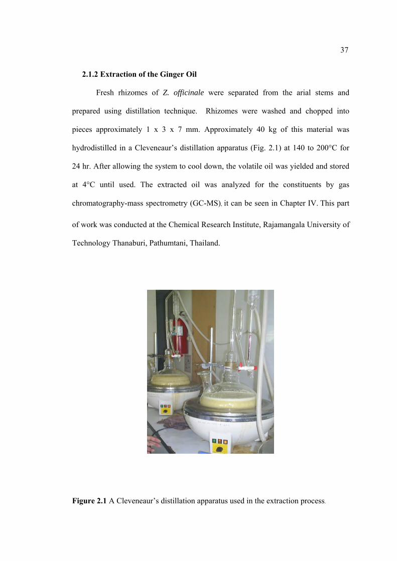
2.1.2 Extraction of the Ginger Oil
Fresh rhizomes of Z. officinale were separated from the arial stems and
prepared using distillation technique. Rhizomes were washed and chopped into
pieces approximately 1 x 3 x 7 mm. Approximately 40 kg of this material was
hydrodistilled in a Cleveneaur’s distillation apparatus (Fig. 2.1) at 140 to 200°C for
24 hr. After allowing the system to cool down, the volatile oil was yielded and stored
at 4°C until used. The extracted oil was analyzed for the constituents by gas
chromatography-mass spectrometry (GC-MS), it can be seen in Chapter ΙV. This part
of work was conducted at the Chemical Research Institute, Rajamangala University of
Technology Thanaburi, Pathumtani, Thailand.
Figure 2.1 A Cleveneaur’s distillation apparatus used in the extraction process.

38
2.1.3 Making Stock Solution
A stock solution was obtained by dissolving small aliquots of the oil in hexane
(1:1 v/v). The stock solution was further diluted using Krebs’ solution to attain the
final concentration. The final concentration of hexane in any dilution was less than
0.15%.
2.2 Animal Preparations
2.2.1 Housing
Rats were maintained in accordance with the guidelines of the Committee on
Care and Use of Laboratory Animal Resources, National Research Council, Thailand.
The experiments performed on rats were conducted in accordance with the advice of
the Institutional Animal Care and Use Committee, SUT.
Twenty five non-pregnant Wistar rats (200–250g) were housed in the animal
house of the SUT under a controlled environment (23–25 oC) and illumination (12 hr
light, 12 hr dark) room. Rats were given tap water and a standard diet ad libitum.
2.2.2 Myometrial Tissue Preparations
The rats were humanely killed by cervical dislocation. Myometrial tissues
were obtained. The uterus was removed and immediately immersed in buffered
physiological Krebs’ solution (pH 7.40) containing (mM): 154 NaCl; 5.4 KCl; 1.2
MgSO4; 12 glucose; 2 CaCl2, and 10 N-[2-hydroxyethyl]piperazine-N’-[2-
ethanesulphonic acid]] [HEPES]. The uterus was then placed in a shallow dissecting
dish containing Krebs’ solution at room temperature under a microscope. The
longitudinal layer was separated from the endometrium and circular layers. Five or six
39
strips (1-2 mm x 0.5 mm x 10 mm) of longitudinal fibres were then dissected. The
strips were either used immediately or stored for a maximum of 12 hr at 4 oC.
2.2.3 Histological Tissue Preparations
The strips were immersed in buffer physiological Krebs’ solution as a control,
in Krebs’ solution containing ginger oil, or ginger oil related compounds for 30 min.
The strips were fixed in 10% formalin until analyzed for histological features.
2.2.4 Myometrial Tissue Staining Preparations for LM and TEM
2.2.4.1 LM Staining Preparations
The horns of rat uteri were cut into short segmenta. These strips were
dehydrated in an ascending ethanol series (75%, 85%, 95%, 100% and 100%, 1 hr
each) and then removed the sections into pure xylene for 2 min. The sections were
embedded with xylene : paraplast (3 : 1 and 1 : 1, 15 min each) with pure paraplast for
1 hr. Post-embedded tissues were cut approximately 5 µm with microtome and moved
into water bath at 60°C for 10 min. The tissues were mounted on slides in the slide
warmer at 60°C and then twice immersed the slides in pure xylene (5 and 2 min,
respectively). Next, tissues were hydrated in a descending ethanol series (100%, 95%,
70%, 50%, 30%, distill water, 2 min each) and stained with heamatoxylin according
to standard protocols. All tissues were dehydrated in a descending ethanol series
(35%, 50%, 70%, 95%, 2 min each) and stained with eosin for 1 min. Finally, the
slides were immersed in 95% ethanol (2-3 min) and 100% ethanol (1 min), pure
xylene (5 min) and then covered with cover slip after xylene clearing for LM study.

40
2.2.4.2 TEM Staining Preparations
The horns of rat uteri were cut into short segmenta. These strips were fixed in
2.5% glutaraldehyde in 0.1 M phosphate buffer, pH 7.2 overnight at 4°C then washed
in the same buffer 3 times. Post-fixed with 1% osmium tetroxide for 2 hr then rinsed
in distill water 3 times. Gold coated uterus on stubs were examined for TEM, after
post-fixing the samples were dehydrated in graded acetone at concentrations of 20%,
40%, 60%, 80%, 100% and 100%, then infiltrated and embedded in Spur’s resin.
Upon identifying suitable areas from the semithin sections, ultrathin sections (<100
nm thick) of mainly transverse and longitudinal orientation to the uterine tissues were
cut with diamond knife, mounted on copper grids then stained with lead citrate and
uranyl acetate. The ultrathin sections on copper grids were examined using TEM
(JEM 1230) at Central Instrumentation Unit, Faculty of Science, Mahasarakham
University (Phungnoi and Narkkong, 2007).
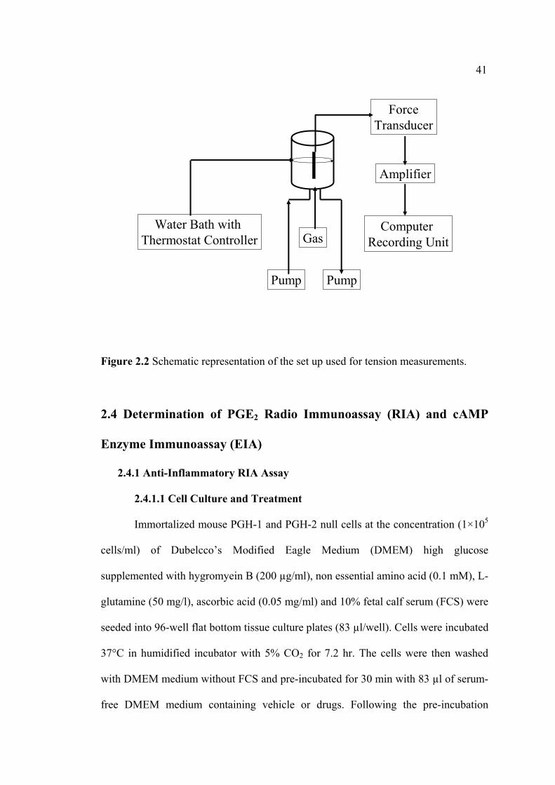
2.3 Measurements of Tension
The uterine strip was mounted vertically under resting tension of 1g in a single
chamber (25 ml) tissue bath connected to a force transducer (see Fig. 2.2).
The organ bath contained Krebs’ solution maintained at pH 7.4, temperature
of 37 oC, and gassed with carbogen (95% O2 and 5% CO2). The myometrial strip was
attached at each end to metal hooks and another hook was fixed to a transducer. Then
the electrical signal has been recorded from the transducer and converted to the digital
signal on a computer using Chart software (Kupittayanant et al., 2002).
41
ForceTransducer
Amplifier
Computer Recording Unit
Water Bath with Thermostat Controller
Pump Pump
Gas
Figure 2.2 Schematic representation of the set up used for tension measurements.
2.4 Determination of PGE2 Radio Immunoassay (RIA) and cAMP
Enzyme Immunoassay (EIA)
2.4.1 Anti-Inflammatory RIA Assay
2.4.1.1 Cell Culture and Treatment
Immortalized mouse PGH-1 and PGH-2 null cells at the concentration (1×10⁵
cells/ml) of Dubelcco’s Modified Eagle Medium (DMEM) high glucose
supplemented with hygromyein B (200 µg/ml), non essential amino acid (0.1 mM), L-
glutamine (50 mg/l), ascorbic acid (0.05 mg/ml) and 10% fetal calf serum (FCS) were
seeded into 96-well flat bottom tissue culture plates (83 µl/well). Cells were incubated
37°C in humidified incubator with 5% CO2 for 7.2 hr. The cells were then washed
with DMEM medium without FCS and pre-incubated for 30 min with 83 µl of serum-
free DMEM medium containing vehicle or drugs. Following the pre-incubation
42
period, the medium was removed and the cells were immediately treated with serum-
free medium containing vehicle or drugs and AA (20µM) or A 23187 (2 µM) for 30
min (Kirtikara et al., 1998). Culture supernatants were then collected from wells and
analyzed for PGE2 concentrations by radioimmunoassay (RIA).
2.4.1.2 PGE2 Measurement
The RIA method used for measuring PGE2 concentrations in the culture
supernatant is based on the competition between PGE2 in samples and 3H labeled
PGE2 for anti-PGE2 antibody binding sites. The assay was performed on ice as
following. The supernatant was diluted with DMEM (1:10) for blank and zero %
binding and added approximately 1.5 ml into 50 µl micro-centrifuge tube. Then, 50 µl
of anti-PGE2 antibody in RIA buffer (0.1 mM phosphate buffer, pH 7.4, containing
0.9% sodium chloride, 0.1% sodium azide and 0.1% gelatin) was added to every tube
except for the blanks, in which 50 µl RIA buffer was added. Subsequently, 50 µl of
3H-PGE2 (1.12 µci/ml), was added to each tube, vortexed briefly and incubated
overnight at 4°C. Then, 100 µl at 2% charcoal-dextran suspension in RIA buffer was
added to each tube. After 15 min incubation on ice, the tubes were centrifuged at
3,800 rpm (1,500g) at 4°C for 10 min. Supernatants were then transferred to new 1.5
ml micro-centrifuge tubes containing liquid scintillation cocktail, vortexed and
counted for radioactivity. The resulting radioactive counts were used to calculate %
binding of 3H-PGE2, which were then used for the estimation of PGE2 concentrations
from standard curves. This part of work was conducted by National Center for
Genetic Engineering and Biotechnology (BIOTEC), Pathumthani, Thailand.

43
2.4.2 cAMP EIA Assay
cAMP content of myometrial smooth muscle cells were measured by enzyme
immunoassay (EIA). The uterine strips were incubated with 5mM CaCl2 for 10 min
then removed to Krebs’ solution containing aminophylline (1 nM), ginger oil (50
µl/100 ml), 95% citral (2.2 mM), and 95% camphene (7.5 mM) at 37°C for 30 min.
Post-incubated tissues with various treatments, the strips were rapidly frozen in liquid
nitrogen and stored at -80°C until homogenized in 6% trichloroacetic acid (0.4 ml).
The homogenate was centrifuged at 3000×g for 15 min. The supernatant was washed
with 1.5 ml of water-saturated diethyl ether four times. The cAMP contents were
assayed by using EIA kit (Amersham Pharmacia Biotech, Little Chalfont, UK). cAMP
contents are presented as fmol/g wet weight.
2.5 Chemicals
All chemicals were purchased from Sigma® unless stated otherwise. Details of
stock solutions were given in the individual chapter concerned. These stock solutions
were prepared and kept as recommended by the producer. Dissolved vehicles (e.g.
DMSO, ethanol) did not alter the myometrial contractile ability as judged by the peak
tension, frequency of contractions, and contraction integral (see 3.4.1). The dilutions
were made on the day of the experiment.
2.6 Data Analysis
Statistical Analysis of Tension Measurements
The result data were analyzed using Microcal Origin Software (Massachusetts,
USA). Parameters that were measured include maximum tension development of each

44
contraction, the contraction integral (total tension developed in each contraction),
contraction duration, and contraction frequency. IC50 values (concentrations) of
multiple substance-inhibited 50% of myometrial contraction were calculated.
Data were then presented as mean ± s.e.m. and “n” represents the number of
samples, each one from a different animal. Significance was tested using appropriate t
tests or ANOVA and P values < 0.05 taken to be significant. Results were then
expressed as percentages of control contractions (i.e. the control is 100%).
Statistical Analysis of PGE2 Contents
The paired t test procedure was used to determine the differences in the PGE2
levels between control samples of wild-type. COX-2 cells and among control samples
and samples from cytokine-treated cells. Differences were considered significant if p
< 0.05 (Kirtikara, Swangkul, and Ballon, 2001).
Statistical Analysis of cAMP Assay
Data were calculated as the average optical density (OD) for each set of
replicate wells. The percent bound for each standard and sample (see below) was
calculated.
Abbreviations:
%B/B0 = × 100 (standard or sample OD-NSB OD) (zero standard OD-NSB OD)
OD = optical density
NSB = non specific binding wells
%B/B0 = the percent bound for each standard and sample
45
2.7 References
Hardman, R. (1972). Spices and herbs, their families, secretory tissues and
pharmaceutical aspects. London.
Harris, J. G. and Harris, M. W. (1954). Plant identification terminology: an
illustrated. P 188. Utah: Spinh Lake Publishing.
Heywood, V. H. (1993). Flowering plants of the world. p 336. BT Batsford Ltd.
London.
Kirtikara, K., et al. (1998). Compensatory prostaglandin E2 biosynthesis in
cyclooxygenase 1 or 2 null cells. Journal of Experimental Medicine. 187 :
517 – 523.
Kirtikara, K., Swangkul, S. and Ballon, L. R. (2001). The analysis of nonsteroidal
anti-infammatory drug delectivity in prostaglandin G/H synthase (PGHs)-
null cells. Inflammatory Research. 50 : 327 – 332.
Kupittayanant, S., Luckas, M. J. M. and Wray, S. (2002). Effect of inhibiting the
sarcoplasmic reticulum on spontaneous and oxytocin-induced contractions of
myometrium. Journal of Obstetrics and Gynaecology. 109 : 289 – 296.
Medicinal Plant Information Center. (1992). Thai medicinal plants. Prachachon Co.,
Ltd. p 397.
Phungnoi, Y. and Narkkong, N. (2007). Ultrastructure of eupyrene and apyrene
spermatozoa in pila angelica. Journal of Microscopy Society Thailand. 21
(1) : 116 – 120.
Purseglove, J. W., Brown, E. G., Green, C. L. and Robbins, S. R. K. (1981). Spices
Vol. 2. New York: Longman.
46
…………………………………fully…………………………….
Kessler, O. J., Keisari, Y., Servadio, C. (1998). Role of chronic inflammation in the
promotion of prostatic hyperplasia in rats. Journal of Urology. 159 ; 1049 –
1053.
Bode, A. M., Ma, W – Y., surh, Y. J. Dong. Z. (2001). Inhibition of epidermal growth
factor-induced cell transformtion and activator protein activation by [6]-
gingerol. Cancer Research. 61 : 850 – 853.
Hardman, R. (1972). Spices and herbs, their families, secretory tissues and
pharmaceutical aspects. London.
Ingold, C. K. (1969). Structure and mechanism in organic chemistry. 2nd ed. p 769.
Cornell University Press. Ithaca, New York.
……………………………………….move to Chapter 4………………….

47
2.1.5 Pharmacological Study of Ginger-Related Compounds
As shown in Table 2.1, the major active compounds found in ginger oil are the
important role for evaluating potential mechanism to treatments.
Several studies have been investigated the effectiveness of ginger in the
prevention and used in traditional medicine to treat such symptoms as inflammation,
rheumatic disorders and gastrointestinal discomforts for long time (Peng, 1992;
Aeschbach et al., 1994; Habsah et al., 2000; Surh, 1998; 2003). Its extract and major
pungent principles, such as [6]-ginger and [6]-paradol, have recently been shown to
exhibit a variety of biological activities including anticancer (Katiyar, Agarwak, and
Makhtar, 1996; Surh and Lee 1998; Surh, 2003; Bode, Ma, Surh and Dong, 2001;
Keum et al., 2002). The pungency inhibited on nitric oxide synthesis (Ippoushi, Itou,
Azuma, and Higashio, 2003), COX-2 activity (Tjendraputra, Tran, Liu-Brennan,
Roufogalis and Duke, 2001) and protecting neuronal cells from β-amyloid injure (D.
Kim, and J. Kim, 2004). In contrast, Wei et at. (2005) reported that the study of
Chinese ginger, gingerol-related compounds, has been shown to possess significant
cytotoxicity against HL-60 cells (IC50 < 50 µM) and its cytotoxic activity was
associated with the cell apoptosis.
The most important compounds, responsible for ginger’s theraputic activity
have been found such compounds include zingiberene, -ar-curcumene, β-
sesquiphelandrene, α-pinene, camphene and citral (Mustafa, Srivastava, Jensen,
1993). Citral, a widely used natural ingredient, was added to foods and cosmetics as a
flavoring and fragrance agent (Trasarti, Marchi and Apestegua, 2004). They have
tested for citral conversion to menthols, Ni/Al-MCM-41; the best catalysis, which
yielded menthol and menthol in the menthol mixture (Trasarti et al., 2004). In
48
addition, citral was used as a chemical intermediate in the synthesis of vitaminA,
ionone and methylionone (Budavari, 1989). On the other hand, citral was selected for
carcinogen studies because of its widespread use as a flavoring and fragrance
ingredient. Citral administrered dermally has previously been studied skin severally
irritating to albino angora rabbits, male Hartley guinea pigs, and humans (Basketter
and Scholes, 1992; Cardullo, Ruszkowski, and Deleo, 1989; Motoyoshi, Toyoshima,
Sato, and Yoshimura, 1979). Although it was positive in the local lymph node assay
(Basketter and Scholes, 1992), citral also has been reported to induce benign and
atypical prostatic hyperplasia in rats when applied dermally for one or more months
(Engelstein, Shueli, Bruhis, Servadio, and Abramovici, 1996; Kessler, Keisari, and
Servadio, 1998). In addition, citral at a high exposure (31,300 ppm); thymic atrophy
in males and females has been revealed to inanition and the moribund condition of the
rodent studies (Ress, 2003). There was no citral-related effect on the occurrence of
either internally visible myometrial structural abnormalities, or uterine malformations.
Camphene is one of the attractive by its used on pharmaceutical and cosmetic
applications. This is one of ginger compound obtained from pinene. Camphene
hydrochloride has played an important historical role in the chemistry of the terpenes
of carbonium ion rearrangements (Simonsen and Owen, 1957; Berson, 1963; Barlett,
1965; Eastman and Noller, 1953; Ingold, 1969). It was used in the manufacture of
camphor and its related compounds (Comelli, Ponzi, E. N and Ponzi, M., 2005).
References Aesehbach, R., et al. (1994). Antioxidant actions of thymol, carbacrol, 6-gingerol,
zingerone and hydroxytyrosol. Food and Chemical Toxicology. 32 : 31 – 36.
49
Bartlett, P. D. (1965). Non-classical Ions’. p 48. W. A. Benjamin, New York.
Basketter, D. A. and Scholes. E. W. (1992). Comparison of the local lymph mode
assay with the guinea-pig maximization test for the detection of a range of
contract allergens. Food Chemistry Toxicology. 30 : 65 – 69.
Berson, J. A. (1963). In : Molecular Rearrangements. 1 : p 113. (Ed. P. de Mayo).
Interscience : New York.
Budavari, S. (1989). The Merl: Index: An Encyclopedia of chemicals, drugs, and
biologicals. 11th ed. Merk and Co, Rahway, N J, USA.
Cardullo, A. C., Ruszkowski, A. M. and Deleo, V. A. (1989). Allergic contact
dermatitis resulting from sensitivity to citrus peel. Geraniol, and citral.
Journal of American Academy Dermatology. 21 : 395 – 397.
Comelli, N. A., Ponzi, E. N. and Ponzi, M. (2005). α-pinene isomerization to
camphene effect of thermal treatment on sulfated zirconia. Chemical
Engineering Journal. 117 : 93 – 99.
Eastman, R. H. and Noller, C. R. (1953). Organic chemistry. Quoted in An advanced
treatise. (Ed. H. Gilman). 4 : p 652. John Wiley. New York.
Engelstein, D., Shmueli, J., Bruhis, S., Servadio, C. and Abramovici, A. (1996). Citral
and testosterone interactions in inducing benign and atypical prostatic
hyperplasia in rats. Comp. Biochemistry Physiology. 115 : 169 – 177.
Habsah, M., et al. (2000). Screening of Zingiberaceae extracts for antimicrobial and
antioxidant activities. Journal of Ethnopharmacology. 72 : 403 – 410.
Ippoushi, K., Itou, H., Azuma, K. and Higashio, H. (2003). Effect of naturally
occurring organosulfur compounds on nitric oxide production in
lipopolysaccharide activated macrophages. Life Science. 71 : 411 – 419.
50
Katiyar, S. K., Agarwal, R. and Makhtar, H. (1996). Inhibition of tumor promotion in
Senkar mouse skin by ethanol extract of Zingiber officinale rhizome. Cancer
Research. 56 : 1023 – 1030.
Kessler, O. J., Keisari, Y., Servadio, C. (1998). Role of chronic inflammation in the
promotion of prostatic hyperplasia in rats. Journal of Urology. 159 : 1049 –
1053.
Keum,Y. S., et al. (2002). Induction of apoptosis and caspase-3 activation by
chemopreventive [6]-paradol and structurally related compounds in KB cells.
Cancer Letters. 177 : 41 – 47.
Kim, D. S. H. L. and Kim, J. Y. (2004). Side-chain length is important for shogaols in
protecting neuronal cells from β-amyloid insult. Bioorganic and Medicinal
Chemistry Letters. 14 : 1287 – 1289.
Motoyoshi, K., Toyoshima, Y., Sato, M. and Yoshimura, M. (1979). Comparative
studies on the irritancy of oils and synthetic perfumes to the skin of rabbit,
guinea pig, rat, miniature swine and man. Cosmetic Toilet. 94 : 41 – 48.
Mustafa, T., Srivastava, K. C. and Jensen, K. B. (1993). Drug development report:
pharmacology of ginger, Zingiber officinale, Journal of Drug Development.
6 : 25 – 39.
Peng, P.J. (1992). Pharmoclogical and clinical applications of ginger. Journal of
Chineses Medicine. 17 : 370 – 373.
Ress, N. B., et al. (2003). Toxicology and carcinogenesis studies of
microencapsulated citral in rats and mice. Toxicological Sciences. 71 : 198 –
206.
51
Simonsen, J. and Owen, L. N. (1957). The terpene. 2nd ed. (2) p 307. Cambridge
University Press.
Surh, Y. J. and Lee, J. M. (1998). Chemoprotective properties of some pungent
ingredients present in red pepper and ginger. Muttion Research. 402 : 259 –
267.
Surh, Y. J. (2003). Cancer chemoprevention with dietary phytochemicals. Nature
Reviews Cancer. 3 : 768 – 780.
Trasarti, A. F., Marchi, A. J. and Apestegua, C. R. (2004). Highly selective synthesis
of menthols from citral in a one-step process. Journal of Catalysis. 224 : 484
– 488.
Tjendraputra, E. Tran, V. H., Liu-Brennan, D., Roufogalis, B. D. and Duke, C. C.
(2001). Effect of ginger constituents and synthetic analogues on
cyclooxygenase-2 enzyme in intact cells. Bioorganic Chemistry. 29 : 156 –
163.
Wei, Q. Y., Ma, J-P., Cai, Y-S., Yand, L. and Liu, Z-L. (2005). Cytotoxic and
apoptotic activities of diaryheptanoids and gingerol-related compounds from
the rhizome of Chinese ginger. Journal of Ethnopharmacology. 102 : 177 –
184.
CHAPTER ΙII
EFFECTS OF GINGER OIL ON SPONTANEOUS AND
PGs-INDUCED RAT MYOMETRIAL CONTRACTION
3.1 Abstract
Ginger rhizomes (Zingiber officinale Rocsoe) have been widely used not only
as food, but also as medicine for intestinal disorders and relaxation of the smooth
muscles. In addition, the essential oil and oleoresin from the ginger rhizomes were
utilized as flavour additives in foods and beverages. The purpose of this study was to
investigate the inhibitory effects of ginger oil on rat uterine contraction arising either
spontaneously or by PGs (PGF2α and PGE2) stimulation and further investigated the
underlying mechanisms.
Ginger rhizomes were provided and hydrodistilled to obtain the oil yield. Its
structural constituents were then analyzed by GC/MS. Rats were humanely killed by
asphyxiation with CO2 and myometrial strips isolated. The effects of ginger oil on
uterine contraction were measured and recorded. The results showed that ginger oil
(10–150 µl/100 ml) inhibited spontaneous and PGs-induced contraction. Its effects
were reversible upon elevation (from 2 mM to 5 mM) of external Ca2+ concentration.
Its IC50 was 50 µl/100 ml. In summary, ginger oil is a potent inhibitor of myometrial
contraction in rat uterus. This is probably due to the inhibition of L-type calcium
channels.

47
3.2 Introduction
Zingiber officinale Roscoe, known as “Ginger,” is widely used in foods as a
spice around the world. Beside in Asia, ginger is used as household remedy for
aromatherapy. In addition, it is used in medicine as a folk remedy to calm nausea
associated with motion sickness (Lien et al., 2003) and to treat the digestive tract
(Gruenwald et al., 2000). One of the most interesting properties of ginger rhizome is
its inhibitory effect on rat ileal motility in vitro (Borrellia et al., 2004) and to relax the
tracheal or ileal smooth muscle (Reiter and Brandt, 1985). An aqueous extract of a
low dose of ginger rhizome (50 mg/kg) could be used in the rats as an cholesterol-
lowering, antithrombotic, anti-inflammatory agent inhibitory effect on platelet
aggregation. Furthermore, it has been reported that the levels of serum PGE2 were
significantly reduced in the presence of ginger (Srivastava, 1984, 1986; Thomson et
al., 2002).
In vitro experiment, shogoals and 6-, 8-, and 10-gingerols, isolated from the
methanolic extract of Z. officinale rhizome exhibited anti-emetic principles (Kawai et
al., 1994). Both 6-shogaol and 6-gingerol have been shown to have a number of
pharmacological activities including antipyretic, analgesic, antitussive and
hypotensive effects (Suekawa et al., 1986). Moreover, anti-inflammatory data were
obtained from the metabolic profiling analysis of Z. officinale samples. The oil
derived from different origins show no qualitative differences in major volatile
compounds, [6]-, [8]-, and [10]-gingerols (Park et al., 1998). The most active anti-
inflammatory components were evaluated (Park et al., 1998; Young et al., 2005). The
ability of all extracts from the Zingiber species was shown to inhibit LPS-induced
PGE2 and TNF-α production (Jiang et al., 2006). In addition, ginger has shown to
48
exhibit antithrombotic activity and inhibit prostaglandin-E2 from arachidonic acid,
gingerol, and dehydroparadol favored the inhibition of cyclooxygenase (Srivastava,
1984, 1986; Flynn et al., 1986).
However, the study of the effects of ginger constituents on the uterine
contraction has not yet been elucidated. Therefore, the aims of this chapter were to 1)
demonstrate the effects of ginger oil on spontaneous contractions and 2) PGF2α-and
PGE2-induced contractions in isolated rat myometrium in the presence or in the
absence of external Ca2+ concentration. In addition, its effect on Ca2+- CaM-MLCK
pathway and non Ca2+- CaM-MLCK pathway was investigated.
3.3 Materials and Methods
3.3.1 Chemicals and Physiological Solution
All chemicals were purchased from Sigma® unless state otherwise.
Agonists/antagonists for the investigation of physiological pathways used were;
PGF2α, PGE2, nifedipine, and BSA. PGF2α and PGE2 were dissolved in ethanol at a
concentration of 1 µM. Bovine serum albumin (BSA, 5%) was dissolved in distilled
water. Nifedipine was dissolved in DMSO at a concentration of 10 µM.
3.3.2 Ginger Oil Preparations
As described in 2.1.3, a stock solution of the oil was dissolved by hexane in
Krebs’ solution just before use. The final concentration of hexane in any dilution was
less than 0.15%.
49
3.3.3 Myometrial Tissue Preparations
Tissue preparations are essentially the same as those described in Chapter II.
Non-pregnant Wistar rats (200-250 g) were used in this study and maintained in
accordance with the guidelines of the committee on Care and Use of Laboratory
Animal Resources, National Research Council, Thailand. The experiments performed
on rats were conducted in accordance with the advice of the Institutional Animal Care
and Use Committee, Suranaree University of Technology (SUT), Thailand.
Myometial tissue preparations were dissected and provided for tension measurements
as those described in 2.2.2.
3.3.4 Measurements of Tension
The uterine strip was mounted vertically under resting tension of 1g in a single
chamber (25 ml) tissue bath connected to a force transducer (as described in 2.3).
The organ bath contained Krebs’ solution maintained at pH 7.4, temperature
of 37 oC, and gassed with carbogen (95% O2 and 5% CO2). The myometrial strip was
attached at each end to metal hooks and another hook was fixed to a transducer. The
electrical signal has been recorded from the transducer and converted to the digital
signal on a computer using Chart software (Kupittayanant, 2003).
3.3.5 Data Analysis
Data were presented as mean ± S.E.M. and “n” represents the number of
samples, each one from a different animal. Significance was tested using appropriate t
tests or ANOVA and p values < 0.05 taken to be significant. Results were then
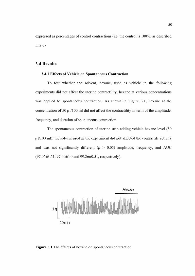
50
expressed as percentages of control contractions (i.e. the control is 100%, as described
in 2.6).
3.4 Results
3.4.1 Effects of Vehicle on Spontaneous Contraction
To test whether the solvent, hexane, used as vehicle in the following
experiments did not affect the uterine contractility, hexane at various concentrations
was applied to spontaneous contraction. As shown in Figure 3.1, hexane at the
concentration of 50 µl/100 ml did not affect the contractility in term of the amplitude,
frequency, and duration of spontaneous contraction.
The spontaneous contraction of uterine strip adding vehicle hexane level (50
µl/100 ml), the solvent used in the experiment did not affected the contractile activity
and was not significantly different (p > 0.05) amplitude, frequency, and AUC
(97.06±3.51, 97.00±4.0 and 99.86±0.51, respectively).
Figure 3.1 The effects of hexane on spontaneous contraction.
1 g
10 min
Hexane
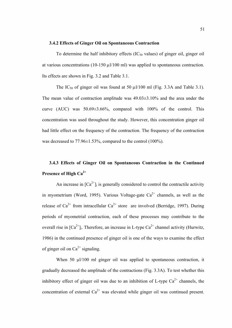
51
3.4.2 Effects of Ginger Oil on Spontaneous Contraction
To determine the half inhibitory effects (IC50 values) of ginger oil, ginger oil
at various concentrations (10-150 µl/100 ml) was applied to spontaneous contraction.
Its effects are shown in Fig. 3.2 and Table 3.1.
The IC50 of ginger oil was found at 50 µl/100 ml (Fig. 3.3A and Table 3.1).
The mean value of contraction amplitude was 49.03±3.10% and the area under the
curve (AUC) was 50.69±3.66%, compared with 100% of the control. This
concentration was used throughout the study. However, this concentration ginger oil
had little effect on the frequency of the contraction. The frequency of the contraction
was decreased to 77.96±1.53%, compared to the control (100%).
3.4.3 Effects of Ginger Oil on Spontaneous Contraction in the Continued
Presence of High Ca2+
An increase in [Ca2+]i is generally considered to control the contractile activity
in myometrium (Word, 1995). Various Voltage-gate Ca2+ channels, as well as the
release of Ca2+ from intracellular Ca2+ store are involved (Berridge, 1997). During
periods of myometrial contraction, each of these processes may contribute to the
overall rise in [Ca2+]i. Therefore, an increase in L-type Ca2+ channel activity (Hurwitz,
1986) in the continued presence of ginger oil is one of the ways to examine the effect
of ginger oil on Ca2+ signaling.
When 50 µl/100 ml ginger oil was applied to spontaneous contraction, it
gradually decreased the amplitude of the contractions (Fig. 3.3A). To test whether this
inhibitory effect of ginger oil was due to an inhibition of L-type Ca2+ channels, the
concentration of external Ca2+ was elevated while ginger oil was continued present.
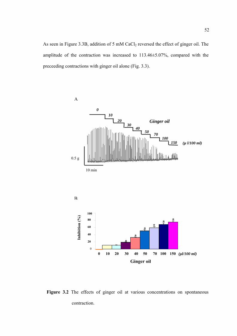
52
As seen in Figure 3.3B, addition of 5 mM CaCl2 reversed the effect of ginger oil. The
amplitude of the contraction was increased to 113.46±5.07%, compared with the
preceeding contractions with ginger oil alone (Fig. 3.3).
Figure 3.2 The effects of ginger oil at various concentrations on spontaneous
contraction.
B
0
20
40
60
80
100
0
Ginger oil
Inhi
bitio
n (%
)
150 100 70 50 40 30 20 10
***
**
*
(µl/100 ml)
0.5 g
10 min
(µ l/100 ml)
Ginger oil
150 100
7040
3020
10
50
0
A

53
Table 3.1
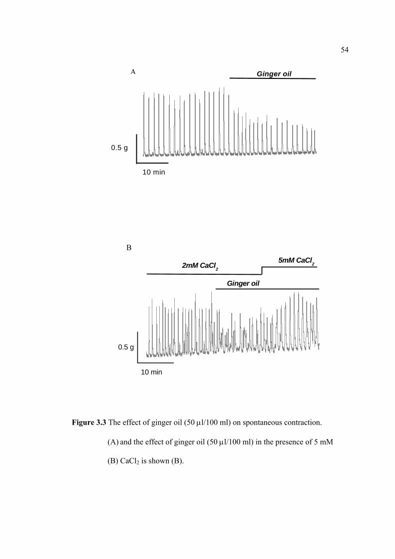
54
Figure 3.3 The effect of ginger oil (50 µl/100 ml) on spontaneous contraction.
(A) and the effect of ginger oil (50 µl/100 ml) in the presence of 5 mM
(B) CaCl2 is shown (B).
B
0.5 g
10 min
2mM CaCl2
5mM CaCl2
Ginger oil
0.5 g
10 min
Ginger oil
A
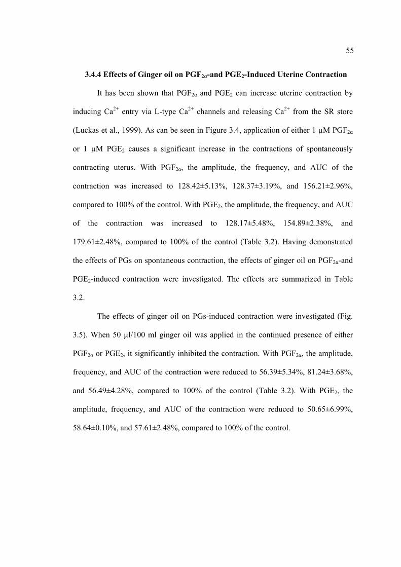
55
3.4.4 Effects of Ginger oil on PGF2α-and PGE2-Induced Uterine Contraction
It has been shown that PGF2α and PGE2 can increase uterine contraction by
inducing Ca2+ entry via L-type Ca2+ channels and releasing Ca2+ from the SR store
(Luckas et al., 1999). As can be seen in Figure 3.4, application of either 1 µM PGF2α
or 1 µM PGE2 causes a significant increase in the contractions of spontaneously
contracting uterus. With PGF2α, the amplitude, the frequency, and AUC of the
contraction was increased to 128.42±5.13%, 128.37±3.19%, and 156.21±2.96%,
compared to 100% of the control. With PGE2, the amplitude, the frequency, and AUC
of the contraction was increased to 128.17±5.48%, 154.89±2.38%, and
179.61±2.48%, compared to 100% of the control (Table 3.2). Having demonstrated
the effects of PGs on spontaneous contraction, the effects of ginger oil on PGF2α-and
PGE2-induced contraction were investigated. The effects are summarized in Table
3.2.
The effects of ginger oil on PGs-induced contraction were investigated (Fig.
3.5). When 50 µl/100 ml ginger oil was applied in the continued presence of either
PGF2α or PGE2, it significantly inhibited the contraction. With PGF2α, the amplitude,
frequency, and AUC of the contraction were reduced to 56.39±5.34%, 81.24±3.68%,
and 56.49±4.28%, compared to 100% of the control (Table 3.2). With PGE2, the
amplitude, frequency, and AUC of the contraction were reduced to 50.65±6.99%,
58.64±0.10%, and 57.61±2.48%, compared to 100% of the control.
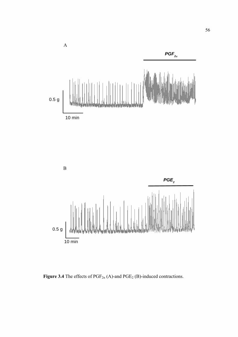
56
Figure 3.4 The effects of PGF2α (A)-and PGE2 (B)-induced contractions.
10 min
0.5 g
PGF2α
A
10 min
0.5 g
PGE2
B
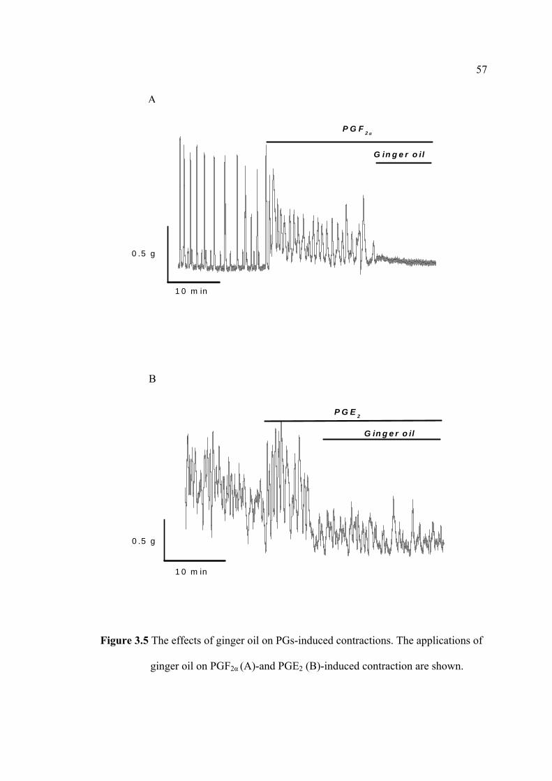
57
Figure 3.5 The effects of ginger oil on PGs-induced contractions. The applications of
ginger oil on PGF2α (A)-and PGE2 (B)-induced contraction are shown.
0 .5 g
1 0 m in
G in g e r o i l
P G F 2 α
A
0 .5 g
1 0 m in
G in g e r o il
P G E 2
B

58
Table 3.2
59
3.4.5 Effects of Ginger Oil on PGF2α-and PGE2-Induced Uterine Contraction
in the Continued Presence of High Ca2+
As shown in Figure 3.5, ginger oil inhibited both PGF2α and PGE2 contraction.
Next, the underlying mechanisms were investigated. To test whether the inhibitory
effect of ginger oil on the agonist-induced contraction was via the inhibition of L-type
Ca2+ channels, the effects of ginger oil were tested in the continued presence of high
Ca2+.
As seen in Figure 3.6, the effects of ginger oil can be reversed upon an
increase in external Ca2+ concentration. This was also the case for PGE2 (data not
shown). The effect of ginger oil on PGF2α-and PGE2-induced contraction in the
presence of high Ca2+ are summarized in Table 3.3.
3.4.6 Effects of Ginger Oil on PGF2α-and PGE2-Induced Uterine Contraction
in the Absence of External Ca2+
It has been reported that PGs can cause small contraction independent of
external Ca2+, presumably via non-Ca2+-CaM MLCK pathway (Luckas et al., 1999).
As the previous report, in the present study showed that both PGF2α and PGE2 caused
the small rise in contraction when external Ca2+ entry was inhibited by nifedipine
(Fig. 3.7A and 3.8A, respectively). To test whether, ginger oil could inhibit this
contraction, it was applied in the continued of these agonists.
As can be seen in Figure 3.7B and C, the application of ginger oil before and
after the application of PGF2α abolished the contraction. This was also the case of
PGE2 (Fig. 3.8B and C).

60
Figure 3.6 The effects of ginger oil on PGs-induced contractions in the presence of
high Ca2+ concentration (5 mM CaCl2). (A) An application of ginger oil on
PGF2α-induced contraction is shown. (B) An application of ginger oil on
PGE2-induced contraction is shown.
10 min
0.5g
2mM CaCl25mM CaCl2
Ginger oil
PGE2
0.5 g
10 min
2m CaCl25m CaCl2
Ginger oil
PGF2α
A
B

61
Table 3.3
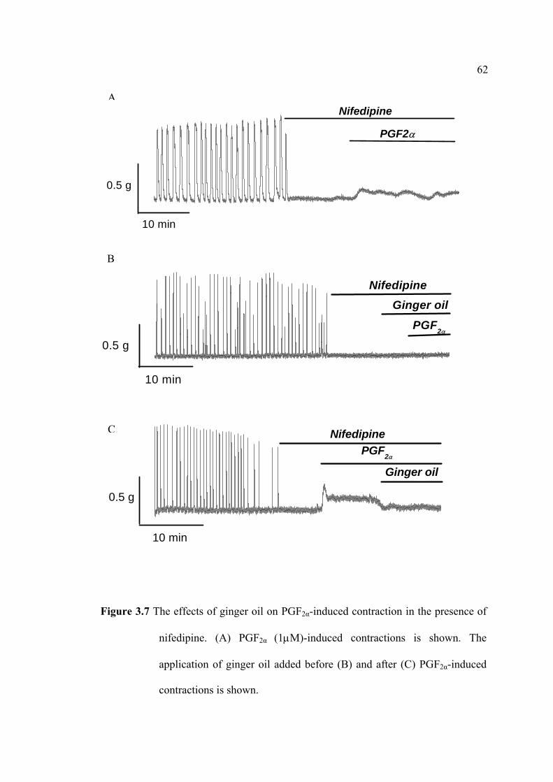
62
Figure 3.7 The effects of ginger oil on PGF2α-induced contraction in the presence of
nifedipine. (A) PGF2α (1µM)-induced contractions is shown. The
application of ginger oil added before (B) and after (C) PGF2α-induced
contractions is shown.
0.5 g
10 min
PGF2α
Nifedipine
0.5 g
10 min
Ginger oil
PGF2α
NifedipineC
A
B
0.5 g
10 min
PGF2α
Ginger oilNifedipine
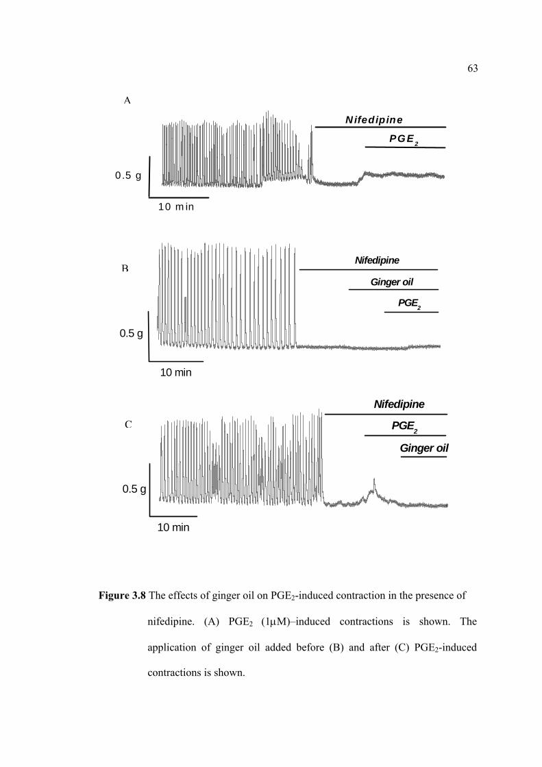
63
Figure 3.8 The effects of ginger oil on PGE2-induced contraction in the presence of
nifedipine. (A) PGE2 (1µM)–induced contractions is shown. The
application of ginger oil added before (B) and after (C) PGE2-induced
contractions is shown.
0.5 g
10 min
Ginger oil
PGE2
Nifedipine
0.5 g
10 min
Nifedipine
PGE2
Ginger oilB
C
0.5 g
10 m in
PGE 2
Nifedipine
A
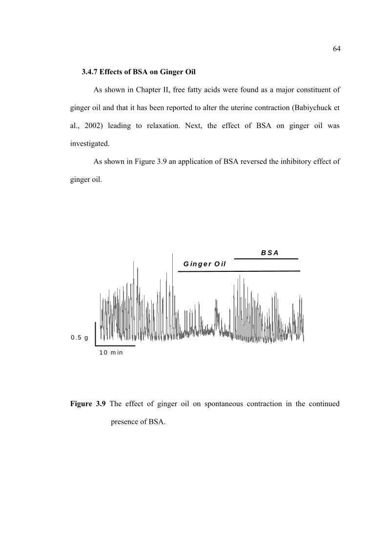
64
3.4.7 Effects of BSA on Ginger Oil
As shown in Chapter II, free fatty acids were found as a major constituent of
ginger oil and that it has been reported to alter the uterine contraction (Babiychuck et
al., 2002) leading to relaxation. Next, the effect of BSA on ginger oil was
investigated.
As shown in Figure 3.9 an application of BSA reversed the inhibitory effect of
ginger oil.
Figure 3.9 The effect of ginger oil on spontaneous contraction in the continued
presence of BSA.
0 .5 g
1 0 m in
B S AG in g e r O il
65
3.5 Discussion
The data indicated that the effects of ginger oil on rat myometrial contractile
activity may occur by inhibitory of Ca2+ influx via the L-type Ca2+ channels. This was
also the case for both spontaneous and PGs-induced contractions.
Ginger oil statistically significant (p < 0.05) effect in reducing contractile
activity of rat myometrium. However, low dose of ginger oil (10 – 20 µl/100 ml) did
not more effectively and not significantly (p > 0.05) different amplitude, frequency
and AUC than the spontaneous contraction (control) in non-pregnant rat uterus.
Although, the inhibitory effect of ginger oil (as seen in Fig. 3.9) could be reduced to
half-maximum contraction, it was induced by BSA similarly to the control.
An increase the external Ca2+ concentration can reversed the effect of ginger
oil on spontaneous contraction to compare with the control. Furthermore, the effect of
ginger oil in the presence of high Ca2+ concentration and in PGs-induced contractions
are also returned to a rise the recovering contractility (Fig. 3.6). Because of these
agonists, PGs are also most likely to play an important role in these conduction to
induce Ca2+ from the SR store via IP3 binding to receptors on the SR membrane,
known as IP3–induced Ca2+ release (IICR; Berridge, 1997). Thus, it has been clearly
demonstrated that agonists induced the release of Ca2+ and contraction had been
rapidly abolished by nifedipine (10 µM) due to inhibit the L-type Ca2+ channels
(Coleman et al., 2000; Kupittayanant, 2000). As a result of the same concentration of
PGs-induced contraction in the absence (nifedipine) of external Ca2+ did not reverse
the inhibitory effect of ginger oil (50 µl/100 ml, Fig. 3.7B, C). Although, the lone of
PGs in this condition also increased amplitude and AUC of spontaneous contraction
66
but did not induce spontaneously return to contraction while adding ginger oil (50
µl/100 ml) in the absence of Ca2+ concentration (Figs. 3.7B, 3.8B).
These parameters are similarly shown to the effect of Melisa officinale extract,
the strong constituent of citral from essential oil could reduce contraction on the rat
isolated ileum in the presence of high KCl (Sadraei et al., 2003). Such the result of
this experiment, citral has been the major compound that found in hydro-distillation
from fresh ginger rhizome.
In conclusion, the inhibitory effect of ginger oil may be elucidated to reduce
both spontaneous and PGF2α/PGE2-induced contractile activity via closing the L-type
Ca2+ channels with citral. In addition, it is clear that ginger oil has effect on Ca2+ rise
in smooth muscle and also cause to specific effect on Ca2+ CaM-MLCK pathways.
3.6 References
Berridge, M. J. (1997). Elementary and global aspects of calcium signaling. Journal
of Physiology. 499 : 291 – 306.
Borrellia, F., Capassoa, R., Pinto, A. and Izzoa, A. A. (2004). Inhibitory effect of
ginger (Zingiber officinale) on rat ileal motility in vitro. Life Sciences. 74 :
2889 – 2896.
Bubiychuck, E. B., Draeger, A., Burdyga, T. V. and Wray, S. (2002). Extraction of
cholesterol abolishes phasic contraction of rat and guinea-pig ureter. Journal
of physiology. 543 : 82.
Coleman, H. A., Hart, J. D. E., Tonta, M. A. and Parkington, H. C. (2000). Changes in
the mechanisms involved in uterine contractions during pregnancy in guinea-
pigs. Journal of Physiology. 523 (3) : 785 – 798.
67
Flynn, D. L., Fafferty, M. F. and Bactor, A. M. (1986). Inhibition of human neutrophil
5-lipooxygenase activity by gingerdione, shogoal, capsaicin and related
pungent compounds. Prostaglandins Leukotrienes and Medicine. 24 : 195 –
198.
Gruenwald, J., Brendler, T. and Jaenicke, C. (2000). PDR for herbal medicines.
Medical Economics Company Montvale. New Jersey USA.
Hurwitz, L. (1986). Pharmacology of calcium channels and smooth muscle.
Pharmacology and Toxicology. 26 : 225 – 258.
Jiang, H., et al. (2006). Metabolic profiling and phylogenetic analysis of medicinal
Zingiber species: Tools for authentication of ginger (Zingiber officinale Rosc).
Phytochemistry. 67 : 1673 – 1685.
Kawai, T., Kinoshita, K., Koyama, K., Takahashi, K. (1994). Anti-emetic principles
of Magnolia obovata bark and Zingiber officinale rhizome. Planta Medica.
60 (1) : 17 – 20.
Kupittyanant, S., Luckas, M. J. M. and Wray, S. (2000). Inhibition the sarcoplasmic
reticulum in human uterus dose not decrease contraction. Journal of
Physiology. [Hungarian]. 233 – 224.
Kupittayanant, S. (2003).The role of calcium and sigalling pathways in the control
and medulation of uterine contraction: with emphasis on human
myometrium. Ph.D. Dissertation. The University of Liverpool. UK.
Lien, H. C., et al. (2003). Effects of ginger on motion sickness and gastric slow-wave
dysrhythmics induced by circular vection. American Journal of Physiology
Gastrointestinal and Liver Physiology. 284 (3) : G481 - 489.
68
Luckas, M. J., Taggart, M. J. and Wray, S. (1999). Intracellular calcium stores and
agonist-induced contractions in isolated human myometrium. American
Journal of Obstetrics and Gynecology. 181 : 486 – 476.
Park, K. K., Chun, K. S., Lee, J. M., Lee, S. S. and Surh, Y. J. (1998). Inhibitory
effects of [6]-gingerol, a major pungent principle of giger, on phorbol ester-
induced inflammation, epidermal ornithine decarboxylase activity and skin
tumor promotion in ICR mice. Cancer Letters. 129 : 139 – 144.
Reiter, M. and Brandt, W. (1985). Relaxant effects on tracheal and ireal smooth
muscle of the guinea pig. Arzneim Forschung. 35 : 408 – 414.
Sadraei, H., Ghannadi, A. and Malekshahi, K. (2003). Relaxant effect of essential oil
of Melissa officinalis and citral on rat ileum contractions. Fitoterapia. 74 :
445 – 452.
Srivastava, K. C. (1984). Effects of aqueous extracts of onion, garlic and ginger on
platelet aggregation and metabolism of arachidonic acid in the blood vascular
system, in vitro study. Prostaglandins Leucotrienes and Medicine. 13 : 227
– 235.
Srivastava, K. C. (1986). Isolation and effects of some ginger components on platelet
aggregation and eicosanoid biosynthesis. Prostaglandins Leucotrienes and
Medicine. 25: 187 – 198.
Suekawa, M., et al. (1986). Plarmacological studies on ginger. 1. Pharmacological
actions of pungent constituents, (6)-gingerol and (6)-shogaol. Journal of
Pharmacobiological and Dynamic. 7 : 836 – 848.

69
Thomson, M., et al. (2002). The use of ginger (Z. officinale) as a potential anti-
inflammateory and anti-thrombotic agent. Prostaglandins, Leukotrienes and
Essential Fatty Acids. 67 (6) : 475 – 478.
Young, H. Y., et al. (2005). Analgesic and anti-inflammatory activities of [6]-
gingerol. Journal of Ethnopharmacology. 96 : 207 – 210.
Word, R. A. (1995). Myosin phosphorylation and the control of myometrial
contraction/ relaxation. Seminars in Perinatal. 19 : 3 – 14.
CHAPTER ΙV
EFFECTS OF CITRAL AND CAMPHENE ON
SPONTANEOUS AND PGs-INDUCED RAT
MYOMETRIAL CONTRACTION
4.1 Abstract
As citral and camphene are the most constituents found in ginger oil and are
commercial available, the aims of this Chapter were therefore to study the effects of
their agents on uterine contractility. The effects on both spontaneous and PGs-induced
contraction were particularly investigated. The results showed that the effects of citral
and camphene were similar indistinguishable to those of ginger oil. Thus, they
decreased uterine contractions, irrespectively of how they were produced, via the
inhibitory of L-type Ca2+ channels.
4.2 Introduction
The effects of pure citral and camphene have not been investigated. As they
were abundant found in ginger oil (Table 4.1). It is worth investigating whether the
effect of ginger oil found in Chapter III was due to the effect of camphene or citral.
4.2.1 Citral
Citral is a naturally-occurring mixture of the cis (neral) and trans (geranial)
genometric isomers of 3,7-dimethyl-2,6-octadien-l-al. It imparts a strong “lemony”
scent of edible vegetables and fruits, including lemon, lime, orange, grapefruit,
tomatoes, and ginger (Miyazawa and Kameoka, 1988). In addition, citral by far is the
highest constituent (75% - 85%) of lemongrass (Cymbopogon citratus) oil, as well as
of ginger oil (Wohlmuth et al., 2006).
Several previous investigations have been reported that citral was found to
possess anticancer effect against prostate gland tumor in various strains of rats
(Scolnid et al., 1994). It was known to possess antiseptic, antimicrobial, anti-
inflammatory, carminative, diuretic and central nervous system stimulating effects
(Carbajal et al., 1989). Besides, the antioxidant activity of citral was tested in human
intestinal homogenates in vitro. It inhibited the conversion of β-carotene to retinoic
acid by preventing the oxidation process (Wang et al., 1992). It was found that citral
is devoid of major toxicity and carcinogenic potential in both mice and rats (Ress et
al., 2003) and that it is devoid of mutagenic effect in vitro models (Viniketkumnuen et
al., 1994).
4.2.2 Camphene
Camphene is one of volatile oil consisted of mainly monoterpene in ginger oil.
It has several pharmacological properties. For example, Iranian folk medicine has
reported its antispasmodic, carminative, stomachic effects. The oil has been used as a
spasmolytic remedy for abdominal cramps in some parts of Iran (Sdraei et al., 2003).
According to its recommended clinical application it was tested on the respiratory
72
system as well as by its application on epilated skin. In addition, its combination with
menthol exerts spasmolytic activity in intestinal smooth muscle by blocking calcium
channels (Hawthorn et al., 1988), and attenuates bronchoconstriction and cough by
combined anaesthetic action on sensory nerves and bronchial smooth muscle (Wright
et al., 1997). Essential oil contained camphene was given twice daily for four weeks
has been shown to improve asthma control (Tamaoki et al., 1995).
There are number of pungent compounds found in fresh ginger rhizomes as
shown in Figure 4.1 and Table 4.1. These include citral (14.19% geranial, 10.00%
neral) and camphene (7.74%), which are commercially available compounds. The
main aims of this Chapter were to study the effects of citral and camphene on uterine
smooth muscle contraction. The effects on spontaneous or PGs-induced contractions
were particularly examined as well as the effects of these compounds in the presence
or the absence of external Ca2+ concentrations.
4.3 Materials and Methods
4.3.1 Identification of the Ginger Oil Constituents
The extracted oil was analyzed for the constituents by gas chromatography-
mass spectrometry (GC-MS), the analysis was performed on a Hewlett-Packard 5973
(IE) MS selective detector coupled with Hewlett Packcard 6890 gas chromatography
equipped with a cross-linked 5% PHME siloxane HP-5MS capillary column (30
m×2.25 mm; film thickness, 0.25 µm). The gas chromatography condition was as
follows: carrier gas, helium with a flow rate of 1.0 ml/min; column temperature, 50°C
at 6°C/min; injector temperature, 250°C; volume injected, 1.0 µl of the oil; split ratio,
250:1.
73
Identification of constituents was based on computer matching against the
library spectra with components of known constituents. MS literature data and
evaluation of fragmentation patterns of compounds were confirmed by their gas
chromatography retention time indices of authentic reference compounds where
possible. This part of work was conducted by Science and Technology Service
Center, Chaing Mai University, Thailand.
4.3.2 Chemicals and Physiological Solution
All chemicals were purchased from Sigma® unless state otherwise.
Agonists/antagonists for the investigation of physiological pathways were used,
including citral (95%), camphene (95%), PGF2α, PGE2, nifedipine, and BSA. Citral
and camphene were dissolved by hexane in Krebs’ solution just before used. The final
concentration of hexane in any dilution was less than 0.15%. PGF2α and PGE2 were
dissolved in ethanol at a concentration of 1 µM. BSA (5%) was disolved in distilled
water. Nifedipine was dissolved in DMSO to give a final concentration of 10 µM.
4.3.3 Myometrial Tissue Preparations
Tissue preparations are essentially the same as those described in Chapter II.
Non-pregnant Wistar rats (200-250 g) were used in this study and maintained in
accordance with the guidelines of the committee on Care and Use of Laboratory
Animal Resources, National Research Council, Thailand. The experiments performed
on rats were conducted in accordance with the advice of the Institutional Animal Care
and Use Committee, Suranaree University of Technology (SUT), Thailand.
74
Myometial tissue preparations were dissected and provided for tension measurements
as those described in 2.2.2.
4.3.4 Measurements of Tension
The uterine strips were mounted vertically under resting tension of 1g in a
single chamber (25 ml) tissue bath connected to a force transducer (as described in
2.3). The strip was allowed to contract spontaneously. Any chemical used in the study
was applied after an equilibrium period was taken. The data were recorded as the
electrical signal from the transducer and converted to a digital signal on a computer
using Chart software.
4.3.5 Data Analysis
Data were then presented as mean ± S.E.M. and “n” represents the number of
samples, each one from a different animal. Significance was tested using appropriate t
tests or ANOVA and P values < 0.05 taken to be significant. Results were then
expressed as percentages of control contractions (i.e. the control is 100%).
4.4 Results
4.4.1 Chemical Constituents of Ginger Oil
The ginger oil was found to contain high mono-and sesqui-terpenes and others
as described in the Table 4.1.
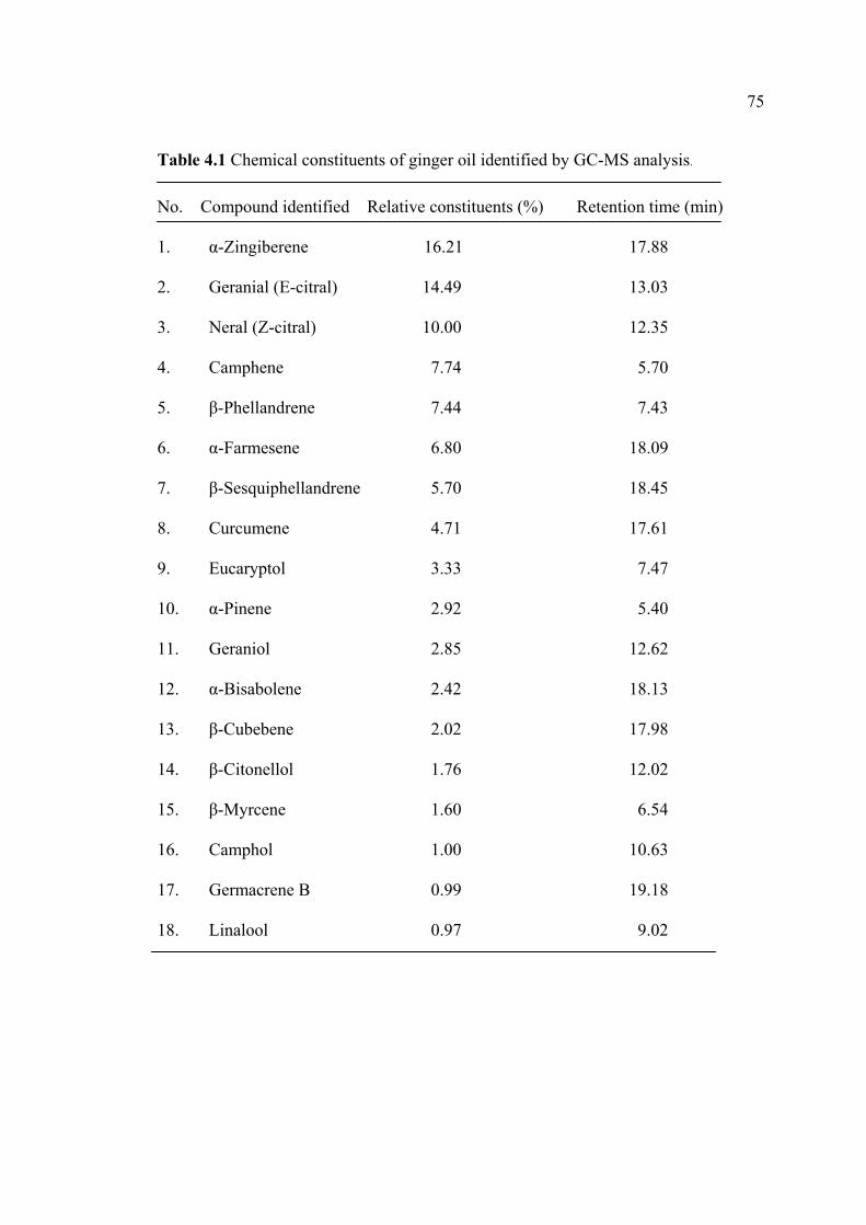
75
Table 4.1 Chemical constituents of ginger oil identified by GC-MS analysis.
No. Compound identified Relative constituents (%) Retention time (min)
1. α-Zingiberene 16.21 17.88
2. Geranial (E-citral) 14.49 13.03
3. Neral (Z-citral) 10.00 12.35
4. Camphene 7.74 5.70
5. β-Phellandrene 7.44 7.43
6. α-Farmesene 6.80 18.09
7. β-Sesquiphellandrene 5.70 18.45
8. Curcumene 4.71 17.61
9. Eucaryptol 3.33 7.47
10. α-Pinene 2.92 5.40
11. Geraniol 2.85 12.62
12. α-Bisabolene 2.42 18.13
13. β-Cubebene 2.02 17.98
14. β-Citonellol 1.76 12.02
15. β-Myrcene 1.60 6.54
16. Camphol 1.00 10.63
17. Germacrene B 0.99 19.18
18. Linalool 0.97 9.02
76
Table 4.1 (continued).
No. Compound identified Relative constituents (%) Retention time (min)
19. α-Terpineol 0.93 11.20
20. Geranyl acetate 0.84 15.44
21. Sesquisabinene hydrate 0.71 2 0.54
22. Zingiberenol 0.68 20.21
23. Nonyl 0.59 13.48
24. Unknown 0.56 21.61
25. Unknown 0.47 10.94
26. β-citronellal 0.46 10.25
27. s-Heptyl alcohol 0.44 4.71
28. l-Phellandrene 0.42 6.86
29. β-Pinene 0.37 6.28
30. Citronellol acetate 0.37 14.78
31. α-Terpinolene 0.21 8.77

77
Figure 4.1 Constituents of ginger oil
(A) The most constituents of ginger oil are Shown. (B) Chemical
structures of camphene (a), geranial (b), and neral (c).
78
4.4.2 Effects of Citral and Camphene on Spontaneous Contraction
Effects of Citral on Spontaneous Contraction
Under control conditions, spontaneous contractions of consistent amplitude
AUC and frequency could be recorded for several hours; allowing different
concentrations of citral to be tested in the rat uterus. Citral, in a dose-dependent
response (0.5 to 8.3 mM), significantly inhibited the myometrail contraction arising
spontaneously (Fig. 4.2A and Table 4.2, n = 5). The IC50 of citral occurred at the
concentration of 2.2 mM. Thus this concentration was used throughout the
experiments.
Effects of Camphene on Spontaneous Contraction
The effects of camphene on spontaneous contraction was tested at various
concentrations under the same condition (Fig. 4.2B and Table 4.3). As could be
observed, the effects of camphene was not as strong as of citral. The IC50 occurred at
the concentration of 7.5 mM (Fig. 4.2B and Table 4.3, n = 5). A typical effects of
camphene on spontaneous contraction is shown in Figure 4.2B. Camphene (7.5 mM)
decreased the contraction amplitude, frequency, and AUC to 53.42±3.52%,
50.64±2.52%, and 53.42%±3.21%, respectively (Fig. 4.3B).
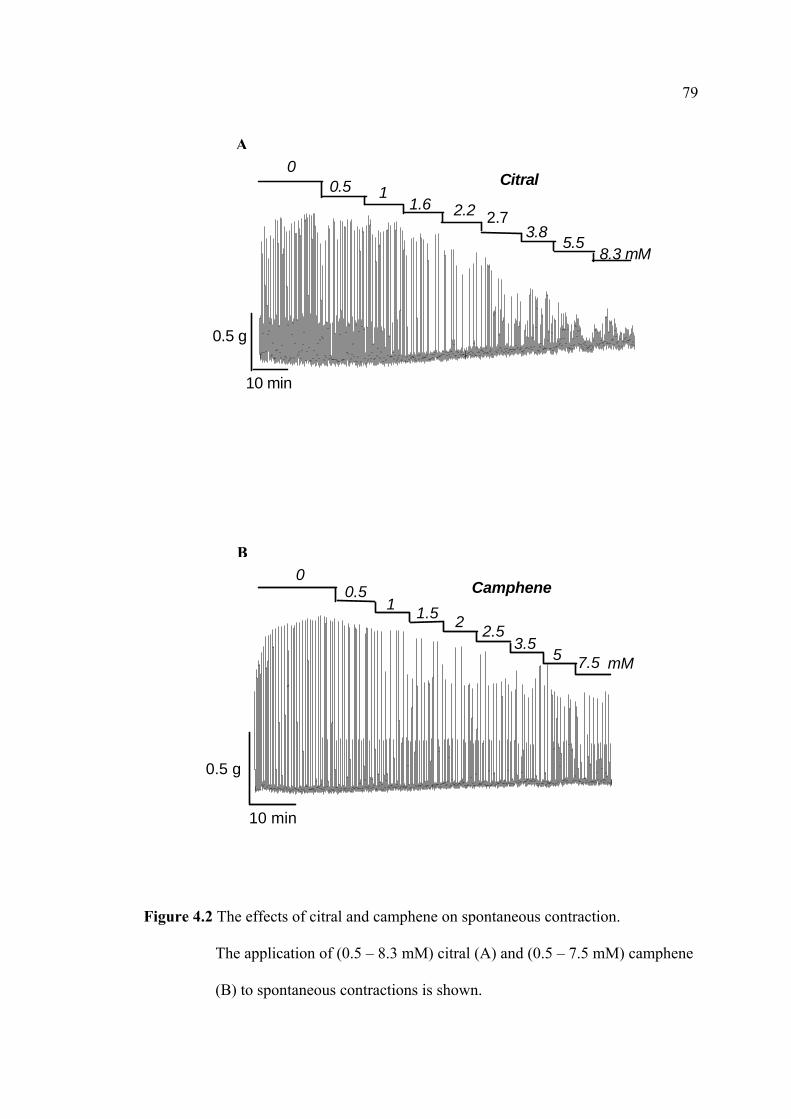
79
Figure 4.2 The effects of citral and camphene on spontaneous contraction.
The application of (0.5 – 8.3 mM) citral (A) and (0.5 – 7.5 mM) camphene
(B) to spontaneous contractions is shown.
0Citral
10 min
0.5 g
8.3 mM5.5
3.82.72.21.6
10.5
A
Camphene0
10 min
0.5 g
mM7.5 53.5
2 2.5
10.5
1.5
B
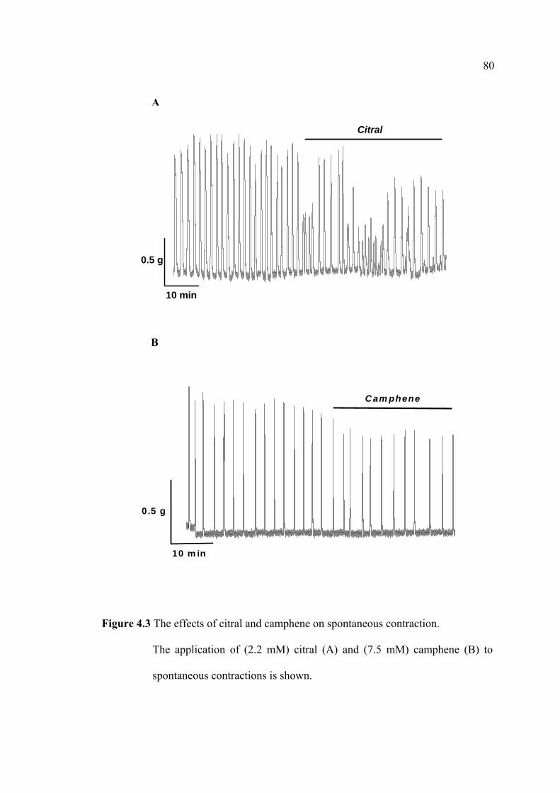
80
Figure 4.3 The effects of citral and camphene on spontaneous contraction.
The application of (2.2 mM) citral (A) and (7.5 mM) camphene (B) to
spontaneous contractions is shown.
10 min
0.5 g
Citral
A
10 m in
0.5 g
C am phene
B

81
Table 4.2

82
Table 4.3
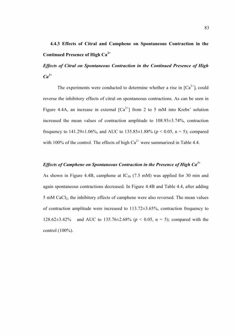
83
4.4.3 Effects of Citral and Camphene on Spontaneous Contraction in the
Continued Presence of High Ca2+
Effects of Citral on Spontaneous Contraction in the Continued Presence of High
Ca2+
The experiments were conducted to determine whether a rise in [Ca2+]i could
reverse the inhibitory effects of citral on spontaneous contractions. As can be seen in
Figure 4.4A, an increase in external [Ca2+] from 2 to 5 mM into Krebs’ solution
increased the mean values of contraction amplitude to 108.93±3.74%, contraction
frequency to 141.29±1.06%, and AUC to 135.85±1.88% (p < 0.05, n = 5); compared
with 100% of the control. The effects of high Ca2+ were summarized in Table 4.4.
Effects of Camphene on Spontaneous Contraction in the Presence of High Ca2+
As shown in Figure 4.4B, camphene at IC50 (7.5 mM) was applied for 30 min and
again spontaneous contractions decreased. In Figure 4.4B and Table 4.4, after adding
5 mM CaCl2, the inhibitory effects of camphene were also reversed. The mean values
of contraction amplitude were increased to 113.72±3.65%, contraction frequency to
128.62±3.42% and AUC to 135.76±2.68% (p < 0.05, n = 5); compared with the
control (100%).
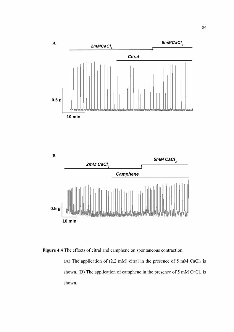
84
Figure 4.4 The effects of citral and camphene on spontaneous contraction.
(A) The application of (2.2 mM) citral in the presence of 5 mM CaCl2 is
shown. (B) The application of camphene in the presence of 5 mM CaCl2 is
shown.
10 min
0.5 g
2mMCaCl2
5mMCaCl2
Citral
A
10 min
0.5 g
Camphene
2mM CaCl2
5mM CaCl2
B

85
Table 4.4
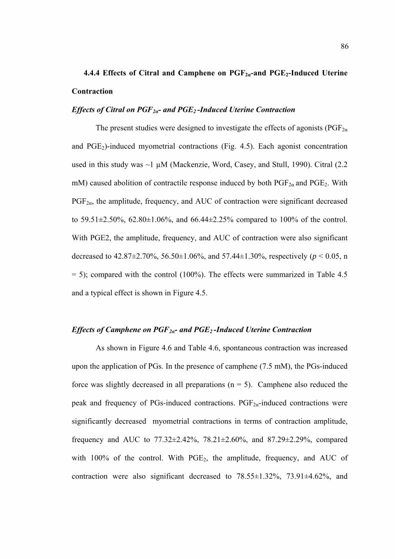
86
4.4.4 Effects of Citral and Camphene on PGF2α-and PGE2-Induced Uterine
Contraction
Effects of Citral on PGF2α- and PGE2 -Induced Uterine Contraction
The present studies were designed to investigate the effects of agonists (PGF2α
and PGE2)-induced myometrial contractions (Fig. 4.5). Each agonist concentration
used in this study was ~1 µM (Mackenzie, Word, Casey, and Stull, 1990). Citral (2.2
mM) caused abolition of contractile response induced by both PGF2α and PGE2. With
PGF2α, the amplitude, frequency, and AUC of contraction were significant decreased
to 59.51±2.50%, 62.80±1.06%, and 66.44±2.25% compared to 100% of the control.
With PGE2, the amplitude, frequency, and AUC of contraction were also significant
decreased to 42.87±2.70%, 56.50±1.06%, and 57.44±1.30%, respectively (p < 0.05, n
= 5); compared with the control (100%). The effects were summarized in Table 4.5
and a typical effect is shown in Figure 4.5.
Effects of Camphene on PGF2α- and PGE2 -Induced Uterine Contraction
As shown in Figure 4.6 and Table 4.6, spontaneous contraction was increased
upon the application of PGs. In the presence of camphene (7.5 mM), the PGs-induced
force was slightly decreased in all preparations (n = 5). Camphene also reduced the
peak and frequency of PGs-induced contractions. PGF2α-induced contractions were
significantly decreased myometrial contractions in terms of contraction amplitude,
frequency and AUC to 77.32±2.42%, 78.21±2.60%, and 87.29±2.29%, compared
with 100% of the control. With PGE2, the amplitude, frequency, and AUC of
contraction were also significant decreased to 78.55±1.32%, 73.91±4.62%, and
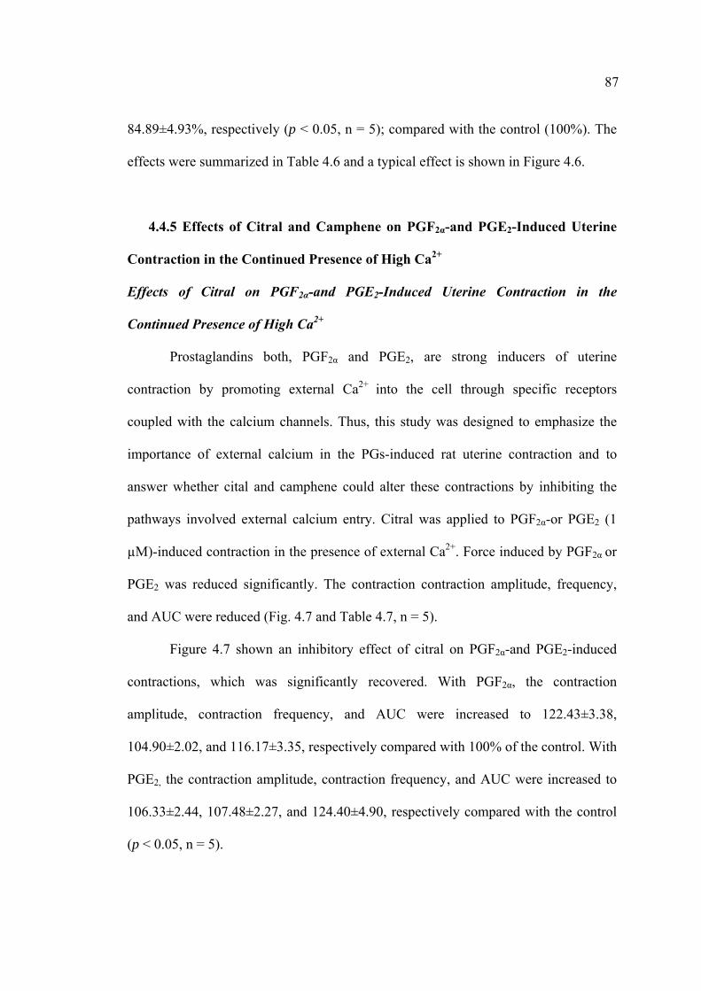
87
84.89±4.93%, respectively (p < 0.05, n = 5); compared with the control (100%). The
effects were summarized in Table 4.6 and a typical effect is shown in Figure 4.6.
4.4.5 Effects of Citral and Camphene on PGF2α-and PGE2-Induced Uterine
Contraction in the Continued Presence of High Ca2+
Effects of Citral on PGF2α-and PGE2-Induced Uterine Contraction in the
Continued Presence of High Ca2+
Prostaglandins both, PGF2α and PGE2, are strong inducers of uterine
contraction by promoting external Ca2+ into the cell through specific receptors
coupled with the calcium channels. Thus, this study was designed to emphasize the
importance of external calcium in the PGs-induced rat uterine contraction and to
answer whether cital and camphene could alter these contractions by inhibiting the
pathways involved external calcium entry. Citral was applied to PGF2α-or PGE2 (1
µM)-induced contraction in the presence of external Ca2+. Force induced by PGF2α or
PGE2 was reduced significantly. The contraction contraction amplitude, frequency,
and AUC were reduced (Fig. 4.7 and Table 4.7, n = 5).
Figure 4.7 shown an inhibitory effect of citral on PGF2α-and PGE2-induced
contractions, which was significantly recovered. With PGF2α, the contraction
amplitude, contraction frequency, and AUC were increased to 122.43±3.38,
104.90±2.02, and 116.17±3.35, respectively compared with 100% of the control. With
PGE2, the contraction amplitude, contraction frequency, and AUC were increased to
106.33±2.44, 107.48±2.27, and 124.40±4.90, respectively compared with the control
(p < 0.05, n = 5).

88
Effects of Camphene on PGF2α-and PGE2-Induced Uterine Contraction in the
Presence of High Ca2+
As shown in Figure 4.8, myometrial contraction induced by PGs is dependent
external calcium as camphene or extracellular calcium depletion abolished their
effects. It was observed that camphene (7.5 mM) also inhibited PGs-induced force
and contractions. The inhibitory effects of camphene on PGF2α-induced force and
contractions were significantly increased the contraction amplitude, frequency, and
AUC to 106.62±1.67, 115.43±2.35, and 108.79±0.61, respectively compared with
100% of the control. With PGE2, the amplitude, frequency, and AUC of contraction
were increased to 104.60±1.80, 109.39±3.68, and 110.63±3.38, respectively
compared with control in the presence of high external Ca2+. The data were
summarized in Table 4.8 (p < 0.05, n = 5).
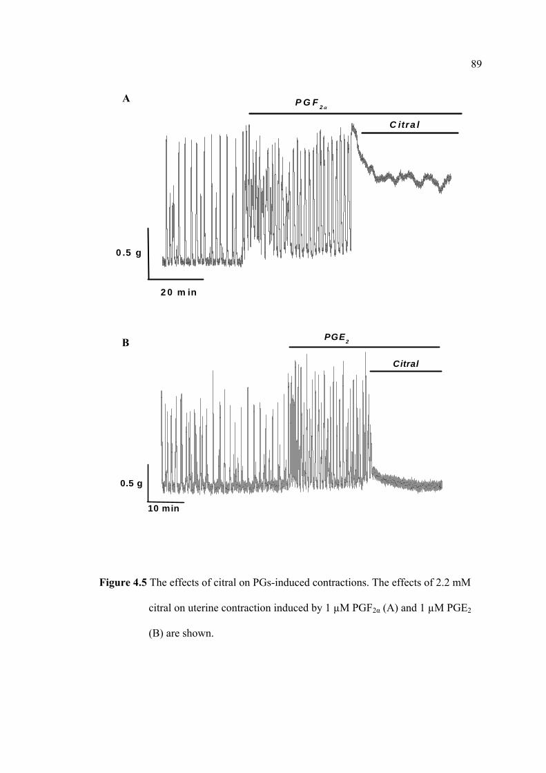
89
Figure 4.5 The effects of citral on PGs-induced contractions. The effects of 2.2 mM
citral on uterine contraction induced by 1 µM PGF2α (A) and 1 µM PGE2
(B) are shown.
10 min
0.5 g
Citral
PGE2B
2 0 m in
0 .5 g
C itra l
P G F 2 αA

90
Table 4.5
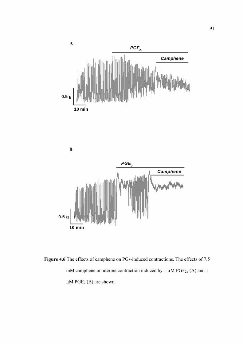
91
Figure 4.6 The effects of camphene on PGs-induced contractions. The effects of 7.5
mM camphene on uterine contraction induced by 1 µM PGF2α (A) and 1
µM PGE2 (B) are shown.
Camphene
PGF2α
10 min
0.5 g
A
Camphene
PGE2
10 min
0.5 g
B

92
Table 4. 6
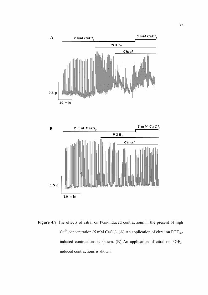
93
Figure 4.7 The effects of citral on PGs-induced contractions in the present of high
Ca2+ concentration (5 mM CaCl2). (A) An application of citral on PGF2α-
induced contractions is shown. (B) An application of citral on PGE2-
induced contractions is shown.
10 min
0.5 g
2 mM CaCl2
5 mM CaCl2
PGF2α
Citral
A
2 m M C a C l2
1 0 m in
0 .5 g
5 m M C a C l2
C itra l
P G E 2
B

94
Table 4.7
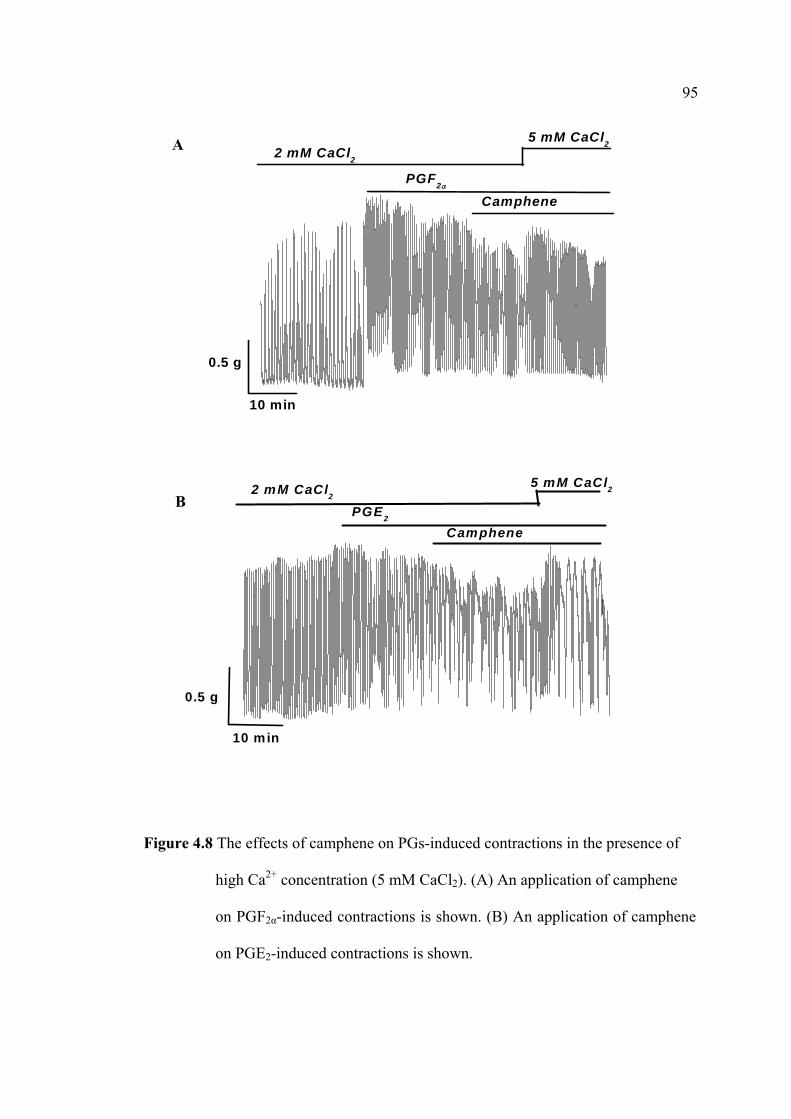
95
Figure 4.8 The effects of camphene on PGs-induced contractions in the presence of
high Ca2+ concentration (5 mM CaCl2). (A) An application of camphene
on PGF2α-induced contractions is shown. (B) An application of camphene
on PGE2-induced contractions is shown.
5 mM CaCl22 mM CaCl2
10 min
0.5 g
Camphene
PGF2α
5 mM CaCl22 mM CaCl2
10 min
0.5 g
CamphenePGE2
B
A

96
Table 4.8
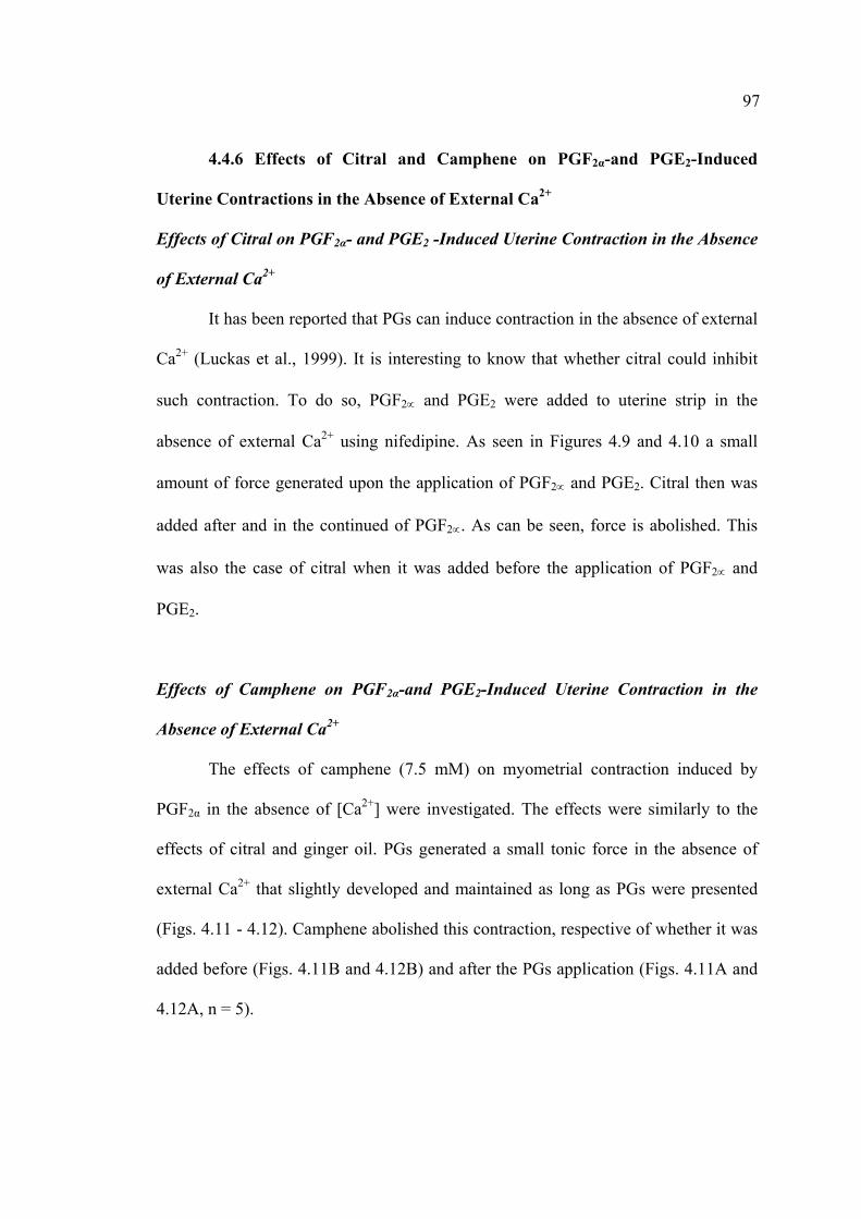
97
4.4.6 Effects of Citral and Camphene on PGF2α-and PGE2-Induced
Uterine Contractions in the Absence of External Ca2+
Effects of Citral on PGF2α- and PGE2 -Induced Uterine Contraction in the Absence
of External Ca2+
It has been reported that PGs can induce contraction in the absence of external
Ca2+ (Luckas et al., 1999). It is interesting to know that whether citral could inhibit
such contraction. To do so, PGF2∝ and PGE2 were added to uterine strip in the
absence of external Ca2+ using nifedipine. As seen in Figures 4.9 and 4.10 a small
amount of force generated upon the application of PGF2∝ and PGE2. Citral then was
added after and in the continued of PGF2∝. As can be seen, force is abolished. This
was also the case of citral when it was added before the application of PGF2∝ and
PGE2.
Effects of Camphene on PGF2α-and PGE2-Induced Uterine Contraction in the
Absence of External Ca2+
The effects of camphene (7.5 mM) on myometrial contraction induced by
PGF2α in the absence of [Ca2+] were investigated. The effects were similarly to the
effects of citral and ginger oil. PGs generated a small tonic force in the absence of
external Ca2+ that slightly developed and maintained as long as PGs were presented
(Figs. 4.11 - 4.12). Camphene abolished this contraction, respective of whether it was
added before (Figs. 4.11B and 4.12B) and after the PGs application (Figs. 4.11A and
4.12A, n = 5).

98
Figure 4.9 The effects of citral in the absence of external Ca2+ using 10 µM
nifedipine. Citral was added after (A) and before (B) PGF2α-induced
contraction.
10 min
0.5 g
PGF2α
Citral
Nifedipine
B
10 min
0.5 g
Citral
PGF2α
Nifedipine
A
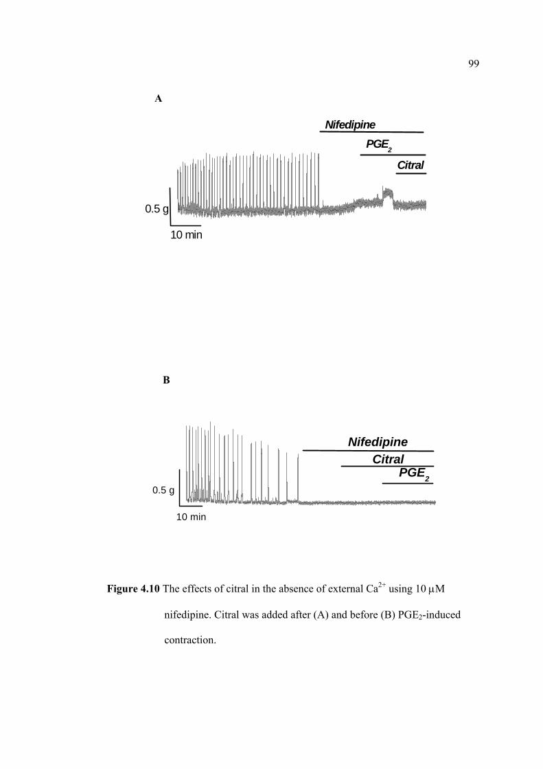
99
Figure 4.10 The effects of citral in the absence of external Ca2+ using 10 µM
nifedipine. Citral was added after (A) and before (B) PGE2-induced
contraction.
10 min
0.5 g
Nifedipine
PGE2
Citral
B
Citral
PGE2
Nifedipine
10 min
0.5 g
A
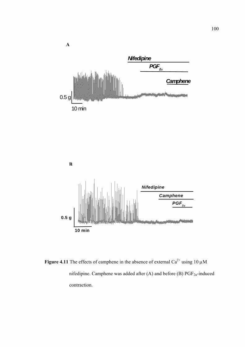
100
Figure 4.11 The effects of camphene in the absence of external Ca2+ using 10 µM
nifedipine. Camphene was added after (A) and before (B) PGF2α-induced
contraction.
10 min
0.5 g
PGF2α
Camphene
Nifedipine
B
10 min
0.5 g
PGF2α
Nifedipine
Camphene
A
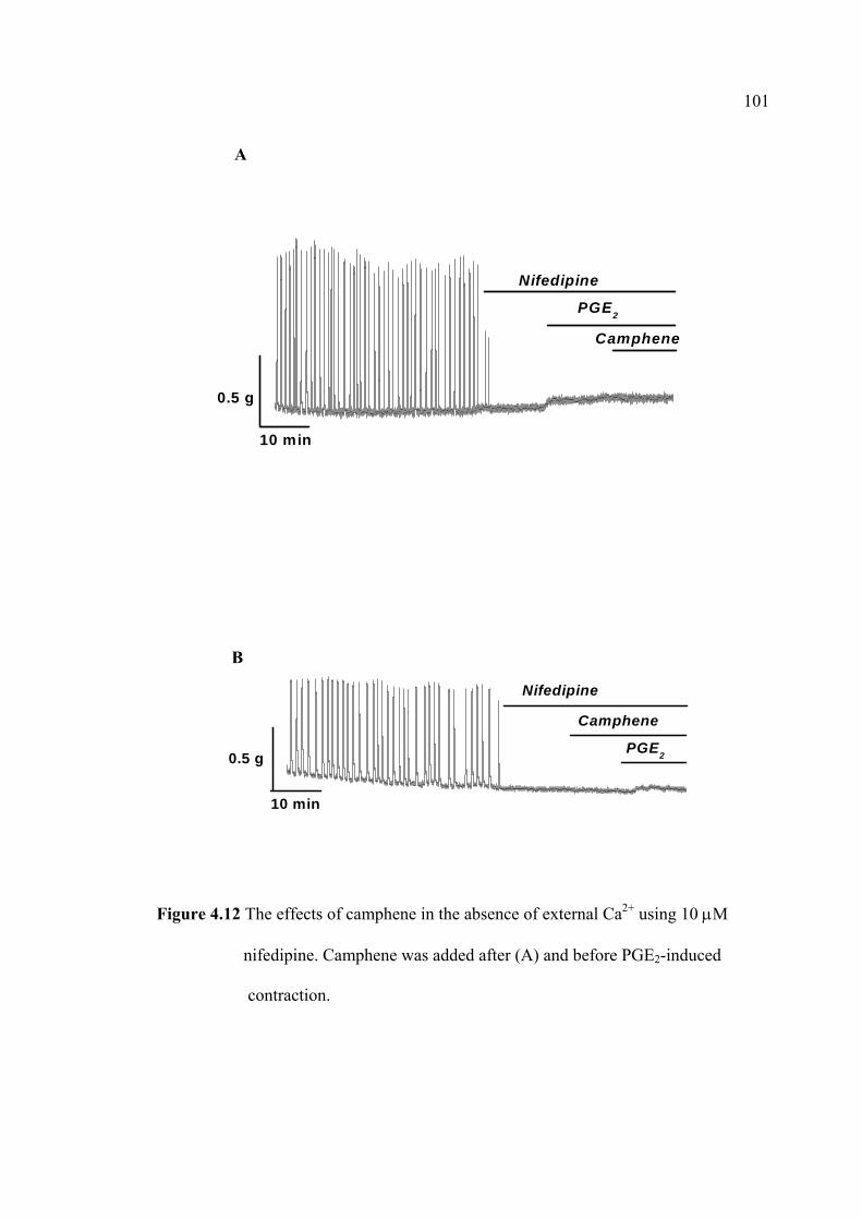
101
Figure 4.12 The effects of camphene in the absence of external Ca2+ using 10 µM
nifedipine. Camphene was added after (A) and before PGE2-induced
contraction.
B
10 min
0.5 g
Camphene
PGE2
Nifedipine
10 min
0.5 g
PGE2
Camphene
Nifedipine
A
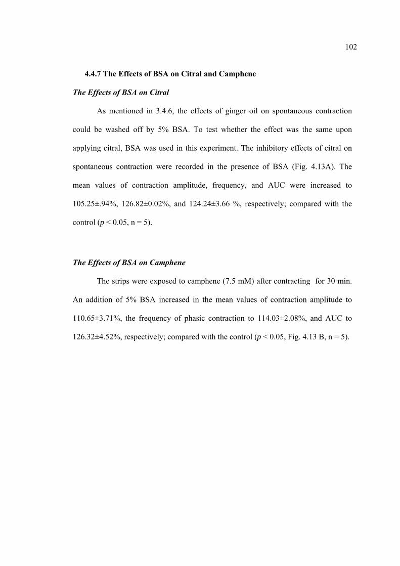
102
4.4.7 The Effects of BSA on Citral and Camphene
The Effects of BSA on Citral
As mentioned in 3.4.6, the effects of ginger oil on spontaneous contraction
could be washed off by 5% BSA. To test whether the effect was the same upon
applying citral, BSA was used in this experiment. The inhibitory effects of citral on
spontaneous contraction were recorded in the presence of BSA (Fig. 4.13A). The
mean values of contraction amplitude, frequency, and AUC were increased to
105.25±.94%, 126.82±0.02%, and 124.24±3.66 %, respectively; compared with the
control (p < 0.05, n = 5).
The Effects of BSA on Camphene
The strips were exposed to camphene (7.5 mM) after contracting for 30 min.
An addition of 5% BSA increased in the mean values of contraction amplitude to
110.65±3.71%, the frequency of phasic contraction to 114.03±2.08%, and AUC to
126.32±4.52%, respectively; compared with the control (p < 0.05, Fig. 4.13 B, n = 5).
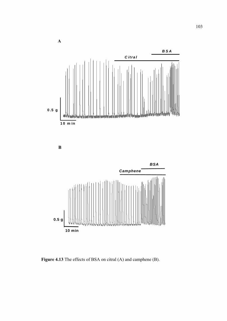
103
Figure 4.13 The effects of BSA on citral (A) and camphene (B).
10 min
0.5 g
BSA
Camphene
B
1 0 m in
0 .5 g
B S AC it ra l
A
104
4.5 Discussion
Citral and camphene were found as the main constituents of ginger oil as
shown in Table 4.1. The inhibitory effects of both compounds on myometrial
contraction have not been investigated. Therefore, the aims of this Chapter were to
evaluate the effects of citral or camphene on myometrial contractile activities arising
spontaneously and by PGs-induced contractions. The results showed that the effects
of both citral and camphene were similarly indistinguishable to these of ginger oil;
suggesting the effects of ginger oil that found could be due to the effects of citral and
camphene.
4.5.1 The Effects of Citral
Citral can inhibit both the contraction arising either spontaneously or by PGs-
induced contraction. Its effects were dose dependent. The IC50 is at 2.2 mM. The
effects found were the same as those observed in ginger oil. Interestingly, its effects
were likely to be via inhibition of L-type Ca2+ channels. This because, an elevation of
external [Ca2+] can reverse the effects of citral. This study showed that both PGF2α
and PGE2 significantly increased spontaneous contractile activity in uterine strips
(Carolyn et al., 2002), which are associated with a rise [Ca2+]i by external Ca2+ entry
and intracellular Ca2+ stores. They also induced a tonic contraction in the absence of
external Ca2+. Interestingly, citral inhibited the contraction occurred in both the
presence and absence of external Ca2+. This is similar to the effect of ginger oil as
shown in Chapter III.
Thus, PGs involved Ca2+ influx through L-type Ca2+ channels (Coleman et al.,
2000) were also blocked by citral on their activities (Figs. 4.5 and 4.6). However an
105
arising of external Ca2+ (5 mM CaCl2) or PGs-stimulated myometrial contraction
could be reappeared to force and contract again in the presence of citral at IC50
concentration (Figs. 4.4 and 4.7). As can be seen in Figures 4.9 and 4.10, there have
no contraction when external Ca2+ entry was blocked by nifedipine (10 µM) and
abolished PGs-induced force in the presence of citral.
4.5.2 The Effects of Camphene
Another known compound of ginger constituent, camphene, was also studied.
This is the first study of its inhibitory effects on myometrial contraction in vitro. No
other known compounds such as zingeberene, or curcumene, are commercially
available, and so could not be investigated.
These expreiments were designed to investegate and to compare the inhibitory
effects of camphene on spontaneous or PGs-induced contraction with the effects of
citral and ginger oil. The results indicated that (0.5 to 1.5 mM) camphene had no
significant different of contraction frequency, amplitude and AUC when compared to
control (100%). These findings suggest that its effective seem to be weaker compared
with the effects of citral and ginger oil. However, at the same concentration, the
inhibitory effects of citral is the strongest as it can inhibit both spontaneous and PGs-
induced myometrial contractions. Also, camphene at IC50 was found to inhibit Ca2+
influx in myometrial strips (Fig. 4.4B). And there was no response contraction when
external Ca2+ was blocked by nefedipine (10 µM) in the presence of camphene (Figs.
4.11-12). However, the amplitude contraction, frequency, and AUC were increased
higher than spontanneous contraction once adding BSA (Fig. 4.13B). Those
experiments conclude that its effects were in accordance with the inhibition of volatile
106
oil of Teucrium polium in vitro of isolated rabbit intestine. Thus, the effect was likely
to be due to camphene inhibits Ca2+ influx through the potential dependent Ca2+
channels (Agel and Gharibeh, 1990).
In conclusion the present study shown that two known commercial available
compounds may be to the active compounds of ginger oil. Particularly, the results
indicated that their inhibitory effects may act on Ca2+ dependent pathways. Based on
the present finding, citral showed the higher potent inhibition on external Ca2+
required for myometrial contractile activities. In addition, it is clearly demonstrated
that the inhibitory effects of both citral and camphene may be involved in the pathway
of non-Ca CaM MLCK.
4.6 References
Agel, M. B. and Gharibeh, M. N. (1990). The calcium antagonistic effect of the
volatile oil of Teucrium polium. Pharmaceutical Biology. 28 : 201 – 207.
Carolyn, A. L., Baillie, Y. P., Vedernilow, M. D., Saade, G. R. and Garfield, R. E.
(2002). Prostaglandin-induced activation of uterine contractility in pregnant
rats does not involve potassium channels. American Journal of Obstetrics
and Gynecology. 186 : 453 – 457.
Carbajal, D., Casaco, A., Arruzazabala, L., Gonzalez, R. and Tolon, Z. (1989).
Pharmacological study of Cymbopogon citrates leaves. Journal of
Ethnopharmacology. 25 : 103 – 107.
Coleman, H. A., Hart, J. D. E., Tonta, M. A. and Parkington, H. C. (2000). Changes in
the mechanisms involved in uterine contractions during pregnancy in guinea-
pigs. Journal of Physiology. 523(3) : 785 – 798.
107
Hawthorn, M., et al. (1988). The actions of peppermint and menthol on calcium
channel dependant processes in intestinal, neural and cardiac preparations.
Ailment Pharmacology Therapy. 2 : 101 – 118.
Luckas, M. J., Taggart, M. J. and Wray, S. (1999). Intracellular calcium stores and
agonist-induced contractions in isolated human myometrium. American
Journal of Obstetrics and Gynecology. 181(2) : 468 – 476.
Mackenzie, L. W., Word, R.A., Casey, M. L. and Stull, J. T. (1990). Myosin light
chain phosphorylation in human myometrial smooth muscle. American
Journal of Physiology. 258 : C92 – 98.
Miyazawa, M. and Kameoka, H. (1988). Volatile flavor components of Zingiberis
rhizome (Zingiber officinale Roscoe). Agricultural Biological Chemistry. 52
: 2961 – 2963.
Ress N. B., et al. (2003). Toxicology and carcinogenesis studies of microencapsulated
citral in rats and mice. Toxicological Sciences. 71 : 198 – 206.
Sadraei, H., Ghannadi, A. Malekshahi, K. (2003). Composition of the essential oil of
Ferula assa-feotida L. and its spasmolytic action. Pharmaceutical Journal.
11(3) : 136 – 140.
Scolnid, M. D., Servadio, C., Abramovici, A. (1994). Comparative study of
experimentally induced benign and atypical hyperplasia in the ventral
prostrate of different rat strains. Journal of Andrology. 15 : 287 – 297.
Tamaoki, J., et al. (1995). Effect of menthol vapour on airway responsiveness in
patients with mild asthma. Respiratory Medicine. 108 : 589 – 593.

108
Viniketkumnuen, U., Puatanchokchai, R. and Kongtawelert, P. (1994). Anti-
mutagenicity of lemongrass (Cymbopogon citrats stapf) to various known
mutagens in salmonella mutation assay. Mutation Research. 341 : 71 – 75.
Wang, X. D., Krinsky, N., Tang, G. W. and Russell, R. M. (1992). Retinoic acid can
be produced from excentric cleavage of β-carotene in human intestinal
mucosa. Archives of Biochemistry and Biophysics. 293 : 298 – 304.
Wohlmuth, H., Smith, M. K., Brooks, L. O., Myers, S. P. and Leach, D. N. (2006).
Essential oil composition of diploid and tetraploid clones of ginger (Zingiber
officinale Roscoe). Journal of American and Food Chemistry. 54 : 1414 –
1419.
Wright, C. E, et al. (1997). Capsaicin and neurokinin A-induced bronchoconstriction
in the anaesthetized guinea–pig: evidence for a direct action of menthol on
isolated bronchial smooth muscle. British Journal Pharmacology. 121: 1645
– 1650.
CHAPTER V
EFFECTS OF GINGER OIL, CITRAL, AND CAMPHENE
ON INFLAMMATORY MEDIATED PATHWAYS IN RAT
MYOMETRIUM
5.1 Abstract
Several studies have been reported that cyclooxygenase (COX) catalysis is the
first step of arachidonic acid (AA) mechanisms of PGs synthesis. An increased
expression of COX-2 enzyme participates to a dramatic PG rates rising in
inflammation tissues. Previous studies suggested that an anti-inflammatory process
was depressed by ginger constituents. Therefore, the aims of this chapter were to
examine whether the inhibitory effects of ginger oil, citral, and camphene on
spontaneous contraction in non-pregnant rat myometrium are via COX-2 or via
phosphodiesterase (PDE) pathways. There were three main aims; 1) to assess the
inhibitory effects on AA-induced myometrial contraction 2) to measure PGE2 and
cAMP contents and 3) to determine the inhibitory effects contribution of
indomethacin in the myometrial contraction. The rat myometrial strips were prepared.
The effects of exposing the strips to AA, indomethacin and aminophylline were
investigated. Exposure of AA increased the amplitude, frequency, and AUC of
spontaneous contraction. AA-induced contraction was inhibited in the presence of
ginger oil and its pure compounds. As with ginger oil, indomethacin inhibited the
amplitude of spontaneous contraction, but did not affect the frequency and AUC of
110
contractions. Assay of COX-2 enzyme showed no effect of its activities in this
condition. Aminophylline (1 nM), ginger oil, citral, and camphene remarkably
inhibited PDE enzyme but did not cause an accumulation of cAMP content. These
results indicate that the inhibitory effects on spontaneous contraction of ginger oil and
others did not occur via inflammatory process and that this neither involved with
COX-2 pathway nor cAMP production.
5.2 Introduction
Inflammation is associated with a large range of mediators that initiates
inflammatory response for AA release by cell biosynthesis of prostaglandins (PGs)
via the cyclooxygenase (COX) pathway. The COX isoenzymes, COX-1 and COX-2
(His et al., 1994; O’Banion et al., 1992) pathway produces PGs (Simmons et al.,
2004), prostacyclins and thromboxanes. PGs are important biological mediators
essential for physiological functions in the body that control inflammatory responses,
pain, and fever (De Witt, 1991). An increase production of prostaglandins during an
inflammatory response in achieved by induction of COX-2 (Ramsay et al., 2003).
Activation of COX results in the synthesis of a wide spectrum of prostaglandins
(Kuby, 1997) the critical mediators of the inflammatory process, with various
activities leading to increased vascular permeability, increased vasodilation (Kuby,
1997). In addition, the role of PG activities are involved in various processes
associated with reproduction (Vane et al., 1988), ovulation (Lemaire and Marsh,
1975), fertilization (Lim et al., 1997), implantation (Chakraborty et al., 1996),
maintenance of normal pregnancy (Kennedy, 1977; Trautman et al., 1996), and
induction of labor (Hoffman, 1978). COX converts AA to PGG2 through oxygenase
111
activity, and this unstable product is reduced to PGH2 through peroxidase activity
(Smith and Marnett, 1991).
A second messenger role of AA in signal transduction is widely accepted
(Axelrod, 1990; Khan et al., 1995). As the evidence of an experiment suggested that
changes in intracellular AA concentration could cause pathological effects such as
ischemia and neurotrauma (Bazan et al., 1995; Oe et al., 1994; Katsura et al., 1993).
However, the various kinds of drugs had been developed to inhibit these
enzyme (COX-1, COX-2), non-steroidal anti-inflammatory drugs (NSAIDs), are
devided into two groups: COX-1/-2 non-selective inhibitors (Meade et al., 1993;
O’Neill et al., 1994; Cromlish et al., 1994) and COX-2 selective inhibitors (Copeland
et al., 1994; Seibert et al., 1994; Futaki et al., 1994). COX-2-selective-NSAIDs have
been used to inhibit pain, fever, inflammation and intestinal neoplasia in mice and
human (Mann and DuBios, 2004) by inhibiting the COX pathway. COX inhibitors,
nonstelective drugs or COX-2 NSAIDs such as piroxicam, aspirin, diclofenac,
particularly indomethacin, are among the most potent tocolytic agents available
(Vermillion and Landen, 2001). However, like other classes of drugs for tocolytic,
their efficacy is limited. These effects have been focused on finding lead anti-
inflammatory compounds from natural products because of NSAIDs side effects.
Determining for avoiding the NSAIDs side effects or problem in therapeutics that
damage with protracted use include dizziness, headache, nausea, vomiting and
diarrhea gastrointestinal tract (Cahill, 1986; Bjarnason et al., 1986). The experiments
are therefore design to suppress inflammation without their side effects of the present
range of NSAIDs.
112
Several studies have indicated that compounds found in ginger are effective in
relief of symptoms from chronic inflammatory diseases and rheumatoid arthritis
(Srivastana and Mustafa, 1992). On the one hand, it was in vitro observed that
gingerdione inhibited the production of 5-HETE (leucotrienes) and PGE2.
Additionally, it way proved that shogaol inhibited the production of leucotrienes.
Gingerol, as well as dehydroparadol, favored the COX inhibition (Srivastava, 1984;
Kiuchiet et al., 1992; Mustafa et al., 1993; Tjendraputra et al., 2001). It was
demonstrated that ginger was a potent inhibitor of the prostaglandin synthesis, even
stronger than indomatacin (Mustafa et al., 1993). On the other hand, it was in vivo
observed that ginger acted by inhibiting the prostaglandin synthesis and the
production of free radical during metabolism of the AA (Mustafa et al., 1993).
Recently study, ginger is one of most valuable effects its ability to reduce and relieve
inflammatory pain as effectively as aspirin, ibupofen, and other NSAIDs, without
their adverse side effects. Ginger does this by partial blocking COX-2 enzymes,
which are necessary for inflammation. Moreover, an inhibitor of PG synthesis, ginger
root has been used for thousand of years for its anti-inflammatory properties
(Srivastava and Mustafa, 1989; Taymor et al., 1964). In additional medicine, ginger
has been used to treat many inflammatory conditions and associated pain.
Furthermore, ginger constituents, (8)-paradol and (8)-shogaol, as well as to synthetic
analogues, 3-hydroxy-1(-4-hydroxy-3-methoxyphenyl), decane and 5-hydroxy-1-(4-
hydroxy-1-3-methoxyphenyl) dedecane, showed strong inhibitory effects on COX-2
enzyme activity (Tjendraputra et al., 2001).
However, the mechanisms of AA action on smooth muscle remain to be
identified. Adding AA leading to contraction activated from the intracellular side of
113
the plasma membrane, in accordance with other researchs, different protein kinase C
isoforms can be activated by AA (Lester et al., 1991; Khan et al., 1995). AA
stimulated protein kinase C binding to actin filaments (Prekeris et al., 1996) and
related lipid messengers were reported to sensitize the contractile smooth muscle
apparatus to basal Ca2+ concentrations (Gong et al., 1992). Moreover, Filipeanu et al.
(1998) showed that different mechanisms are involved in the contractile effect of
intracellular and in the effect of extracellular AA in rat aorta. Previous studies have
found that it also functions by prostaglandin independent mechanisms, such as
inhibition of nitric oxide synthase and phosphodiesterase 4 (Bruch et al., 1983;
Bevilacqua and Magni, 1993) related cAMP. In addition, the study on uterine smooth
muscle suggested that papaverine inhibited smooth muscle contraction mainly by
inhibition of mitochondrial respiration and did not increase in cAMP level (Shimizu et
al., 2000). In smooth muscle, cAMP modulation has been postulated to involve
stimulation of Ca2+ and Na+ pumps (Scheid et al., 1979; Bulbring and den Hertog,
1980), partial inhibition of potential-operated Ca2+ channels (Meisheri and van
Breemen, 1982). It also decreased in the affinity of myosin light chain kinase for the
Ca-calmodulin complex (Adelstein et al., 1978, 1982).
Nevertheless, there have been a few reports indicating that PDE3, 4, and 5
inhibitors caused relaxation in canine colonic smooth muscle, and that PDE3 and 4
inhibitors decreased the hydrolysis of cAMP, and PDE5 inhibitor decreased the
hydrolysis of cGMP (Barnette et al., 1993). At the cellular level, the intensity and the
duration of the intracellular cAMP and cGMP signals are partly regulated by the PDE
enzymes whose functions are to degrade cyclic nucleotides into their inactive
metabolites. PDE4 is selsctive for cAMP hydrolysis did, and it has been widely

114
evidenced that the elevation of cAMP levels by PDE4 inhibition relaxes various types
of smooth muscle fibers (Conti et al., 1995). The role of the cAMP-specific PDE 4
family has seldom been investigated (Ahn et al., 1992). However, the relaxant effects
of pregnant rat uteri were not mediated by cAMP when the arising of external Ca2+
concentration by using aminophylline, an inhibitor of cAMP-PDE enzyme (Apaydin
et al., 1998). Additionally, with regard to uterine smooth muscle of non-pregnant rats,
the cAMP-independent mechanisms remain unclear.
The aims of this Chapter were to determine whether the inhibitory effect of
ginger oil and its active compounds involved in inflammatoty process. AA was used
to induce contraction. The effects of ginger oil and its pure compounds together with
aminophylline, the non-specific PDE enzyme inhibitor, and indomethacin, the COX-2
inhibitor, on uterine contraction were studied. The production of cAMP was also
investigated.
5.3 Materials and Methods
5.3.1 Chemicals and Physiological Solution
All chemicals were purchased from Sigma® unless state otherwise.
Agonists/antagonists for the investigation of inflammatory pathways were 95% citral,
95% camphene, indomethacin, aminophylline and AA, cAMP EIA kit (cAMP Biotrak
EIA system, Amersham Pharmacia Biotech, UK) and PGE2 assay kit. Ginger oil and
its pure compounds were dissolved by hexane. The final concentration of hexane in
any dilution was less than 0.15%. DMSO was used for PGE2 assay.
115
5.3.2 Myometrial Tissue Preparations
Tissue preparations are essentially the same as those described in Chapter II.
Non-pregnant Wistar rats (200-250 g) were used in this study and maintained in
accordance with the guidelines of the committee on Care and Use of Laboratory
Animal Resources, National Research Council, Thailand. The experiments performed
on rats were conducted in accordance with the advice of the Institutional Animal Care
and Use Committee, SUT, Thailand. Myometial tissue preparations were dissected
and provided for tension measurements as described in 2.2.2.
5.3.3 Measurements of Tension
The uterine strips were mounted vertically under resting tension of 1 g in a
single chamber (25 ml) tissue bath connected to a force transducer (as described in
2.3). Tissues were allowed to contract spontaneously. After an equilibrium period for
30 min, either AA or indomethacin was applied. The data were recorded as the
electrical signal from the transducer and converted to a digital signal on a computer
using Chart software.
5.3.4 Anti-Inflammatory RIA Assay
5.3.4.1 Cell Culture and Treatment
Immortalized mouse PGH-1 and PGH-2 null cells at the concentration (1×10⁵
cells/ml) of Dubelcco’s Modified Eagle Medium (DMEM) high glucose
supplemented with hygromyein B (200 µg/ml), non essential amino acid (0.1 mM), L-
glutamine (50 mg/l), ascorbic acid (0.05 mg/ml) and 10% fetal calf serum (FCS) were
seeded into 96-well flat bottom tissue culture plates (83 µl/well). Cells were incubated
116
37°C in humidified incubator with 5% CO2 for 7.2 hr. The cells were then washed
with DMEM medium without FCS and pre-incubated for 30 min with 83 µl of serum-
free DMEM medium containing vehicle or drugs. Following the pre-incubation
period, the medium was removed and cells were immediately treated with serum-free
medium containing vehicle or drugs and AA (20 µM) or A 23187 (2 µM) for 30 min.
Culture supernatants were then collected from wells and analyzed for PGE2
concentrations by radioimmunoassay (RIA).
5.3.4.2 PGE2 Measurement
The RIA method used for measuring PGE2 concentrations in the culture
supernatant is based on the competition between PGE2 in samples and 3H labeled
PGE2 for anti-PGE2 antibody binding sites. The assay was performed on ice as
following. The supernatant was diluted with DMEM (1:10) for blank and zero %
binding and added approximately 1.5 ml into 50 µl micro-centrifuge tube. Then, 50 µl
of anti-PGE2 antibody in RIA buffer (0.1 mM phosphate buffer, pH 7.4, containing
0.9% sodium chloride, 0.1% sodium azide and 0.1% gelatin) was added to every tube
accept for the blanks, in which 50 µl RIA buffer was added. Subsequently, 50 µl of
3H-PGE2 (1.12 µci/ml), was added to each tube and vortexed briefly, then incubated
overnight at 4°C. Then, 100 µl at 2% charcoal-dextran suspension in RIA buffer was
added to each tube. After 15 min incubation on ice, the tubes were centrifuged at
3,800 rpm (1,500 g) at 4°C for 10 min. Supernatants were then transferred to new 1.5
ml microcentrifuge tubes containing liquid scintillation cocktail, vortexed, and
counted for radioactivity. The resulting radioactive counts were used to calculate a
117
certain percentage of 3H-PGE2, which were then used for the estimation of PGE2
concentrations from standard curves.
5.3.5 cAMP EIA Assay
In the muscle strips, cAMP content were measured by enzyme immunoassay
(EIA) as reported previously (Shimizu et al., 200; Kaneda et al., 2005). The uterine
strips were incubated with 5 mM CaCl2 for 10 min then with aminophylline, ginger
oil and its pure compounds (citral and camphene) for 30 min at 37°C. After
incubation with various treatments, the strips were rapidly frozen in liquid nitrogen
and stored at -80°C until homogenized in 6% trichloroacetic acid (0.4 ml). The
homogenate was centrifuged at 3000×g for 15 min and the supernatant was washed
with 1.5 ml of water-saturated diethylether four times; the cAMP contents were
assayed by using EIA kit (Amersham Pharmcia Biotech, Little Chalfont, UK). cAMP
contents were expressed as fmol/g wet weight.
5.3.6 Data Analysis
5.3.6.1 Statistic Analysis for Measurements of Tension
All data were then presented as mean ± S.E.M. and “n” represents the number
of samples, each one from a different animal. Significance was tested using
appropriate t tests or ANOVA and P values < 0.05 taken to be significant. Results
were then expressed as percentages of control contractions (i.e. the control is 100%).
118
5.3.6.2 Statistic Analysis of PGE2 Contents
The paired t-test procedure was used to determine the differences in the PGE2
levels between control samples of wild-type. COX-2 cells and among control samples
and samples from cytokine-treated cells were collected. Differences were considered
significant if p < 0.05.
5.3.6.3 The cAMP EIA Assay
Data were calculated as the average optical density (OD) for each set of
replicate wells. The percent bound for each standard and sample (see below) was
calculated.
5.4 Results
5.4.1 Measurements of PGE2 Contents (COX-2 Assay)
PGE2 biosynthesis is regulated by successive metabolic steps involving the
phospholipase A2 mediating release of AA and its conversion to PGE2 by COX,
hydroperoxyidase and isomerase (Coetzi et al., 1995). Following the preincubation
period, the medium containing vehicle or drugs and AA (20 µM) was prepared for 30
min. Samples were then collected to the RIA method (Kirtikara et al., 1998; Kirtikara
et al., 2001) used for measuring PGE2 contents were measured by RIA method in the
cell culture based on the competition between PGE2 in the samples and 3H label PGE2
for antibody binding sites. Anti-inflammation of ginger oil and its pure compounds
was tested by RIA to compare with control (aspirin 0.9 – 1.0×10⁻⁵ g/ml, the COX-2
%B/B0 = × 100 (standard or sample OD-NSB OD) (zero standard OD-NSB OD)
119
inhibitor). 10-5 g/ml such as ginger oil, 95% citral, and 95% camphene were inactive
for anti-COX-2 (the inhibition less than 50%, containing DMSO vehicle, Table 5.1).
Table 5.1 Effects of ginger oil, citral, and camphene on COX-1, COX-2 pathways.
Anti-COX-2 Anti-COX-1 Samples Activity IC50 Activity IC50
Citral (95%) Inactive - - -
Ginger oil Inactive - - -
Camphene (95%) Inactive - - - IC50 of positive control: Aspirin = 0.9 - 10×10-5 g/ml (COX-2), and 0.4 – 0.5×10⁻⁵
g/ml (COX-1), maximum final concentration of tested samples: 10-5 g/ml, negative
control: 0.1% DMSO
Interpretation: % inhibition Activity
< 50% Inactive
≥ 50% Active (IC50)
5.4.2 Effects of Ginger Oil, Citral, Camphene, and AA on the Spontaneous
Contraction
Spontaneous contraction of rat myometrial tissues was stimulated and
increased after equilibrium with AA (20 µM) for 30 min. It was found that AA
induced significantly increase in the mean amplitude, frequency and AUC of
contraction up to 134.61±1.65, 114.52±5.02, 134.61±1.65, and 133.024±3.91%,
respectively (p < 0.05, n=10), compare with the control (Figs. 5.1 and 5.3A).
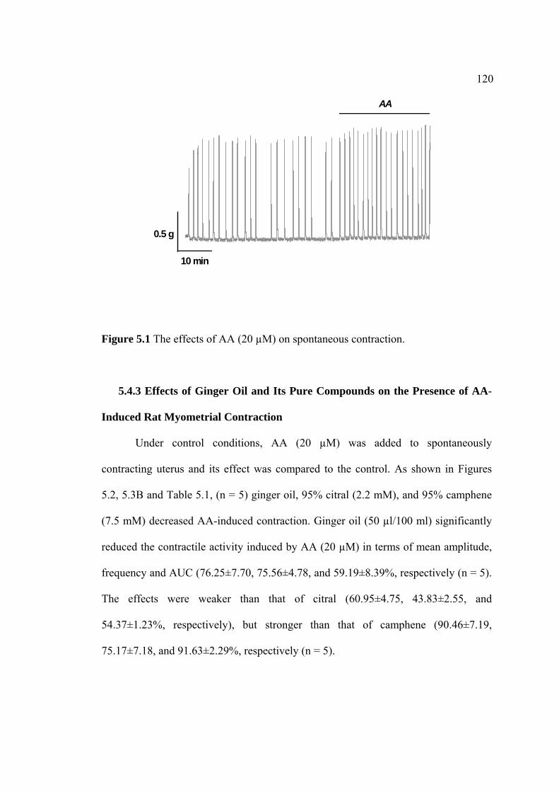
120
Figure 5.1 The effects of AA (20 µM) on spontaneous contraction.
5.4.3 Effects of Ginger Oil and Its Pure Compounds on the Presence of AA-
Induced Rat Myometrial Contraction
Under control conditions, AA (20 µM) was added to spontaneously
contracting uterus and its effect was compared to the control. As shown in Figures
5.2, 5.3B and Table 5.1, (n = 5) ginger oil, 95% citral (2.2 mM), and 95% camphene
(7.5 mM) decreased AA-induced contraction. Ginger oil (50 µl/100 ml) significantly
reduced the contractile activity induced by AA (20 µM) in terms of mean amplitude,
frequency and AUC (76.25±7.70, 75.56±4.78, and 59.19±8.39%, respectively (n = 5).
The effects were weaker than that of citral (60.95±4.75, 43.83±2.55, and
54.37±1.23%, respectively), but stronger than that of camphene (90.46±7.19,
75.17±7.18, and 91.63±2.29%, respectively (n = 5).
AA
10 min
0.5 g
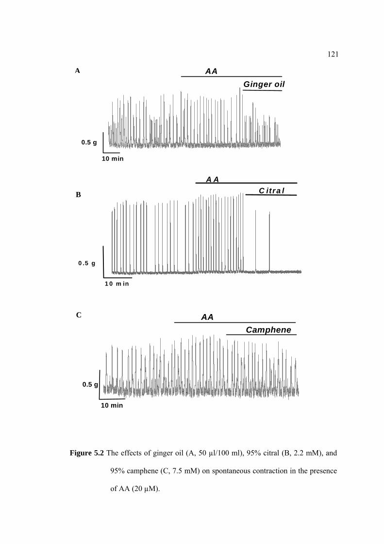
121
Figure 5.2 The effects of ginger oil (A, 50 µl/100 ml), 95% citral (B, 2.2 mM), and
95% camphene (C, 7.5 mM) on spontaneous contraction in the presence
of AA (20 µM).
10 min
0.5 g
Ginger oil
AAA
1 0 m in
0 .5 g
C itra lA A
B
10 min
0.5 g
CampheneAAC
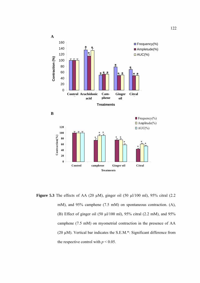
122
Figure 5.3 The effects of AA (20 µM), ginger oil (50 µl/100 ml), 95% citral (2.2
mM), and 95% camphene (7.5 mM) on spontaneous contraction. (A),
(B) Effect of ginger oil (50 µl/100 ml), 95% citral (2.2 mM), and 95%
camphene (7.5 mM) on myometrial contraction in the presence of AA
(20 µM). Vertical bar indicates the S.E.M.*: Significant difference from
the respective control with p < 0.05.
A
0
20
40
60
80
100
120
140
160
1 2 3 4 5
Treatments
Frequency(%) Ampletude(%) AUC(%)
***
**
*
***
**
*
Control Cam- phene
Arachidonicacid
Ginger oil
Citral
Con
trac
tion
(%)
0
20
40
60
80
100
120
Control camphene Ginger oil CitralTreatments
Con
trac
tion(
%)
Frequency(%)Amplitude(%)AUC(%)
**
*
***
* * *
B

123
Table 5.1
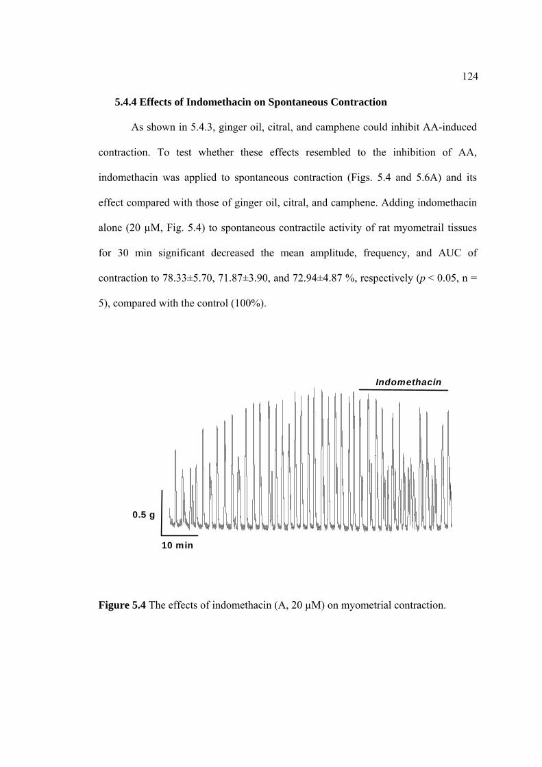
124
5.4.4 Effects of Indomethacin on Spontaneous Contraction
As shown in 5.4.3, ginger oil, citral, and camphene could inhibit AA-induced
contraction. To test whether these effects resembled to the inhibition of AA,
indomethacin was applied to spontaneous contraction (Figs. 5.4 and 5.6A) and its
effect compared with those of ginger oil, citral, and camphene. Adding indomethacin
alone (20 µM, Fig. 5.4) to spontaneous contractile activity of rat myometrail tissues
for 30 min significant decreased the mean amplitude, frequency, and AUC of
contraction to 78.33±5.70, 71.87±3.90, and 72.94±4.87 %, respectively (p < 0.05, n =
5), compared with the control (100%).
Figure 5.4 The effects of indomethacin (A, 20 µM) on myometrial contraction.
0.5 g
10 min
Indomethacin
125
The effects of combination of indomethacin and ginger oil, citral, and
camphene were also tested. Indomethacin combined with ginger oil (50 µl/100 ml)
significant decreased mean amplitude, frequency, and AUC of contraction to
60.92±3.65, 36.51±8.87, and 61.71±7.14% (p < 0.05, n = 5), respectively. As with
ginger oil, indomethacin combined with 95% citral (2.2 mM) significant decreased in
mean amplitude, frequency, and AUC of contraction to 58.37±8.55, 63.51±6.12, and
50.78±4.00% (p < 0.05, n = 5), respectively. Again, indomethacin combined with
95% camphene (2.2 mM) significant decreased mean amplitude, frequency, and AUC
of contraction to 68.78±8.47, 50.04±5.16, and 64.08±3.39% (p < 0.05, n = 5),
respectively. The data were summarized in Table 5.2 and a typical effect is shown in
Figures 5.5 and 5.6B.

126
Figure 5.5 The effects of ginger oil (A, 50 µl/100 ml), 95% citral (B, 2.2 mM), and
95% camphene (C, 7.5 mM) on spontaneous contraction in the presence
of indomethacin (20 µM).
10 min
0.5 g
Indomethacin
Ginger oil
A
10 min
0.5 g
Citral
Indomethacin
B
10 min
0.5 g
Indomethacin
Camphene
C
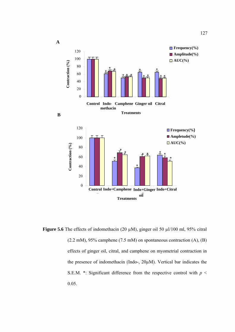
127
Figure 5.6 The effects of indomethacin (20 µM), ginger oil 50 µl/100 ml, 95% citral
(2.2 mM), 95% camphene (7.5 mM) on spontaneous contraction (A), (B)
effects of ginger oil, citral, and camphene on myometrial contraction in
the presence of indomethacin (Indo-, 20µM). Vertical bar indicates the
S.E.M. *: Significant difference from the respective control with p <
0.05.
A
0
20
40
60
80
100
120
Con
trac
tion
(%)
Frequency(%)Amplitude(%)AUC(%)
Control Indo- Camphene Ginger oil Citral methacin Treatments
* * * * * *
* * *
* * *
0
20
40
60
80
100
120
1 3 4 Treatments
Con
trac
tion
(%)
Frequency(%)Ampletude(%)AUC(%)
* *
* * * * * *
*
Control Indo+Citral Indo+Camphene
Indo+Gingeroil
B

128
Table 5.2
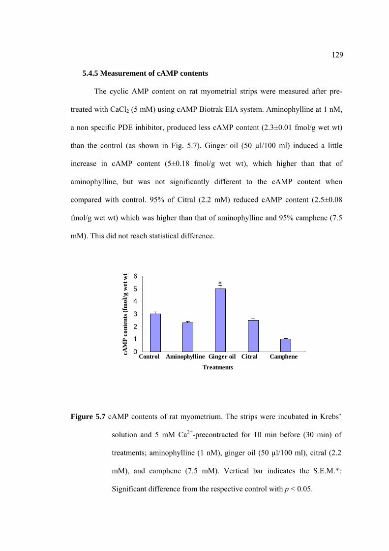
129
5.4.5 Measurement of cAMP contents
The cyclic AMP content on rat myometrial strips were measured after pre-
treated with CaCl2 (5 mM) using cAMP Biotrak EIA system. Aminophylline at 1 nM,
a non specific PDE inhibitor, produced less cAMP content (2.3±0.01 fmol/g wet wt)
than the control (as shown in Fig. 5.7). Ginger oil (50 µl/100 ml) induced a little
increase in cAMP content (5±0.18 fmol/g wet wt), which higher than that of
aminophylline, but was not significantly different to the cAMP content when
compared with control. 95% of Citral (2.2 mM) reduced cAMP content (2.5±0.08
fmol/g wet wt) which was higher than that of aminophylline and 95% camphene (7.5
mM). This did not reach statistical difference.
Figure 5.7 cAMP contents of rat myometrium. The strips were incubated in Krebs’
solution and 5 mM Ca2+-precontracted for 10 min before (30 min) of
treatments; aminophylline (1 nM), ginger oil (50 µl/100 ml), citral (2.2
mM), and camphene (7.5 mM). Vertical bar indicates the S.E.M.*:
Significant difference from the respective control with p < 0.05.
0
1
2
3
4
5
6
Treatments
cAM
P co
nten
ts (f
mol
/g w
et w
t)
AminophyllineControl Ginger oil Citral Camphene
*
130
5.5 Discussion
The aims of this chapter were to examine the inhibitory effects of ginger oil
and its pure compounds on COX pathway. To do so, AA was used in the study to
generate PGs. Ginger oil and its pure compounds were examined and compared with
indomethacin (a specific COX-2 inhibitor). The major measurement of PDE
inhibition was measured using a drug, aminophylline (1nM, a PDE inhibitor). The
cAMP content was examined after the strips were treated with Ca2+ (5 mM CaCl2).
5.5.1 Arachidonic Acid (AA)
The data demonstrated that AA increased myometrial contraction of non-
pregnant rat uteri. Ginger oil (50 µl/100 ml), 95% citral (2.2 mM), and 95%
camphene (7.5 mM) were able to reduce myometrial contraction induced by AA (20
µM). This suggests that ginger oil and its pure compounds may act on a messenger
promoting protein phosphorylation through inhibition of myosin phosphatase enzyme.
It is also possible that its metabolites may involve in Rho-Rho-associated Kinase
pathway.
5.5.2 COX-2 Pathway
The RIA method was used for measuring PGE2 concentration occurred via
COX-2 pathway in the presence of ginger oil (10-5 – 10-3 g/ml), 95% citral (10-5 g/ml),
and 95% camphene (10-5 g/ml). The result clearly demonstrated that the hydro-/-steam
distillation of ginger rhizomes containing particular citral and camphene, the
monoterpenoids: using DMSO solvent, did not inhibit COX-2 enzyme (activity <
50%) of PGE2 synthesis.
131
As described previously ginger hydro-distillated containing either
predominant citral (neral and geranial), curcumene, α-zingiberene, α-farnesne and β-
sesquiphellandrene was identified by GC-MS. The hydro-distillated oil, 95% citral,
and 95% camphene: the high content of ginger oil extracted by steam distillation,
were not capable to inhibiting effect on COX-2 expression. According to the major
constituents of ginger extracts of low concentration of shogaol, had no effect on
COX-2 pathway of PG: synthesis (Lantz et al., 2006). Whereas, the extracts from
ginger rhizomes containing gingerols or shogaols (identified by HPLC, IC50 < 0.1
µg/ml) were highly active at inhibiting LPS-induced PGE2 production (Lantz et al.,
2006). Similarly to the effects of ginger extracts with dichloro-methanol (1:1 v/v) was
able to inhibit LPS induced production of PGE2 and compared with indomethacin at
IC50 (< 0.1 µg/ml) that suggested by Kuo et al. (2004). Additionally, the ginger
extracts have been reported as an anti-inflammatory that it has been suggested by
Altman and Marcussen (2001) for effecting at reduction of the ginger extracts on
symptoms of arthritis in humans.
5.5.3 Indomethacin
The non-steroidal anti-inflammatory drug (NSAID), indomethacin (20µM)
inhibited rat myometrial contraction in vitro. Combination of indomethacin with
ginger oil and its pure compounds on the following were assessed in rat myometrium.
Combined drugs are significantly decreased the contractile response. Similarly, the
great responsibility of indomethacin (10-5 - 10-3 mol/l) administration inhibits the
uterine activity by slow release system (Garza et al., 2004). In addition, indomethacin
was previously reported that it reduced PGE2 production by 82% and 90% at the same
132
concentration (Sawdy et al., 1997). Recently, Sawdy et al. (1998) reported that both
drugs, nimesulide (100 µM) and indomethacin (300 µM), inhibited myometrial
contractility via mechanisms independent of COX inhibition and also blockaded the
Ca2+ current. Additionally, these data agree with several previous reports which
showed that indomethacin although more potent against COX-1, also inhibits COX-2
with micromolar (Mitchell et al., 1993).
In conclusion, the results suggested that the inhibitory effect of indomethacin
combined with ginger oil and its pure compounds on rat myometrial contractile
activity could be due to a dual mechanisms of action; 1) inhibiting uterine
contractility through the Ca2+ channel blocking mechanism and/or 2) prostaglandin
synthesis through inhibition of COX-2 pathway with indomethacin.
5.5.4 PDE Pathway
cAMP plays an important for secondary messenger in regulating muscle
contraction. In smooth muscles, an increase in cAMP stimulated by a β2-adrenoceptor
agonists causes relaxation. There are three machanisms of relaxation; 1) activation
ATPase pump on SR membrane, 2) inhibition of Ca2+ slow channels, and 3) inhibition
of MLCK (Adelstein et al., 1978; 1982). These are similar to the nature of ralaxing
effect attributed to several mechanisms leading to a decrease in both the cytoplasmic
free Ca2+ concentration and the Ca2+ sensitivity of the contractile activity (Savineau
and Mironneau, 1990). The inhibitory effects of antagonists on adenosine receptor of
PDE enzyme in smooth muscle depressed lower cellular cAMP levels (Laifer et al.,
1986; Wolffada et al., 1981).
133
Several investigations have reported that cAMP inhibited Ca2+-induced
contraction of smooth muscles. This is due to the activation of cAMP-dependent
protein kinase, producing phosphorylation of the MLCK. It results in an inhibition of
actin-myosin interaction (Kerrick and Hoar, 1981; Ruegg et al., 1981; Ruegg and
Paul, 1982). However, the inhinitory effect of cAMP was demonstrated under special
conditions using high concentrations of cAMP (> 10-4 M) by the use of protein kinase.
In contrast, the cAMP (3 µM) had no effect on the Ca2+-induced contraction in the
smooth muscle and in the presence of exogenous protein kinase. Since this problem is
very important for understanding the Ca2+ regulation in smooth muscle, further
investigations are requirement for studies.
In summary, the major aim of this Chapter was to examine whether the
inhibitory effects of ginger oil and its pure compounds on PDE enzyme (the regulator
of cAMP production). To detect cAMP content in the treatment of aminophylline,
ginger oil, citral, and camphene expressed to 2.3, 5, 2.5, and 1 (fmol/g wet wt,
respectively) compared with the control (3 fmol/g wet wt). These results showed
inhibitory effects of ginger oil and its pure compounds on rat myometrial contraction
may be not mediated by cAMP.
5.6 References
Adelstein, R. S., Conti, M. A., Hathaway, D. R. and Klee, C. B. (1978).
Phosphorylation of smooth muscle myosin light chain kinase by the catalytic
subunit of adenosine 3´, 5´–monophosphate-dependent protein kinase.
Journal of Chemistry. 253 : 8347 – 8350.

134
Adelstein, R. S., Pato, M. D. Seller, J. R., de Lanerolle, P. and Conti, M. A. (1982).
Regulation of actin-myosin interaction by reversible phosphorylation of
myosin and myosin kinase. Cold Spring Harbor Symposium. Quanternary
Biology. 106 : 921 – 928.
Ahn, H. S., Crim, W., Pitts, B. and Sybertz, E. J. (1992). Calcium-calmodulin-
stimulated and cyclic-GMP-specific phosphodiesterases. Tissue distribution,
drug sensitivity, and regulation of cyclic GMP levels. Advance of Secondary
Messenger Phosphoprotein Research. 25 : 271 – 288.
Altman, R. D. and Marcussen, K. C. (2001). Effects of a ginger extract on knee pain
in patients with osteoarthritis, Arthritis Rheumatism. 44 : 2531 – 2538.
Apaydin, S., Gonen, C. and Guven, H. (1998). The probable role of nitric oxide on the
relaxations obtained by caffeine and aminophylline in rat uterus.
Pharmacological Research. 38 : 387 – 392.
Axelrod, J. (1990). Receptor-mediated activation of phospholipase A2 and
arachidonic acid release in signal transduction. Biochemistry Socciety
Transactions. 18 : 504 – 507.
Barnette, M. S., Manning, C. D., Price, W. J. and Barone, F. B. (1993).Initial
biochemical and functional characterization of cyclic nucleotide
phosphodiesterase isozymes in canie colonic smooth muscle. Journal
Pharmacological Experimental Therapy. 264 : 801 – 812.
Bazan, A. D., Standish, M. M., Watkins, J. C. (1995). Diffusion of univalent ions
across the lamellae of swollen phospholipids. Journal of Molecular Biology.
13 : 138 – 252.
135
Bevilacqua, M. and Magni, E. (1993). Recent contributions to knowledge of the
mechanism of action of nimesulide. Drugs. 46 : 40 – 47.
Bjarnason, I., et al. (1986). Effect of non-steroidal anti-inflammatory drugs and
prostaglandins on the permeability of the human small intestine. Gut. 27 :
1292 – 1297.
Bruch, R. M., Wise, W. C. and Halushka, P. V. (1983). Prostaglandin- independent
inhibition of calcium transport by nonosteroidal anti- inflammatory drugs:
Differntial effects of carboxylic acids and piroxicam. Journal of
Pharmacology of Experimental Therapeutics. 227 : 48 – 91.
Bulbring, E. and den Hertog, A. (1980). The action of isoprenaline on the smooth
muscle of the guinea-pig taenia coli. Journal of Physilogy. 304 : 277 – 296.
Cahill, M. (ed.). (1986). Signs and symptoms. Springhouse, PA: Springhouse.
Chang, C. –P., et al. (1995). The effect of Chinese medicinal herb Zingiberis
rhizoma extract on cytodine secretion by hyman peripheral blood
monomuclear cells. Journal of Ethnopharmacology. 48 : 13 – 19.
Chakraborty, I., Das, S. K. Wang, J. and Dey, S. K. (1996). Developmental
expression of the cyclo-oxygenase-1 and cyclooxygenases in the peri-
implantation mouse uterus and thir differenial regulation by the blastocyst and
ovarian steroids. Journal of Molecular Endocrinology. 16 : 107 – 112.
Coetzi, E. J., An, S. and Smith, W. L. (1995). Specificity of expression of eicosanoid
mediation in normal physiology and human diseases. The Federation of
American Soceities for experimental Biology. 9 : 1051 – 1058.

136
Conti, M. Nemoz, G. Sette, C. and et Vicini, E. (1995). Recent progress in
understanding the hormonal regulation of phosphodiesteraes. Endocrine
Review. 16 : 370 – 389.
Copeland, R. A. et al. (1994). Mechanism of selective inhibition of the inducible
isoform of protaglandin G/H synthase. Proceeding of the United National
Science Academy. 91 : 11202 – 112026.
Cromlish, W. A., et al. (1994). High-level expression of active human
cyclooxygenase-2 in insect cells. Arch Biochemistry and Biophysics. 314 :
193 – 199.
De Witt, D. L. (1991). Prostaglandin endoperoxide synthase: regulation of Enzyme
expression. Biochemistry Biophysics Acta. 1083 : 121 – 134.
Filipeanu, C. M., Brailoiu, E., Petrescu, G. and Nelemans, S. A. (1998). Extracellular
and intracellular arachidonic acid-induced contractions in rat aorta. European
Journal of Pharmacology. 349 : 67 – 73.
Futaki, N., et al. (1994). NS-398, a new anti-inflammatory agent, selectively inhibits
prostaglandind G/H synthase/cyclooxygenase (COX-2) activity in vitro.
Prostaglandins. 47 : 55 – 59.
Garza, J., et al. (2004). In situ inhibition of uterine activity by indomethacin: Possible
relevance to preterm labor after fetal surgery. Journal of Pediatric Surgery.
39 (8) : 1173 – 1175.
Gong, M. C., et al. (1992). Arachidonic acid inhibits myosin light chain Phosphatase
and sentitizes amooth muscle to Ca2+. Journal Biology Chemistry. 267 :
21492 – 21498.
137
His, L. C., Hoganson, C. W., Babcock, G. T. and Smith, W. L. (1994).
Characterization of a tyrosyl radical in prostaglandin endoperoxide synthase-2.
Biochemistry Biophysical Research Communication. 202 : 1592 – 1598.
Hoffman, L. H. (1978). Antifertility effects of indomethacin during early Pregnancy
in rabbit. Biology Reproduction. 18 : 148 – 155.
Kaneda, T., et al. (2005). Inhibitory mechanism of papaverine on carbachol- Induced
contraction in bovine trachea. Journal of pharmacology. 98 : 275 – 282.
Khan, W. A., Blobe, C. G., Hannun, Y. A. (1995). Arachidonic acid and free fatty
acids as second messengers and the role of protein kinase C. Cell Signaling. 7
: 171 – 184.
Katsura, K., et al. (1993). Coupling among energy failure, loss of ion homeostasis,
and phospholipase A2 and C activation during ischemia. Journal of
Neurochemistry. 61 : 1677 – 1684.
Kennedy,T. G. (1977). Evidence for a role for prostaglandins in the initiation of
blastocyst implantation in the rat. Biology Reproduction. 16 : 286 – 291.
Kerrick, W. G. and Hoar, F. E. (1981). Inhibition of smooth muscle tension by cyclic
AMP-dependent protein kinase. Nature. 292 : 253 – 255.
Kirtikara, K., et al. (1998). Compensatory Prostaglandin E2 Biosynthesis in
Cyclooxygenase 1 or 2 null cells. Journal Experimental Medicine. 187 : 517
– 523.
Kirtikara, K., Swangkul, S. and Ballon, L. R. (2001). The analysis of nonsteroidal
anti-infammatory drug selectivity in prostaglandin G/H synthase (PGHS)-null
cells. Inflammatory Research. 50 : 327 – 332.
Kuby, J. (1997). Immunology. pp 357 – 376. W. H. New York: Freeman.
138
Kuo, C. L., et al. (2004). The effects of berberine on COX-1 and COX-2 enzyme
activities as expressed by PGE2 synthesis. Cancer Letters. 203 : 127 – 137.
Laifer, S. A. Ghodgaongar, R. B. Zaeur, H. A. and Dubin, N. H. (1986). The effect of
aminopylline on uterine smooth muscle contractility and prostaglandin
production in the pregnant rat uterus in vitro. American Journal of Obsterict
Gynecology. 155 (1) : 212 – 215.
Lantz, R. C., et al. (2006). The effect of extracts from ginger rhizome on
inflammatory mediator production. Phytomedicine. 14 (2-3) : 123 – 128.
Lester, D. S., Collin, C., Etcheberrigaray, R., Alkon, D. L. (1991). Arachidonic acid
and diacylglycerol act synergistically to activate protein kinase C in vitro and
in vivo. Biochem. Biophys. Research Community. 179 : 1522 – 1528.
Lim, M., et al. (1997). Multiple female reproductive failures in cyclooxygenase-2
defecient mice. Cell. 91 : 197 – 208.
Mann, J. R. and DuBios, R. N. (2004). Cyclooxygenase-2 and gastrointestinal cancer.
Cancer Journal. 10 : 145 – 152.
Meade, E. A., Smith, W. L. and De Witt, D. L. (1993). Differential inhibition of
prostaglandin endoperoxide synthaes (cyclooxygenase) isozumes by aspirin
and other non-steroidal anti-inflammatory drugs. Journal of Biological
Chemistry. 268 : 6610 – 6614.
Meisheri, K. D. and van Breemen, C. (1982). Effects of β-adrenergic stimulation on
calcium movements in rabbit aortic smooth muscle: relationship with cyclic
AMP. Journal of Physiology (London). 331 : 429 – 441.
Mitchell, J. A., Akarasereenont, P., Thienermann, C., Flower, R. J. and Vane, J. R.
(1993). Selectivity of nonsteroidal antiinflammatory drugs as inhibitors of
139
constitutive and inducible cyclooxygenase. Proceeding of the United
National Science Academy. 90 : 11693 – 11697.
Mustafa, T., Srivastava, K. C., Jensen, K. B. (1993). Drug development report:
pharmacology of ginger, Zingiber officinale, Journal of Drug Development.
6 : 25 – 39.
O’Banion, M. K., Winn, V. D. and Yong, D. A. (1992). Cloning and functional
activity of a glucocorticoid-regulated inflammatory cyclooxygenase.
Proceeding of the United National Science Academy.. 89 : 4888 – 4889.
Oe, H., et al. (1994). Calcium overload and cardiac myocyte cell damage by
arachidonate lipoxygenation. American Journal Physiology. 267 : H1396 –
1402.
O’Neill, G. P., et al. (1994). Overexpression of hyman prostaglandin G/H synthase-1
and -2 by recombinant vaccinia virus: inhibition by nonsteroidal anti-
inflammatory drugs and biosyhtesis of 15-hydroxyeicosatetraenoic acid.
Molecular Pharmacology. 45 : 245 – 254.
Ramsay, R. G., et al. (2003). Transcriptional regulation of cyclooxygenase
expression: three pillars of control. Journal 0f International
Immunopathology and Pharmacology. 16 : 59 – 67.
Ruegg, J. C., Sparrow, M. P. and Mrwa, U. (1981). Cyclic: AMP mediated relaxation
of chemically skinned fibers of smooth muscle. Pfûgers Archive Europain
Journal of Physiology. 390 : 198 – 201.
Ruegg, J. C. and Paul, R. J. (1982). Vascular smooth muscle: calmodulin and cyclic
AMP dependent protein kinase alter calcium sensitivity in porcine carotid
skinned fibers. Circulatory Research. 50 : 394 – 399.
140
Savineau, J. P. and Mironneau, J. (1990). Caffeine acting on pregnant rat
myometrium: analysis of its relaxant action and its failure to release Ca2+ from
intracellular stors. British Journal of Pharmacology. 99 : 261 - 266.
Sawdy, R., Slater, D., Jones, G., Poston, L. and Bennett, P. R. (1997). Potential of the
cyclooxygenase type-2 selective inhibitor nimesulide in prevention of preterm
labour. Journal of Soc. Gynecology. 172 : 77 – 82.
Sawdy, R., et al. (1998). Effect of nimesulide and indomethacin on contractility and
the Ca2+ channel current in myometrial smooth muscle from pregnant women.
Britidh Journal of Pharmacology. 125 : 1212 – 1217.
Scheid, C. R., Honeyman, T. W. and Fay, F. S. (1979). Mechanism of β- adrenergic
relaxation of smooth muscle. Nature (London). 277 : 32 – 36.
Seibert, K., et al. (1994). Pharmacological and biochemical demonstration of the role
of cyclooxygenase 2 in inflammation and pain. Proc Natl Acad Sci USA. 91 :
12013 – 12017.
Shimizu, K., et al. (2000). Mechanism of relaxant response to papaverine on the
smooth muscle of non-pregnant rat uterus. Journal of Muscle Research. 36 :
83 – 91.
Simmons, D. L., Botting, R. M. and Hla, T. (2004 ). Cyclooxygenase isozymes: the
biology of prostaglandin synthesis and inhibition. Pharmacological Review.
56. 387 – 437.
Smith, W. L. and Marnett, L. J. (1991). Prostaglandin endoperoxide synthase:
structure and catalysis. Biochem Biophysiology Act. 1083 : 1 – 17.
Srivastava, K. C. (1984). Effects of aqueous extracts of onion, garlic and ginger on
platelet aggregation and metabolism of arachidonic acid in the blood vascular
141
system: in vitro study. Prostaglandins Leukotrienes and Medicine. 13 : 227
– 235.
Srivastava, K. C. and Mustafa, T. (1989). Ginger (Zingiber officinale) and rhematic
disorders. Medical Hypotheses. 29 : 25 – 28.
Srivastava, K. C. and Mustafa, T. (1992). Ginger (Zingiber officinale) in rheumatism
and musculoskeletal disorders. Medicine Hypothesis. 39 : 342 – 348.
Taymor, M. L., Sturgis, S. H.and Yahia, C. (1964). The etiological role of chronic
iron deficiency in production of menorrhagia. The Journal of American
Medical Association. 187 : 323 – 327.
Tjendraputra, E., Tran, V. H., Liu-Brennan, D., Roufogalis, B. D. and Duke, C. C.
(2001). Effect of ginger constituents and synthetic analogues on
cyclooxygenase-2 enzyme in intact cells. Bioorganic Chemistry. 29 : 156 –
163.
Trautman, M. S., et al. (1996). Prostaglandin 2 synthase-2 in human gestational
tissues: regulation in amnion. Placenta. 17 : 239 – 245.
Vane, J. R., Bakhle, Y. S. and Botting, R. M. (1988). Cyclooxygenase 1 and 2.
Research Pharmacology Toxicology. 38 : 97 – 120.
Vermillion, S. T. and Landen, C. N. (2001). Prostaglandin inhibitors as tocolytic
arents. Seminars in Perinatology. 25 : 256 – 262.
Wolffada, J., Londos, C. and Westfall, D. P. (1981). Adenosine receptors and the
regulation of cyclase. Cyclic Nucleotide Research. 14 : 199.
CHAPTER VI
EFFECTS OF GINGER OIL, CITRAL, AND CAMPHENE
ON RAT MYOMETRIAL HISTOLOGICAL CHANGES
6.1 Abstract
As shown in previous Chapters, ginger oil and its pure compounds altered
uterine contraction, irrespective how it was produced. To assure that the inhibitory
effects of ginger oil and its pure compounds did not occur by damaging of myometrial
tissue, the aim of this Chapter was therefore to investigate the effect of ginger oil and
its pure compounds on uterine histology.
To do so non-pregnant Wistar rats (200 – 250 g) were humanely killed by
cervical dislocation. Myometrial tissues were removed from the rat uteri and
immediately immersed in buffered physiological Krebs’ solution as a control and
others in Krebs’ solution containing ginger oil (50 µl/100 ml), 95% citral or 95%
camphene, respectively. The tissues were incubated for 30 min at room temperature.
These strips were then immediately fixed in 10% formaldehyde and stained to routine
histological techniques for light microscopic (LM) and transmission electron
microscopic (TEM) studies. The results showed that ginger oil and its pure
compounds did not affect myometrial histology in terms of smooth muscle cells,
density of intracellular organelles, and subcellular distribution of myofilaments.
However, the two pure compounds seemed to have little effect on uterine histology. It
was found that few cells of myometrial tissues had slight changes in the density of
143
extracellular myofilaments, but they exhibited considerable enveloping cellular
organelles as sarcoplamic reticulums (SRs) or intracellular myofilaments. Thus,
ginger oil and its pure compounds may not affect on myometrial morphological
changes as detected with LM and TEM.
6.2 Introduction
As described in Chapter I, most of the uterine wall is composed of myometrial
smooth muscle cells, known as myometrial layer. The presence or absence of
myometrial lesion may reveal morphological signs of smooth muscle cell changes and
can be observed using LM and TEM. The ultrastructural characteristics of the smooth
cells, especially the subcellular distribution of, and relationship between, the
myofilaments and cellular organelles are of prime interest. The spatial distribution of
the SR, the main intracellular store thought to provide Ca2+ for myofilament
activation in smooth muscle cells, is particular importance relation to contractile
properties of the cell (Sweeney et al., 2005; Taggart and Morgan, 2007). The muscles
are composed of many subunits known as myofibrils, which can be observed under
the electron microscopy (EM). These myofibrils are approximately 1 micrometer
(1µm) in diameter and extend in parallel rows from one end of the other organells,
such as mitochondria and intracellular membranes, are restricted to the narrow
cytoplasmic spaces that remain between adjacent myofibrils. Each myofibril contains
even smaller structures called myofilaments, or simply filaments. The thick filaments
are primarily composed of the protein myosin, and the thin filaments are primarily
composed of the protein actin.

144
Ginger is pharmacologically safe regarding the investigated aspects (Weidner
and Sigwart, 2000). Chinese ginger extract and major pungent principles have been
shown to exhibit a verity of biological activities. They found that five new
diarylheptanoids and gingerol-related compounds possess significant cytotoxicity
against human promyelocytic leukemia (HL-60) cells. An in vitro study on the
cytotoxic and apoptotic activities of these compounds is associated with the cell
apoptosis (Wei et al., 2005). They suggested that the inhibitions of ginger-related
compounds had also possessed of activator protein 1 (Lee and Surh, 1998; Surh and
Lee, 1998; Bode et al., 2001). Furthermore, the pungent, [6]-paradol and its synthetics
[10]-paradol, [3]-dehydroparadol, [6]-dehydroparadol and [10]-dehydroparadol, could
induce apoptosis of an oral squamous carcinoma KB cells (Keum et al., 2002). In
addition, super-critical CO2 extracts of ginger against human cancer cell lines (Leal et
al., 2003) and antioxidant activity (Tjendraputra et al., 2001). Indeed, most ginger
constituents with biological activities posses such a functionality (Surh et al., 1998;
Keum et al., 2002; Kim and Kim, 2004).
As mentioned in Chapter I, there are two main organelles related with
myometrial contractility, the myofilament and the SR. Myosin, the thick filament
consists of MLC that it involve in the regulatory of Ca2+-Ca MLCK phosphorylation
and response to diverse contractile stimuli. The thin filament, actin can activate
myosin ATPase activity in Ca2+-dependent manner indicating an alternative pathway
to MLC20 phosphorylation. SR is abundant in uterine smooth muscle cells. Although
the major source of activator Ca2+ in the myometrium is extracellular fluid, SR is a
minor one of its role in excitation-contraction coupling in smooth muscle (Peachey
and Porter, 1959; Shmygol et al., 1998; Wray et al., 2001). In order to assess the
145
morphological changed in the biopsy of the same piece using TEM and LM were used
in subsequent procedure of the study (Romanini, 1994). Ultrastucturally, the
sarcoplasm of the myometrium is dominated by dense longitudinally aligned
contractile filaments containing the contractile proteins, actin and myosin (Romanini,
1994). Structural integrity of the myometium is thus fundamental to normal uterine
contractions. Normal myometria, fibromyomata and host myometria are all similar in
that do not undergo ultrastructural change during the normal menstrual cycle.
Estrogen and progesterone may induce some structural change in the myometrium,
during the pre-and post-ovulated phases respectively (Friederici and Decloux, 1968).
In contrast, the structural abnormality of the plasmalemmal dense bands that occurs in
the presence of fibromyomata may cause to cell disruption. This affected in the
normal calcium cycling of the effected myocytes resulting in a disruption of the
rhythmical contraction process (Richard et al., 1998). Further suggests that the normal
contractile mechanisms and patterns may be disrupted in the presence of myometial
irritability (Richard et al., 1998).
However, the effect of ginger oil and its pure compounds on myometrial
histological changes has not been elucidated. It is unclear whether the inhibitory
effects of ginger oil and pure compounds as shown in previous Chapters occurred by
tissue damaging or by physiological mechanisms. The aim of this study was to
examine the effects of these treatments on rat myometrial smooth muscle cell
histological changes using LM and TEM techniques.
146
6.3 Materials and Methods
Whole uterus from Wistar rats (as described in Chapter II) were washed with
normal saline and incubated in Krebs’ solution (control group), Krebs’ containing
ginger oil (50 µl/100 ml), citral (2.2 mM) or camphene (7.5 mM) at room temperature
for 30 min and then fixed in 10% formalin until analyzed. This part of work was
conducted at Department of Biology, Faculty of Science, Mahasarakham University,
Thailand.
6.3.1 LM Staining Preparations
The horns of rat uteri were cut into short segmenta. These strips were
dehydrated in an ascending ethanol series (75%, 85%, 95%, 100% and 100%, 1 hr
each) and then removed the sections into pure xylene for 2 min. The sections were
embedded with xylene : paraplast (3 : 1 and 1 : 1, 15 min each) and then followed by
the pure paraplast for 1 hr. Post-embedded tissues were cut approximately 5 µm with
microtome and moved these sections into water bath for incubation (60°C). The
tissues were mounted on slides in the slide warmer at 60°C and then twice immersed
the slides in pure xylene (5 and 2 min, respectively). Next, tissues were hydrated in a
descending ethanol series (100%, 95%, 70%, 50%, 30%, distill water, 2 min each)
and stained with heamatoxylin according to standard protocols. All tissues were
dehydrated in a descending ethanol series (35%, 50%, 70%, 95%, 2 min each) and
stained with eosin for 1 min. Finally, the slides were immersed into 95% ethanol (2-3
min) and 100% ethanol (1 min), pure xylene (5 min) and then covered with cover slip
after xylene clearing for LM study.
147
6.3.2 TEM Staining Preparations
The horns of rat uteri were cut into short segmenta. These strips were fixed in
2.5% glutaraldehyde in 0.1 M phosphate buffer, pH 7.2 overnight at 4°C then
washed in the same buffer 3 times. Post-fixed with 1% osmium tetroxide for 2 hr then
rinsed in distill water 3 times, Gold coated uterus on stubs were examined for TEM,
after post-fixing the samples were dehydrated in graded acetone at concentrations of
20%, 40%, 60%, 80%, 100% and 100%, then infiltrated and embedded in Spur’s
resin. Upon identifying suitable areas from the semithin sections, ultrathin sections
(<100 nm thick) of mainly transverse and longitudinal orientation to the uterine
tissues were cut with diamond knife, mounted on copper grids then stained with lead
citrate and uranyl acetate. The ultrathin sections on copper grids were examined with
TEM (JEM 1230) at Central Instrumentation Unit, Faculty of Science, Mahasarakham
University (Phungnoi and Narkkong, 2007).
6.4 Results
6.4.1 LM Observation and Uterine Morphological Feature
The rat uteri were stained (with heamatoxylin, eosin) and examined by LM
(Fig. 6.1), the only myometrial layer is presented at a higher magnification in Fig. 6.2
– 6.4. It can be seen that the uterus is a thick-walled organ, whose wall consists of
three layers; 1) The external serosa (S) is unremarkable and is not damaged by the
effects of ginger oil and its pure compounds (Fig. 6.1), 2) The thick myometrium (M)
is composed of smooth muscle, subdivided into three poorly delineated layers (outer
longitudinal, middle circular and inner longitudinal) and will be described in 6.4.1.1,
148
3). The endometrium (E) is subdivided into a basal layer and a functional layer as
shown in 6.4.1.2.
6.4.1.1 The Thick Myometrium
The basic functional unit of the myometrium is the smooth muscle cell, but
these in turn are arranged into bundles of connective tissue interspersed with
microvasculature (Wetzstein and Renn, 1970; Word et al., 1993). The thin outer and
inner layers are mostly longitudinal or oblique fibers. As seen in Figure 6.1B the
tissue treated with ginger oil (50 µl/100 ml) is depicted to dominate by dense
longitudinally aligned myofilaments as normal tissue (Fig. 6.1A). Observation of
section-treated with 95% citral (2.2 mM, Fig. 6.1 C) contained mildly myofilaments
and loose connective tissue under light microscope. Structural integrity of the
myometrium-treated with 95% camphene (7.5 mM) is slightly appeared longitudinal
myofilaments (Fig. 6.1D).
As described above in Figure 6.1B, the tissue treated with ginger oil (50
µl/100 ml) is densely packed with cells and its illustrated similarly to normal
untreated tissue (Fig. 6.1A). The epithelial lining is clearly evident, composed of
different cell features, a thin peg cell, which bears no cilia. The photograph (Fig.
6.1C) is lower to show the cellular epithelially lined loose connective tissue, but
higher dense cellular epithelium is shown in Fig. 6.2D.
6.4.1.2 The Endometrium
The endometrium is an inner layer defined with the light micoscopy. The
endometrial surface consists of non-ciliated epithelium. The sections having taken

149
during diestrous stage are separated into a deeper basal and a more superficial
functional layer, each with its own blood supply. The basal layer, which remains
intact during estrous phase, is served by short straight arteries and is occupied by the
base of uterine glands. The functional layer served by the arteries, undergoes
hormonally modulated cycle changes (Gartner and Hiatt, 2000). The breakdown of
the functionalism produces the clinical presentation of the menstrual cycle. Which is a
precipitated by luteolysis of the corpus luteum, with drawal of hormonal support, and
changes in the vascular supply of the endometrium (Kerr, 1999).
Figure 6.1B, the tissue treated with ginger oil (50 µl/100 ml) is a glandular
epithelia layer and located by the simple columnar epithelial cells. Fig. 6.1C, the
tissue treated with 95% citral (2.2 mM) is a single fibrotic gland with one layer under
columnar epithelial cells. Figure 6.1D, the tissue treated with 95% camphene (7.5
mM) is luminal and glandular epithelial tissues to compare with control (untreated,
Fig. 6.1A).
6.4.2 Ultrastructeral Observation and Myometrial Morphological Feature
The ultrathin sections were successfully obtained, involving the structures that
the myometrail cells are illustrated by TEM analysis (Fig. 6.2 – 6.4). Ultrastructurally,
smooth muscle cells are surrounded by an external lamina (EL) and have placed
centrally or perinuclear (N) containing myofilaments. As seen in Figure 6.2A, the
normal cell in the Krebs’ solution (untreated) is shown by an atypical accumulation of
intermediate filaments and perinuclear strips. According to the ultrathin section (in
Fig. 6.2B) treated with ginger oil (50 µl/100 ml) contained by the centrally placed
nuclei. In contrast to the normal cell, the ultrathin section treated with 95% citral (2.2
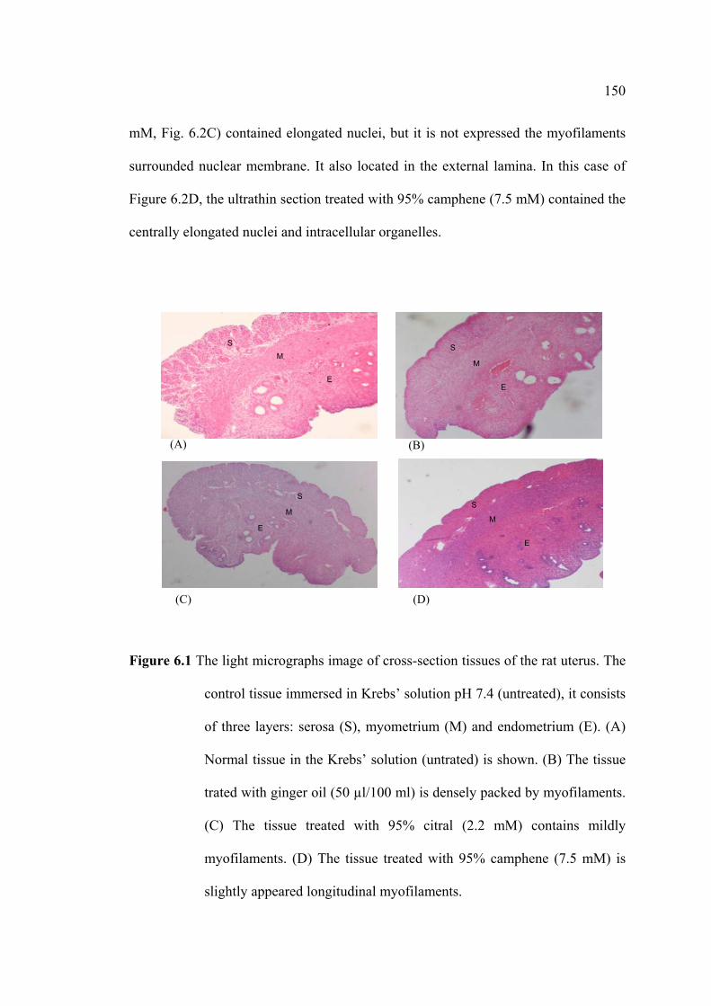
150
mM, Fig. 6.2C) contained elongated nuclei, but it is not expressed the myofilaments
surrounded nuclear membrane. It also located in the external lamina. In this case of
Figure 6.2D, the ultrathin section treated with 95% camphene (7.5 mM) contained the
centrally elongated nuclei and intracellular organelles.
Figure 6.1 The light micrographs image of cross-section tissues of the rat uterus. The
control tissue immersed in Krebs’ solution pH 7.4 (untreated), it consists
of three layers: serosa (S), myometrium (M) and endometrium (E). (A)
Normal tissue in the Krebs’ solution (untrated) is shown. (B) The tissue
trated with ginger oil (50 µl/100 ml) is densely packed by myofilaments.
(C) The tissue treated with 95% citral (2.2 mM) contains mildly
myofilaments. (D) The tissue treated with 95% camphene (7.5 mM) is
slightly appeared longitudinal myofilaments.
E
M S S
E
M
S M
E
(A) (B)
(C) (D)
S M
E
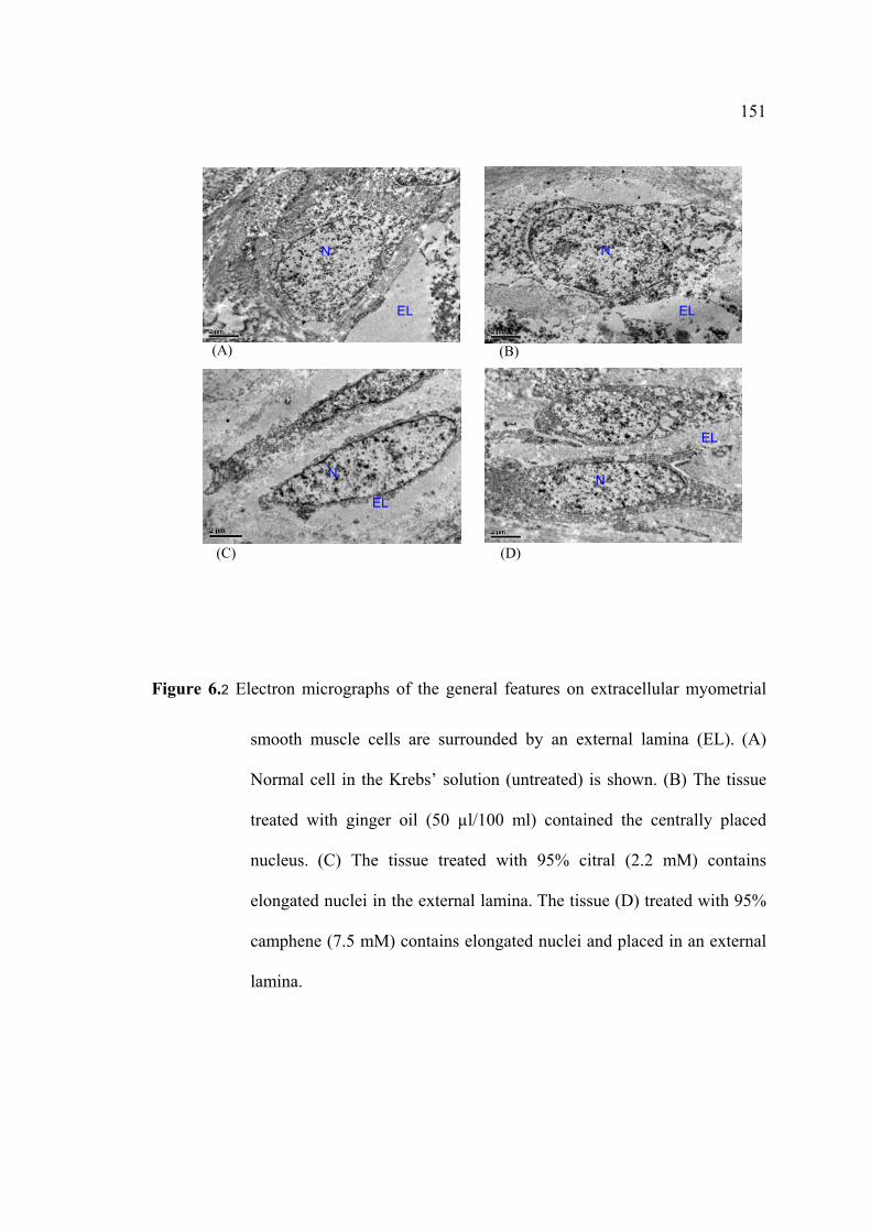
151
Figure 6.2 Electron micrographs of the general features on extracellular myometrial
smooth muscle cells are surrounded by an external lamina (EL). (A)
Normal cell in the Krebs’ solution (untreated) is shown. (B) The tissue
treated with ginger oil (50 µl/100 ml) contained the centrally placed
nucleus. (C) The tissue treated with 95% citral (2.2 mM) contains
elongated nuclei in the external lamina. The tissue (D) treated with 95%
camphene (7.5 mM) contains elongated nuclei and placed in an external
lamina.
(A) (B)
(C) (D)
N
N N
EL
EL
EL
N EL
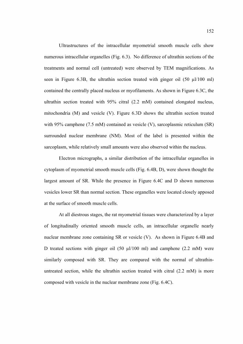
152
Ultrastructures of the intracellular myometrial smooth muscle cells show
numerous intracellular organelles (Fig. 6.3). No difference of ultrathin sections of the
treatments and normal cell (untreated) were observed by TEM magnifications. As
seen in Figure 6.3B, the ultrathin section treated with ginger oil (50 µl/100 ml)
contained the centrally placed nucleus or myofilaments. As shown in Figure 6.3C, the
ultrathin section treated with 95% citral (2.2 mM) contained elongated nucleus,
mitochondria (M) and vesicle (V). Figure 6.3D shows the ultrathin section treated
with 95% camphene (7.5 mM) contained as vesicle (V), sarcoplasmic reticulum (SR)
surrounded nuclear membrane (NM). Most of the label is presented within the
sarcoplasm, while relatively small amounts were also observed within the nucleus.
Electron micrographs, a similar distribution of the intracellular organelles in
cytoplasm of myometrial smooth muscle cells (Fig. 6.4B, D), were shown thought the
largest amount of SR. While the presence in Figure 6.4C and D shown numerous
vesicles lower SR than normal section. These organelles were located closely apposed
at the surface of smooth muscle cells.
At all diestrous stages, the rat myometrial tissues were characterized by a layer
of longitudinally oriented smooth muscle cells, an intracellular organelle nearly
nuclear membrane zone containing SR or vesicle (V). As shown in Figure 6.4B and
D treated sections with ginger oil (50 µl/100 ml) and camphene (2.2 mM) were
similarly composed with SR. They are compared with the normal of ultrathin-
untreated section, while the ultrathin section treated with citral (2.2 mM) is more
composed with vesicle in the nuclear membrane zone (Fig. 6.4C).
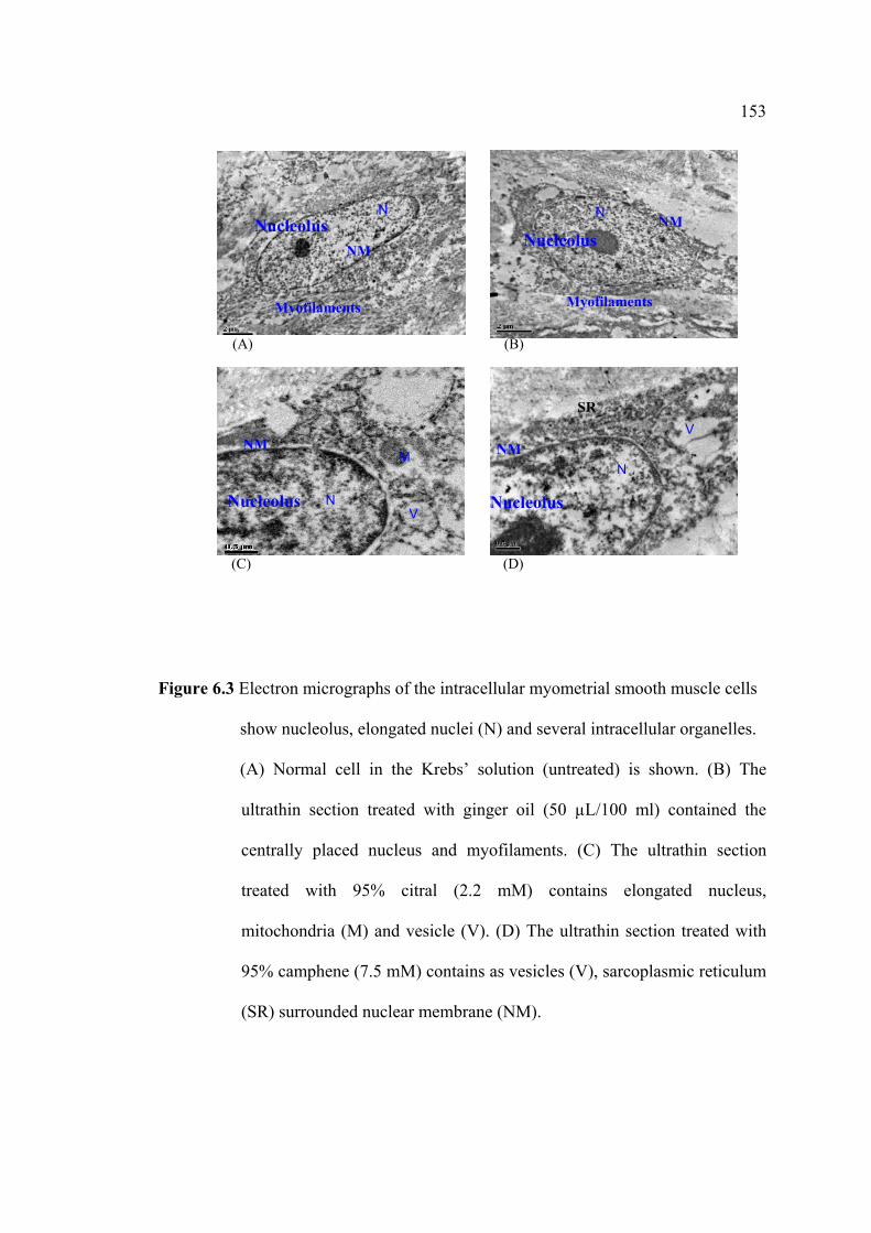
153
Figure 6.3 Electron micrographs of the intracellular myometrial smooth muscle cells
show nucleolus, elongated nuclei (N) and several intracellular organelles.
(A) Normal cell in the Krebs’ solution (untreated) is shown. (B) The
ultrathin section treated with ginger oil (50 µL/100 ml) contained the
centrally placed nucleus and myofilaments. (C) The ultrathin section
treated with 95% citral (2.2 mM) contains elongated nucleus,
mitochondria (M) and vesicle (V). (D) The ultrathin section treated with
95% camphene (7.5 mM) contains as vesicles (V), sarcoplasmic reticulum
(SR) surrounded nuclear membrane (NM).
Nucleolus
Nucleolus
Nucleolus
Nucleolus
N
(A) (B)
(C) (D)
N N
N M
Myofilaments
V
V
Myofilaments
SR
NM
NM
NM
N
NM
Nucleolus
Nucleolus
Nucleolus
Nucleolus
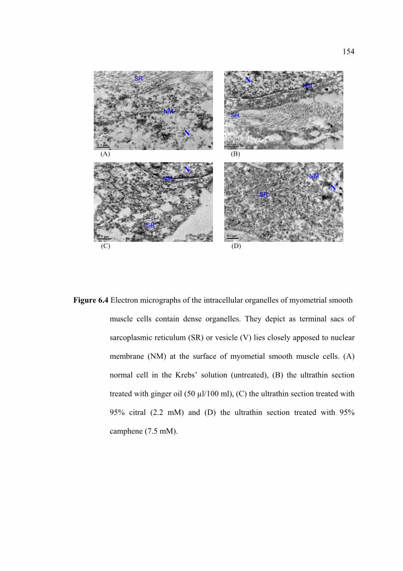
154
Figure 6.4 Electron micrographs of the intracellular organelles of myometrial smooth
muscle cells contain dense organelles. They depict as terminal sacs of
sarcoplasmic reticulum (SR) or vesicle (V) lies closely apposed to nuclear
membrane (NM) at the surface of myometial smooth muscle cells. (A)
normal cell in the Krebs’ solution (untreated), (B) the ultrathin section
treated with ginger oil (50 µl/100 ml), (C) the ultrathin section treated with
95% citral (2.2 mM) and (D) the ultrathin section treated with 95%
camphene (7.5 mM).
V
(A) (B)
(C) (D)
SR
SR
SR
SR
NM NM
NM
NM N
N
N
N
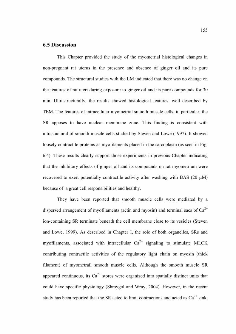
155
6.5 Discussion
This Chapter provided the study of the myometrial histological changes in
non-pregnant rat uterus in the presence and absence of ginger oil and its pure
compounds. The structural studies with the LM indicated that there was no change on
the features of rat uteri during exposure to ginger oil and its pure compounds for 30
min. Ultrastructurally, the results showed histological features, well described by
TEM. The features of intracellular myometrial smooth muscle cells, in particular, the
SR apposes to have nuclear membrane zone. This finding is consistent with
ultrastuctural of smooth muscle cells studied by Steven and Lowe (1997). It showed
loosely contractile proteins as myofilaments placed in the sarcoplasm (as seen in Fig.
6.4). These results clearly support those experiments in previous Chapter indicating
that the inhibitory effects of ginger oil and its compounds on rat myometrium were
recovered to exert potentially contractile activity after washing with BAS (20 µM)
because of a great cell responsibilities and healthy.
They have been reported that smooth muscle cells were mediated by a
dispersed arrangement of myofilaments (actin and myosin) and terminal sacs of Ca2+
ion-containing SR terminate beneath the cell membrane close to its vesicles (Steven
and Lowe, 1999). As described in Chapter I, the role of both organelles, SRs and
myofilaments, associated with intracellular Ca2+ signaling to stimulate MLCK
contributing contractile activities of the regulatory light chain on myosin (thick
filament) of myometrail smooth muscle cells. Although the smooth muscle SR
appeared continuous, its Ca2+ stores were organized into spatially distinct units that
could have specific physiology (Shmygol and Wray, 2004). However, in the recent
study has been reported that the SR acted to limit contractions and acted as Ca2+ sink,
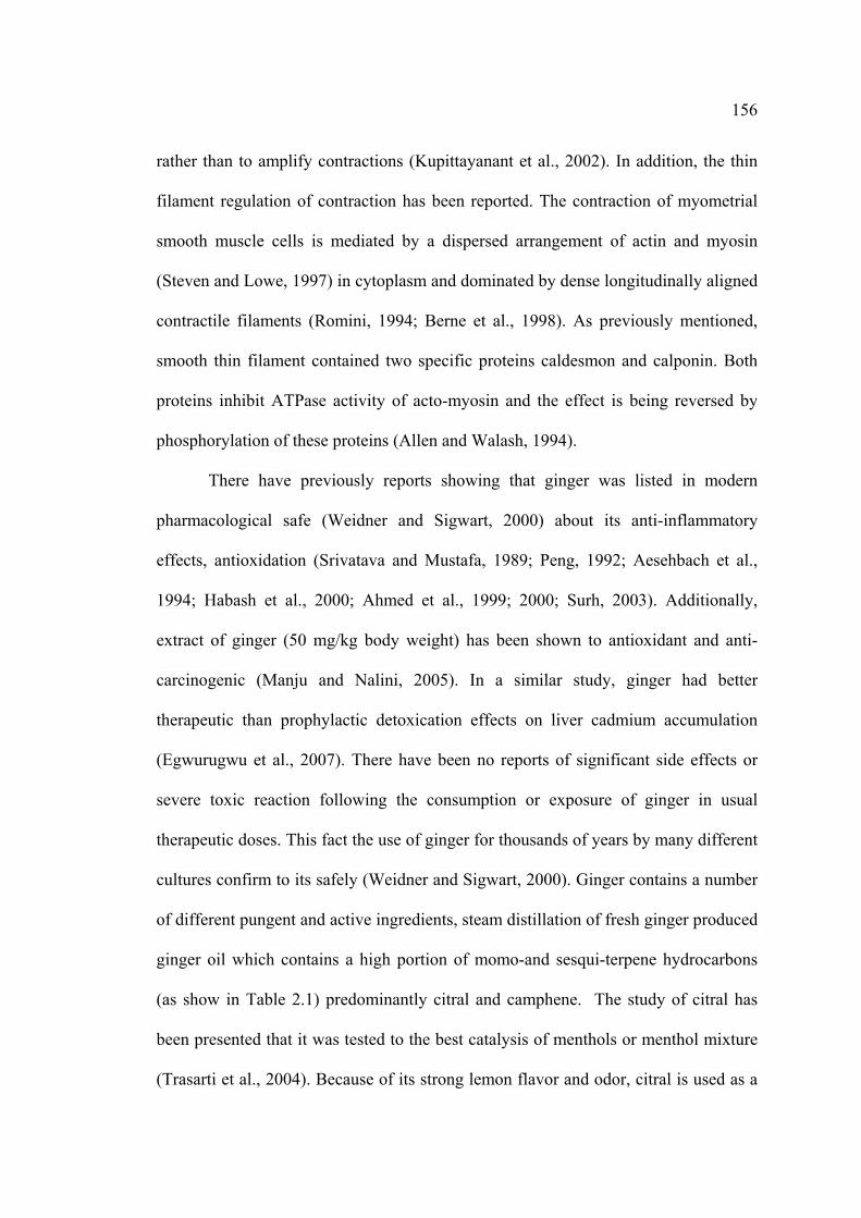
156
rather than to amplify contractions (Kupittayanant et al., 2002). In addition, the thin
filament regulation of contraction has been reported. The contraction of myometrial
smooth muscle cells is mediated by a dispersed arrangement of actin and myosin
(Steven and Lowe, 1997) in cytoplasm and dominated by dense longitudinally aligned
contractile filaments (Romini, 1994; Berne et al., 1998). As previously mentioned,
smooth thin filament contained two specific proteins caldesmon and calponin. Both
proteins inhibit ATPase activity of acto-myosin and the effect is being reversed by
phosphorylation of these proteins (Allen and Walash, 1994).
There have previously reports showing that ginger was listed in modern
pharmacological safe (Weidner and Sigwart, 2000) about its anti-inflammatory
effects, antioxidation (Srivatava and Mustafa, 1989; Peng, 1992; Aesehbach et al.,
1994; Habash et al., 2000; Ahmed et al., 1999; 2000; Surh, 2003). Additionally,
extract of ginger (50 mg/kg body weight) has been shown to antioxidant and anti-
carcinogenic (Manju and Nalini, 2005). In a similar study, ginger had better
therapeutic than prophylactic detoxication effects on liver cadmium accumulation
(Egwurugwu et al., 2007). There have been no reports of significant side effects or
severe toxic reaction following the consumption or exposure of ginger in usual
therapeutic doses. This fact the use of ginger for thousands of years by many different
cultures confirm to its safely (Weidner and Sigwart, 2000). Ginger contains a number
of different pungent and active ingredients, steam distillation of fresh ginger produced
ginger oil which contains a high portion of momo-and sesqui-terpene hydrocarbons
(as show in Table 2.1) predominantly citral and camphene. The study of citral has
been presented that it was tested to the best catalysis of menthols or menthol mixture
(Trasarti et al., 2004). Because of its strong lemon flavor and odor, citral is used as a
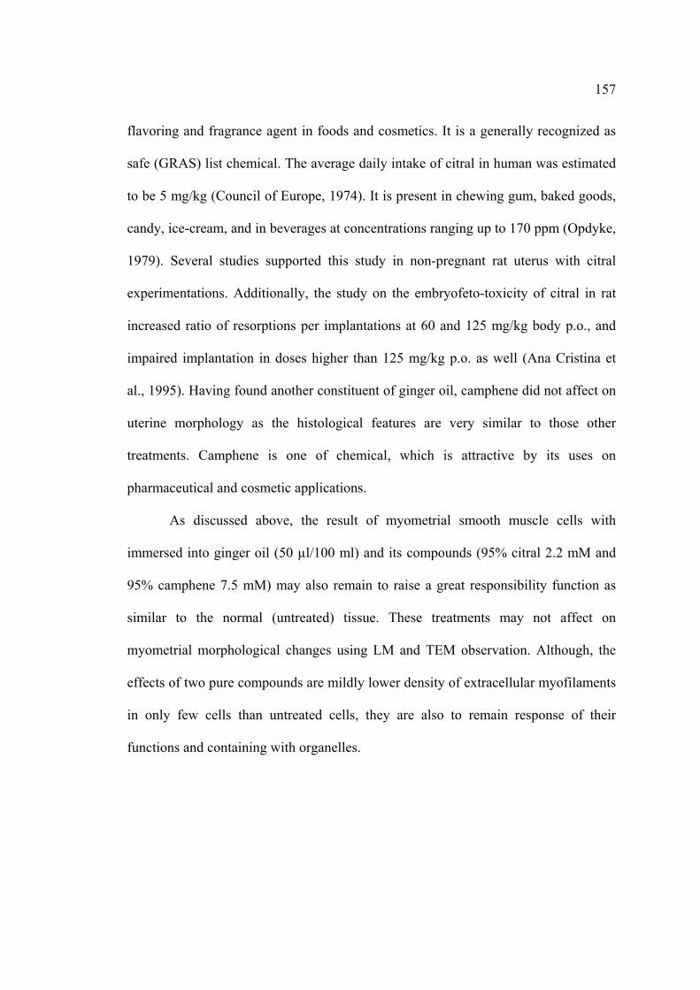
157
flavoring and fragrance agent in foods and cosmetics. It is a generally recognized as
safe (GRAS) list chemical. The average daily intake of citral in human was estimated
to be 5 mg/kg (Council of Europe, 1974). It is present in chewing gum, baked goods,
candy, ice-cream, and in beverages at concentrations ranging up to 170 ppm (Opdyke,
1979). Several studies supported this study in non-pregnant rat uterus with citral
experimentations. Additionally, the study on the embryofeto-toxicity of citral in rat
increased ratio of resorptions per implantations at 60 and 125 mg/kg body p.o., and
impaired implantation in doses higher than 125 mg/kg p.o. as well (Ana Cristina et
al., 1995). Having found another constituent of ginger oil, camphene did not affect on
uterine morphology as the histological features are very similar to those other
treatments. Camphene is one of chemical, which is attractive by its uses on
pharmaceutical and cosmetic applications.
As discussed above, the result of myometrial smooth muscle cells with
immersed into ginger oil (50 µl/100 ml) and its compounds (95% citral 2.2 mM and
95% camphene 7.5 mM) may also remain to raise a great responsibility function as
similar to the normal (untreated) tissue. These treatments may not affect on
myometrial morphological changes using LM and TEM observation. Although, the
effects of two pure compounds are mildly lower density of extracellular myofilaments
in only few cells than untreated cells, they are also to remain response of their
functions and containing with organelles.
158
6.6 References
Aesehbach, R., et al. (1994). Antioxidant actions of thymol, carbacrol, 6-gingerol,
zingerone and hydroxytyrosol. Food and Chemical Toxicology. 32 : 31 – 36.
Ahmed, R. S., Seth, V. and Banerjee, B. D. (1999). Influence of dietary ginger
(Zingiber officinale Rosc) on the antioxidant defense system in rat :
comparison with ascorbic acid. Indian Journal of Experimental Biology. In
Press.
Ahmed, R. S., Seth, V., Pasha, S. T. and Bannejee, B. D. (2000). Influence of dietary
ginger (Zingiber officinales Rosc) on oxidative stress induce by malathion in
rats. Food and chemical toxicology. 38 : 443 – 450.
Allen, B. G. and Walash, M. P. (1994). The biochemical basis of regular of smooth-
muscle contraction. Trends in Biochemical Science. 19 : 362 – 368.
Ana Cristina, M. A., et al. (1995). Study on the embryofeto-toxicity of citral in the rat.
Toxicology. 96 : 105 – 113.
Berne, R., Leney, M. N., Koeppen, B. M. and Stanton, B. A. (1998). Physiology. 4th.
Mossby.
Bode, A. M., Ma, W – Y., surh, Y. J. Dong. Z. (2001). Inhibition of epidermal growth
factor-induced cell transformtion and activator protein activation by [6]-
gingerol. Cancer Research. 61 : 850 – 853.
Council of Europe. (1974). Natural Flavouring substances, their sources, and
added artificial flavouring substances. P 147. Partial Agreement in the
Social and Public Health Field. List 1. no. 109. Strasbourg : France.
Egwurugwu, J. N., et al. (2007). Effects of ginger (Zingiber officinale) on cadmium
toxicity. African Journal of Biotechnology. 6 (18) : 2078 – 2082.
159
Friederici, H. H. R. and Decloux, R. J. (1968). The early response of immature rat
myometrium to estrogenic stimulation. Journal of Ultrastuctural Research.
22 : 402 – 412.
Gartner, L. P. and Haiatt, J. L. (2000). Corlor atlas of histology. 3rd ed. pp. 334 –
355. Williams and Wilkins. Pennsylvanis.
Habsah, M., et al. (2000). Screening of Zingiberaceae extracts for antimicrobial and
antioxidant activities. Journal of Ethnopharmacology. 72 : 403 – 410.
Kerr, J. (1999). Atlas of functional histology. pp 318 – 322. Mosby. London.
Keum,Y. S., et al. (2002). Induction of apoptosis and caspase-3 activation by
chemopreventive [6]-paradol and structurally related compounds in KB cells.
Cancer Letters. 177 : 41 – 47.
Kim, D. S. H. L. and Kim, J. Y. (2004). Side-chain length is important for shogaols in
protecting neuronal cells from β-amyloid insult. Bioorganic and Medicinal
Chemistry Letters. 14 : 1287 – 1289.
Kupittayanant, S., Luckas, M. J. and Wray, S. (2002). Effect of inhibiting the
sarcoplasmic reticulum on spontaneous and oxytocin-induced contractions of
human myometrium. British Journal of Obstetric Gynecology. 109 : 289 –
296.
Lee, E., Surh, Y-J. (1998). Induction of apoptosis in HL-60 cells by pungent
vanilloids, [6]-gingerol and [6]-parado. Cancer Letters. 134 : 163 – 168.
Leal, P. E., et al. (2003). Functional properties of spice extracts obtained via
supercritical fluid extraction. Journal of Agricultural and Food Chemistry.
51 : 2520 – 2525.
160
Manju, V. and Natini, N. (2005). Chemopreventive efficacy of ginger, a naturally
occurring anticarcinogen during the initiation, post-initiation stages of 1, 2
dimethyldrazine-induced colon cancer. China Acta Chimica. 358 : 60 – 67.
Opdyke, D. L. J. (1979). Monographs on fragrance raw materials. Citral, Food
Cosmetic Toxicology. 17 : 259 – 266.
Peachey, L. D. and Porter, K. R. (1959). Intracellular impulse conduction in muscle
cells. Science. 129 : 721 – 722.
Peng, P.J. (1992). Pharmoclogical and clinical applications of ginger. Journal of
Chineses Medicine. 17 : 370 – 373.
Phungnoi, Y. and Narkkong, N. (2007). Ultrastructure of eupyrene and apyrene
spermatozoa in pila angelica. Journal of Microscopy Society Thailand. 21
(1) : 116 – 120.
Richard, P. A. Richard, P. D. and Tiltman, A. J. (1998). The ultrastructure of
fibromyomatous myometrium and its relationship to infertility. Human
Reproduction Update. 4(5) : 520 – 525.
Romamini, C. (1994). Measurement of uterine contractions. Quoted in Chard, T.,
Grudzinskas, J. G. (Eds). The uterus, pp 337 – 355.
Shmygol, A. Eisner, D. A. and Wray, S. (1998). Properties of voltage-activated
[Ca2+]ii transients in single smooth muscle cells isolated from pregnant rat
uterus, Journal of Physiology. 511 : 803 – 811.
Shmygol, A. and Wray, S. (2004). Functional architecture of the SR calcium store in
uterine smooth muscle. Cell Calcium. 35 : 501 – 508.
Srivastava, K. C. and Mustafa, T. (1989). Ginger (Zingiber officinale) and rheumatic
disorders. Medical Hypotheses. 29 : 25 – 28.
161
Steven, A. and Lowe, J. (1997). Human histology. 2nd ed. pp 56 – 76. Mosby.
Toronto.
Surh, Y. J. and Lee, J. M. (1998). Chemoprotective properties of some pungent
ingredients present in red pepper and ginger. Muttion Research. 402 : 259 –
267.
Sweeney, M., Jones, C. U. P., Greenwood, S. L., Baker, P. N. and Taggart, M. J.
(2005). Ultrastructural fratures of smooth muscle and endothelial cells of
isolated isobaric human placental and maternal arteries. pp 1 – 15. The
University of Manchester. United Kingdom.
Taggart, M. J. and Morgan, K. G. (2007). Regulation of the uterine contractile
apparatus and cytoskeleton. Seminars in Cell and Developmental Biology.
18 : 296 – 304.
Trasarti, A. F., Marchi, A. J. and Apestegua, C. R. (2004). Highly selective synthesis
of menthols from citral in a one-step process. Journal of Catalysis. 224 : 484
– 488.
Tjendraputra, E., Tran, V. H., Liu-Brennan, D., Roufogalis, B. D. and Duke, C. C.
(2001). Effect of ginger constituents and synthetic analogues on
cyclooxygenase-2 enzyme in intact cells. Bioorganic Chemistry. 29 : 156 –
163.
Wei, Q. Y., Ma, J-P., Cai, Y-S., Yand, L. and Liu, Z-L. (2005). Cytotoxic and
apoptotic activities of diaryheptanoids and gingerol-related compounds from
the rhizome of Chinese ginger. Journal of Ethnopharmacology. 102 : 177 –
184.
162
Weidner, M. S. and Sigwart, K. (2000). The safety of a ginger extract in the rat.
Journal of Ethnopharmacology. 73 : 513 – 520.
Wetzstein, R. and Renn, K. H. (1970). Arrangement of smooth muscle in the human
uterus. Verhandlugen der Anatomistes Gesellschaftfϋr. 64 : 461 – 468.
Word, R. A., et al. (1993). Contractile elements and myosin light chain
phosphorylation in myometrial tissue from nonpregnant and pregnant women.
Journal of Clinical Investigation. 92 : 29 – 37.
Wray, S., Kupittayanant, S., Shmygol, A., Smith, R.D. and Burdyga, T. (2001). The
physiological basis of uterine contractility: a short review. Experimental
Physiology. 86 : 239 – 246.
CHAPTER VII
CONCLUSION
The main aims of this thesis were to investigate the effects of ginger oil and its
pure compounds on uterine contraction and determine the mechanisms where by
ginger oil and its pure compounds exert their effects. The major findings are
summarized as follows:
7.1 Identification of Ginger Oil
Ginger oil was analyzed by GC/MS. It contains mainly eight compounds with
retention times of 5.70 (7.74%), 7.43 (7.44%), 12.35 (10.00%), 13.03 (14.49%), 17.61
(4.71%), 17.88 (16.22%), 18.09 (6.80%), 18.09 (6.80%) and 18.45 (5.70%). They are
camphene, β-phellandrene, neral, geranial, curcumene, α-zingiberene α-farnesene and
β-sesquiphelladrene, respectively. Traces of twenty-three other known compounds
and two unknown compounds were found.
7.2 Effects of Ginger Oil and Its Pure Compounds on Spontaneous
Contraction and PGs
Under control conditions, the spontaneous contraction of rat myometrial
smooth muscle can be inhibited by the effects of the different concentrations of ginger
oil and some of its pure compounds (citral 2.2 mM and camphene 7.5 mM). Ginger
oil in a concentration dependent manner (10 – 150 µl/100 ml) inhibited myometrial
164
contraction arising either by spontaneously or PGs-induced contraction. It showed
similar effects to citral on spontaneous and PGs-induced contraction. Camphene, one
of ginger oil constituents, can weakly inhibit uterine contraction. IC50 of ginger oil,
citral, and camphene was found at 50 µl/100 ml, 2.2 mM, and 7.5 mM, respectively.
In the absence of external Ca2+, PGs (PGF2α/PGE2) elicited a small force. This force
was inhibited by ginger oil, citral and camphene. Thus the inhibitory effects of ginger
oil and its pure compounds on uterine contraction may be via inhibition of Ca2+-CaM
MLCK pathway. In addition in the absence of external Ca2+, they can inhibit force,
presumably via inhibition of non-Ca2+-CaM MLCK pathway.
7.3 Effects of Ginger Oil and Its Pure Compounds on AA
Arachidonic acid (AA) increased, at constant Ca2+, the levels of force and 20
KDa myosin light chain (MLC20) phosphorylation in permeabilized (Gong et al.,
1992). Moreover, AA itself can activate protein kinase C (Sward et al., 2000), having
Ca2+-sensitizing effects on smooth muscle.
AA (20 µM) induced myometrial contraction. In the presence of ginger oil (50
µl/100 ml) and 95% citral (2.2 mM) and 95% camphene (7.5 mM), the contractions
were decreased. This inhibition of force may be via inhibition of PKC or ROK
pathway.
7.4 Effects of Ginger Oil and Its pure compounds on the Mechanism
of COX-2 Pathway
PGE2 biosynthesis is regulated by successive metabolic steps involving the
phospholipase A2-mediated release of arachidonic acid (AA) and its conversion to
165
PGE2 by cyclooxygenase (COX) activities. The conversion of AA to PGs in
biosynthesis step is mediated by COX-1 and COX-2 (Smith and Marnett, 1991). A
common precursor AA, the increase in PGE2/PGF2α ratio indicates that its synthesis
directed preferentially towards the PGF compounds (Downie et al., 1974).
The result show that PGE2 inhibitor, aspirin, inhibited both COX-1 and COX-
2 enzyme, but this was not the case for the ginger oil and its pure compounds. The
COX-2 inhibitor, indomethacin, is a non-steroidal anti-inflammatory drug (NSAID)
used to reduce PG productions (Sawdy et al., 1998; DeWitt et al., 1991). It inhibited
myometrial contractility by blocking Ca2+ current. It was found that ginger oil and its
pure compounds further inhibited contraction in the presence of indomethacin. This
supports that the inhibitory effects of ginger oil, citral and camphene on rat
myometrial contractile may contribute to indomethacin mediated COX-2 pathways as
well as the blockade to the Ca2+ L-type channels as shown in previous Chapters.
7.5 Effects of Ginger Oil and Its Pure Compounds on the Mechanism
of PDE Pathway
In most cell types, rapid changes in [Ca2+]i directly control cellular functions,
while slower fluctuations in cAMP levels modulate the Ca2+ control system. cAMP is
synthesized by adenylyl cyclase and PDE. PDE4 is specific for the hydrolysis of
cAMP (Beavo, 1995). It is well known that aminophylline is non-specific PDE
enzyme and has been reported to inhibit pregnant and non-pregnant rat uterine smooth
muscle contraction (Apadin et al., 1998). The nature of this relaxing effect is
attributed to several mechanisms leading to a decrease in both the cytoplasmic free
166
Ca2+ concentration and the Ca2+ sensitivity of the contractile machinery (Savineau and
Mironneau, 1990).
In uterine smooth muscle, ginger oil, citral, and camphene caused an
accumulation of cAMP in low level (5±0.18, 2.5±0.08, 1±0.33 fmol/g wet wt;
respectively). This, however, was, higher than that of aminophylline, the inhibitor of
PDE (2.3±0.01 fmol/g wet wt). It has been suggested that the concentration of cAMP
should be ~10-6 M to confirm the effects of PDE pathway.
Thus, the inhibitory effects of ginger oil and its pure compounds on
myometrial contraction (with 5mM CaCl2-pretreated) may not be mediated by PDE
pathway as they produced lower quantity of cAMP level than 10-6 M.
7.6 Effects of Ginger Oil and Its Pure Compounds on Myometrial
Histological Changes
Ginger oil and its pure compounds do not affect uterine morphological
changes, after incubated the strips with the oil or its pure compounds. This is clearly
shown by routine histological procedure and observed qualitatively under light
microscope (LM) and transmission electron microscope (TEM). Observation of
microscopic feature of the myometrail smooth muscle cells shows that the cytoplasm
of myometrial smooth muscle cells is dominated by dense longitudinally aligned
contractile filaments containing the contractile proteins, actin and myosin. Structural
integrity of the sections is thus fundamental to normal uterus. Thus it is similar to the
control section. The ultra-structure, as observed by TEM in tissues that exposing to
ginger oil and camphene, are normal. However, there is a sign of change in
extracellular myofilament, SR, and intracellular myofilaments with citral.

167
Thus, the results in this study, indicate that expression of SR and
myofilaments in myometrial smooth muscle cells generally function. Ginger oil and
its pure compounds do not damage myometrial tissues as they inhibit contraction.
This was also confirmed by the effect of BSA as described previously.
Future Work
In future studies, it would be interesting to further investigate some possible
future experiments, for example, to investigate the role of ginger oil on prevention of
morning sickness in pregnant women. Morning sickness is one of the most common
complaints during pregnancy, a combination of nausea, headache and dizziness that is
experienced by about half of pregnant women during their first few months of
pregnancy. To relieve this problem, can also make a tasty morning sickness treatment
by combining ginger with lemon juice against nausea during the day.
However, the safety of use in pregnant women is not known. This study
showed that ginger oil can inhibit uterine contraction in non-pregnant, but not in
pregnant rats. Thus, it is worth examining the effects in pregnant rats and women to
answer its safety use.
7.7 References
Apadin, S., Gonen, C. and Guven, H. (1998). The problem role of nitric oxide on the
relaxations obtained by caffeine and aminophylline in rat uterus.
Pharmacological Research. 38 : 387 – 392.
Beavo, J. A. (1995). Cyclic nucleotide phosphodiesterase: functional implications of
multiple isoforms. Physiological Review. 75 : 725 – 748.
168
De Witt, D. L. (1991). Prostaglandin endoperoxide synthase: regulation of Enzyme
expression. Biochemica et Biophysica Acta. 1083 : 121 – 134.
Downie, J., Poyser, N. L. and Wunderlich, M. (1974). Levels of prostaglandins in
human endometrium during the normal menstrual cycle. Journal of
Physiology. 236 : 465 – 472.
Gong, M. C., et al. (1992). Arachidonic acid inhibits myosin light chain phosphatase
and sensitizes smooth muscle to Ca2+. Journal of Biology Chemistry. 267 :
21492 – 21498.
Savineau, J. P. and Mironneau, J. (1990). Caffeine acting on pregnant rat
myometrium: analysis of its relaxant action and its failure to release Ca2+ from
intracellular stores. British Journal of Pharmacology. 99 : 261 –266.
Sawdy, R. J., Knock, G. P. R., Poston, L. and Aranson, P. (1998). The effect of
nimesulide and indomethacin in contractility and calcium current in
myometrial smooth muscle from pregnant women. British Journal of
Pharmacology. 125 : 1212 – 1217.
Smith, W. L. and Marnett, L. J. (1991). Prostaglandin endoperoxide synthase:
structure and catalysis. Biochemica et Biophysiologica Acta. 1083 : 1 – 17.
Sward, K., et al. (2000). Inhibition of Rho-associated kinase blocks agonist-induced
Ca2+ sensitization of myosin phosphorylation and force in guinea-pig ileum.
Journal of Physiology (London). 522 : 33 – 39.
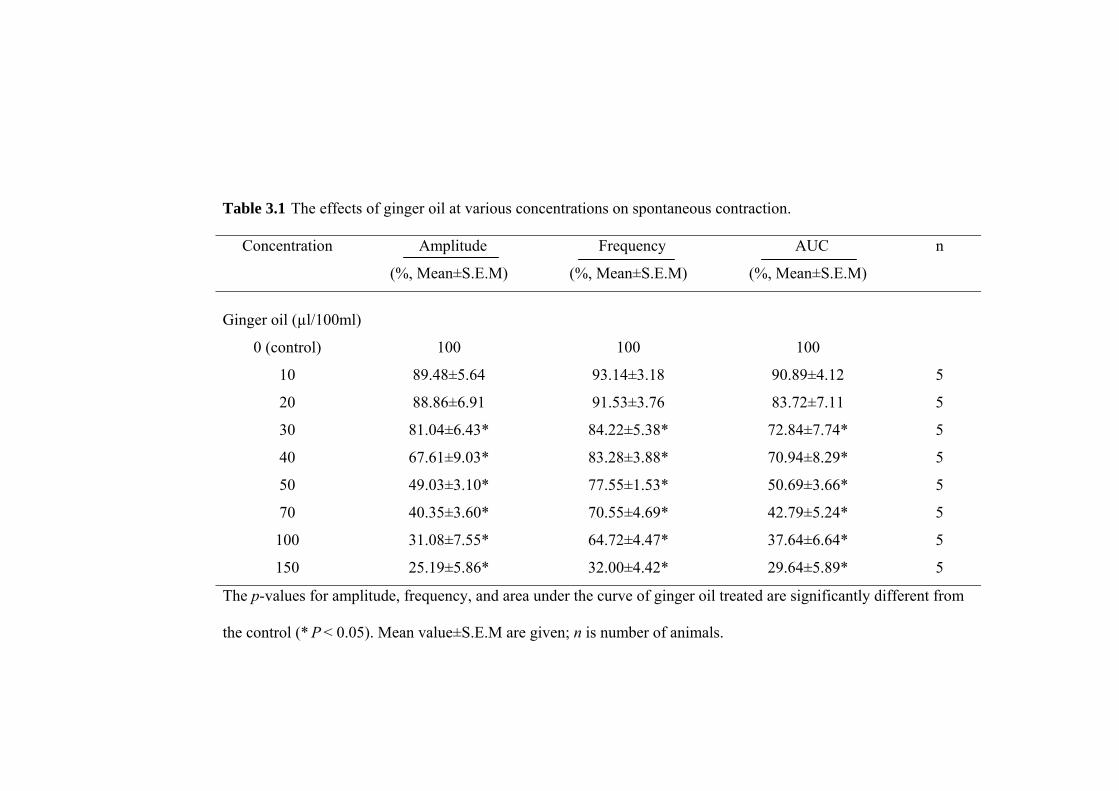
Table 3.1 The effects of ginger oil at various concentrations on spontaneous contraction.
Concentration Amplitude Frequency AUC n
(%, Mean±S.E.M) (%, Mean±S.E.M) (%, Mean±S.E.M)
Ginger oil (µl/100ml)
0 (control) 100 100 100
10 89.48±5.64 93.14±3.18 90.89±4.12 5
20 88.86±6.91 91.53±3.76 83.72±7.11 5
30 81.04±6.43* 84.22±5.38* 72.84±7.74* 5
40 67.61±9.03* 83.28±3.88* 70.94±8.29* 5
50 49.03±3.10* 77.55±1.53* 50.69±3.66* 5
70 40.35±3.60* 70.55±4.69* 42.79±5.24* 5
100 31.08±7.55* 64.72±4.47* 37.64±6.64* 5
150 25.19±5.86* 32.00±4.42* 29.64±5.89* 5
The p-values for amplitude, frequency, and area under the curve of ginger oil treated are significantly different from
the control (* P < 0.05). Mean value±S.E.M are given; n is number of animals.
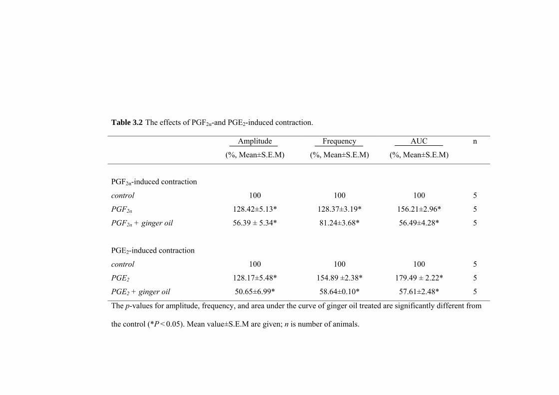
Table 3.2 The effects of PGF2α-and PGE2-induced contraction.
Amplitude Frequency AUC n
(%, Mean±S.E.M) (%, Mean±S.E.M) (%, Mean±S.E.M)
PGF2α-induced contraction
control 100 100 100 5
PGF2α 128.42±5.13* 128.37±3.19* 156.21±2.96* 5
PGF2α + ginger oil 56.39 ± 5.34* 81.24±3.68* 56.49±4.28* 5
PGE2-induced contraction
control 100 100 100 5
PGE2 128.17±5.48* 154.89 ±2.38* 179.49 ± 2.22* 5
PGE2 + ginger oil 50.65±6.99* 58.64±0.10* 57.61±2.48* 5
The p-values for amplitude, frequency, and area under the curve of ginger oil treated are significantly different from
the control (*P < 0.05). Mean value±S.E.M are given; n is number of animals.
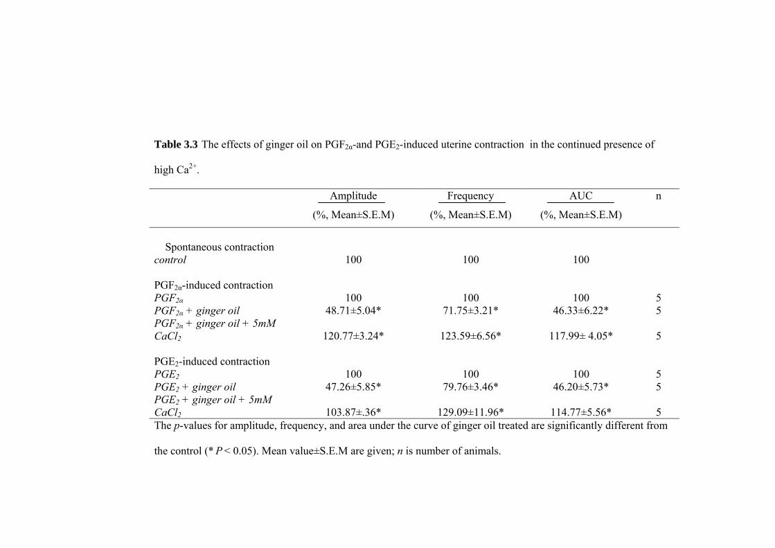
Table 3.3 The effects of ginger oil on PGF2α-and PGE2-induced uterine contraction in the continued presence of
high Ca2+.
Amplitude Frequency AUC n
(%, Mean±S.E.M) (%, Mean±S.E.M) (%, Mean±S.E.M)
Spontaneous contraction
control 100 100 100
PGF2α-induced contraction PGF2α 100 100 100 5 PGF2α + ginger oil 48.71±5.04* 71.75±3.21* 46.33±6.22* 5 PGF2α + ginger oil + 5mM CaCl2 120.77±3.24* 123.59±6.56* 117.99± 4.05* 5 PGE2-induced contraction PGE2 100 100 100 5 PGE2 + ginger oil 47.26±5.85* 79.76±3.46* 46.20±5.73* 5 PGE2 + ginger oil + 5mM CaCl2 103.87±.36* 129.09±11.96* 114.77±5.56* 5 The p-values for amplitude, frequency, and area under the curve of ginger oil treated are significantly different from
the control (* P < 0.05). Mean value±S.E.M are given; n is number of animals.
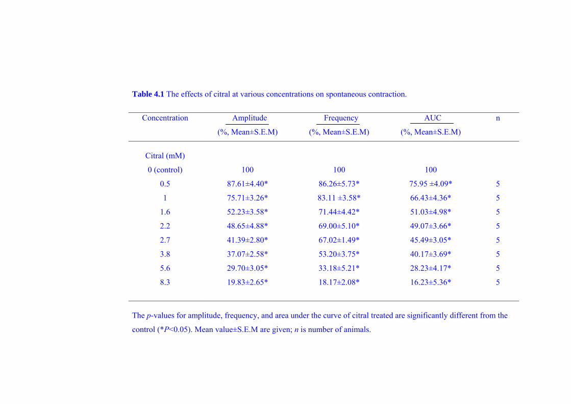
Table 4.1 The effects of citral at various concentrations on spontaneous contraction.
Concentration Amplitude Frequency AUC n
(%, Mean±S.E.M) (%, Mean±S.E.M) (%, Mean±S.E.M)
Citral (mM)
0 (control) 100 100 100
0.5 87.61±4.40* 86.26±5.73* 75.95 ±4.09* 5
1 75.71±3.26* 83.11 ±3.58* 66.43±4.36* 5
1.6 52.23±3.58* 71.44±4.42* 51.03±4.98* 5
2.2 48.65±4.88* 69.00±5.10* 49.07±3.66* 5
2.7 41.39±2.80* 67.02±1.49* 45.49±3.05* 5
3.8 37.07±2.58* 53.20±3.75* 40.17±3.69* 5
5.6 29.70±3.05* 33.18±5.21* 28.23±4.17* 5
8.3 19.83±2.65* 18.17±2.08* 16.23±5.36* 5
The p-values for amplitude, frequency, and area under the curve of citral treated are significantly different from the
control (*P<0.05). Mean value±S.E.M are given; n is number of animals.
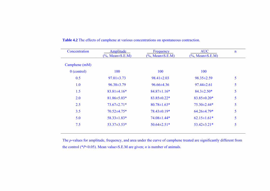
Table 4.2 The effects of camphene at various concentrations on spontaneous contraction.
Concentration Amplitude Frequency AUC n
(%, Mean±S.E.M) (%, Mean±S.E.M) (%, Mean±S.E.M)
Camphene (mM)
0 (control) 100 100 100
0.5 97.01±3.73 98.41±2.03 98.35±2.59 5
1.0 96.38±3.79 96.66±4.36 97.44±2.61 5
1.5 83.81±4.16* 84.87±1.16* 84.3±2.50* 5
2.0 81.86±5.03* 83.85±0.22* 83.85±0.20* 5
2.5 73.67±2.71* 80.78±1.63* 75.30±2.44* 5
3.5 70.52±4.75* 78.43±0.19* 64.26±4.79* 5
5.0 58.33±1.83* 74.08±1.44* 62.15±1.61* 5
7.5 53.37±3.53* 50.64±2.51* 53.42±3.21* 5
The p-values for amplitude, frequency, and area under the curve of camphene treated are significantly different from
the control (*P<0.05). Mean value±S.E.M are given; n is number of animals.

Table 4.3 The effects of citral and camphene on spontaneous contraction in the continued presence of high Ca2+.
Concentration Amplitude Frequency AUC n
(%, Mean±S.E.M) (%, Mean±S.E.M) (%, Mean±S.E.M)
Spontaneous contraction
control 100 100 100
citral 41.39±2.80* 67.02±1.49* 45.49±3.05* 5
citral + 5mM CaCl2 108.93±3.74* 141.29±1.06* 135.85±1.88* 5
Spontaneous contraction
control 100 100 100
camphene 73.67±2.71* 80.78±1.63* 75.30±2.44* 5
camphene + 5mM CaCl2 113.72±3.65* 128.62±3.42* 135.76±2.68* 5
The p-values for amplitude, frequency, and area under the curve of citral / camphene treated are significantly different
from the control (*P<0.05). Mean value±S.E.M are given; n is number of animals.
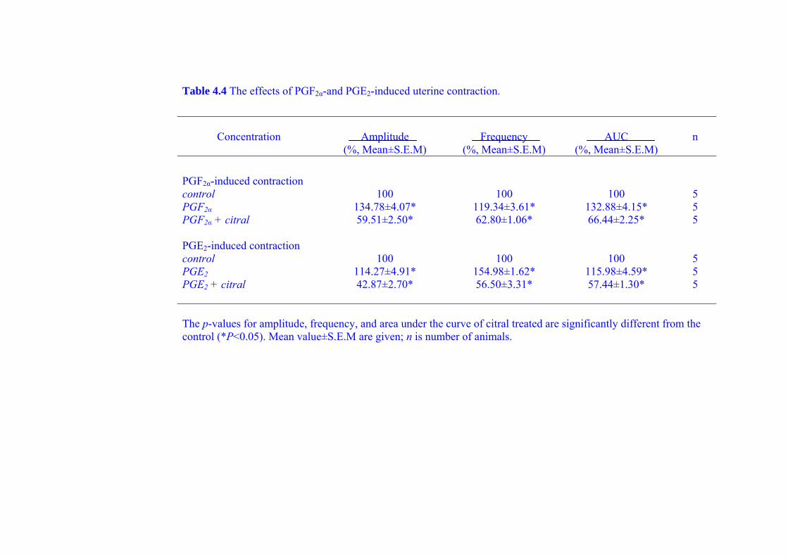
Table 4.4 The effects of PGF2α-and PGE2-induced uterine contraction.
Concentration Amplitude Frequency AUC n (%, Mean±S.E.M) (%, Mean±S.E.M) (%, Mean±S.E.M)
PGF2α-induced contraction control 100 100 100 5 PGF2α 134.78±4.07* 119.34±3.61* 132.88±4.15* 5 PGF2α + citral 59.51±2.50* 62.80±1.06* 66.44±2.25* 5
PGE2-induced contraction control 100 100 100 5 PGE2 114.27±4.91* 154.98±1.62* 115.98±4.59* 5 PGE2 + citral 42.87±2.70* 56.50±3.31* 57.44±1.30* 5 The p-values for amplitude, frequency, and area under the curve of citral treated are significantly different from the control (*P<0.05). Mean value±S.E.M are given; n is number of animals.
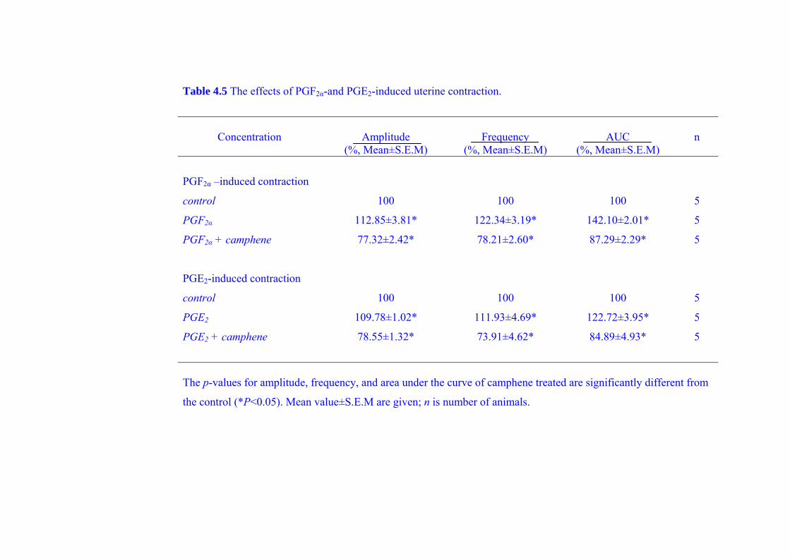
Table 4.5 The effects of PGF2α-and PGE2-induced uterine contraction.
Concentration Amplitude Frequency AUC n (%, Mean±S.E.M) (%, Mean±S.E.M) (%, Mean±S.E.M)
PGF2α –induced contraction
control 100 100 100 5
PGF2α 112.85±3.81* 122.34±3.19* 142.10±2.01* 5
PGF2α + camphene 77.32±2.42* 78.21±2.60* 87.29±2.29* 5
PGE2-induced contraction
control 100 100 100 5
PGE2 109.78±1.02* 111.93±4.69* 122.72±3.95* 5
PGE2 + camphene 78.55±1.32* 73.91±4.62* 84.89±4.93* 5
The p-values for amplitude, frequency, and area under the curve of camphene treated are significantly different from
the control (*P<0.05). Mean value±S.E.M are given; n is number of animals.

Table 4.6 The effects of citral on PGF2α-and PGE2-induced uterine contraction in the continued presence of high Ca2+.
Concentration Amplitude Frequency AUC n
(%, Mean±S.E.M) (%, Mean±S.E.M) (%, Mean±S.E.M)
PGF2α-induced contraction
PGF2α 100 100 100 5
PGF2α + citral 57.78±4.12* 60.65±4.43* 55.18±4.58* 5
PGF2α + citral + 5mM CaCl2 122.43±3.38* 104.90±2.02 116.17±3.35* 5
PGE2-induced contraction
PGE2 100 100 100 5
PGE2 + citral 42.87±4.70* 56.50±3.31* 57.44±1.30* 5
PGE2 + citral + 5mM CaCl2 106.33±2.44* 107.48±2.27* 124.40±4.90* 5
The p-values for amplitude, frequency, and area under the curve of citral treated are significantly different from the
control (*P<0.05). Mean value±S.E.M are given; n is number of animals.
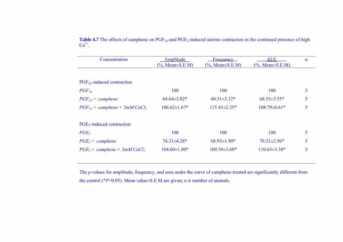
Table 4.7 The effects of camphene on PGF2α-and PGE2-induced uterine contraction in the continued presence of high Ca2+.
Concentration Amplitude Frequency AUC n (%, Mean±S.E.M) (%, Mean±S.E.M) (%, Mean±S.E.M)
PGF2α-induced contraction
PGF2α 100 100 100 5
PGF2α + camphene 64.64±3.82* 60.51±3.12* 68.53±3.55* 5
PGF2α + camphene + 5mM CaCl2 106.62±1.67* 115.43±2.35* 108.79±0.61* 5
PGE2-induced contraction
PGE2 100 100 100 5
PGE2 + camphene 74.31±4.28* 68.93±1.90* 70.21±2.96* 5
PGE2 + camphene + 5mM CaCl2 104.60±1.80* 109.39±3.68* 110.63±3.38* 5
The p-values for amplitude, frequency, and area under the curve of camphene treated are significantly different from
the control (*P<0.05). Mean value±S.E.M are given; n is number of animals.
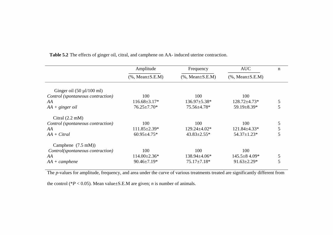
Table 5.2 The effects of ginger oil, citral, and camphene on AA- induced uterine contraction.
Amplitude Frequency AUC n
(%, Mean±S.E.M) (%, Mean±S.E.M) (%, Mean±S.E.M)
Ginger oil (50 µl/100 ml) Control (spontaneous contraction) 100 100 100 AA 116.68±3.17* 136.97±5.38* 128.72±4.73* 5 AA + ginger oil 76.25±7.70* 75.56±4.78* 59.19±8.39* 5
Citral (2.2 mM) Control (spontaneous contraction) 100 100 100 5 AA 111.85±2.39* 129.24±4.02* 121.84±4.33* 5 AA + Citral 60.95±4.75* 43.83±2.55* 54.37±1.23* 5 Camphene (7.5 mM)) Control(spontaneous contraction) 100 100 100 AA 114.00±2.36* 138.94±4.06* 145.5±8 4.09* 5 AA + camphene 90.46±7.19* 75.17±7.18* 91.63±2.29* 5 The p-values for amplitude, frequency, and area under the curve of various treatments treated are significantly different from
the control (*P < 0.05). Mean value±S.E.M are given; n is number of animals.
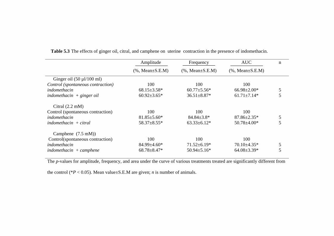
Table 5.3 The effects of ginger oil, citral, and camphene on uterine contraction in the presence of indomethacin.
Amplitude Frequency AUC n
(%, Mean±S.E.M) (%, Mean±S.E.M) (%, Mean±S.E.M)
Ginger oil (50 µl/100 ml) Control (spontaneous contraction) 100 100 100 indomethacin 68.15±3.58* 60.77±5.56* 66.98±2.00* 5 indomethacin + ginger oil 60.92±3.65* 36.51±8.87* 61.71±7.14* 5 Citral (2.2 mM) Control (spontaneous contraction) 100 100 100 indomethacin 81.85±5.60* 84.84±3.8* 87.86±2.35* 5 indomethacin + citral 58.37±8.55* 63.33±6.12* 50.78±4.00* 5 Camphene (7.5 mM)) Control(spontaneous contraction) 100 100 100 indomethacin 84.99±4.60* 71.52±6.19* 70.10±4.35* 5 indomethacin + camphene 68.78±8.47* 50.94±5.16* 64.08±3.39* 5 The p-values for amplitude, frequency, and area under the curve of various treatments treated are significantly different from the control (*P < 0.05). Mean value±S.E.M are given; n is number of animals.