Monte Carlo simulations of proteins in cages: influence of confinement on the stability of...
28
arXiv:0711.0916v2 [q-bio.BM] 4 Aug 2008 Monte Carlo simulations of proteins in cages: influence of confinement on the stability of intermediate states Pedro Ojeda & Martin E. Garcia 1 Theoretische Physik, FB 18, and Center for Interdisciplinary Nanostructure Science and Technology (CINSaT), Universit¨at Kassel, Germany, Aurora Londo˜ no Department of Molecular Biology, Instituto Potosino de Investigaci´on Cient´ ıfica y Tecnol´ogica, Camino a la presa San Jos´ e 2055, 78216 San Luis Potos´ ı, Mexico. Nan-Yow Chen Institute of Physics, Academic Sinica, Nankang, Taiwan 1 Corresponding author. Email: [email protected]
-
Upload
independent -
Category
Documents
-
view
0 -
download
0
Transcript of Monte Carlo simulations of proteins in cages: influence of confinement on the stability of...
arX
iv:0
711.
0916
v2 [
q-bi
o.B
M]
4 A
ug 2
008
Monte Carlo simulations of proteins in cages:
influence of confinement on the stability of
intermediate states
Pedro Ojeda & Martin E. Garcia1 Theoretische Physik, FB 18, and Centerfor Interdisciplinary Nanostructure Science and Technology (CINSaT),
Universitat Kassel, Germany,
Aurora LondonoDepartment of Molecular Biology, Instituto Potosino
de Investigacion Cientıfica y Tecnologica, Caminoa la presa San Jose 2055, 78216 San Luis Potosı, Mexico.
Nan-Yow ChenInstitute of Physics, Academic Sinica, Nankang, Taiwan
1Corresponding author. Email: [email protected]

Abstract
We present a theoretical study of the folding of small proteins insideconfining potentials. Proteins are described in the framework of an effec-tive potential model which contains the Ramachandran angles as degrees offreedom and does not need any a priori information about the native state.Hydrogen bonds, dipole-dipole- and hydrophobic interactions are taken ex-plicitly into account. An interesting feature displayed by this potential isthe presence of some intermediates between the unfolded and native states.We consider different types of confining potentials in order to study thestructural properties of proteins folding inside cages with repulsive or at-tractive walls. Using the Wang-Landau algorithm we determine the densityof states (DOS) and analyze in detail the thermodynamical properties of theconfined proteins for different sizes of the cages. We show that confinementdramatically reduces the phase space available to the protein and that thepresence of intermediate states can be controlled by varying the propertiesof the confining potential. Cages with strongly attractive walls lead to thedisappearance of the intermediate states and to a two-state folding into aless stable configuration. However, cages with slightly attractive walls makethe native structure more stable than in the case of pure repulsive poten-tials, and the folding process occurs through intermediate configurations. Inorder to test the metastable states we analyze the free energy landscapes asa function of the configurational energy and of the end-to-end distance asan order parameter.
Key words: Protein Folding; Simulation; Chaperones; Confinement;Wang-Landau; ; Monte Carlo
Proteins under confining potentials 2
I. INTRODUCTION
Protein folding is one of the most intensively studied and still unsolvedproblems in biology. Many diseases such as Alzheimer and Parkinson arebelieved to be caused by the misfolding and aggregation of certain pro-teins (1, 2, 3). Although in the last years several aspects related to theLevinthal’s paradox have been clarified with the help of lattice models andother approaches (4, 5, 6, 7), many questions regarding details of the foldingand misfolding mechanisms still remain open.
In this paper we focus on the problem of protein folding assisted by Chap-erones, which is one of the mechanisms present in nature to avoid aggregationand misfolding. Chaperones are molecules in the form of a cage inside whichproteins fold correctly. Recently, some progress has been achieved in the un-derstanding of the folding of protein inside chaperones. These studies haveshown that stability and folding kinetics are strongly correlated with the ge-ometry and the degree of confinement inside the cage (8, 9, 10, 11, 12, 13).However, many details of the folding under confinement still remain uncov-ered.
In this work we focus on the folding of the peptide V3-loop, ProteinData Bank ID 1NJ0, and analyze it under two kinds of time-independentconfining potentials. The first potential simulates the confining effects ofa cage being composed by rigid walls, while the second potential describesa cage with an attractive inner surface. The effect of both potentials arereflected in the thermodynamical properties, which we calculate using theWang-Landau algorithm (14, 15).
As one of the main results of this work we obtain that the folding processof V3-loop occurs through metastable intermediate states (16), and that thepresence of those states can be controlled by the confining potential.
For the description of the protein we use a force field which does notdepend on the previous knowledge of the native structure and is also able todescribe folding of proteins into both helices and β-sheets with the same setof parameters (17). In addition to this improvement, two new features notreported previously are included: (i) the dipole-dipole interaction betweenthe CO-NH pairs lying on the amide plane, and (ii) the local hydrophobicinteraction between neighboring residues, which takes into account the hy-drophobic and hydrophilic properties of the side chains. The sequence ofthe amino acids is the only input of the force field.
The paper is organized as follows. In section II we describe the modelused and the Monte Carlo method applied to calculate the thermodynamicalproperties of the protein. In section III we present our results and make a

Proteins under confining potentials 3
careful analysis of our simulations. Finally, we present a summary in sectionIV.
II. THEORY
The model
As mentioned in the previous section, the structure of the protein is simu-lated using the reduced off-lattice model developed by one of us in Ref. (17).The amino acids are represented by means of backbones. Each backbonecontains the atoms N, Cα, C’, O and H. The residues are modeled as spher-ical beads, R, attached to the Cα’s. The only remaining degrees of freedomare the Ramachandran angles ψ and φ. The values for the bond lengths andangles are given in Ref. (18).
The force field containing all relevant interactions in the protein is givenby
EProtein = ESteric + EHB + EDD + EMJ + ELocalHP (1)
where ESteric represents hard-core interparticle-potentials to avoid unphys-ical contacts, EHB accounts for the hydrogen bonding and EDD describesthe dipole-dipole interactions. EMJ is a distance-dependent version of theMiyazawa-Jerningan (MJ) matrix (19), which describes the interactions be-tween residues. ELocalHP accounts for local hydrophobic effects. The roleof the presence of water molecules is taken into account both by the termEMJ and ELocalHP . It is important to point out that EMJ partially includesthe effect of water polarization (20). The values of the parameters of thispotential are given in the original work by Chen et al. (17).
In addition to EProtein we add a term to simulate the confinement ofthe protein into a cage. This is accomplished in the present work by us-ing two different kinds of spherically symmetric potentials depending on aradius Rc, which is a measure of the size of the cage. In a first approach,we use an external potential V1 which allows the protein to fold freely fordistances smaller than Rc, but has a strongly repulsive part for larger dis-tances, simulating the presence of the walls of the cage. The potential V1
reads (11),
V1 =0.01
Rc
[
er−Rc(r − 1) − r2
2
]
, (2)
where r = |~R| denotes the position of each residue. V1 represents, however,
Proteins under confining potentials 4
a too simple description of the confining potential of a cage. Therefore, wealso investigate the effect of a second external potential V2, which accountsfor attractive walls (21) and reads
V2 = 4ǫhπRc
r
(
1
5
[
(
σ
r −Rc
)10
−(
σ
r +Rc
)10]
− ǫ2
[
(
σ
r −Rc
)4
−(
σ
r +Rc
)4])
. (3)
The physical meaning of the different parameters in Eq. (3) can be de-scribed as follows. A uniform distribution of beads spreads out on the surfaceof the cage with a number density 1/σ2. The parameter ǫ is used to simulatethe degree of attraction of the inner surface of the cage. A wall with a purelyattractive lining has a value of ǫ = 1 whereas a purely repulsive lining hasa value ǫ = 0. In Eq. (3) we set ǫh = 1.25 kcal/mol and σ = 3.8 A. Theexternal potential V1 has the only effect of confining the protein inside thecage whereas the external potential V2 interacts with the protein by slightlyreducing its energy as ǫ increases. As a consequence, the residues tend tobe far apart of each other in the region close to the walls of the cage.
Simulations
Various methods based on Monte Carlo (MC) simulations have been pro-posed to compute the thermodynamical properties of finite systems. Theyinclude, for instance, multi-canonical simulations (22) and simulated anneal-ing (23). In the present work we use the Wang-Landau algorithm (14), alsoincluding a recent improvement introduced by Pereyra et al. (15). One ofthe main advantages of Wang-Landau simulations is that they allow to ob-tain directly the density of states (DOS) of the system, which is, of course,independent of the simulation temperature. Once the DOS is known, onecan obtain all the thermodynamical properties of the system at any tem-perature. Within this framework, the transition probability between twoconformations before and after a MC trial move, X1 and X2 respectively, iscalculated as
P (X1 → X2) = min
[
1,g(X1)
g(X2)
]
, (4)
where g(X) is the DOS of the system and X is a generalized coordinate,which in our case is represented by a vector with two entries X = (E,Q),

Proteins under confining potentials 5
being E the configurational energy and Q the end-to-end distance. Notethat Q can be interpreted as an order parameter for the folding (unfolding)transition.
The original scheme developed by Landau (14) can be briefly describedas follows: one sets the initial function g(X) together with an auxiliaryhistogram H(X) to be equal to 1. Then, each time the bin X is visited, oneupdates the histogram H(X) and modifies g(X) as g(X) → g(X) × f , withf = e = 2.718281... . This procedure is continued until a ”flat” histogram(with a certain significance, i.e. 80%) is obtained. At this step the histogramH(X) is reseted and the factor f is reduced. The usual way to perform thisreduction is by taking fi+1 =
√fi. Convergence is achieved when a value
for fi+1 close enough to 1 is obtained. The last step must be compatiblewith the desired accuracy, for example f = exp(10−7).
As mentioned before, we have adopted in this paper a modification pro-posed in Ref. (15), which has been demonstrated to speed up the simulationsalso to partially avoid the problem of saturation error. According to the newscheme, one does not need to wait until the histogram H(X) is ”flat”, but itis enough to require that all the entries of H(X) are visited. Then H(X) = 0is reseted fi+1 =
√fi is updated.
Following Ref. (15) we employ a second histogram H2(X) which is neverreseted during the whole simulation and define the Monte Carlo time-step ast = j/N , N being the number of points in the energy axis and j the numberof trial moves performed. If fi+1 ≤ t−1 then fi+1 = f(t) = t−1 and from thispoint on f(t) is updated at each Monte Carlo time step. H(X) is not usedduring the rest of the calculation. Convergence is achieved when f(t) <ffinal. In the present simulations we used ffinal = exp(10−7). Finally,the thermodynamical properties of the system such as the free energy F (T ),internal energy U(T ), entropy S(T ) and specific heat C(T ) can be calculatedfrom g(X) as
F (T ) = −kBT ln
(∫
DXg(X)e−βE
)
(5)
U(T ) = 〈E〉T =
∫
DXEg(X)e−βE
∫
DXg(X)e−βE
(6)
S(T ) =U(T ) − F (T )
T(7)
Proteins under confining potentials 6
C(T ) =
⟨
U2⟩
T− 〈U〉2T
kBT 2(8)
where β = 1/kBT and kB is the Boltzmann constant. The free energylandscape as a function of E and Q can be computed as
F (E) = −kBT ln
(∫
dQg(X)e−βE
)
, (9)
and
F (Q) = −kBT ln
(∫
dEg(X)e−βE
)
, (10)
respectively.
III. RESULTS AND DISCUSSION
As mentioned in Section I, we focused our attention on a peptide composedof 16 amino acids with PDB code 1NJ0 to study the folding mediated by con-fining potentials. This peptide conforms the V3-loop of the exterior mem-brane glycoprotein (GP120) of the Human Immunodeficiency Virus type 1(HIV-1).
To explore the phase space we have chosen an energy window between-132.0 kcal/mol and -30 kcal/mol and the end-to-end distance ranging from5 A to 50 A. This region in the X space is enough to cover both the highlyordered structures present (T ∼ 0) and the fully disordered random coils(stable for T ∼ ∞). The MC search was generated by changing each pair ofRamachandran angles ψi and φi at each MC step using cutoffs with values|∆ψc| ≤ 40o and |∆φc| ≤ 40o. In order to reach ffinal = exp(10−7) 8 × 10⁹
trial moves were necessary.The obtained ground state structure of the V3-loop is depicted in Fig. 1.
It consists of a β−sheet structure with energy ∼ −132.0 Kcal/mol and anend-to-end distance of ∼5.5 A.
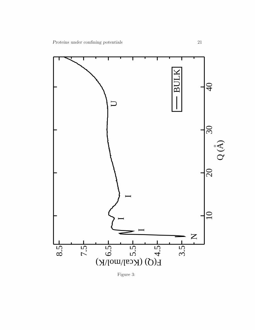
A new feature described by our force field is the presence of interme-diates (I) between the native (N) and the unfolded (U) states as shown inFig. 2 (16). We obtain two intermediate states in the free energy landscapeF (E) at the transition temperature. Intermediates are also observed whenthe energy landscape is calculated as a function of the order parameter Q(end-to-end distance). F (Q) is plotted in Fig. 3.
Interestingly, in F (Q) three intermediates can be clearly observed, whereasin F (E) only two intermediate states can be distinguished. This shows the
Proteins under confining potentials 7
importance of choosing the adequate order parameters to plot the free en-ergy. In order to analyze the nature of the intermediate states we havesplitted the energetic and entropic parts of the free energy. Results indi-cate that intermediates are mainly due to energy minima and that only theunfolded state is stabilized by entropic effects.
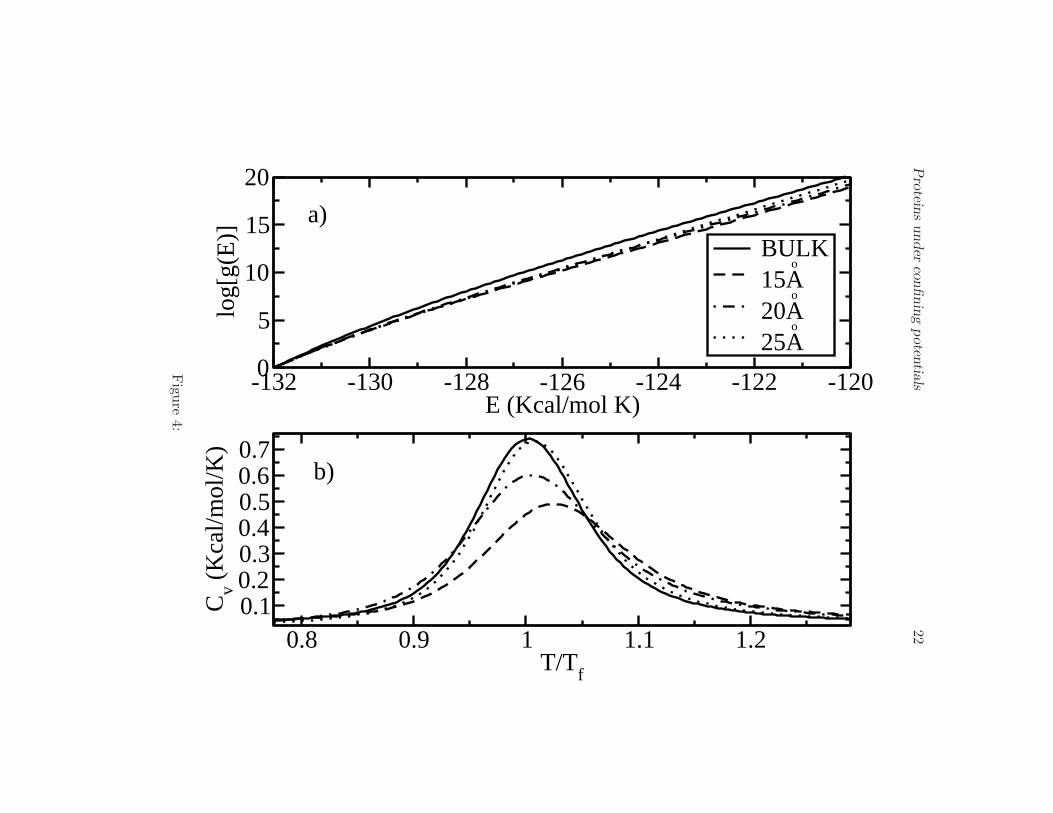
The next problem to address is the influence of confinement on the be-havior of the intermediates. For this purpose, we calculated the DOS andthe specific heat of the protein assuming the rigid-wall confining potentialV1 described above. We considered different diameters of the cage (Rc =15 A, 20 A, and 25 A). In Fig. 4(a) we show influence of V1 on the behaviorof the DOS. Note that, due to confinement, log[g(E)] considerably decreasesat high energies (temperatures) compared to the bulk case (Rc → ∞). Forenergies close to the ground state, log[g(E)] does not exhibit any notice-able change because the protein is almost folded. Since its gyration radiusin the ground state is Rg ∼ 13 A, barriers of radii equal or larger than15 A almost do not affect folded structures. This result is consistent withthe intuitive picture that cages, for instance chaperones, restrict the oth-erwise huge phase space for high energies, making the number of availablestructures considerably smaller than in absence of a cage.
The effect of confinement can be also observed in the specific heat ofthe V3-loop, which we show in Fig. 4(b). There, we plot the specific heatfor different radii of the barriers, 15 A, 20 A, 25 A, and for the bulk case(Rc → ∞) as function of T/T 0
f , T 0f = 321 K being the transition (unfolding)
temperature in absence of a cage. The effect of the rigid-wall potential V1
is to increase the transition temperature, see Table 1, and make the curveof the specific heat broader as the radius of the cage decreases. A broadercurve means that there are more structures with energies close to the nativestate than in the bulk-case where only the native state is the most importantstructure. For radii larger than 25 A the transition temperatures are equalto T 0
f within the statistical error of our simulations. We conclude that theprotein is more stable as the radius of the cage decreases. This results arein agreement with Ref. (11), in which Monte Carlo simulations were used,and with Refs. (9) and (21), where Langevin simulations were performed.It is important to mention, however, that in those cited simulations a sim-plified Go-type potentials was used. Thirumalai (24) made a considerableimprovement in the potential by introducing the effect of the non-nativeinteractions. However, important interactions such as dipole-dipole and hy-drogen bonds were not taken into account. The presence of intermediateswas not reported either in those studies.
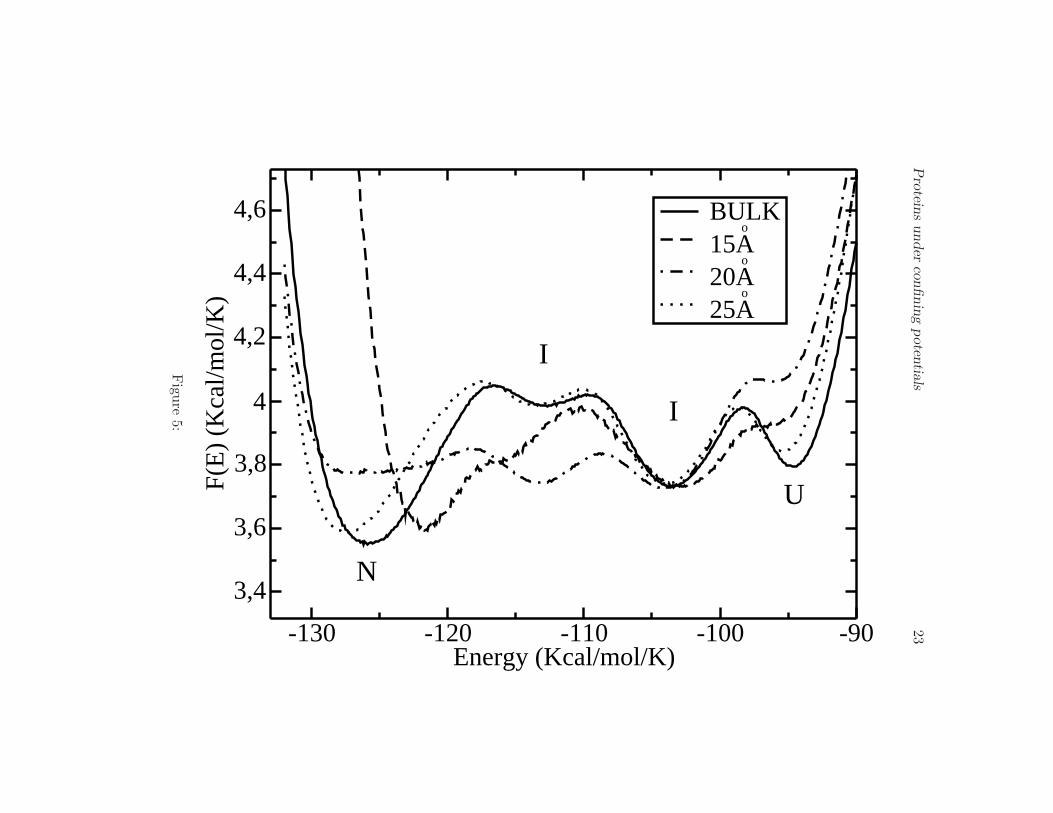
Notice that also in the presence of a confining potential V1 the native

Proteins under confining potentials 8
state (N) and the unfolded state (U) are separated by two metastable states(I), as can be seen in Fig. 5. The largest effect is observed for a cage withradius 20 A, for which the native state becomes unstable. In this case, onlymetastable structures and unfolded states are present. For all values of Rc
we observe the presence of intermediates. The free energy as a function ofthe order parameter Q for the bulk, and barriers of 15 A and 30 A is showedin Fig. 6. The most dramatic changes occur for the smallest barrier studied(15A), for which one of the minima corresponding to the intermediate statesbecomes extended.
Now, we introduce the attraction effects of the surface by using the con-fining potential V2 (Eq. 3 ) with radius Rc = 30 A. The degree of attractionis described by the coefficient ǫ. A completely attractive barrier is obtainedwhen ǫ = 1.0, whereas ǫ = 0.0 corresponds to a completely repulsive or neu-tral inner wall of the cage (potential V1). The effect of ǫ can be visualized inthe following way: as ǫ increases from 0 to 1, the walls of the cage tend toattract more the residues because of the relative minimum generated by thepotential V2. The minimum of V2(ǫ) is reached when ǫ = 1.0 and correspondsto V min
2 ∼ 5 Kcal/mol. This energy is comparable to the energy requiredto break one hydrogen bond, ∆EHB ∼ 4.8 Kcal/mol. Therefore, for ǫ ∼ 1.0the potential is able to destroy the structure of the protein (denaturation).The density of states for different degrees of attraction and for the bulk caseis shown in Fig. 7(a). The difference between a purely repulsive barrierand an attractive one can be observed in the DOS for energies close to theground state. As ǫ grows these states increase in number with respect tothe bulk and determine the folding at the transition temperature (see thecurves for ǫ > 0.6).
One can clearly observe a dramatic reduction of g(E) by ∼ 13 ordersof magnitude as ǫ goes from 0 to 1. However, this remarkable reduction ofthe phase space in this case does not help the protein to fold correctly butforces it to acquire a denatured conformation. This effect occurs becausethe peptide decreases its energy by placing some of the residues close to theborder of the cage. Then, the number of accessible states at those energiesdecreases and residues are no longer allowed to be far apart from the border,since it would cost much energy. As a consequence, the peptide sticks tothe wall of the cage. As ǫ increases, the curve of the specific heat becomesbroader (see Fig. 7(b)). The transition temperatures for different values ofǫ and for the bulk case are presented in Table 2. Interestingly, for ǫ = 0−0.3we obtain an increase of the transition temperature compared to the bulkcase. ǫ = 0.3 seems to be the optimal value. For that range of ǫ the proteinis more stable than in the absence of a cage. For higher values of ǫ the
Proteins under confining potentials 9
transition temperatures become lower. For ǫ = 1.0 the curve of the specificheat is extremely broad and attenuated, reflecting the fact that the proteinis almost denatured.
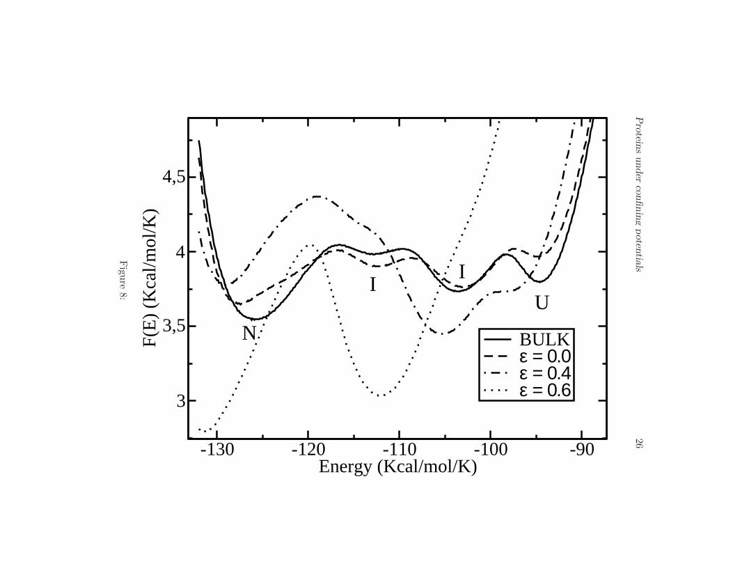
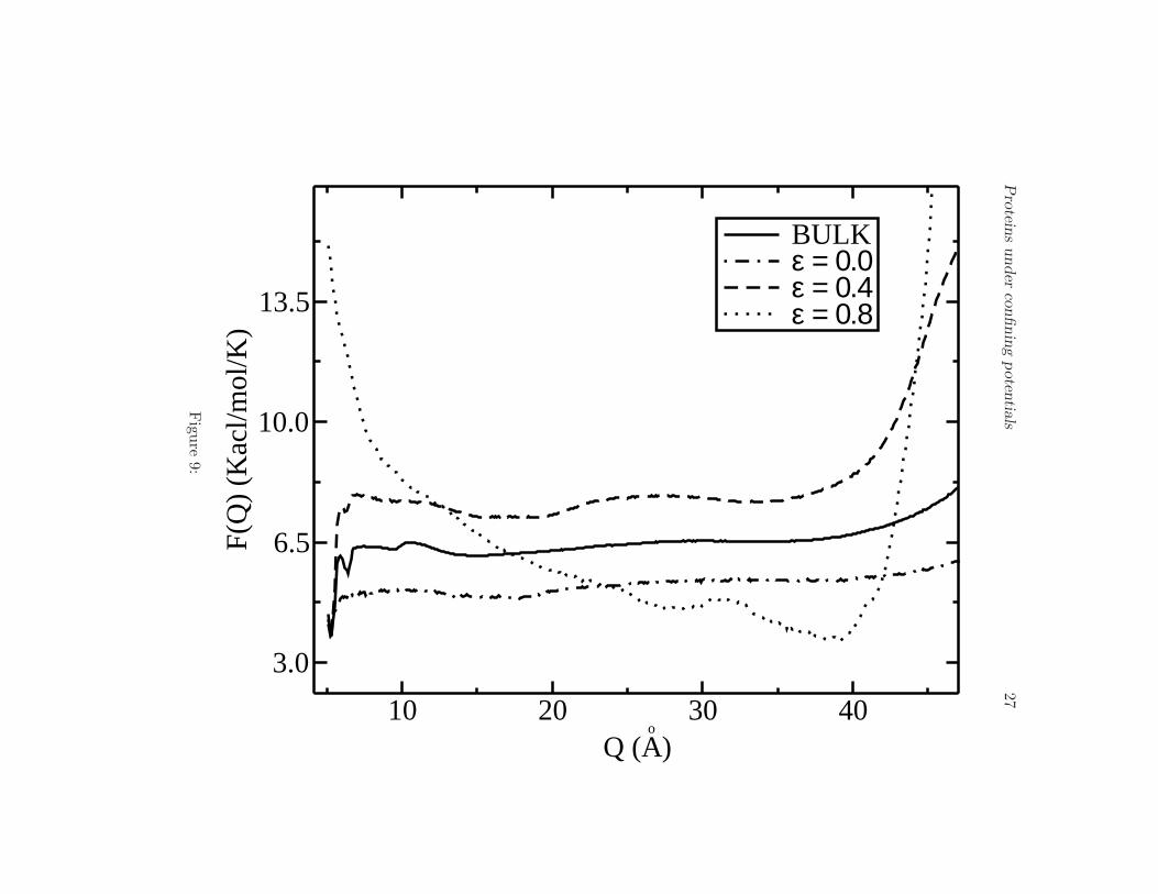
One of the main results of the present paper is illustrated in Fig. 8,where we plot the free energy profile F (E) for different values of ǫ andfor the bulk (30 A barrier-radius in all cases). The same native state (N),intermediates (I) and unfolded state (U) are observed as long as ǫ < 0.6.However, for 0.6 < ǫ < 0.8 the intermediate states (I) disappear leadingto a two-state landscape. This effect is very important because it showsthat one can remove in principle metastable states by simply changing theelectrostatic properties of the confining surface. Shea and coworkers (13)showed that for a particular protein one metastable state might exist in thepresence a weakly hydrophobic barrier. In this work and for the peptide V3-loop we obtain the opposite result, namely, that the protein shows a foldingbehavior through intermediates in the bulk, but an attractive barrier canlead to eliminate the intermediate states and to induce a two-state foldingprocess. As a function of the order parameter Q the free energy shows asimilar behavior compared to the bulk case but for ǫ ∼ 0.8 the intermediatestend to dissappear and two main minima are observed (see Fig. 9).
IV. CONCLUSION
We have studied the folding of the 16 amino acids peptide 1NJ0 underconfining and attractive potentials. We have improved previous works byintroducing a more complete potential which is independent on the nativestructure and includes relevant interactions such as the dipole-dipole andhydrogen bonds. Also, we demonstrate the presence of intermediates notreported before. We analyzed the confining effects of a protein inside aChaperone by using two kinds of potentials, one in the form of a rigid-wall inert barrier and the other one describing a cage with an attractiveinner wall. In the first case we found that the presence of the cage tends todecrease the number of accessible states by allowing only those with are closeto the native state. The transition temperatures increase as the radius ofthe barrier decreases as seen in the curves of the specific heat. These resultsare in agreement with previous simulations on other peptides (9, 11). Ourpeptide shows a folding through intermediate states (16), which are presentfor any radius of the cage. In the second case we considered the effects ofattraction inside the barrier ranging from a completely attractive (ǫ = 1.0)to an entirely repulsive (ǫ = 0.0) cage wall. We performed the simulations

Proteins under confining potentials 10
on a single barrier of radius 30 A. For fully attractive walls (ǫ = 1.0) weobserved a decrease of ∼ 13 orders of magnitude of the density of statescompared to the bulk which can be interpreted as a denaturation processof the peptide. A strongly attractive potential V2(ǫ) is able to break somehydrogen bonds of the peptide as ǫ → 1.0, thus decreasing the magnitudeof the specific heat peak strongly. However, for the interval of ǫ between0 and 0.3 we observe that the correct folding of the protein occurs. Theincreasing transition temperatures and the lower average end to end distancealso allow us to conclude that the protein is more stable as ǫ increases inthis particular interval. The analysis of the free energy profile at differentvalues of the parameter ǫ, shows that it is possible to eliminate metastableintermediate states in a controlled way. For the interval 0.6 < ǫ < 0.8 weobtained a two-state folding instead of the folding through intermediates inthe bulk. The simulations were performed as a function of the energy Eand the end-to-end distance Q, which turned out to be an appropriate orderparameter.
P. Ojeda thanks the DAAD for the financial support for his PhD.
References
1. Etienne, M., J. Aucoin, Y. Fu, R. McCarley, and R. Hammer, 2006.Stoichiometric Inhibition of Amyloid -Protein Aggregation with Pep-tides Containing Alternating A,A ,-Disubstituted Amino Acids. J. Am.
Chem. Soc. 128:3522–3523.
2. Kelly, J., 1998. The alternative conformations of amyloidogenic pro-teins and their multi-step assembly pathways. Curr. Opin. Struct. Biol.
8:101–106.
3. Lynn, D., and S. Meredith, 2000. Review: Model Peptides and thePhysicochemical Approach to Beta-Amyloids. J. Struc. Biol. 130:153–173.
4. Skolnick, J., and A. Kolinski, 1990. Simulations of the Folding of aGlobular Protein. Science 250:1121–1125.
5. Abkevich, V., A. Gutin, and E. Shakhnovich, 1994. Specific Nucleusas the Transition State for Protein Folding: Evidence from the LatticeModel. Biochemistry 33:10026–10036.
Proteins under confining potentials 11
6. Duan, Y., L. Wang, and P. Kollman, 1998. The early stage of folding ofvillin headpiece subdomain observed in a 200-nanosecond fully solvatedmolecular dynamics simulation. Proc. Nat. Aca. Sci. USA 95:9897–9902.
7. Guo, Z., and D. Thirumalai, 1998. Kinetics and Thermodynamics ofFolding of ade NovoDesigned Four-helix Bundle Protein. Proc. Nat.
Aca. Sci. USA 263:9897–9902.
8. Fang, F., and L. Szleifer, 2006. Controlled release of proteins frompolymer-modified surfaces. Proc. Nat. Aca. Sci. USA 103:5769–5774.
9. Takagi, F., N. Koga, and S. Takada .
10. Thirumalai, D., D. Klimov, and G. Lorimer, 2003. Caging helps proteinsfold. Proc. Nat. Aca. Sci. USA 100:11195–11197.
11. Rathore, N., T. Knotts, and J. Pablo, 2005. Confinement Effects on theThermodynamics of Protein Folding: Monte Carlo Simulations. Bio-
phys. Jour. 90:1767–1773.
12. Netto, A., C. Silva, and A. Caparica, 2006. Wang-Landau Sampling inThree-Dimensional Polymers. Braz. Jour. Phys. 36:619–622.
13. Jewett, A., A. Baumketner, and J. Shea, 2004. Accelerated folding inthe weak hydrophobic environment of a chaperonin cavity: Creation ofan alternate fast folding pathway. Proc. Nat. Aca. Sci. USA 101:13192–13197.
14. Wang, F., and D. Landau, 2001. Efficient, Multiple-Range RandomWalk Algorithm to Calculate the Density of States. Phys. Rev. Lett.
86:2050–2053.
15. Belardinelli, R. E., and V. D. Pereyra, 2007. Fast algorithm to calculatedensity of states. Phys. Rev. E 75:046701–046705.
16. Schnabel, S., M. Bachmann, and W. Janke, 2007. Two-State Fold-ing, Folding through Intermediates, and Metastability in a MinimalisticHydrophobic-Polar Model for Proteins. Phys. Rev. Lett. 98:048103.
17. Chen, N.-Y., Z.-Y. Su, and C.-Y. Mou, 2006. Effective Potentials forFolding Proteins. Phys. Rev. Lett. 96:078103–078107.
18. Solomons, G., and C. Fryhle, 2000. Organic Chemistry. John Wiley &Sons, Oxford, 7th. edition.

Proteins under confining potentials 12
19. Miyazawa, S., and R. Jerningan, 1996. Residue Residue Potentialswith a Favorable Contact Pair Term and an Unfavorable High PackingDensity Term, for Simulation and Threading. Jour. Mol. Biol. 256:623–644.
20. Wang, Z., and H. Lee, 2000. Origin of the Native Driving Force forProtein Folding. Phys. Rev. Lett. 84:574 – 577.
21. Lu, D., Z. Lu, and J. Wu, 2006. Structural Transitions of ConfinedModel Proteins: Molecular Dynamics Simulation and Experimental Val-idation. Biophys. Jour. 90:3224–3238.
22. Berg, B., and T. Neuhaus, 1991. Multicanonical algorithms for firstorder phase transitions. Phys. Lett. B 267:249–253.
23. Kirkpatrick, S., C. Gelat, and M. Vecci, 1983. Optimization by Simu-lated Annealing. Science 220:671–680.
24. Thirumalai, D., D. Klimov, and G. Lorimer, 2006. Nanopore-ProteinInteractions Dramatically alter Stability and Yield of the Native Statein Restricted Spaces. Jour. Mol. Biol. 357:632–643.

Proteins under confining potentials 13
TABLES
Proteins under confining potentials 14
Temperature (K) Radius (A)
329.2 15323.4 20323.2 25321.0 ∞
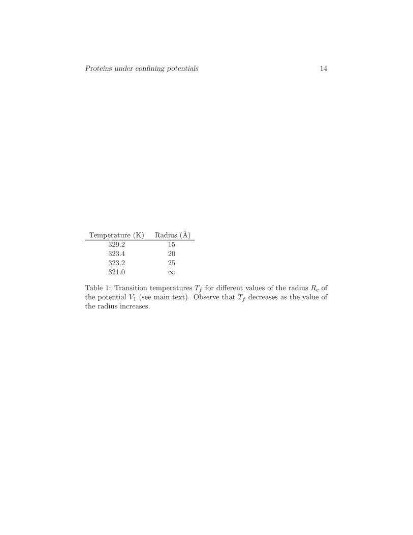
Table 1: Transition temperatures Tf for different values of the radius Rc ofthe potential V1 (see main text). Observe that Tf decreases as the value ofthe radius increases.
Proteins under confining potentials 15
Temperature (K) Radius (A)
321.0 BULK324.2 ǫ = 0.0324.1 ǫ = 0.2314.5 ǫ = 0.4292.1 ǫ = 0.6253.5 ǫ = 0.8
– ǫ = 1.0
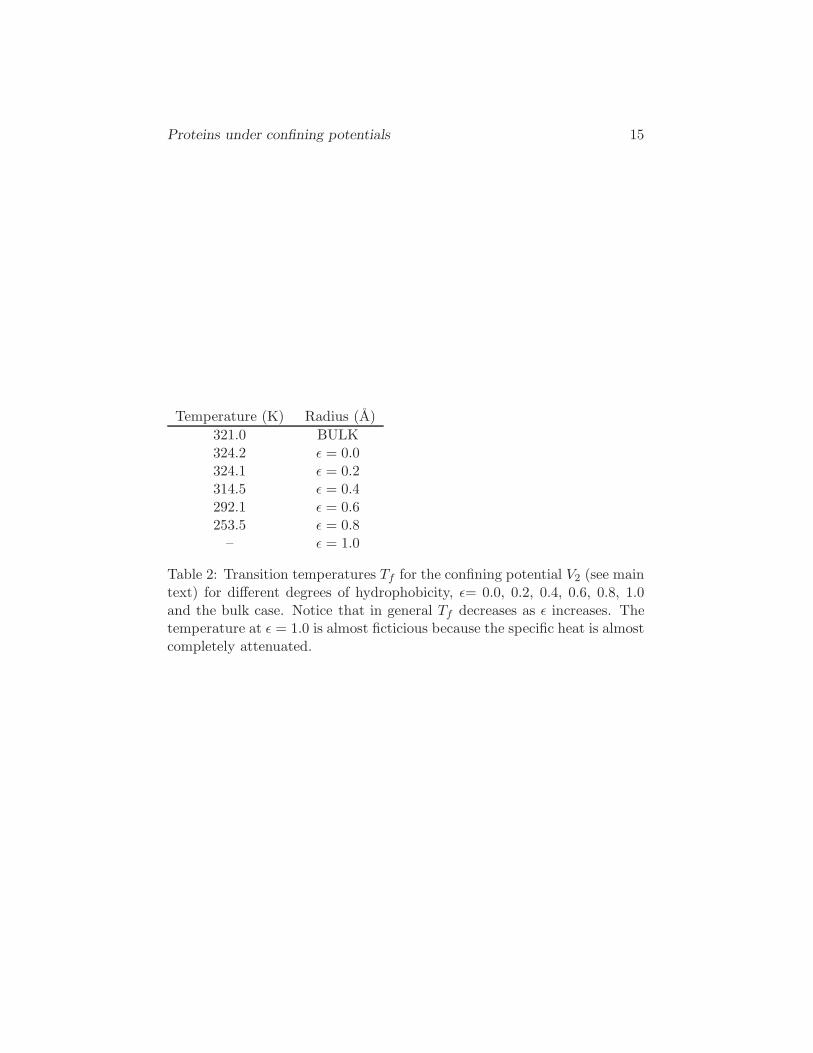
Table 2: Transition temperatures Tf for the confining potential V2 (see maintext) for different degrees of hydrophobicity, ǫ= 0.0, 0.2, 0.4, 0.6, 0.8, 1.0and the bulk case. Notice that in general Tf decreases as ǫ increases. Thetemperature at ǫ = 1.0 is almost ficticious because the specific heat is almostcompletely attenuated.

Proteins under confining potentials 16
FIGURE LEGENDS

Proteins under confining potentials 17
Figure 1.
Ground state structure (β-sheet) of the peptide 1NJ0 (Eg ∼ −132.0).
Figure 2.
Free energy as a function of the configurational energy E for the bulk (Rc →∞) showing the native (N), intermediates (I) and unfolded (U) states.
Figure 3.
Free energy as a function of the end-to-end distance Q for the bulk (Rc →∞). We observe the native (N), unfolded (U) and three intermediate states(I).
Figure 4.
(a) Logarithm of the density of states g(E) for the potential barrier V1 andfor different values of Rc, 15 A, 20 A, 25 A and for the bulk case. Onenotices the remarkable decrease of the density of states for decreasing Rc.
(b) Specific heat for the bulk-case and for barriers with radii 15 A, 20 A,and 25 A. Tf = 321 K is the transition temperature for the bulk. Thetransition temperature increases as the the radius Rc decreases.
Figure 5.
Free energy profile F (E) at the transition temperature Tf for the bulk, andbarriers of 15 A, 20 A and 25 A, showing the folding through intermediates(I) behavior. The two metastable intermediate states are present in all thecases.
Figure 6.
Free energy profile F (Q) at the transition temperature Tf for the bulk, andbarriers of 15 A and 30 A, the same features are shown in all these threecases but smoothing out is more appreciated for the smallest barrier.
Figure 7.
(a) Logarithm of g(E) for different degrees of hydrophobicity ǫ = 0.0, 0.2,0.4, 0.6, 0.8 and 1.0, and for the bulk case. We observed an abrupt decay of
Proteins under confining potentials 18
g(E) of ∼ 13 orders of magnitude as ǫ goes from 0.0 to 1.0. For high valuesof ǫ the protein tends to be in the unfolded state.
(b) Specific heat of the protein for different values of ǫ, 0.0, 0.2, 0.4,0.6, 0.8, 1.0, compared to the bulk case. Tf = 321 K is the transitiontemperature for the bulk. Notice how the transition temperatures and thepeak of the specific heat decrease as we go from a purely repulsive barrierǫ = 0.0 to a strongly attractive one ǫ = 1.0.
Figure 8.
Free energy profile F (E) at the transition temperature Tf for the bulk, andvalues of ǫ 0.0, 0.4 and 0.6. Observe how the intermediate states (I) presentin the bulk are destroyed by an attractive surface at ǫ = 0.6 causing atwo-state folding process.
Figure 9.
Free energy profile F (Q) at the transition temperature Tf for the bulk, andvalues of ǫ 0.0, 0.4 and 0.8. For small values of ǫ the free energy is similarto the bulk but for high values of ǫ the intermediates tend to dissappear.

Proteins under confining potentials 19
Figure 1:

Pro
teins
under
confinin
gpoten
tials
20-130 -120 -110 -100 -90Energy (Kcal/mol/K)
3,4
3,6
3,8
4
4,2
4,4
4,6F(
E)
(Kca
l/mol
/K)
BULK
N
U
I
I
Figu
re2:
Proteins under confining potentials 21
1020
3040
Q (
Ao
)
3.5
4.5
5.5
6.5
7.5
8.5
F(Q) (Kcal/mol/K)
BU
LK
N
II
U
I
Figure 3:
Pro
teins
under
confinin
gpoten
tials
220.8 0.9 1 1.1 1.2T/T
f
0.10.20.30.40.50.60.7
Cv (
Kca
l/mol
/K)
BULK15A
o
20Ao
25Ao
-132 -130 -128 -126 -124 -122 -120E (Kcal/mol K)
0
5
10
15
20
log[
g(E
)]a)
b)
Figu
re4:
Pro
teins
under
confinin
gpoten
tials
23-130 -120 -110 -100 -90Energy (Kcal/mol/K)
3,4
3,6
3,8
4
4,2
4,4
4,6F(
E)
(Kca
l/mol
/K)
BULK15A
o
20Ao
25Ao
N
U
I
I
Figu
re5:

Pro
teins
under
confinin
gpoten
tials
24
10 20 30 40Q (A
o
)
3.5
4.5
5.5
6.5
7.5
8.5F(
Q)
(Kca
l/mol
/K)
BULK15A
o
30Ao
N
U
U
UFigu
re6:

Pro
teins
under
confinin
gpoten
tials
25
0.6 0.7 0.8 0.9 1 1.1 1.2 1.3 1.4T/T
f
0.2
0.4
0.6
Cv (
Kca
l/mol
/K)
-160 -140 -120 -100 -80Energy (Kcal/mol)
0
20
40
60
80lo
g[g(
E)]
BULKε = 1.0ε = 0.8ε = 0.6ε = 0.4ε = 0.2ε = 0.0
a)
b)
Figu
re7:
Pro
teins
under
confinin
gpoten
tials
26
-130 -120 -110 -100 -90Energy (Kcal/mol/K)
3
3,5
4
4,5
F(E
) (K
cal/m
ol/K
)
BULKε = 0.0ε = 0.4ε = 0.6
N
II
U
Figu
re8:
Pro
teins
under
confinin
gpoten
tials
27
10 20 30 40Q (A
o
)
3.0
6.5
10.0
13.5
F(Q
) (K
acl/m
ol/K
)BULKε = 0.0 ε = 0.4ε = 0.8
Figu
re9:


![Inclusion of methano[60]fullerene derivatives in cavitand-based coordination cages](https://static.fdokumen.com/doc/165x107/631ed46f4c5c8fb3a00e625a/inclusion-of-methano60fullerene-derivatives-in-cavitand-based-coordination-cages.jpg)