
Monochrome Camera Conversion: Effect on Sensitivity ... - MDPI
13
Citation: Crowther, J. Monochrome Camera Conversion: Effect on Sensitivity for Multispectral Imaging (Ultraviolet, Visible, and Infrared). J. Imaging 2022, 8, 54. https://doi.org/ 10.3390/jimaging8030054 Academic Editors: Edoardo Provenzi and Raimondo Schettini Received: 15 January 2022 Accepted: 23 February 2022 Published: 25 February 2022 Publisher’s Note: MDPI stays neutral with regard to jurisdictional claims in published maps and institutional affil- iations. Copyright: © 2022 by the author. Licensee MDPI, Basel, Switzerland. This article is an open access article distributed under the terms and conditions of the Creative Commons Attribution (CC BY) license (https:// creativecommons.org/licenses/by/ 4.0/). Journal of Imaging Article Monochrome Camera Conversion: Effect on Sensitivity for Multispectral Imaging (Ultraviolet, Visible, and Infrared) Jonathan Crowther JMC Scientific Consulting Ltd., Egham TW20 8LL, UK; jonathan@jmcscientificconsulting.com Abstract: Conversion of standard cameras to enable them to capture images in the ultraviolet (UV) and infrared (IR) spectral regions has applications ranging from purely artistic to science and research. Taking the modification of the camera a step further and removing the color filter array (CFA) results in the formation of a monochrome camera. The spectral sensitivities of a range of cameras with different sensors which were converted to monochrome were measured and compared with standard multispectral camera conversions, with an emphasis on their behavior from the UV through to the IR regions. Keywords: UV; IR; monochrome; multispectral; calibration; validation; sensor 1. Introduction “Do you prefer to shoot in color or monochrome?” Few questions in photography can prompt discussion the way the one of whether color or monochrome is your preferred approach to imaging does. The first photographs were, of course, monochrome, but by the beginning of the 20th century, commercial processes for creating color photographs were starting to be developed [1]. In the early 1990s, with the first consumer-orientated digital camera, the Kodak DCS 100, a monochrome camera option appeared alongside a color version [2]. However as digital-camera manufacturers have continued to push the boundaries with sensor design, and increasing pixel count and dynamic range, outside of the machine vision and scientific camera areas, relatively few have created cameras with dedicated monochrome sensors [3,4]. Most consumer cameras use color sensors and include a way of creating monochrome images within the camera software, or the users can convert their color images to monochrome later, using a wide range of software packages. However, it should be remembered that the sensors themselves are originally monochrome, and the processes applied to them during manufacturing enable color images to be generated, typically through the use of a Bayer or color filter array (CFA) [5] or by relying on the depth of penetration of different wavelengths of light within the sensor [6]. If a monochrome image is the desired end result, the processing first to create a color image, and then to remove all that color information again, would seem to be overly complex, and could impact the quality of the final image [7]. These extra steps can be especially important factors if the photographer is interested in imaging in the regions beyond visible light, such as the ultraviolet (UV) and infrared (IR), and for multispectral imaging using band-pass optical filters or tightly defined light sources [8]. This article describes how the sensitivity of a range of monochrome converted digital cameras compares to both standard and multispectral converted (UV–Visible–IR) cameras. Sensitivity was assessed between 300 and 800 nm for a variety of test cameras with tra- ditional sensor designs and backside illuminated (BSI) sensors. Enhanced UV sensitivity for the monochrome converted cameras was quantified by using a newly defined method, using narrow band-pass optical filters. Images in the UV and IR are also included here to demonstrate the observed sensitivity results. The relative contributions of the CFA and microlenses to the observed sensitivity are also discussed. J. Imaging 2022, 8, 54. https://doi.org/10.3390/jimaging8030054 https://www.mdpi.com/journal/jimaging
-
Upload
khangminh22 -
Category
Documents
-
view
2 -
download
0
Transcript of Monochrome Camera Conversion: Effect on Sensitivity ... - MDPI

�����������������
Citation: Crowther, J. Monochrome
Camera Conversion: Effect on
Sensitivity for Multispectral Imaging
(Ultraviolet, Visible, and Infrared). J.
Imaging 2022, 8, 54. https://doi.org/
10.3390/jimaging8030054
Academic Editors: Edoardo Provenzi
and Raimondo Schettini
Received: 15 January 2022
Accepted: 23 February 2022
Published: 25 February 2022
Publisher’s Note: MDPI stays neutral
with regard to jurisdictional claims in
published maps and institutional affil-
iations.
Copyright: © 2022 by the author.
Licensee MDPI, Basel, Switzerland.
This article is an open access article
distributed under the terms and
conditions of the Creative Commons
Attribution (CC BY) license (https://
creativecommons.org/licenses/by/
4.0/).
Journal of
Imaging
Article
Monochrome Camera Conversion: Effect on Sensitivity forMultispectral Imaging (Ultraviolet, Visible, and Infrared)Jonathan Crowther
JMC Scientific Consulting Ltd., Egham TW20 8LL, UK; [email protected]
Abstract: Conversion of standard cameras to enable them to capture images in the ultraviolet (UV)and infrared (IR) spectral regions has applications ranging from purely artistic to science and research.Taking the modification of the camera a step further and removing the color filter array (CFA) resultsin the formation of a monochrome camera. The spectral sensitivities of a range of cameras withdifferent sensors which were converted to monochrome were measured and compared with standardmultispectral camera conversions, with an emphasis on their behavior from the UV through to theIR regions.
Keywords: UV; IR; monochrome; multispectral; calibration; validation; sensor
1. Introduction
“Do you prefer to shoot in color or monochrome?” Few questions in photographycan prompt discussion the way the one of whether color or monochrome is your preferredapproach to imaging does. The first photographs were, of course, monochrome, but bythe beginning of the 20th century, commercial processes for creating color photographswere starting to be developed [1]. In the early 1990s, with the first consumer-orientateddigital camera, the Kodak DCS 100, a monochrome camera option appeared alongside acolor version [2]. However as digital-camera manufacturers have continued to push theboundaries with sensor design, and increasing pixel count and dynamic range, outsideof the machine vision and scientific camera areas, relatively few have created cameraswith dedicated monochrome sensors [3,4]. Most consumer cameras use color sensorsand include a way of creating monochrome images within the camera software, or theusers can convert their color images to monochrome later, using a wide range of softwarepackages. However, it should be remembered that the sensors themselves are originallymonochrome, and the processes applied to them during manufacturing enable color imagesto be generated, typically through the use of a Bayer or color filter array (CFA) [5] or byrelying on the depth of penetration of different wavelengths of light within the sensor [6].If a monochrome image is the desired end result, the processing first to create a color image,and then to remove all that color information again, would seem to be overly complex,and could impact the quality of the final image [7]. These extra steps can be especiallyimportant factors if the photographer is interested in imaging in the regions beyond visiblelight, such as the ultraviolet (UV) and infrared (IR), and for multispectral imaging usingband-pass optical filters or tightly defined light sources [8].
This article describes how the sensitivity of a range of monochrome converted digitalcameras compares to both standard and multispectral converted (UV–Visible–IR) cameras.Sensitivity was assessed between 300 and 800 nm for a variety of test cameras with tra-ditional sensor designs and backside illuminated (BSI) sensors. Enhanced UV sensitivityfor the monochrome converted cameras was quantified by using a newly defined method,using narrow band-pass optical filters. Images in the UV and IR are also included here todemonstrate the observed sensitivity results. The relative contributions of the CFA andmicrolenses to the observed sensitivity are also discussed.
J. Imaging 2022, 8, 54. https://doi.org/10.3390/jimaging8030054 https://www.mdpi.com/journal/jimaging
J. Imaging 2022, 8, 54 2 of 13
2. Materials and Methods2.1. Camera Spectral Sensitivity Measurement
Camera spectral sensitivity was measured by using a custom-built device, using amethod previously described in [9]. The sensitivity measurements are quantitative, in thatthey can be compared between different wavelengths and cameras, but they should not beconfused with quantum efficiency values. The cameras discussed here are listed below:
• Standard unmodified Nikon d850.• Nikon d850 UV–Vis–IR monochrome conversion with the CFA/microlenses and inter-
nal IR filter stack removed, and sensor cover glass replaced with a fused silica window.• Standard unmodified Nikon d800.• Nikon d800 UV–Vis–IR monochrome conversion with the CFA/microlenses and internal
IR filter stack removed, and sensor cover glass replaced with a Schott WG280 window.• Standard unmodified Sony A7III.• Sony A7III multispectral conversion with the internal IR filter stack removed and the
sensor cover glass replaced with a fused silica window.• Standard unmodified Canon EOS 5DS R.• Canon EOS 5DS R multispectral conversion with the internal IR filter stack removed
and the sensor cover glass replaced with a Schott WG280 window.• Canon EOS 5DS R UV–Vis–IR monochrome conversion with the CFA/microlenses
and internal IR filter stack removed, and sensor cover glass replaced with a SchottWG280 window.
The multispectral and monochrome conversions were carried out by Llewellyn DataProcessing LLC, Carlstadt, NJ, USA. The lens used for the sensor sensitivity tests was aNikon Rayfact 105 mm f4.5 UV macro lens with appropriate lens mount adapter (the lensadapter did not contain any additional optics). Any variations for changes in ISO speedsetting during the sensitivity measurements were corrected for during the data analysis.All images were saved as RAW files for analysis.
Error bars have not been included on the sensitivity graphs for sake of clarity; however,typically, the measured sensor-response values were within 3% of each other for multiplerepeated measurements. However increased errors were observed for the multispectralcameras below 340 nm due to their relatively low sensitivity in that region (the unmodifiedcameras had no measurable sensitivity at 380 nm or below).
2.2. UVB/UVA Sensitivity Comparison between 313 and 365 nm
Measurement of camera sensitivity at 313 and 365 nm (UVB and UVA regions) wascarried out by using a Hamamatsu LC8 light source with a 200 W mercury xenon (HgXe)lamp, fitted with a custom made UV collimator. The lamp was run unfiltered and wasleft for at least 30 min to warm up before any imaging was carried out. A Labsphere 80%diffuse reflectance standard made from Spectralon® was used as the test target. The testcamera was fitted with a Rayfact 105 mm f4.5 UV macro lens with appropriate lens mountadapter (the lens adapters did not contain any additional optics). All images were capturedat ISO400, and with a lens aperture of f8. Images were captured at a range of exposuretimes, using Edmund Optics 10 nm UV band-pass filters (313 and 365 nm OD4 hard-coatedband-pass filters) mounted in a set of 52 mm filter adapter rings. Channel response of thecameras was plotted against exposure time.
2.3. UVA and UVB Images of Common Dandelion
Flower images at 365 and 313 nm were captured by using the monochrome and multi-spectral converted Canon EOS 5DS R cameras, and monochrome Nikon d850 camera usedfor the other experiments outlined in this article. The flower was a common dandelion(Taraxacum officinale) placed in a glass vase and imaged against white paper. A 20% Lab-sphere Spectralon® diffuse reflectance standard was also imaged at the same settings as theflower to enable exposure to be compared at the two wavelengths between the cameras.Exposure times were based on the results of the sensitivity test. The light source was a

J. Imaging 2022, 8, 54 3 of 13
Hamamatsu LC8 light source with a 200 W mercury xenon (HgXe) lamp, fitted with acustom made UV collimator. The lamp was run unfiltered and was left for at least 30 min towarm up before any imaging was carried out. A Rayfact 105 mm f4.5 UV macro lens withappropriate lens mount adapter (the lens adapter did not contain any additional optics).All images were captured at ISO400, and with a lens aperture of f16. Images were capturedat a range of exposure times, using Edmund Optics 10 nm UV band-pass filters (313 and365 nm OD4 hard coated filters) mounted in a set of 52 mm filter adapter rings. Images areshown in monochrome to enable visual comparison.
Filter-transmission spectra between 250 and 800 nm were measured by using an OceanInsight DH-2000-BAL light source, Ocean Insight FX spectrometer using 600 µm extremesolarization fiber optic patch cords for the input and output fibers and 74-UV collimatinglenses, mounted in an Ocean Insight RTL-T stand. The light source irradiance spectrawere also measured by using the Ocean Insight FX spectrometer, using a 600 µm extremesolarization fiber optic patch cord and cosine corrector (measurement distance 50 cm fromlight source collimator).
2.4. IR Imaging Using Different Long-Pass Filters
IR sensitivity was compared for the Canon 5DS R monochrome and multispectralcameras, and the Nikon d800 and d850 monochrome cameras (the multispectral Sony A7IIIcould not be tested as the test lens did not fit the available lens adapter). A Noct-Nikkor58 mm f1.2 lens was used at f16, as this lens is reported to have good IR behavior [10]. FourHeliopan long-pass IR filters were used to assess different wavelength regions; Heliopan715, 780, 830, and 1000. Images were taken at ISO400. A Labsphere Spectralon® 60% diffusereflectance standard was included with each image in order to check channel responseat the different wavelengths. Lighting was overcast daylight (no direct sunlight), and allimages were captured within a 15-min period. Exposure times were normalized for theCanon 5DS R multispectral camera with the different filters to ensure consistency (1/20 sfor the 715 filter, 1/15 s for the 780 filter, 1/5 s for the 830 filter, and 4 s for the 1000 filter).These exposure times were then used for the other 3 test cameras.
2.5. Sensitivity Testing of Canon EOS 450d with Half the CFA/Microlens Array Removed
In addition to the cameras described above for tested for spectral sensitivity, thefollowing was also assessed for sensitivity at 313 and 365 nm and in the 400–800 nm region;Canon EOS 450d with the internal IR filter stack and sensor cover glass removed, and theCFA/microlens array removed from half of the sensor, using the scraping method [11].The sensitivity was measured in the three different regions of the sensor: (a) CFA andmicrolenses present, (b) CFA present but microlenses removed, and (c) CFA and microlensesremoved (monochrome). The results were then used to calculate the transmission throughthe CFA and microlenses individually.
2.6. Image Analysis
Channel sensor response from the RAW files was carried out by using the approachoutlined in [9], using RawDigger x64 Research Edition (LibRaw LLC, Potomac, MD, USA,version 1.2.23), and analyzed as RAW composite files (not RGB renders). All data wererecorded, processed, and plotted, using Microsoft Excel 2010.
3. Results and Discussion
The camera conversions carried out for this research can be divided into two types.The first is multispectral conversion, where the internal IR filter stack is removed, alongwith the sensor cover glass (which is then replaced with a clear window of either fusedsilica or Schott WG280). The resulting camera can still capture color images, as the CFAis intact, but it can do so in wavelengths from UV to IR. The second is a monochromeconversion, which, similar to the multispectral conversion, removes the IR filter stack andsensor cover glass, but then it also removes the microlenses and CFA from the surface of

J. Imaging 2022, 8, 54 4 of 13
the sensor. This produces a sensor which captures monochrome images from UV to IR, butlacks the color information.
There are two different sensor designs in the cameras being assessed. The CanonEOS 5DS R and Nikon d800 both have a conventional CMOS sensor design, while theNikon d850 and Sony A7III both have backside illuminated (BSI) sensors. Cameras withboth types of sensor were assessed to try to determine differences in behavior based onsensor design.
The camera spectral response was measured between 300 and 800 nm, in 20 nmintervals, using the method reported in [9]. Measured sensor responses for the test camerasfor the Red, Green, Blue, and Green 2 color channels between 300 and 800 nm are given inFigure 1 (note the individual channel response data for the Canon EOS 5DS R cameras hasbeen shown before in [9] and is not repeated here).
Figure 1. Measured sensor response curves between 300 and 800 nm for (a) standard Nikon d850,(b) monochrome-converted Nikon d850, (c) standard Nikon d800, (d) monochrome-converted Nikond800, (e) standard Sony A7III, and (f) multispectral converted Sony A7III.
While with the manufacturers do not always release spectral response characteristicsfor their cameras, the shape of the red, green, and blue curves for the standard unmodifiedcameras in Figure 1, between 400 and 700 nm are comparable to those available in the liter-ature [9,12–16]. The unmodified Nikon and Sony cameras show similar spectral responsesfor the different color channels. This is not surprising, as the both the Nikon d800 andNikon d850 are reported to be using a Sony sensor [17,18]. The spectral-response curves

J. Imaging 2022, 8, 54 5 of 13
for the two monochrome converted cameras shown in Figure 1 display the same profileshape, although they differ in the degree of sensitivity to the monochrome Canon EOS 5DSR discussed in [9]. The multispectral converted Sony A7III is similar to the multispectralCanon EOS 5DS R reported in [9]. With the monochrome conversions, sensitivity starts tobecome apparent below 300 nm, rising to a maximum at around 480 nm and then droppingagain at longer wavelengths. The multispectral conversion has reduced sensitivity below340 nm compared to the monochrome conversions. It should be noted that the monochromeconverted cameras do not give an absolutely identical response in each of the color channelsfor all of the wavelengths; however, the differences between them are small, typically lessthan 0.3 stops. Possible reasons for why this is the case were discussed in [9].
Average channel response as a function of wavelength for the different cameras, withand without modification, is shown in Figure 2. Note that different y-axis scales are usedfor the different test cameras.
Figure 2. Averaged sensor channel response curves between 300 and 800 nm for (a) standard andmonochrome Nikon d850; (b) standard and monochrome Nikon d800; (c) standard and multispectralSony A7III; and (d) standard, monochrome, and multispectral Canon EOS 5DS R.
As can be seen in the graphs in Figure 2, the standard unmodified versions of all thecameras had a similar average sensitivity as a function of wavelength. After conversion tomultispectral, the Canon EOS 5DS R and Sony A7III cameras both showed a slight dropin average channel response at around 500 nm. The reason for this is not clear, althoughit could have to do with the nature of any anti-reflection coatings on the original internalIR filter stack and sensor cover glass. These would be optimized for light transmission inthe visible region, so their replacement with a plain clear window with no anti-reflectioncoating could reduce the camera response, as less light would reach the sensor. Themultispectral Sony A7III with its BSI sensor showed a greater IR response above 700 nmthan the multispectral Canon EOS 5DS R.
Conversion to monochrome resulted in approximately the same maximum sensitivityfor the Canon EOS 5DS R camera when compared to the standard unmodified cameraand the multispectral converted camera. However, with the Nikon cameras, conversion tomonochrome resulted in a large increase in sensitivity at all wavelengths, with the muchlarger improvement seen for the Nikon d850 with its BSI sensor. All three monochrome
J. Imaging 2022, 8, 54 6 of 13
converted cameras showed a significant increase in the sensitivity in the UV region, and asthe wavelength reduced to 300 nm, they were behaving similarly.
The principle differences between the standard and monochrome converted cam-eras are the absence of the internal IR filter stack, the sensor cover glass, and the mi-crolenses/CFA. Removal of the CFA (which absorbs some of the incoming light) wouldbe expected to increase sensitivity with a monochrome conversion; however, removal ofthe microlenses would reduce sensitivity, as light is no longer being collected from thenon-sensitive parts of the sensor. Based on the results observed here, this would suggestthat the removal of the microlenses is having a larger effect on the Canon EOS 5DS R thanfor the Nikon cameras. The Nikon d850 with its BSI sensor shows a much greater increasein maximum sensitivity compared to the Nikon d800, which has a conventionally designedCMOS sensor. In a BSI sensor, the sensitive part of the pixel is closer to the front surface ofthe sensor, rather than being inside a well [19]. Therefore, the removal of the microlenseswould be expected to have less of a detrimental effect on sensor sensitivity for a BSI sensorthan for the conventional sensor [20].
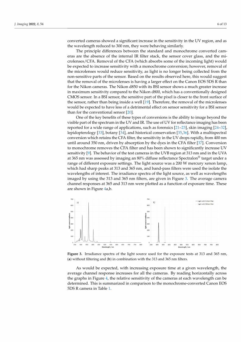
One of the key benefits of these types of conversions is the ability to image beyond thevisible part of the spectrum in the UV and IR. The use of UV for reflectance imaging has beenreported for a wide range of applications, such as forensics [21–23], skin imaging [24–32],lepidopterology [33], botany [34], and historical conservation [35,36]. With a multispectralconversion which retains the CFA filter, the sensitivity in the UV drops rapidly, from 400 nmuntil around 350 nm, driven by absorption by the dyes in the CFA filter [37]. Conversionto monochrome removes the CFA filter and has been shown to significantly increase UVsensitivity [9]. The behavior of the test cameras in the UVB region at 313 nm and in the UVAat 365 nm was assessed by imaging an 80% diffuse reflectance Spectralon® target under arange of different exposure settings. The light source was a 200 W mercury xenon lamp,which had sharp peaks at 313 and 365 nm, and band-pass filters were used the isolate thewavelengths of interest. The irradiance spectra of the light source, as well as wavelengthsimaged by using the 313 and 365 nm filters, are given in Figure 3. The average camerachannel responses at 365 and 313 nm were plotted as a function of exposure time. Theseare shown in Figure 4a,b.
Figure 3. Irradiance spectra of the light source used for the exposure tests at 313 and 365 nm,(a) without filtering and (b) in combination with the 313 and 365 nm filters.
As would be expected, with increasing exposure time at a given wavelength, theaverage channel response increases for all the cameras. By reading horizontally acrossthe graphs in Figure 4, the relative sensitivity of the cameras at each wavelength can bedetermined. This is summarized in comparison to the monochrome-converted Canon EOS5DS R camera in Table 1.

J. Imaging 2022, 8, 54 7 of 13
Table 1. Relative sensitivity of test cameras at 313 and 365 nm compared to the monochromeconverted Canon EOS 5DS R.
Test Camera 313 nm (UVB) 365 nm (UVA)
Canon EOS 5DS R multispectral 5.3 stops slower 1.7 to 2 stops slowerNikon d850 monochrome Same 0.7 to 1 stop fasterNikon d800 monochrome 0.3 stops slower 0 to 0.3 stops slowerSony A7III multispectral 4.7 to 5 stops slower 2.3 stops slower
Figure 4. Average camera channel responses at (a) 365 nm and (b) 313 nm plotted as a function ofexposure time (shutter speed).
At 365 nm, the Nikon d800 monochrome camera is almost identical to the monochromeCanon EOS 5DS R, while the Nikon d850 monochrome camera is almost 1 stop faster. Themultispectral cameras, with the CFA and microlenses still in place, are much slower at365 nm than the monochrome cameras (2.3 stops slower for the Sony A7III and 1.7 to 2 stopsslower for the Canon EOS 5DS R). At 313 nm, the difference between the multispectraland monochrome converted cameras becomes much more significant, with about 5 stopsdifference between them. Moreover, at 313 nm, the difference between the three differentmonochrome cameras essentially disappears, with at most 0.3 stops between them.
Example images of a Common Dandelion (Taraxacum officinale) in a glass vase, usingthe 313 and 365 nm filters and three of the test cameras, and using the mercury xenon lamp,are given in Figure 5 (note the exposure times between the different cameras). All the UVimages are shown as monochrome to enable comparison.
Figure 5. Images of a common dandelion (Taraxacum officinale) taken in (a) UVA (365 nm) and UVB(313 nm) with the test cameras and including exposure times; (b) visible light image of the subjectand diffuse reflectance target.

J. Imaging 2022, 8, 54 8 of 13
In the visible spectrum, the dandelion is a similar tone across the flower; however,under UV illumination, the central part of the dandelion, as with many flowers, looks darkas it strongly absorbs UV. Given its strong UV absorbance, dandelion-flower extract hasbeen evaluated as a UV-protecting ingredient for use on skin [38], highlighting the usefulrole that UV photography can play in screening for potential candidates for UV-absorbingactives. The glass vase appears very different at 365 nm compared to 313 nm, going fromtransparent to opaque, as the glass strongly absorbs the short wavelength UVB light. Insummary, conversion of the camera sensors to monochrome by removing the CFA resultedin a large increase in sensitivity in the UV region, and especially in the short wavelengthUVB region. The monochrome converted Nikon d850 with its BSI sensor demonstratedincreased sensitivity in the UVA region when compared with the other two test cameras;however, in the UVB region at 313 nm, this difference is no longer apparent.
At the other end of the visible spectrum to the UV and above 700 nm is the IR region.Normal silicon based camera sensors are sensitive up to around 1100 nm [39], and IRimaging (i.e., above 700 nm) is often used by landscape photographers to darken skiesand lighten foliage [40], and in forensics analysis of skin [41]. The method used to derivethe spectral sensitivity curves in Figures 1 and 2 can only be used up to 800 nm with theequipment available to the author. Imaging up to and beyond 800 nm was carried outby using a range of long-pass IR filters (Heliopan 715, 780, 830, and 1000) which let lightthrough at increasingly longer wavelengths. A comparison of the test cameras while usingeach of these filters to capture a garden scene in daylight is shown in Figure 6.
Figure 6. IR images taken with test cameras, using different IR filters: (a) direct image comparison;(b) average channel response of the diffuse reflectance target with the different cameras comparedwith the Canon EOS 5DS R multispectral camera for the different IR filters.
In Figure 6a, the exposure times were set by using the Canon EOS 5DS R multispectralconverted camera, and a Spectralon® diffuse reflectance standard in the lower left partof each image. This was performed to ensure that the exposure was consistent with the

J. Imaging 2022, 8, 54 9 of 13
different filters. The exposure times were then used to determine the sensitivity of each ofthe three monochrome cameras in relation to the multispectral Canon EOS 5DS R. Notethat the Sony A7III multispectral camera could not be tested here, as the lens used forthe test would not fit the adapter available for the camera. Visually, there are differencesbetween the cameras in their response to IR. The monochrome Canon EOS 5DS R andNikon d800 behave similarly to each other and show reduced sensitivity with the 780, 830,and 1000 filters compared to the multispectral Canon EOS 5DS R. The monochrome Nikond850 has a much brighter image than the multispectral Canon EOS 5DS R with the all thefilters, and this would be expected given the sensitivity data in the 700–800 nm region inFigures 1 and 2. It should also be noted that the Noct-Nikkor 58 mm lens showed someevidence of “hot spotting” in the middle of the IR images, and this has been commented onbefore when the lens is used stopped down to f16 [10].
A quantification of the sensitivities of the cameras with each IR filter and a comparisonof them against the multispectral Canon EOS 5DS R are shown in Figure 6b. As can be seen,the monochrome Canon EOS 5DS R and Nikon d800 both show a drop in response withthe 780, 830, and 1000 filters. The monochrome Nikon d850 with its BSI sensor shows amuch higher response than the other cameras. Above around 800 nm, the dyes used inthe CFA become more and more transparent, blocking less and less of the incoming light.Therefore, the difference between the monochrome and multispectral cameras would beexpected to reduce in the IR. However, with the monochrome conversion, the microlensesare being removed, which, as discussed above, would reduce sensitivity. It is the overallcombined effect of these two factors which contributes to the lowering in sensitivity ofthe monochrome cameras with the standard CMOS sensors for the longer wavelengthfilters compared to the 715 filter. Interestingly, both monochrome Nikon cameras show aslight increase in sensitivity when using the longest wavelength 1000 filter when comparedwith the multispectral Canon EOS 5DS R. The reason for this is not clear; thus, it requiresfurther investigation.
As discussed above, the key difference between the multispectral and monochromeconversions was the CFA and microlenses on top of the sensor. During the monochromeconversions tested in this article so far, both the CFA and microlenses were removed, so itwas not possible to individually assess the impact they had on sensitivity. Some peoplehave carried out their own monochrome conversions by gently scraping the surface of thesensor with a wooden stick to remove the CFA and microlenses [11]. This is an extremelyhigh-risk process which can easily damage the sensor, and, of course, it is very difficult toaccurately control. While researching monochrome conversions, the author requested asensor which had the CFA removed from half of a sensor, using this method. Essentially,half of the sensor was converted to monochrome, and the other half was left intact withthe CFA and microlenses untouched. As a by-product of the scraping method, there was aregion in the middle of the sensor which had the microlenses removed, but it retained theCFA. A photograph of the sensor showing the different regions is shown in Figure 7a. Byusing the approach outlined for Figure 1, and measuring the channel response at differentwavelengths, it was possible to calculate how much light was absorbed by the CFA andmicrolenses separately. The average response for the different color channels is shown forthe 400 to 800 nm region in Figure 7b. It should be noted that the transmission goes above100% when the microlenses are present. This is because the microlenses are gathering lightwhich would normally hit a non-sensitive part of the sensor, allowing it to be collectedand imaged.

J. Imaging 2022, 8, 54 10 of 13
Figure 7. Sensor with half the CFA/microlens array removed; (a) appearance of the sensor; and per-centage transmission between for the CFA+ microlenses, CFA only, and microlenses only, calculatedfrom the average channel response; (b) 400 and 800 nm (c) at 313 and 365 nm.
Unfortunately, the approach outlined above could not be used to generate data from300 to 400 nm for this camera, as it was an older design with a relatively low maximum ISOspeed setting. However, by testing with the 313 and 365 nm band-pass filters, using themethod described for Figure 4, the relative sensitivity at these wavelengths for the differentregions was measured, and it is shown in Figure 7c.
As can be seen from Figure 7b, across the visible and IR region, the microlenses actto increase the sensitivity compared to the CFA alone, which is what would be expected.Moreover, as discussed above, by 800 nm, the transmission of all three colors of the CFAis starting to increase, as the dyes used for the different color channels do not block theIR [42]. Although the measurements carried out here cannot go beyond 800 nm, the CFAessentially becomes equally transparent for red, green, and blue beyond approximately800 nm. This explains the observed behavior with IR imaging while using the multispectraland monochrome cameras in Figure 6, where the multispectral camera (with the CFA andmicrolenses intact) showed a brighter IR image than the monochrome cameras with thesame sensor type with the longer wavelength IR filters.
Below 400 nm, the CFA starts to block the UV (Figure 7c), with the UVB regionshowing more blocking than the UVA region. The microlenses are still working effectivelyto improve channel response in both the UVA and UVB, as shown by their transmissionbeing above 100%. This was unexpected, as these are typically formed from polymericmaterial which could absorb the UV light [43], but it demonstrates that their presence canstill be of benefit for UV photography. The improvement to the sensitivity offered by themicrolenses was greater in the UVA region than the UVB region, presumably due to greaterlight absorption by the polymeric material at shorter wavelengths.
At this stage, it is only fair to highlight the limitations of this work described here. Thesensor cover glasses were not the same on all the converted cameras. The monochromeconverted Nikon d850 and multispectral Sony A7III had their sensor windows replacedwith fused silica, while the monochrome converted Nikon d800 (and Canon EOS 5DS Rconversions) had the sensor window replaced with Schott WG280. Given the spectraltransmission characteristics of the Schott WG280 compared to those of fused silica, this

J. Imaging 2022, 8, 54 11 of 13
would only be expected to make a difference at 280 nm or shorter wavelengths and shouldnot influence the work presented here. The multispectral Canon EOS 5DS R and Sony A7IIImultispectral conversions were more extreme than a typical multispectral conversion, asthey also had their sensor cover glass removed and replaced—most standard multispectralconversions leave the existing cover glass in place to minimize risk to the sensor duringconversion. While it has not been possible to go into the details of internal camera filters inthis article, the author has discovered that these sensor cover glasses can themselves behighly UV absorbing, especially in the UVB region. Therefore, the improvement offered bythe monochrome conversions in the UV region could be even greater when compared witha multispectral camera, which retains the original sensor cover glass. Some commercialmonochrome cameras exist, such as the Leica Monochrom and Phase One IQ4 Achromatic;however, the author was not able to obtain examples of these for comparative testing.Likewise, no commercially available monochrome machine-vision cameras were tested.
4. Conclusions
Although conversion to monochrome is a serious modification to make to a cam-era, removal of the CFA and microlens array can offer enhanced sensitivity to differentwavelengths of light, especially in the UV region. This improvement will be of interest toboth artistic and scientific photographers, and especially those interested in multispectralimaging. The behavior of the dyes in the CFA, the microlenses, and sensor design (eitherconventional CMOS or BSI) all play a part in the overall change in sensitivity offered bythe monochrome conversion.
By using a controlled light source and band-pass optical filters, the behavior of a rangeof converted cameras was tested in the UVA and UVB regions, and quantification of theirsensitivity was here reported. Moreover, it was possible to characterize the camera responsein the IR region by using a set of long-pass filters with different cutoff wavelengths. Finally,the relative role of microlenses and CFA filter in determining the light transmission tothe sensor, from the UV to the IR, was determined and quantified. While monochromeconversion of a Canon EOS 5DS R and Nikon d800, which both have conventional CMOSsensors, provided some improvement to sensitivity, it was the camera with the BSI sen-sor (the Nikon d850) which saw the largest increase in sensitivity to light as a result ofthe conversion.
Further work is planned to look more deeply into how the conversion to monochromeinfluences aspects of the image, such as resolution and tonality.
Funding: This research has no external funding.
Institutional Review Board Statement: Not applicable.
Informed Consent Statement: Not applicable.
Data Availability Statement: Data from this research are not available elsewhere. Please contact theauthor for more information, if required.
Conflicts of Interest: The author declares no conflict of interest.
References1. Allen, R.L.M. Chapter 21: Colour photography. In Colour Chemistry. Studies in Modern Chemistry; Springer: Boston, MA, USA, 1971.2. Jackson, T.A.; Bell, C.S. A 1.3-megapixel-resolution portable CCD electronic still camera. In Proceedings of the Volume 1448,
Camera and Input Scanner Systems, San Jose, CA, USA, 1 June 1991. [CrossRef]3. New Gear: Leica M-Monochrom Has a Full-Frame Black and White Sensor. Popular Photography. Available online: https:
//www.popphoto.com/gear/2012/05/new-gear-leica-m9-bw/ (accessed on 12 January 2022).4. Gibson, A.; Piquette, K.E.; Bergmann, U.; Christens-Barry, W.; Davis, G.; Endrizzi, M.; Fan, S.; Farsiu, S.; Fitzgerald, A.; Griffiths,
J.; et al. An assessment of multimodal imaging of subsurface text in mummy cartonnage using surrogate papyrus phantoms.Herit. Sci. 2018, 6, 7. [CrossRef]
5. Stojkovic, A.; Shopovska, I.; Luong, H.; Aelterman, J.; Jovanov, L.; Philips, W. The Effect of the Color Filter Array Layout Choiceon State-of-the-Art Demosaicing. Sensors 2019, 19, 3215. [CrossRef] [PubMed]

J. Imaging 2022, 8, 54 12 of 13
6. Fai, C.S. Detecting Near-UV and Near-IR Wavelengths with the Foveon Image Sensor. Master’s Thesis, Naval PostGraduate School, Monterey, CA, USA, 2004. Available online: http://www.dtic.mil/dtic/tr/fulltext/u2/a429699.pdf(accessed on 12 January 2022).
7. Kanan, C.; Cottrell, G.W. Color-to-Grayscale: Does the Method Matter in Image Recognition? PLoS ONE 2012, 7, e29740.[CrossRef] [PubMed]
8. Lenk, S.; Chaerle, L.; Pfündel, E.E.; Langsdorf, G.; Hagenbeek, D.; Lichtenthaler, H.K.; van der Straeten, D.; Buschmann, C.Multispectral fluorescence and reflectance imaging at the leaf level and its possible applications. J. Exp. Bot. 2007, 58, 807–814.[CrossRef] [PubMed]
9. Crowther, J.M. Understanding colour reproduction in multispectral imaging: Measuring camera sensor response in the ultraviolet,visible and infrared. Imaging Sci. J. 2019, 67, 268–276. [CrossRef]
10. Normal Lenses For Nikon ‘F’ Mount. Available online: http://www.naturfotograf.com/lens_norm.html (accessed on 12 January 2022).11. Scratching the Color Filter Array Layer off a DSLR Sensor for Sharper B&W Photos. Available online: https://petapixel.com/20
13/08/04/scratching-the-color-filter-array-layer-off-a-dslr-sensor-for-sharper-bw-photos/ (accessed on 12 January 2022).12. Mauer, C. Measurement of the Spectral Response of Digital Cameras with a Set of Interference Filters. Diploma Thesis, Cologne,
Germany. 2009. Available online: https://www.image-engineering.de/content/library/diploma_thesis/christian_mauer_spectral_response.pdf (accessed on 12 January 2022).
13. Sigernes, F.; Dyrland, M.; Peters, N.; Lorentzen, D.A.; Svenøe, T.; Heia, K.; Chernouss, S.; Deehr, C.S.; Kosch, M. The absolutesensitivity of digital colour cameras. Opt. Express 2009, 17, 20211–20220. [CrossRef] [PubMed]
14. Darrodi, M.M.; Finlayson, G.; Goodman, T.; Mackiewicz, M. Reference data set for camera spectral sensitivity estimation. J. Opt.Soc. Am. A 2015, 32, 381–391. [CrossRef]
15. Hoot, J.E. Photometry with DSLR cameras. In Proceedings of the Society for Astronomical Sciences 26th Annual Symposium onTelescope Science, Big Bear, CA, USA, 22 May 2007; The Society for Astronomical Sciences: Big Bear, CA, USA, 2007; pp. 67–72.Available online: http://adsabs.harvard.edu/full/2007SASS...26...67H (accessed on 12 January 2022).
16. Cosentino, A. Multispectral Imaging of Pigments with a digital camera and 12 interferential filters. E-Preserv. Sci.2015, 12, 1–7. Available online: https://chsopensource.org/multispectral-imaging-of-pigments-with-a-digital-camera/(accessed on 12 January 2022).
17. Confirmed: The Sensor Inside the Nikon D800 Is Made by Sony. Available online: https://nikonrumors.com/2012/08/29/confirmed-the-sensor-inside-the-nikon-d800-is-made-by-sony.aspx (accessed on 12 January 2022).
18. The Nikon D850’s Sensor Is Made by Sony: Report. Available online: https://petapixel.com/2018/06/15/the-nikon-d850s-sensor-is-made-by-sony-report/ (accessed on 12 January 2022).
19. BSI Sensors Demystified. Available online: https://www.dtcommercialphoto.com/bsi-sensors-demystified/(accessed on 12 January 2022).
20. Djazovski, O. Chapter 37: Focal Plane Arrays for Optical Payloads. In Optical Payloads for Space Missions; Qian, S.-E., Ed.; Wiley:Chichester, UK, 2015. [CrossRef]
21. Garcia, J.E.; Wilksch, P.A.; Spring, G.; Philp, P.; Dyer, A. Characterization of digital cameras for reflected ultraviolet photography;implications for qualitative and quantitative image analysis during forensic investigation. J. Forensic Sci. 2014, 59, 117–122.[CrossRef]
22. Marin, N.; Buszka, J. Alternative Light Source Imaging Forensic Photography Techniques; Elsevier Inc.: Abingdon, UK, 2013.23. Leintz, R.; Bond, J.W. Can the RUVIS imaging system visualize fingerprint corrosion on brass cartridge casings postfiring? J.
Forensic Sci. 2013, 58, 772–775. [CrossRef]24. Boyers, L.; Karimkhani, C.; Gamble, R.; Dellavalle, R.P. Novel and promising sun safety interventions: UV photography and
shade structures. OA Dermatol. 2014, 2, 6.25. Dykstra, J.L. Avoiding Reactance: The Utility of Ultraviolet Photography, Persuasion, and Parental Protectiveness in Improving
the Effectiveness of a UV Exposure Intervention. Ph.D. Dissertation, Iowa State University, Ames, IA, USA, 2007. Availableonline: http://lib.dr.iastate.edu/cgi/viewcontent.cgi?article=16923&context=rtd (accessed on 12 January 2022).
26. Daniel, L.C.; Heckman, C.J.; Kloss, J.D.; Manne, S.L. Comparing alternative methods of measuring skin color and damage. CancerCauses Control 2009, 20, 313–321. [CrossRef] [PubMed]
27. Niamtu, J., III. Digitally processed ultraviolet images: A convenient, affordable, reproducible means of illustrating ultravioletclinical examination. Dermatol. Surg. 2001, 27, 1039–1042. [CrossRef] [PubMed]
28. Jones, T.; Baceviciene, R.; Vukmer, T.; Karimkhani, C.; Boyers, L.; Dellavalle, R.; Gamble, R. Impact of ultraviolet photography onsun safety practices of snow sport industry conference attendees. Open Dermatol. J. 2014, 8, 8–11. [CrossRef]
29. Crowther, J.M. Understanding sunscreen SPF performance using cross polarized UVA reflectance photography. Int. J. Cosmet. Sci.2018, 40, 127–133. [CrossRef]
30. Crowther, J.M. UV reflectance photography of skin: What are you imaging? Int. J. Cosmet. Sci. 2020, 42, 136–145. [CrossRef][PubMed]
31. Reflected Ultraviolet Photography. Available online: http://medicalphotography.com.au/Article_01/11.html(accessed on 12 January 2022).
32. Welch, M.; Chang, P.; Taylor, M.F. Photoaging photography: Mothers’ attitudes toward adopting skin-protective measures pre-and post-viewing photoaged images of their and their child’s facial sun damage. SAGE Open 2016, 6, 1–11. [CrossRef]
J. Imaging 2022, 8, 54 13 of 13
33. Ayre, E.; Bevan, G. Calibrated UV reflectance photography of Hebomoia Glaucippe Sulphurea. Collect. Forum 2016, 30, 34–50.[CrossRef]
34. Cronin, T.W.; Bok, M.J. Photoreception and vision in the ultraviolet. J. Exp. Biol. 2016, 219, 2790–2801. [CrossRef] [PubMed]35. Verri, G.; Saunders, D. Xenon flash for reflectance and luminescence (multispectral) imaging in cultural heritage applications. Br.
Mus. Tech. Res. Bull. 2014, 8, 83–92.36. Dyer, J.; Verri, G.; Cupitt, J. Multispectral Imaging in Reflectance and Photo-Induced Luminescence Modes: A User Manual. Avail-
able online: http://www.britishmuseum.org/research/research_projects/all_current_projects/charisma/technical_imaging.aspx(accessed on 12 January 2022).
37. Prutchi, D. Exploring Ultraviolet Photography; Amherst Media Inc.: Buffalo, NY, USA, 2017; p. 26.38. Yang, Y.; Li, S. Dandelion Extracts Protect Human Skin Fibroblasts from UVB Damage and Cellular Senescence. Oxid. Med. Cell.
Longev. 2015, 2015, 619560. [CrossRef] [PubMed]39. Vollmer, M.; Möllmann, K.P.; Shaw, J.A. The optics and physics of near infrared imaging. In Proceedings of the Education and
Training in Optics and Photonics 2015, Bordeaux, France, 29 June–2 July 2015. [CrossRef]40. Davies, A. Digital Ultraviolet and Infrared Photography; Routeledge: Abingdon, UK, 2018; p. 127.41. Cain, M.D.; Roper, D.; Atherton, D.S. Use of Infrared Photography to Visualize a Tattoo for Identification in Advanced Decompo-
sition. Acad. Forensic Pathol. 2016, 6, 338–342. [CrossRef] [PubMed]42. Miyauchi, K.; Mori, K.; Otaka, T.; Isozaki, T.; Yasuda, N.; Tsai, A.; Sawai, Y.; Owada, H.; Takayanagi, I.; Nakamura, J. A Stacked
Back Side-Illuminated Voltage Domain Global Shutter CMOS Image Sensor with a 4.0 µm Multiple Gain Readout Pixel. Sensors2020, 20, 486. [CrossRef] [PubMed]
43. Nussbaumdag, P.; Völkeldag, R.; Herzigdag, H.P.; Eisnerddag, M.; Haselbeck, S. Design, fabrication and testing of microlensarrays for sensors and microsystems. Pure Appl. Opt. 1997, 6, 617–636. [CrossRef]