Monitoring the Biodegradation of Dendritic Near-Infrared Nanoprobes by in Vivo Fluorescence Imaging
15
Monitoring the biodegradation of dendritic near infrared nanoprobes by in vivo fluorescence imaging Adah Almutairi + , Walter J. Akers † , Mikhail Y. Berezin † , Samuel Achilefu †,* , and Jean M.J. Fréchet +,* + College of Chemistry, University of California, Berkeley, CA 94720-1460 † Department of Radiology, School of Medicine, Washington University, St Louis, MO 63110 Abstract Synthetic polymers and dendrimers have been widely used by the medical community to overcome biological barriers and enhance in vivo biomedical applications. Despite the widespread use of biomaterials it has been generally extremely difficult to monitor non invasively their fate in vivo. Here we report multilayered nanoprobes, consisting of a near infrared core, nano-encapsulated in a biodegradable dendrimer, and surrounded by a shell of polyethylene oxide. Covalent encapsulation of the near infrared fluorophores in the dendritic scaffold conferred enhanced stability to the nanoprobe with added resistance to enzymatic oxidation and prolonged blood residence time. Insight into the time course of biodegradation of the dendritic aliphatic polyester nanoprobe was gained using non invasive whole body in vivo fluorescence lifetime imaging. As the dendritic shell biodegrades the NIR probe becomes exposed, enabling monitoring of fluorescence lifetime changes in vivo. Keywords Near Infrared fluorescence imaging; whole body; fluorescence lifetime imaging; polymer biodegradation; biodistribution; dendrimer INTRODUCTION Over the last decade, the medical community has made widespread use of synthetic polymers and dendrimers to overcome complex biological barriers and enhance in vivo imaging, 1, 2 drug delivery, 3 gene therapy 4 and tissue engineering. 5 Dendrimers are highly branched macromolecules that have a structural precision approaching that of proteins; 6, 7 their globular structure provides a specific nanoenvironment that surrounds a functional core thereby affecting its molecular properties. 8 The structural features and multivalency of dendrimers translate especially well into pharmacology, and have led to great promise in drug delivery. 9 Aliphatic polyester dendrimers based on the monomer 2,2-bis(hydroxymethyl) propanoic acid (bis-HMPA) 10-12 have low toxicity, low immunogenicity, and a biodegradable structure that * To whom correspondence may be addressed: Jean M. J. Fréchet, College of Chemistry, University of California, 718 Latimer Hall, Berkeley, CA 94720-1460, Tel: (510) 643-3077. Fax: (510) 643-3079E-mail: [email protected] or Samuel Achilefu, Department of Radiology, School of Medicine, Washington University in St. Louis, St. Louis, MO 63110 Tel: (314) 362-8599. Email: [email protected]. Supporting Information Available in vivo fluorescence intensity map, in vivo fluorescence intensity and lifetime maps of cypate-peptide conjugate, fluorescence intensity map post cardiovascular injury, size exclusion chromatograms and dynamic light scattering measurements of NIR dendritic nanoprobes and PEO-polyester dendrimers. NIH Public Access Author Manuscript Mol Pharm. Author manuscript; available in PMC 2009 November 3. Published in final edited form as: Mol Pharm. 2008 ; 5(6): 1103–1110. NIH-PA Author Manuscript NIH-PA Author Manuscript NIH-PA Author Manuscript
Transcript of Monitoring the Biodegradation of Dendritic Near-Infrared Nanoprobes by in Vivo Fluorescence Imaging
Monitoring the biodegradation of dendritic near infrarednanoprobes by in vivo fluorescence imaging
Adah Almutairi+, Walter J. Akers†, Mikhail Y. Berezin†, Samuel Achilefu†,*, and Jean M.J.Fréchet+,*+College of Chemistry, University of California, Berkeley, CA 94720-1460†Department of Radiology, School of Medicine, Washington University, St Louis, MO 63110
AbstractSynthetic polymers and dendrimers have been widely used by the medical community to overcomebiological barriers and enhance in vivo biomedical applications. Despite the widespread use ofbiomaterials it has been generally extremely difficult to monitor non invasively their fate in vivo.Here we report multilayered nanoprobes, consisting of a near infrared core, nano-encapsulated in abiodegradable dendrimer, and surrounded by a shell of polyethylene oxide. Covalent encapsulationof the near infrared fluorophores in the dendritic scaffold conferred enhanced stability to thenanoprobe with added resistance to enzymatic oxidation and prolonged blood residence time. Insightinto the time course of biodegradation of the dendritic aliphatic polyester nanoprobe was gainedusing non invasive whole body in vivo fluorescence lifetime imaging. As the dendritic shellbiodegrades the NIR probe becomes exposed, enabling monitoring of fluorescence lifetime changesin vivo.
KeywordsNear Infrared fluorescence imaging; whole body; fluorescence lifetime imaging; polymerbiodegradation; biodistribution; dendrimer
INTRODUCTIONOver the last decade, the medical community has made widespread use of synthetic polymersand dendrimers to overcome complex biological barriers and enhance in vivo imaging,1, 2 drugdelivery,3 gene therapy4 and tissue engineering.5 Dendrimers are highly branchedmacromolecules that have a structural precision approaching that of proteins;6, 7 their globularstructure provides a specific nanoenvironment that surrounds a functional core therebyaffecting its molecular properties.8 The structural features and multivalency of dendrimerstranslate especially well into pharmacology, and have led to great promise in drug delivery.9Aliphatic polyester dendrimers based on the monomer 2,2-bis(hydroxymethyl) propanoic acid(bis-HMPA)10-12 have low toxicity, low immunogenicity, and a biodegradable structure that
*To whom correspondence may be addressed: Jean M. J. Fréchet, College of Chemistry, University of California, 718 Latimer Hall,Berkeley, CA 94720-1460, Tel: (510) 643-3077. Fax: (510) 643-3079E-mail: [email protected] or Samuel Achilefu, Department ofRadiology, School of Medicine, Washington University in St. Louis, St. Louis, MO 63110 Tel: (314) 362-8599. Email:[email protected] Information Availablein vivo fluorescence intensity map, in vivo fluorescence intensity and lifetime maps of cypate-peptide conjugate, fluorescence intensitymap post cardiovascular injury, size exclusion chromatograms and dynamic light scattering measurements of NIR dendritic nanoprobesand PEO-polyester dendrimers.
NIH Public AccessAuthor ManuscriptMol Pharm. Author manuscript; available in PMC 2009 November 3.
Published in final edited form as:Mol Pharm. 2008 ; 5(6): 1103–1110.
NIH
-PA Author Manuscript
NIH
-PA Author Manuscript
NIH
-PA Author Manuscript
makes them especially appealing for applications in biotechnology.13, 14 We envisioned thatlabeled dendrimers might well be suitable for the non-invasive reporting of the in vivodegradation of this class of aliphatic polyester dendrimers. Radioactive tracers are commonlyattached to pharmaceuticals to monitor their biodistribution and elimination after injection. Apotential limitation of this strategy is that the radiotracer can become separated from themolecule of interest at any time after injection. This separation cannot be detected through non-invasive means. The same is true for optically active (fluorescent) contrast agents in generalwhen using near infrared (NIR) fluorescence intensity for detection. Near infrared light canpass through the skin, without damaging tissue, and with minimal absorption by water,hemoglobin, oxygenated hemoglobin, fat, and melanin.15, 16 Additionally, fluorescence fromintrinsic tissue components (autofluorescence) is low in the NIR region, resulting in highersignal to background ratios.16 The use of fluorescence lifetime in cellular and molecularbiology, and in single molecule spectroscopy is rapidly expanding, because it utilizes theenvironmentally sensitive characteristics of excited state decay. Therefore, the excited statesof certain NIR fluorophores are greatly influenced by their local environment,17 whileremaining independent of the factors that impact fluorescence intensity imaging such asphotobleaching and light path length.18 Fluorescence lifetime measurements have been usedto provide access to functional information on biomolecules,19, 20 tissue microenvironments,18, 21 and, to a much lesser extent, on living animals.22-24 We hypothesized that fluorescencelifetime imaging (FLI) of the fluorescent reporter encapsulated within a dendritic nanoparticlewould enable the non-invasive in vivo monitoring of the degradation of nanoparticle itself.
Herein, we describe the preparation of biodegradable aliphatic polyester dendritic nanoprobesdecorated with a shell of polyethylene oxide (PEO) and a core consisting of a NIR fluorophore.We used whole body in vivo FLI to shed light on the biodegradation of these dendritic NIRnanoprobes. Changes in the decay characteristics of the fluorescent reporter without alterationof the fluorescence intensity enabled us to monitor the progressive biodegradation of thenanoprobes as well as their elimination from the body, a feat not currently possible withradioactive tracers.
RESULTS AND DISCUSSIONWe use the core of a third generation polyester dendrimer, possessing eight orthogonallyprotected branching sites to covalently nano-encapsulate cypate,25 a polymethine dye relatedto indocyanine green (ICG), an FDA approved contrast agent for optical imaging. Cypate iscapable of absorption and emission in the narrow tissue transparency window (750-850 nm),and has been functionalized with high affinity peptides26 and applied to optical molecularimaging of living animals.22, 27 The cypate was encapsulated in a biodegradable polyester9
dendrimer to both dictate fluorescence lifetime properties and prevent its aggregation (seeFigure 1 for the synthetic scheme). The peripheral binding sites of the dendrimer were thenused to graft eight polyethylene oxide (PEO) chains that are known to impart biological stealthand modulate blood retention.14 Indeed, the resulting nanoprobe, with a molecular weight ofca. 40,000 Daltons and a polydispersity index of 1.02, had a prolonged plasma circulation timeof nine hours (Figure 2a) as compared to less than five minutes for ICG.28, 29 Such bloodcirculation times are suitable for cardiovascular and blood pool imaging.30 Furthermore,accumulation of the nanoprobes in normal tissues, besides the blood, was low (Figure 2c).However, preliminary results indicate passive accumulation in traumatized tissue (for detailsplease see methods and SI figure 3). This is in agreement with data from similar systems thatpassively target diseased tissue susceptible to the Enhanced Permeation and Retention (EPR)effect observed in tissue injury and many types of cancer.31 No toxic effects, includingabnormal behavior or weight loss/gain, were observed during an eight-day observation periodafter mice were injected with 1 mg (40 mg/kg) of the biodegradable stealth NIR dendriticnanoprobe.
Almutairi et al. Page 2
Mol Pharm. Author manuscript; available in PMC 2009 November 3.
NIH
-PA Author Manuscript
NIH
-PA Author Manuscript
NIH
-PA Author Manuscript

These NIR dendritic nanoprobes offer a favorable pharmacokinetic profile for multivalentligand presentation. The biological stealth properties imparted by the PEO chains allow thenanoprobes to efficiently evade the Reticulo-Endothelial System (RES) for more than 24 hoursafter injection, leaving a significant circulating concentration. Low accumulation in healthytissue bodes well for targeting efforts. To this end we have successfully synthesized NIRdendritic nanoprobes with heterobifunctional PEO chains (Figure 1) that are amenable tolabeling with ligands at the terminal ends of the PEO chains for multivalent targeting of desiredtissue.
The steady state absorption and emission of these NIR dendritic nanoprobes in buffers showedsubstantially less aggregation than observed with cypate (Figure 3). Molar absorptivitiesremained similar and the quantum efficiencies of cypate and the NIR dendritic nanoprobeswere found to be 2% and 4%, respectively, in water. However, major differences were observedin dynamic fluorescent behavior. Specifically the relaxation rate of the NIR dendriticnanoprobe was unaffected by opsonization remaining mono-exponential at 0.3 nanoseconds(ns), even in the presence of albumin, a ubiquitous serum protein. On the other hand the lifetimeof cypate changes from a mono-exponential lifetime of 0.2 ns in aqueous solutions, to a bis-exponential decay of lifetimes at 0.47 ns, and 0.97 ns in the presence of albumin. The lifetimeof the NIR dendritic nanoprobes also remains constant in solvents such as dimethyl sulfoxide(DMSO) and dichloromethane, while the lifetime of cypate increases to 0.9 ns of mono-exponential decay in DMSO, a more hydrophobic environment better capable of stabilizing itsexcited state.
Furthermore, we observed a significant difference in stability to enzymatic degradationbetween free indocyanine green (ICG) and our NIR dendritic nanoprobe. We tested ICG insteadof cypate because cypate tends to agglomerate on the hydrophobic membrane of themicrosomes used in this study. ICG was found to be moderately stable to degradation withinone hour in pure water, as indicated by an almost horizontal emission line (Figure 4a). Afterthe addition of microsomes and NADPH regenerating systems, the emission of ICGimmediately dropped by 30% and continued to decline through the timeframe of theexperiment. The absorption kinetics curve (Figure. 4b) closely matches the emission data inFigure 4a indicating the loss of the ICG's polymethine chromophore and not emissionquenching. Destruction of the chromophore apparently proceeds via an oxidative pathwaycatalyzed by Cytochrome P450 - a metabolic enzyme in liver microsomes. In contrast to ICG,and as clearly seen in Figure 4, the NIR dendritic nanoprobes are much more stable towardsenzymatic oxidation, due to the protective layer around the chromophore.
The biodegradable structure of the NIR dendritic nanoprobe can only offer this nano-environment independent of in vivo or in vitro factors until its metabolic erosion. Degradationstudies on these aliphatic polyester dendrimers conducted in buffers (pH 5 and 7.4) have shownfull degradation after three weeks.14 There are few techniques for non invasive in vivomonitoring of the biodegradation of biomaterials,32 and to the best of our knowledge, noneusing optical methods. Herein we use FLI to monitor polymer degradation kinetics in vivo.Whole-body fluorescence lifetime maps, shown in Figure 5, were created from time-domainwhole-body fluorescence imaging at indicated time points after injection of the nanoprobes.The NIR dendritic nanoprobes have relatively homogeneous fluorescence lifetime maps. Incontrast, earlier in vivo fluorescence lifetime imaging (FLI) measurements with cypate yieldedheterogeneous lifetime maps22 ranging from 0.7-0.8 ns, with similarly heterogeneous intensitymaps.22 In vivo fluorescence lifetime changes over a seven day period allowed us to monitorthe degradation kinetics of the dendritic nanoprobe in real time. No changes in the fluorescencelifetime images over time were observed when using cypate throughout the duration ofimaging.22 We suspect the hydrolysis of this aliphatic dendrimer leads to exposure of the NIR
Almutairi et al. Page 3
Mol Pharm. Author manuscript; available in PMC 2009 November 3.
NIH
-PA Author Manuscript
NIH
-PA Author Manuscript
NIH
-PA Author Manuscript

fluorophore which is then rapidly opsonized. Opsonization of the NIR fluorophore leads to thechange in fluorescent lifetime indicating the degradation of the dendritic nanoparticle.
CONCLUSIONIn conclusion, we have found that three important advantages arise from incorporating cypatein a biodegradable dendritic scaffold: (i) Nanoprobe degradation kinetics can be monitored invivo by the rate of fluorescence lifetime change, while the metabolic activity of tissue and itsmicroenvironment is probed, (ii) immediate opsonization of the contrast agent and itsdegradation can be prevented, and (iii) accumulation in the RES can be significantly reducedto yield a long circulating NIR contrast nanoprobe useful as a blood pool and cardiovascularsystem imaging agent.
Examples of biodegradable NIR imaging agents capable of prolonged blood residence times,free of non specific accumulation in healthy tissue, are rare.33 Our system can function as auniversal carrier for active multivalent targeting with peptides, as well as for passive targetingof diseased tissue suffering from the EPR effect, seen in cancerous and inflamed tissue. Thebiodegradable nature of this NIR dendritic nanoprobe enables extraction of functionalinformation through fluorescence lifetime changes. Furthermore, this layered approach canprovide a means of sensing physiological differences by changes in the fluorescence lifetimesof NIR probes the biodegradation profile of the encapsulating biomaterial. NIR fluorescencelifetime changes as a function of time could provide valuable functional information on tissuephysiology, metabolism, and protein binding34 in a site-specific and tissue specific manner.
Materials and MethodsGeneral methods
All reagents were purchased from Sigma-Aldrich. and used as received except for the followingpyridine, dichloromethane (DCM), tetrahydrofuran (THF), and N,N-dimethylformamide(DMF) were purchased from Fisher and purified by passing them under nitrogen pressurethrough two packed columns (Glass Contour) of either neutral alumina (pyridine) or activatedmolecular sieves (DMF, DCM, THF). Nanopure® grade water was used for all aqueousformulations. N,N-dimethylaminopyridinium p-toluenesulfonate was prepared according tothe literature. Dendrimer 1 was prepared as reported.11 Monofunctional and heterobifunctionalpolyethylene oxide was purchased from Nektar®. Cypate was prepared as previously reported.25 All glassware were flame dried, evacuated of air and back filled with nitrogen gas unlessotherwise noted. 1H NMR spectra were recorded on a Bruker AVB-400 MHz instrument andall 13C NMR spectra were recorded at 125 MHz. NMR chemical shifts are reported in partsper million (ppm) and coupling constants are reported in Hertz (Hz) and calibrated againstCDCl3 (δ 7.26, 77.00) or CD2Cl2-d6 (δ 5.32) as indicated. Matrix Assisted Laser DesorptionIonization Time-of-Flight (MALDI-TOF) mass spectrometry experiments were performed ona PerSeptive Biosystems Voyager-DE from PerSeptive Biosystems using a nitrogen laser (337nm). Samples were prepared by mixing dilute solutions (∼ 0.1 mM) of the analyte in THF withapproximately equal volumes of a 0.1 M solution of trans-3-indoleacrylic acid in THF,followed by deposition of 1 μL sample solution onto a 100-well gold plate and air drying.Extracts were dried over MgSO4 and solvents were removed with a rotary evaporator ataspirator pressure. Chromatography was carried out with Merck silica gel for flash columns,230-400 mesh. The solvents used for absorbance were spectroscopic grade. Size ExclusionChromatography in DMF solution was carried out at 1.0 mL/min thermostated at 35 °C. TheSEC system consisted of a Waters 510 pump, a Waters 717 autosampler, a Waters 486 UV/vis detector, and a Wyatt Optilab differential refractive index detector.
Almutairi et al. Page 4
Mol Pharm. Author manuscript; available in PMC 2009 November 3.
NIH
-PA Author Manuscript
NIH
-PA Author Manuscript
NIH
-PA Author Manuscript
Synthetic protocolsA convergent synthetic approach was adopted due to the sensitive nature of the NIR dye.
Cbz-G2-acetonide protected (2)—Cbz-2-(2-hydroxy ethoxy)-ethylene amine (1 g, 9.6mmol), dendrimer 1 (3.6 g, 8 mmol), and DPTS (47 mg, 1.6 mmol) were dissolved in DCM(50 mL), and then DCC (1.65 mg, 8 mmol) was added. After 4 h, the reaction was done(MALDI). It was diluted with DCM (200 mL) and washed with 1 M NaHSO4 (3 × 100 mL),1 M NaOH (3 × 100 mL), and water (2 × 100 mL). The organic layer was dried, filtered, andthe solvent was removed under reduced pressure. The desired product was purified by columnchromatography using a 3:2 Hexanes:EtOAc solution as the mobile phase to yield a clearcolorless oil (5.3 g, 91%).1H NMR (400 MHz, CDCl3) δ ppm 1.13 (s, 6H), 1.27 (s, 3H), 1.34(s, 6H), 1.40 (s, 6H), 3.36 (dd, J = 10.35, 5.22 Hz, 2H), 3.53 (t, J = 4.93, 4.93 Hz, 2H), 3.60(s, 2H), 3.64 (m, 4H), 4.14 (d, J = 11.69 Hz, 4H), 4.24 (m, 2H), 4.31 (s 4H), 5.09 (s, 2H), 5.43(bs, 1H), 7.34 (m, 5H). 13C NMR (400 MHz, CDCl3) 173.49, 128.45, 128.04, 98.11, 69.98,68.58, 66.63, 65.91, 65.88, 65.30, 64.10, 46.78, 41.98, 24.99, 22.15, 18.53, 17.63. MALDI-TOF MS: [M+Na]+ 692.7
Cbz-G2-OH (3)—Cbz-G2-acetonide protected (2) (264 mg, 180 mmol) was dissolved inmethanol and a spatula full of Dowex® acidic resin was added, reaction was left to stirovernight. Reaction gave a single clean product by MALDI, then filtered and concentratedunder reduced pressure to yield a clear colorless oil (233 mg, 99%). 1H NMR (400 MHz,CDCl3) δ ppm 1.04 (s, 6H), 1.30 (s, 3H), 3.37-3.39 (m, 2H), 3.55 (t, J = 4.90, 4.90 Hz, 2H),3.67 (m, 2H), 3.76 (dd, J = 44.84, 11.50 Hz, 8H), 4.28 (m, 2.5H), 4.32 (s, 1H), 4.42 (d, J =11.15 Hz, 2H), 5.10 (s, 2H), 5.57 (bs, 1H), 7.35 (m, 5H). MALDI-TOF MS: [M+Na]+ 610.7
Cbz-G2-BOC-Gly (4)—Cbz-G2-OH (3) (74mg, 0.126 mmol), Boc-Gly-OH (174 mg, 1.01mmol), and DPTS (54 mg, 0.201 mmol) were dissolved in DMF (3 mL), and then EDC (192mg, 1.01 mmol) was added, and reaction mixture was left to stir overnight. After 24 hrs, thereaction was done (MALDI, IAA). It was diluted with diethyl ether (35 mL) and washed with1 M NaHSO4 (3 × 25 mL), 1 M NaOH (3 × 25 mL), and water (2 × 25mL). The organic layerwas dried, filtered, and the solvent was removed under reduced pressure to yield a clearcolorless oil (145 mg, 93%). 1H NMR (400 MHz, CDCl3) δ ppm 1.28 (s, 6H), 1.29 (s, 3H),1.47 (s, 36H), 3.41 (dd, J = 10.48, 5.24 Hz, 2H), 3.59 (t, J = 5.02 Hz, 2H), 3.67 (m, 2H), 3.91(d, J = 5.71 Hz, 8H) 4.29 (m, 14H), 5.13 (s, 2H), 5.53 (bs, 4H), 7.38 (m, 5H). 13C NMR (400MHz, CDCl3) 155,643; 149,193; 128,391; 127,992; 52,118; 44,794; 44,607; 42,187; 38,967;35,373; 28,243; 28,215; 14,633;. MALDI-TOF MS: [M+Na]+ 1238.3
NH2-G2-BOC-Gly (5)—Cbz-G2-BOC-Gly (4) (130 mg, 0.105 mmol) was dissolved in dryTHF (2 mL), 20 mg of a mixture of 10% palladium on activated carbon was added, and themixture was pressurized under a hydrogen atmosphere and left to stir overnight, and monitoredby MALDI. The mixture was diluted with THF then filtered through Celite. The combinedfiltrate was concentrated under reduced pressure to obtain 5 as a clear, colorless oil (114 mg,98%) MALDI-TOF MS: [M+Na]+ 1104.6
Di-dendronized cypate-BOC-Gly (6)—Cypate (4 mg, 0.006 mmol) was disolved in dryDMF (2 mL), cooled to 0°C, EDC was added (2.4 mg, 0.013 mmol), and left to stir for 5minutes. HOBt (1.6 mg, 0.012 mmol) was added while maintaining stirring at 0°C and left toreact for 15 minutes. NH2-G2-BOC-Gly (5) (20 mg, 0.019 mmol) was added and reaction wasmonitored by MALDI. After 2 hours all the starting materials were consumed. The reactionwas concentrated down and redissolved in DCM. Organic layer was washed with 1 MNaHSO4 (3 × 15 mL), 1 M NaOH (3 × 15 mL), and water (2 × 15 mL). The organic layer wasdried, filtered, and the solvent was removed under reduced pressure. The desired product was
Almutairi et al. Page 5
Mol Pharm. Author manuscript; available in PMC 2009 November 3.
NIH
-PA Author Manuscript
NIH
-PA Author Manuscript
NIH
-PA Author Manuscript
purified by column chromatography using 9:1 DCM:MeOH as the mobile phase. The desiredproduct was a dark green solid (12.8 mg, 78%) 1H NMR (400 MHz, CDCl3) δ ppm 1.22 (s,6H), 1.25 (s, 3H), 1.42 (s, 36H), 1.77 (bs, 6H), 1.95 (s, 5H), 2.85 (m, 2H), 3.39 (m, 2H), 3.52(m, 2H), 3.59 (m, 2H), 3.88 (d, J = 5.73 Hz, 8H), 4.24 (m, 14H), 4.49 (m, 2H), 5.52 (bs, 4H),6.51 (d, J = 11.86 Hz, 2H), 6.77 (m, 2H), 7.40-7.47 (m, 2H), 7.56-7.62 (m, 3H), 7.77-7.84 (m,2H), 7.92-8.09 (m, 7H). MALDI-TOF MS: [M] 2749.6
General Procedure for grafting Polyethylene chains on to the dendrimers: Di dendronizedPEGylated cypate (8a)
Di-dendronized cypate-Gly (7)—A 4:1 mixture of dry DCM and TFA (2 mL) was cooledto 0°C and used to dissolve di-dendronized cypate-BOC-Gly (6) (25mg, 0.009 mmol). Thesolution immediately turned purple and then biphasic. The reaction was monitored by MALDIand was complete after two hours of stirring. The solvents were removed under reducedpressure yielding a green solid, and taken to the next step without any further purification.MALDI-TOF MS: [M]+ m/z 2068.4 Di-dendronized cypate-Gly (7) was redissolved inanhydrous toluene (2 mL), and NEt3 (10 uL). To this mixture was added NHS-PEO5000-BOC(400 mg, 0.076 mmol) and vortexed to ensure proper mixing. The reaction was left to stir for36 hours and monitored by SEC. A PEO standards calibration curve was established and thepredicted MW was 40,000 Da with a PDI of 1.02. Reaction was concentrated down, redissolvedin nano pure water, and purified by ultrafiltration (membrane MWCO 50 kDa) to remove excessPEO chains. After freeze drying, a fluffy white powder was recovered in 90 % yield (324 mg).DMF SEC showed a peak around 40,000 Da, PDI 1.02, and another at 5000 Da correspondingto the excess PEO (>1%) (see supplemental). Dynamic Light Scattering measurements wereattempted on this nanoparticle to characterize its hydrodynamic volume, however, the core dyeabsorbed the laser light (650 nm) making it difficult to take these measurements. Nanoparticleswith the same branching, chemical compositions, size exclusion chromatograms and molecularweight, were synthesized without the NIR dye and found their hydrodynamic diameters to bein the range of 7-10 nm (see supplemental).
Steady state absorption, fluorescence emission, and lifetime measurementsGround state UV/Vis/NIR absorption spectra were measured on either a Cary 50 UV-Visspectrophotometer or a Shimadzu UV-Vis-NIR spectrophotometer. Samples for absorbanceand emission experiments were measured in standard 1 cm quartz cells. All measurementswere performed at room temperature.
Stabilities of ICG and the dendritic NIR dendritic nanoprobes were assessed by monitoring i)absorption at 780 nm and ii) emission at 820 nm with excitation at 780 nm. In both methods,the sampling was conducted every 20 seconds for one hour. Emission was monitored inantibleaching mode with constant stirring, the emission signal was corrected by lamp intensity(S/R). Experiments were conducted at room temperature.
In the first set of experiments the stability of ICG (instead of cypate which tends to agglomerateon hydrophobic microsome's membrane) (3 mL) was evaluated as described above.
In the next set of experiments fresh solutions ICG and the dendritic NIR dendritic nanoprobesin water (3 mL) were treated in the following order with NADPH regenerating systems B (10uL), NADPH regenerating systems A (50 uL), and a solution of rat liver microsomes (5 uL),purchased from BD Biosciences. The injections of the enzymatic mixtures were made directlyinto the cuvettes after ca. 5 minutes of initial monitoring.
Fluorescence lifetime was measured using Time Correlated Single Photon Counting (TCSPC)technique (Horiba) with excitation source NanoLed 773 nm (Horiba), impulse repetition rate
Almutairi et al. Page 6
Mol Pharm. Author manuscript; available in PMC 2009 November 3.
NIH
-PA Author Manuscript
NIH
-PA Author Manuscript
NIH
-PA Author Manuscript

1MHz at 90° to the detector R928P detector (Hamamatsu). The detector was set to 820 nmwith a 20 nm bandpass. The instrument response function (IRF) was obtained using Rayleighscatter of Ludox - 40 (Aldrich) (0.03% in MQ water) in a quartz cuvette at 773 nm emission.DAS6 v6.1 decay analysis software (Horiba) was used for lifetime calculations. The fit wasjudged by χ2 values and Durbin-Watson parameters and visual observations of fitted line,residuals and autocorrelation function. The lifetime was recorded on 50 ns scale. Total 8200channels were used with time calibration 6.878525E-03 ns/channel.
Three-exponential decay equations were used for data fitting. In all cases, lifetime componentswith insignificant fractional contributions (fi<3%) were discarded and decays were consideredto be mono or two-exponential if the sum of fractional contributions (f1 or f1 + f2,) was higherthan 90%. I(t) is intensity as a function of time, τi – is the decay time of the component i, αi –is the amplitude of the component i at t=0, and n – is the number of components equal to 3.
f1 and f2 are the fractional components (%) with lifetimes τ1 and τ2 (ns) with amplitudes α1 andα2
Animal TreatmentAll animal procedures were conducted in compliance with Washington University AnimalWelfare Committee's requirements for the care and use of laboratory animals in research.
For in vivo imagingMice were anesthetized with ketamine (87 mg/kg) and xylazine (13 mg/kg) via intraperitonealinjection. The nanoprobes (1 mg in PBS) were injected intravenously via lateral tail vein. Invivo mouse images were acquired with a time-domain diffuse optical tomography system(eXplore Optix, GE Healthcare) as reported in the literature. Briefly, the animals werepositioned in ventral recumbency on the heated imaging platform. Images were acquired at 15and 35 minutes, 1, 5, 8 and 25 hours post-injection. The optimal imaging height was assuredby side-view CCD camera. The 2D scanning region of interest was selected by top-view CCD.ROIs were raster-scanned for absorption at 780 nm and emission at 830 nm in 3.0 mmincrements. Laser power was maintained at 0.04 μW for absorption scans. Laser power forfluorescence imaging was adjusted for optimal signal strength after probe administration.
Acquired images were analyzed with Analysis Workstation software provided by ART. Rawfluorescence data were smoothened by the program utilizing photo-information from theabsorption scan. Fluorescence lifetimes for individual data points were obtained from thetemporal point-spread function (TPSF) and used to create a lifetime map by least-squaresanalysis fitting. The average in vivo fluorescence lifetime was determined for the wholescanning region.
Ex vivo biodistributionMice injected with the imaging dose of nanoprobes were sacrificed by cervical dislocationunder anesthesia at 4 hours (n=3), 24 hours (n=3) and 8 days (n=2) after injection. Tissue
Almutairi et al. Page 7
Mol Pharm. Author manuscript; available in PMC 2009 November 3.
NIH
-PA Author Manuscript
NIH
-PA Author Manuscript
NIH
-PA Author Manuscript

samples from muscle, liver, kidney, spleen, brain, and skin were harvested and placed forfluorescence biodistribution measurement. The relative fluorescence intensities of ex vivoorgan tissues were plotted in Graphpad Prism. Biodistribution imaging of the treated mice wasperformed with the Kodak multimodal imaging system (IS4000MM Eastman KodakCompany, New Haven, CT). For fluorescence imaging, broadband illumination from a 150 Whalogen lamp was filtered by 755/35 nm optical bandpass filter (Eastman Kodak Company,New Haven, CT) and emission captured via cooled CCD camera after 830 wide-angle longpassfilter (e830WA, Eastman Kodak Company, New Haven, CT). Excitation images were acquiredwith the same excitation filter, without emission filter for normalization. Fluorescence datawere corrected for excitation power by dividing fluorescence images with the correspondingabsorption images.
Traumatized tissue accumulationThree 16-week-old male NCR nu/nu mice were anesthetized with ketamine/xylazine cocktailas above. The right carotid artery was exposed through ventral cervical incision, bluntdissection and tissue retraction. For two control mice, the incision was closed with tissueadhesive after artery isolation. Carotid arterial injury was performed in the third mouse bystretching the common carotid artery by insertion of a beaded probe and subsequent ligationof the external carotid. In vivo images show high uptake in the surgery site (neck) at 1 and 4hours post-injection.
Supplementary MaterialRefer to Web version on PubMed Central for supplementary material.
Acknowledgments(Support from the National Institutes of Health Program of Excellence in Nanotechnology (1 U01 HL080729-01) isacknowledged with thanks. AA thanks the University of California Office of the President for a postdoctoralfellowship. The authors acknowledge the assistance of Dr. Cameron Lee (UC Berkeley). We thank Dr. Yunpeng Yefor providing cypate used in this study.
References1. Wiener EC, Brechbiel MW, Brothers H, Magin RL, Gansow OA, Tomalia DA, Lauterbur PC.
Dendrimer-Based Metal-Chelates - a New Class of Magnetic-Resonance-Imaging Contrast Agents.Magnetic Resonance in Medicine 1994;31(1):1–8. [PubMed: 8121264]
2. Weissleder R, Tung CH, Mahmood U, Bogdanov A. In vivo imaging of tumors with protease-activatednear-infrared fluorescent probes. Nature Biotechnology 1999;17(4):375–378.
3. Lee CC, Gillies ER, Fox ME, Guillaudeu SJ, Fréchet JMJ, Dy EE, Szoka FC. A single dose ofdoxorubicin-functionalized bow-tie dendrimer cures mice bearing C-26 colon carcinomas.Proceedings of the National Academy of Sciences of the United States of America 2006;103(45):16649–16654. [PubMed: 17075050]
4. KukowskaLatallo JF, Bielinska AU, Johnson J, Spindler R, Tomalia DA, Baker JR. Efficient transferof genetic material into mammalian cells using Starburst polyamidoamine dendrimers. Proceedingsof the National Academy of Sciences of the United States of America 1996;93(10):4897–4902.[PubMed: 8643500]
5. Langer R, Vacanti JP. Tissue Engineering. Science 1993;260(5110):920–926. [PubMed: 8493529]6. Tomalia DA, Naylor AM, Goddard WA. Starburst Dendrimers - Molecular-Level Control of Size,
Shape, Surface-Chemistry, Topology, and Flexibility from Atoms to Macroscopic Matter.Angewandte Chemie-International Edition in English 1990;29(2):138–175.
7. Fréchet JMJ. Functional Polymers and Dendrimers - Reactivity, Molecular Architecture, and InterfacialEnergy. Science 1994;263(5154):1710–1715. [PubMed: 8134834]
Almutairi et al. Page 8
Mol Pharm. Author manuscript; available in PMC 2009 November 3.
NIH
-PA Author Manuscript
NIH
-PA Author Manuscript
NIH
-PA Author Manuscript

8. Hecht S, Fréchet JMJ. Dendritic encapsulation of function: Applying nature's site isolation principlefrom biomimetics to materials science. Angewandte Chemie-International Edition 2001;40(1):74–91.
9. Lee CC, MacKay JA, Fréchet JMJ, Szoka FC. Designing dendrimers for biological applications. NatureBiotechnology 2005;23(12):1517–1526.
10. Ihre H, Hult A, Soderlind E. Synthesis, characterization, and H-1 NMR self-diffusion studies ofdendritic aliphatic polyesters based on 2,2-bis(hydroxymethyl)propionic acid and 1,1,1-tris(hydroxyphenyl)ethane. Journal of the American Chemical Society 1996;118(27):6388–6395.
11. Ihre H, De Jesus OLP, Fréchet JMJ. Fast and convenient divergent synthesis of aliphatic esterdendrimers by anhydride coupling. Journal of the American Chemical Society 2001;123(25):5908–5917. [PubMed: 11414823]
12. Ihre HR, De Jesus OLP, Szoka FC, Fréchet JMJ. Polyester dendritic systems for drug deliveryapplications: Design, synthesis, and characterization. Bioconjugate Chemistry 2002;13(3):443–452.[PubMed: 12009932]
13. De Jesus OLP, Ihre HR, Gagne L, Frechet JMJ, Szoka FC. Polyester dendritic systems for drugdelivery applications: In vitro and in vivo evaluation. Bioconjugate Chemistry 2002;13(3):453–461.[PubMed: 12009933]
14. Gillies ER, Dy E, Fréchet JMJ, Szoka FC. Biological Evaluation of Polyester Dendrimer: Poly(ethylene oxide) “Bow-Tie” Hybrids with Tunable Molecular Weight and Architecture. Mol.Pharmaceutics 2005;2(2):129–138.
15. Rudin M, Weissleder R. Molecular imaging in drug discovery and development. Nature ReviewsDrug Discovery 2003;2(2):123–131.
16. Sevick-Muraca EM, Houston JP, Gurfinkel M. Fluorescence-enhanced, near infrared diagnosticimaging with contrast agents. Current Opinion in Chemical Biology 2002;6(5):642–650. [PubMed:12413549]
17. Berezin MY, Lee H, Akers W, Achilefu S. Near infrared dyes as lifetime solvatochromic probes formicropolarity measurements of biological systems. Biophysical Journal 2007;93(8):2892–2899.[PubMed: 17573433]
18. Gadella TWJ, Jovin TM, Clegg RM. Fluorescence Lifetime Imaging Microscopy (Flim) - Spatial-Resolution of Microstructures on the Nanosecond Time-Scale. Biophysical Chemistry 1993;48(2):221–239.
19. Berezin MY, Lee H, Akers W, Nikiforovich G, Achilefu S. Ratiometric analysis of fluorescencelifetime for probing binding sites in albumin with near-infrared fluorescent molecular probes.Photochemistry and Photobiology 2007;83(6):1371–1378. [PubMed: 18028211]
20. Yang H, Luo GB, Karnchanaphanurach P, Louie TM, Rech I, Cova S, Xun LY, Xie XS. Proteinconformational dynamics probed by single-molecule electron transfer. Science 2003;302(5643):262–266. [PubMed: 14551431]
21. Lakowicz JR, Szmacinski H, Nowaczyk K, Berndt KW, Johnson M. Fluorescence Lifetime Imaging.Analytical Biochemistry 1992;202(2):316–330. [PubMed: 1519759]
22. Bloch S, Lesage F, McIntosh L, Gandjbakhche A, Liang KX, Achilefu S. Whole-body fluorescencelifetime imaging of a tumor-targeted near-infrared molecular probe in mice. Journal of BiomedicalOptics 2005;10(5):0540031–0540038.
23. Cubeddu R, Comelli D, D'Andrea C, Taroni P, Valentini G. Time-resolved fluorescence imaging inbiology and medicine. Journal of Physics D-Applied Physics 2002;35(9):R61–R76.
24. Reynolds JS, Troy TL, Mayer RH, Thompson AB, Waters DJ, Cornell KK, Snyder PW, Sevick-Muraca EM. Imaging of spontaneous canine mammary tumors using fluorescent contrast agents.Photochemistry and Photobiology 1999;70(1):87–94. [PubMed: 10420847]
25. Achilefu S, Dorshow RB, Bugaj JE, Rajagopalan R. Novel receptor-targeted fluorescent contrastagents for in vivo tumor imaging. Investigative Radiology 2000;35(8):479–485. [PubMed:10946975]
26. Achilefu S, Bloch S, Markiewicz MA, Zhong TX, Ye YP, Dorshow RB, Chance B, Liang KX.Synergistic effects of light-emitting probes and peptides for targeting and monitoring integrinexpression. Proceedings of the National Academy of Sciences of the United States of America2005;102(22):7976–7981. [PubMed: 15911748]
Almutairi et al. Page 9
Mol Pharm. Author manuscript; available in PMC 2009 November 3.
NIH
-PA Author Manuscript
NIH
-PA Author Manuscript
NIH
-PA Author Manuscript

27. Achilefu S. Lighting up tumors with receptor-specific optical molecular probes. Technology in CancerResearch & Treatment 2004;3(4):393–409. [PubMed: 15270591]
28. Cuccia DJ, Bevilacqua F, Durkin AJ, Merritt S, Tromberg BJ, Gulsen G, Yu H, Wang J, NalciogluO. In vivo quantification of optical contrast agent dynamics in rat tumors by use of diffuse opticalspectroscopy with magnetic resonance imaging coregistration. Applied Optics 2003;42(16):2940–2950. [PubMed: 12790443]
29. Gurfinkel M, Thompson AB, Ralston W, Troy TL, Moore AL, Moore TA, Gust JD, Tatman D,Reynolds JS, Muggenburg B, Nikula K, Pandey R, Mayer RH, Hawrysz DJ, Sevick-Muraca EM.Pharmacokinetics of ICG and HPPH-car for the detection of normal and tumor tissue usingfluorescence, near-infrared reflectance imaging: A case study. Photochemistry and Photobiology2000;72(1):94–102. [PubMed: 10911733]
30. Jaffer FA, Weissleder R. Seeing within - Molecular imaging of the cardiovascular system. CirculationResearch 2004;94(4):433–445. [PubMed: 15001542]
31. Matsumura Y, Maeda H. A New Concept for Macromolecular Therapeutics in Cancer-Chemotherapy- Mechanism of Tumoritropic Accumulation of Proteins and the Antitumor Agent Smancs. CancerResearch 1986;46(12):6387–6392. [PubMed: 2946403]
32. Mader K, Bacic G, Domb A, Elmalak O, Langer R, Swartz HM. Noninvasive in vivo monitoring ofdrug release and polymer erosion from biodegradable polymers by EPR spectroscopy and NMRimaging. Journal of Pharmaceutical Sciences 1997;86(1):126–134. [PubMed: 9002472]
33. Flaumenhaft R, Tanaka E, Graham GJ, De Grand AM, Laurence RG, Hoshino K, Hajjar RJ, FrangioniJV. Localization and quantification of platelet-rich thrombi in large blood vessels with near-infraredfluorescence Imaging. Circulation 2007;115(1):84–93. [PubMed: 17179017]
34. Almutairi A, Guillaudeu SJ, Berezin MY, Achilefu S, Frechet JMJ. Biodegradable pH-sensingdendritic nanoprobes for near-infrared fluorescence lifetime and intensity imaging. Journal of theAmerican Chemical Society 2008;130(2):444–445. [PubMed: 18088125]
Almutairi et al. Page 10
Mol Pharm. Author manuscript; available in PMC 2009 November 3.
NIH
-PA Author Manuscript
NIH
-PA Author Manuscript
NIH
-PA Author Manuscript

Figure 1.Preparation of nano-encapsulated near infrared imaging agents. i) DCC, DPTS, CH2Cl2, 91%,ii) DOWEX acidic resin, MeOH, 99%, iii) BOC-Gly, EDC, DMAP, DMF, 93%, iv) Pd/C,H2, THF, 18 hrs 98%, v) EDC, HOBt, DMF, 0°C, 78%, vi) TFA, CH2Cl2, 0°C vii) DMF,NEt3, 100%
Almutairi et al. Page 11
Mol Pharm. Author manuscript; available in PMC 2009 November 3.
NIH
-PA Author Manuscript
NIH
-PA Author Manuscript
NIH
-PA Author Manuscript
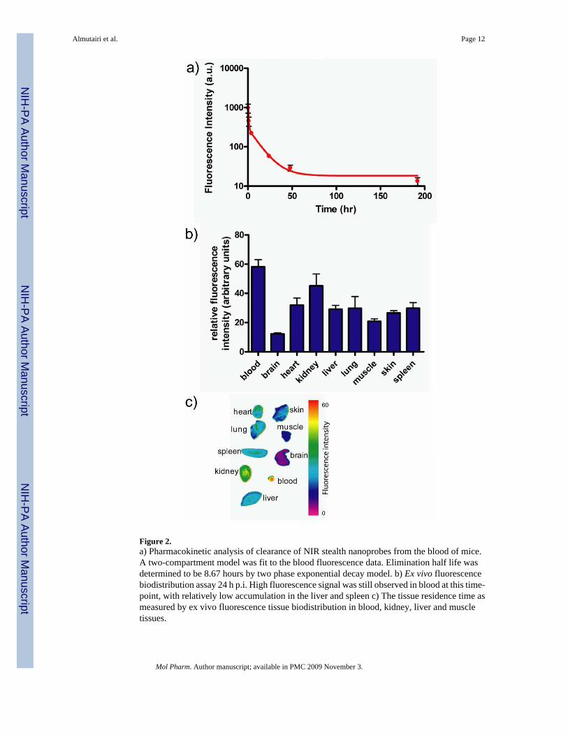
Figure 2.a) Pharmacokinetic analysis of clearance of NIR stealth nanoprobes from the blood of mice.A two-compartment model was fit to the blood fluorescence data. Elimination half life wasdetermined to be 8.67 hours by two phase exponential decay model. b) Ex vivo fluorescencebiodistribution assay 24 h p.i. High fluorescence signal was still observed in blood at this time-point, with relatively low accumulation in the liver and spleen c) The tissue residence time asmeasured by ex vivo fluorescence tissue biodistribution in blood, kidney, liver and muscletissues.
Almutairi et al. Page 12
Mol Pharm. Author manuscript; available in PMC 2009 November 3.
NIH
-PA Author Manuscript
NIH
-PA Author Manuscript
NIH
-PA Author Manuscript
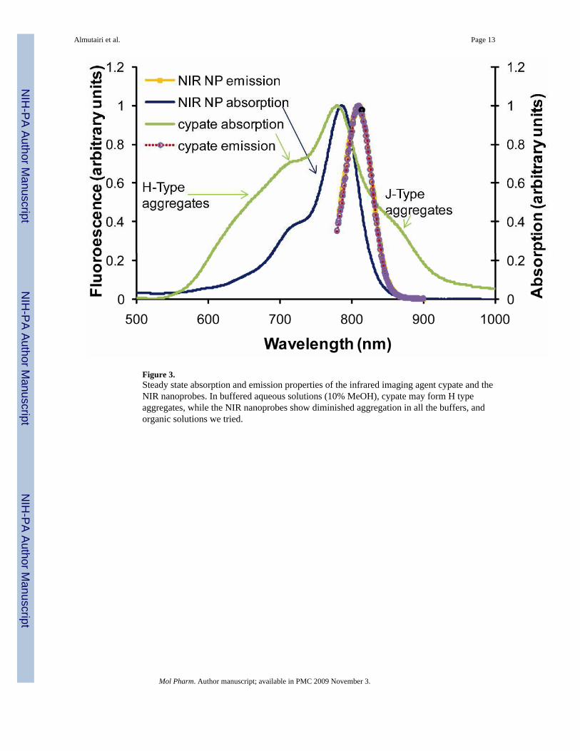
Figure 3.Steady state absorption and emission properties of the infrared imaging agent cypate and theNIR nanoprobes. In buffered aqueous solutions (10% MeOH), cypate may form H typeaggregates, while the NIR nanoprobes show diminished aggregation in all the buffers, andorganic solutions we tried.
Almutairi et al. Page 13
Mol Pharm. Author manuscript; available in PMC 2009 November 3.
NIH
-PA Author Manuscript
NIH
-PA Author Manuscript
NIH
-PA Author Manuscript

Figure 4.ICG was found to be moderately stable to degradation within one hour in pure water, indicatedby an almost horizontal line. (a) After the addition of microsomes and NADPH regeneratingsystems, the emission of ICG immediately dropped by 30% and continued declining withinthe experimental timeframe. The absorption kinetics curve (b) closely matched the emissiondata indicating the loss of the ICG's polymethine chromophore and not the emission quenching.The destruction of the chromophore apparently proceeds via an oxidation pathway and iscatalyzed by Cytochrome P450 - a metabolic enzyme in liver microsomes. In contrast, the NIRdendritic nanoprobes are much more stable toward enzymatic oxidation, likely due to theprotective layer around the chromophore.
Almutairi et al. Page 14
Mol Pharm. Author manuscript; available in PMC 2009 November 3.
NIH
-PA Author Manuscript
NIH
-PA Author Manuscript
NIH
-PA Author Manuscript
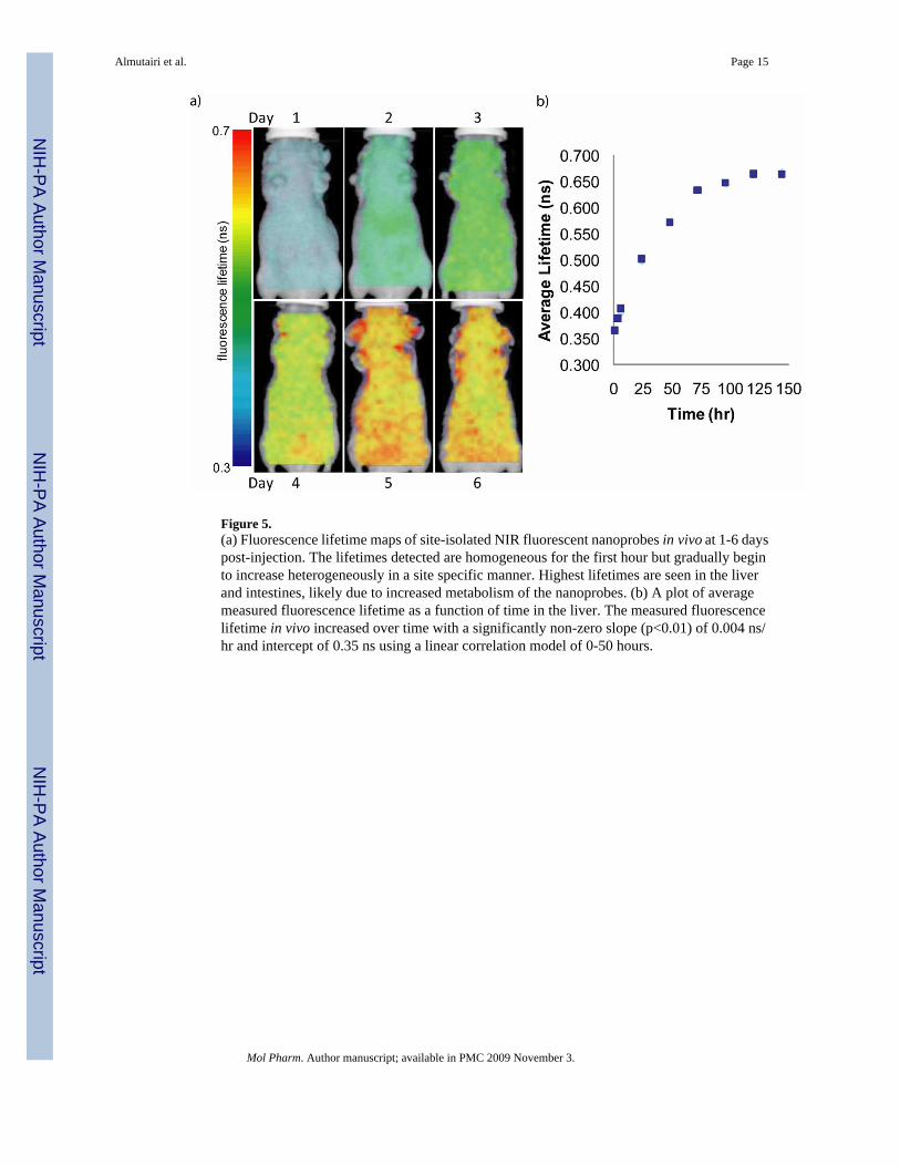
Figure 5.(a) Fluorescence lifetime maps of site-isolated NIR fluorescent nanoprobes in vivo at 1-6 dayspost-injection. The lifetimes detected are homogeneous for the first hour but gradually beginto increase heterogeneously in a site specific manner. Highest lifetimes are seen in the liverand intestines, likely due to increased metabolism of the nanoprobes. (b) A plot of averagemeasured fluorescence lifetime as a function of time in the liver. The measured fluorescencelifetime in vivo increased over time with a significantly non-zero slope (p<0.01) of 0.004 ns/hr and intercept of 0.35 ns using a linear correlation model of 0-50 hours.
Almutairi et al. Page 15
Mol Pharm. Author manuscript; available in PMC 2009 November 3.
NIH
-PA Author Manuscript
NIH
-PA Author Manuscript
NIH
-PA Author Manuscript