Molecular Pharmacology and Ligand Docking Studies Reveal a Single Amino Acid Difference between...
12
1521-0103/347/3/705–716$25.00 http://dx.doi.org/10.1124/jpet.113.208637 THE JOURNAL OF PHARMACOLOGY AND EXPERIMENTAL THERAPEUTICS J Pharmacol Exp Ther 347:705–716, December 2013 Copyright ª 2013 by The American Society for Pharmacology and Experimental Therapeutics Molecular Pharmacology and Ligand Docking Studies Reveal a Single Amino Acid Difference between Mouse and Human Serotonin 5-HT2A Receptors That Impacts Behavioral Translation of Novel 4-Phenyl-2-dimethylaminotetralin Ligands Clinton E. Canal, Tania Cordova-Sintjago, Yue Liu, Myong S. Kim, Drake Morgan, and Raymond G. Booth Center for Drug Discovery (C.E.C., T.C.-S., Y.L., R.G.B.), Department of Pharmaceutical Sciences (C.E.C., T.C.-S., Y.L., R.G.B.), and Department of Chemistry and Chemical Biology (R.G.B.), Northeastern University, Boston, Massachusetts; and Department of Medicinal Chemistry (M.S.K., T.C.-S.) and Department of Psychiatry (D.M.), University of Florida, Gainesville, Florida Received August 7, 2013; accepted September 30, 2013 ABSTRACT During translational studies to develop 4-phenyl-2-dimethyl- aminotetralin (PAT) compounds for neuropsychiatric disorders, the (2R,4S)-trans-(1)- and (2S,4R)-trans-(2)-enantiomers of the analog 6-hydroxy-7-chloro-PAT (6-OH-7-Cl-PAT) demonstrated unusual pharmacology at serotonin (5-HT) 5-HT2 G protein–coupled receptors (GPCRs). The enantiomers had similar affinities (K i ) at human (h) 5-HT2A receptors (∼70 nM). In an in vivo mouse model of 5-HT2A receptor activation [(6)-(2,5)-dimethoxy-4- iodoamphetamine (DOI)–elicited head twitch], however, (2)-6- OH-7-Cl-PAT was about 5-fold more potent than the ( 1)-enantiomer at attenuating the DOI-elicited response. It was discovered that (1)-6-OH-7-Cl-PAT (only) had ∼40-fold-lower affinity at mouse (m) compared with h5-HT2A receptors. Molecular modeling and computational ligand docking studies indicated that the 6-OH moiety of (1)- but not (2)-6-OH-7-Cl-PAT could form a hydrogen bond with serine residue 5.46 of the h5-HT2A receptor. The m5-HT2A as well as m5-HT2B, h5-HT2B, m5-HT2C, and h5- HT2C receptors have alanine at position 5.46, obviating this interaction; (1)-6-OH-7-Cl-PAT also showed ∼50-fold lower affinity than (2)-6-OH-7-Cl-PAT at m5-HT2C and h5-HT2C receptors. Mutagenesis studies confirmed that 5-HT2A S5.46 is critical for (1)- but not (2)-6-OH-7-Cl-PAT binding, as well as function. The (1)-6-OH-7-Cl-PAT enantiomer showed partial agonist effects at h5-HT2A wild-type (WT) and m5-HT2A A5.46S point-mutated receptors but did not activate m5-HT2A WT and h5-HT2A S5.46A point-mutated receptors, or h5-HT2B, h5-HT2C, and m5-HT2C receptors; ( 2)-6-OH-7-Cl-PAT did not activate any of the 5-HT2 receptors. Experiments also included the (2R,4S)-trans-( 1)- and (2S,4R)-trans-(2)- enantiomers of 6-methoxy-7-chloro-PAT to validate hydrogen bonding interactions proposed for the corresponding 6-OH analogs. Results indicate that PAT ligand three-dimensional structure impacts target receptor binding and translational outcomes, supporting the hypothesis that GPCR ligand structure governs orthosteric binding pocket molecular determinants and resulting pharmacology. Introduction Serotonin (5-HT) 5-HT2A, 5-HT2B, and 5-HT2C G protein–coupled receptors (GPCRs) are pharmacotherapeutic targets of interest due in part to their involvement in several psychiatric disorders, as inferred from human genetic studies (Dracheva et al., 2008; Sun et al., 2008; Nichols, 2009; Thanseem et al., 2012). Drug discovery programs targeting 5-HT2 receptors are focused on treating psychosis (5-HT2A antagonists), sleep disorders (5-HT2A antagonists), cluster headaches (5-HT2 agonists), anxiety (5-HT2B/2C antagonists), and obesity (5-HT2C agonists) (Sewell et al., 2006; Abbas and Roth, 2008; Smith et al., 2010). In addition, compounds with combined 5-HT2C agonist plus 5-HT2A/2B antagonist activity, similar to certain trans-4-phenyl-2-dimethylaminotetralins (PATs), may be useful for treating substance abuse and other compulsive behavioral disorders (Booth et al., 2009; Canal et al., 2013; Cunningham et al., 2013; Higgins et al., 2013; Morgan et al., 2013). The (6)-(2,5)-dimethoxy-4-iodoamphetamine (DOI)-elicited head-twitch response (HTR) in mice is an animal model that is This work was supported by the National Institutes of Health National Institute of Mental Health [Grant R01-MH081193]; and National Institutes of Health National Institute on Drug Abuse [Grants R01-DA023928 and R01- DA030989] (to R.G.B. and D.M.). dx.doi.org/10.1124/jpet.113.208637. ABBREVIATIONS: ANOVA, analysis of variance; b 2 AR, b 2 -adrenergic receptor; DMEM, Dulbecco’s modified Eagle’s medium; DOI, (6)-(2,5)- dimethoxy-4-iodoamphetamine; GPCR, G protein–coupled receptor; h5-HT2A, human 5-HT2A receptor; 5-HT, serotonin; HTR, head-twitch response; HTRF, homogeneous time-resolved fluorescence; m5-HT2A, mouse 5-HT2A receptor; MD, molecular dynamics; (1)-6-OH-7-Cl-PAT, (2R,4S)-trans-(1)-6-hydroxy-7-chloro-PAT; (2)-6-OH-7-Cl-PAT, (2S,4R)-trans-(2)-6-hydroxy-7-chloro-PAT; (1)-6-OMe-7-Cl-PAT, (2R,4S)- trans-(1)-6-methoxy-7-chloro-PAT; (2)-6-OMe-7-Cl-PAT, (2S,4R)-trans-(2)-6-methoxy-7-chloro-PAT; PAT, 4-phenyl-2-dimethylaminotetralin (4-phenyl-N,N-dimethyl-1,2,3,4-tetrahydronaphthalene-2-amine); PLC, phospholipase C; TMD, transmembrane domain; WT, wild-type. 705
-
Upload
independent -
Category
Documents
-
view
1 -
download
0
Transcript of Molecular Pharmacology and Ligand Docking Studies Reveal a Single Amino Acid Difference between...

1521-0103/347/3/705–716$25.00 http://dx.doi.org/10.1124/jpet.113.208637THE JOURNAL OF PHARMACOLOGY AND EXPERIMENTAL THERAPEUTICS J Pharmacol Exp Ther 347:705–716, December 2013Copyright ª 2013 by The American Society for Pharmacology and Experimental Therapeutics
Molecular Pharmacology and Ligand Docking Studies Reveala Single Amino Acid Difference between Mouse and HumanSerotonin 5-HT2A Receptors That Impacts BehavioralTranslation of Novel 4-Phenyl-2-dimethylaminotetralin Ligands
Clinton E. Canal, Tania Cordova-Sintjago, Yue Liu, Myong S. Kim, Drake Morgan,and Raymond G. BoothCenter for Drug Discovery (C.E.C., T.C.-S., Y.L., R.G.B.), Department of Pharmaceutical Sciences (C.E.C., T.C.-S., Y.L., R.G.B.),and Department of Chemistry and Chemical Biology (R.G.B.), Northeastern University, Boston, Massachusetts; and Departmentof Medicinal Chemistry (M.S.K., T.C.-S.) and Department of Psychiatry (D.M.), University of Florida, Gainesville, Florida
Received August 7, 2013; accepted September 30, 2013
ABSTRACTDuring translational studies to develop 4-phenyl-2-dimethyl-aminotetralin (PAT) compounds for neuropsychiatric disorders,the (2R,4S)-trans-(1)- and (2S,4R)-trans-(2)-enantiomers of theanalog 6-hydroxy-7-chloro-PAT (6-OH-7-Cl-PAT) demonstratedunusual pharmacology at serotonin (5-HT) 5-HT2Gprotein–coupledreceptors (GPCRs). The enantiomers had similar affinities (Ki) athuman (h) 5-HT2A receptors (∼70 nM). In an in vivo mousemodel of 5-HT2A receptor activation [(6)-(2,5)-dimethoxy-4-iodoamphetamine (DOI)–elicited head twitch], however, (2)-6-OH-7-Cl-PATwas about 5-foldmore potent than the (1)-enantiomerat attenuating the DOI-elicited response. It was discovered that(1)-6-OH-7-Cl-PAT (only) had ∼40-fold-lower affinity at mouse(m) compared with h5-HT2A receptors. Molecular modeling andcomputational ligand docking studies indicated that the 6-OHmoiety of (1)- but not (2)-6-OH-7-Cl-PAT could form a hydrogenbond with serine residue 5.46 of the h5-HT2A receptor. Them5-HT2A as well as m5-HT2B, h5-HT2B, m5-HT2C, and h5-HT2C receptors have alanine at position 5.46, obviating this
interaction; (1)-6-OH-7-Cl-PAT also showed ∼50-fold loweraffinity than (2)-6-OH-7-Cl-PAT at m5-HT2C and h5-HT2Creceptors. Mutagenesis studies confirmed that 5-HT2A S5.46 iscritical for (1)- but not (2)-6-OH-7-Cl-PAT binding, as well asfunction. The (1)-6-OH-7-Cl-PAT enantiomer showed partialagonist effects at h5-HT2A wild-type (WT) and m5-HT2AA5.46S point-mutated receptors but did not activate m5-HT2AWT and h5-HT2A S5.46A point-mutated receptors, or h5-HT2B,h5-HT2C, and m5-HT2C receptors; (2)-6-OH-7-Cl-PAT didnot activate any of the 5-HT2 receptors. Experiments alsoincluded the (2R,4S)-trans-(1)- and (2S,4R)-trans-(2)-enantiomers of 6-methoxy-7-chloro-PAT to validate hydrogenbonding interactions proposed for the corresponding 6-OHanalogs. Results indicate that PAT ligand three-dimensionalstructure impacts target receptor binding and translationaloutcomes, supporting the hypothesis that GPCR ligand structuregoverns orthosteric binding pocket molecular determinants andresulting pharmacology.
IntroductionSerotonin (5-HT) 5-HT2A, 5-HT2B, and 5-HT2C G
protein–coupled receptors (GPCRs) are pharmacotherapeutictargets of interest due in part to their involvement in severalpsychiatric disorders, as inferred from human genetic studies(Dracheva et al., 2008; Sun et al., 2008; Nichols, 2009;Thanseem et al., 2012). Drug discovery programs targeting
5-HT2 receptors are focused on treating psychosis (5-HT2Aantagonists), sleep disorders (5-HT2A antagonists), clusterheadaches (5-HT2 agonists), anxiety (5-HT2B/2C antagonists),and obesity (5-HT2C agonists) (Sewell et al., 2006; Abbas andRoth, 2008; Smith et al., 2010). In addition, compounds withcombined 5-HT2C agonist plus 5-HT2A/2B antagonist activity,similar to certain trans-4-phenyl-2-dimethylaminotetralins(PATs), may be useful for treating substance abuse and othercompulsive behavioral disorders (Booth et al., 2009; Canalet al., 2013; Cunningham et al., 2013; Higgins et al., 2013;Morgan et al., 2013).The (6)-(2,5)-dimethoxy-4-iodoamphetamine (DOI)-elicited
head-twitch response (HTR) inmice is an animal model that is
This work was supported by the National Institutes of Health NationalInstitute of Mental Health [Grant R01-MH081193]; and National Institutes ofHealth National Institute on Drug Abuse [Grants R01-DA023928 and R01-DA030989] (to R.G.B. and D.M.).
dx.doi.org/10.1124/jpet.113.208637.
ABBREVIATIONS: ANOVA, analysis of variance; b2AR, b2-adrenergic receptor; DMEM, Dulbecco’s modified Eagle’s medium; DOI, (6)-(2,5)-dimethoxy-4-iodoamphetamine; GPCR, G protein–coupled receptor; h5-HT2A, human 5-HT2A receptor; 5-HT, serotonin; HTR, head-twitchresponse; HTRF, homogeneous time-resolved fluorescence; m5-HT2A, mouse 5-HT2A receptor; MD, molecular dynamics; (1)-6-OH-7-Cl-PAT,(2R,4S)-trans-(1)-6-hydroxy-7-chloro-PAT; (2)-6-OH-7-Cl-PAT, (2S,4R)-trans-(2)-6-hydroxy-7-chloro-PAT; (1)-6-OMe-7-Cl-PAT, (2R,4S)-trans-(1)-6-methoxy-7-chloro-PAT; (2)-6-OMe-7-Cl-PAT, (2S,4R)-trans-(2)-6-methoxy-7-chloro-PAT; PAT, 4-phenyl-2-dimethylaminotetralin(4-phenyl-N,N-dimethyl-1,2,3,4-tetrahydronaphthalene-2-amine); PLC, phospholipase C; TMD, transmembrane domain; WT, wild-type.
705

dependent on 5-HT2A receptor activation, and the HTR isproduced reliably by most hallucinogenic 5-HT2 agonists(Glennon, 1992; Canal and Morgan, 2012; Halberstadt andGeyer, 2013). Thus, use of the DOI-elicited HTR model isexpedient for translating drug discovery targeting 5-HT2receptors. Although the C57Bl/6 mouse 5-HT2A (m5-HT2A)receptor shares ∼99% sequence identity with the human5-HT2A (h5-HT2A) ortholog in the transmembrane domains(TMDs) where orthosteric ligands bind, there is significantdivergence regarding binding affinities (Ki or KD) of 5-HT2A-targeting ligands at the two receptors (Dougherty and Aloyo,2011). Similar observations are reported for the rat versushuman 5-HT2A receptors (Nelson et al., 1993; Wainscottet al., 1996).The only two residues within the TMDs that differ between
the m5-HT2A and h5-HT2A receptors are at positions(Ballesteros and Weinstein, 1995) 1.42 (alanine in humanand tyrosine in mouse) and 5.46 (serine in human and alaninein mouse). Position 1.42 is far from the orthosteric ligandbinding pocket (Córdova-Sintjago et al., 2012a), and thefunctional and/or structural role of amino acids at position1.42 is unknown. Amino acid residues in TMD5, however,have received attention due to their importance in ligandbinding and function of GPCRs (Almaula et al., 1996a; vanRhee and Jacobson, 1996; Wang et al., 2013). Pointedly,residue 5.46 of aminergic neurotransmitter GPCRs (seroto-nin, dopamine, norepinephrine, and histamine) is an importantcomponent for binding of certain ligands in the orthostericbinding pocket (van Rhee and Jacobson, 1996; Shapiro et al.,2000; Armbruster et al., 2007; Cummings et al., 2010; Istyastonoet al., 2011).Mutagenesis studies have revealed that in vitro pharmaco-
logical properties of some 5-HT2A ligands are impacted by thedifference in residue 5.46 at rodent versus human 5-HT2Areceptors (Kao et al., 1992; Johnson et al., 1994). Few studies,however, have examined species differences in receptorprotein structure-function with regard to in vivo behavioralpharmacology (Miller et al., 2009; Suratman et al., 2011), withmost reports just including a comparison of ligand-receptor invitro pharmacology. Even among in vitro studies, there are noreports that delineate molecular determinants for binding ofdifferent enantiomers of the same compound with 5-HT2Aresidue 5.46.Using a structure-based drug design approach, novel PATs
targeting 5-HT2 receptors are being developed to treat compul-sive behavioral and other neuropsychiatric disorders (Rowlandet al., 2008; Booth et al., 2009; Canal et al., 2013; Morgan et al.,2013). Reported here are results from in silico m5-HT2A andh5-HT2A molecular modeling/ligand docking studies, in vitromolecular pharmacology studies, and in vivo neurobehavioralstudies using the DOI-elicited HTR model for the trans-(2)- andtrans-(1)-enantiomers of two PAT-type drug candidates, 6-OH-7-Cl-PAT and 6-OMe-7-Cl-PAT (Fig. 1). In preliminary studies,it was observed that for 6-OMe-7-Cl-PAT, both enantiomershave similar affinity across h5-HT2 subtypes, and there was nodifference between the enantiomers regarding potency andefficacy for attenuating the DOI-elicited HTR in mice.For 6-OH-7-Cl-PAT, the (2)-enantiomer had significantly
higher affinity than the (1)-enantiomer at h5-HT2B andh5-HT2C receptors, but there was no difference in affinity forthe two enantiomers at the h5-HT2A receptor. Nevertheless,the (2)-enantiomer showed significantly greater potency and
efficacy for attenuating the 5-HT2A-dependent DOI-elicitedHTR in mice. Docking studies of the 6-OH-7-Cl-PAT enan-tiomers at molecular models of 5-HT2A receptors (Córdova-Sintjago et al., 2012a) led to an examination of the putativerole of 5-HT2A residue 5.46 in mediating (1)-6-OH-7-Cl-PATligand affinity and function. The results pinpoint a singleamino acid difference at position 5.46 in the m5-HT2A andh5-HT2A receptor that impacts translation of novel PAT drugcandidates for neuropsychiatric disorders.
Materials and MethodsCompounds
Structures of (1)- and (2)-6-OH-7-Cl-PAT and 6-OMe-7-Cl-PATsused in the studies are shown in Fig. 1. The PATs were synthesizedin our laboratory as racemic mixtures. Single enantiomers wereresolved by chiral stationary-phase high-performance liquid chro-matography and converted to hydrochloride salts as previouslydescribed (Bucholtz et al., 1999; Booth et al., 2009; Vincek andBooth, 2009). DOI and mianserin hydrochloride were purchasedfrom Sigma-Aldrich (St. Louis, MO). 5-HT hydrochloride was purchasedfrom Alfa Aesar (Ward Hill, MA). [3H]Ketanserin, [3H]mesulergine, and[3H]myoinositol were obtained from PerkinElmer Life and AnalyticalSciences (Waltham, MA). All solid compounds were weighed on anXP26 microanalytical balance (Mettler-Toledo, Columbus, OH).
Human and Mouse Receptor Constructs and PointMutations
A description of the wild-type (WT) m5-HT2 and h5-HT2 receptorcDNA constructs used was reported recently (Canal et al., 2013). Pointmutations of residue 5.46 of both m5-HT2A and h5-HT2A receptors weremade by polymerase chain reactions using the QuikChange II Site-Directed Mutagenesis Kit (Agilent Technologies, Santa Clara, CA)according to the manufacturer’s protocol. WT m5-HT2 receptorcDNAs, cloned in the pCMV6 vector, were purchased from OriGene(Rockville, MD). WT h5-HT2 receptor cDNAs, cloned in the pcDNA3.11vector, were purchased from Missouri S&T cDNA Resource Center(Rolla, MO; http://www.cdna.org/). Mutagenesis and sequencing prim-ers were obtained from Life Technologies (Carlsbad, CA). Primersused to make the m5-HT2A A5.46S point mutation were 59-cctca-taggctcttttgtgtcatttttcatccccctaac-39 (sense) and 59-gttagggggatgaaaaat-gacacaaaagagcctatgagg-39 (antisense). Primers used to make theh5-HT2A S5.46A point mutation were 59-cctgatcggctcttttgtggcattttt-cattcccttaac-39 (sense) and 59-gttaagggaatgaaaaatgccacaaaagagcc-gatcagg-39 (antisense). Polymerase chain reaction was performed aspreviously described, with optimization (Canal et al., 2011). ParentalDNA in the reaction mixture was digested using DpnI at 37°C for 1hour. Digestion mixture (2 ml) was transformed into 50 ml of XL1-Bluesupercompetent cells by heat pulse at 42°C for 45 seconds. The
Fig. 1. Structures of PAT ligands tested.
706 Canal et al.
transformed reaction was incubated in Super Optimal broth withCatabolite repression medium (Sigma-Aldrich), then plated onto LBagar plates containing 100 mg/ml kanamycin (mouse 5-HT2AWT andA5.46S mutant receptor) or 100 mg/ml ampicillin (human 5-HT2AWTand S5.46A mutant receptor). Single colonies were selected forsequencing, and point mutations were confirmed by Genewiz (SouthPlainfield, NJ).
Radioligand Binding Assays
Radioligand saturation isotherm and competitive displacementbinding assays were performed in 96-well plates using 5-HT2A,5-HT2B, or 5-HT2C receptors transiently expressed in HEK293 cellsas previously described (Canal et al., 2011, 2013); 10 mg of htr2A,htr2B, or htr2C cDNA and 20 ml of Lipofectamine 2000 (Life Tech-nologies) or TurboFect Transfection Reagent (Thermo Scientific,Waltham, MA) was used for transfections. For saturation bindingassays, 0.02–16 nM [3H]ketanserin was used to obtain KD values atthe WT and mutant 5-HT2A receptors. The BCA protein assay kit(Thermo Scientific) was used to quantify protein concentration inreceptor-expressing cell membranes, for later determination ofm5-HT2A and h5-HT2A WT receptor binding site densities, Bmax
values, calculated using GraphPad software (GraphPad Software, LaJolla, CA; http://graphpad.com/quickcalcs/radcalcform/). For com-petition binding assays, 1–3 nM [3H]ketanserin or [3H]mesulergine(∼KD concentration) was used to obtain the Ki values for unlabeledligands at each of the 5-HT2 receptor subtypes. Nonspecific bindingwas determined in the presence of 10 mM mianserin. Each assay wasperformed aminimum of three times, using at least sample triplicatesfor each ligand concentration.
Phosphoinositide Hydrolysis Assay
Receptor-mediated phosphoinositide hydrolysis by phospholipaseC (PLC) was measured as previously described (Canal et al., 2013). Inbrief, HEK293 cells transiently expressing a 5-HT2 receptor wereincubated with 16 ml inositol-free Dulbecco’s modified Eagle’smedium (DMEM) containing 1 mCi/ml [3H]myoinositol. Cells wereapportioned into 48-well CellBind plates (Corning, Lowell, MA), and[3H]myoinositol incorporation continued for 24 hours in an incubator.Cells were then acclimated to serum-free media, treated for 30minutes with experimental drugs that had been prediluted in mediato contain a final concentration of 50 mM LiCl per well. Media werediscarded, and 50 mM formic acid was added to lyse cells. Plates werestored at 280°C for at least 24 hours. After thawing, solution fromeach well was added to individual anion-exchange columns. Bound[3H]inositol phosphates were eluted with 800 mM ammonium formate,and eluate was added to scintillation fluid (ScintiVerse Cocktail;Thermo Fisher Scientific, Pittsburgh, PA). 3H-Induced scintillationswere counted with a scintillation analyzer. Experiments were per-formed with duplicates for 5-HT and triplicates for all other drugs,and each independent experiment was performed a minimum of threetimes.
IP-One HTRF Assay
5-HT2A receptor–mediated inositol phosphate 1 production wasmeasured using the IP-One HTRF assay kit (Cisbio, Bedford, MA).These assays were performed during a transition from using the time-consuming method of the phosphoinositide hydrolysis assay, asdescribed above. We have found consistent results between the twoassays in the rank order of potency and efficacy of all ligands testedrelative to 5-HT at 5-HT2A receptors, and others also reportcongruency between the IP-One HTRF and Ca21 flux methods at5-HT2A receptors (Jensen et al., 2013). The assay was performedaccording to the Cisbio protocol yet with the following optimization.HEK293 cells transiently transfected with 5-HT2A WT or 5.46mutant receptors were incubated in serum-free DMEM for 1 hour.Cells were then harvested and pelleted by centrifugation at 200g at
37°C for 5 minutes, followed by resuspension in serum-free DMEM.Cell number was counted using a hemacytometer (Hausser Scientific,Horsham, PA), and 500 cells/ml DMEM were used. Five microliters ofcell suspension and 5 ml of test compounds in stimulation buffer(included in kit and containing 50mMLiCl) were added to each well ofa 384-well plate (Greiner Bio-one, Monroe, NC). The plate was incu-bated at 37°C for 2 hours. After incubation, the reaction was termi-nated by adding 5 ml of the donor and acceptor fluorescent conjugatesin lysis buffer. The plate was incubated at room temperature for1 hour to reach equilibrium. Inositol phosphate 1 levels were assessedby the ratio of fluorescence emission at 665 nM and 620 nM (fluor-escence resonance energy transfer, HTRF) using a Synergy H1 platereader with anHTRF filter cube (BioTek,Winooski, VT). Experimentswere performed with duplicates for 5-HT and triplicates for all otherdrugs, and each independent experiment was performed a minimumof three times.
Animal Subjects
Male C57Bl/6J mice, purchased at 60 days old from The JacksonLaboratory (Bar Harbor, ME), were used in all experiments. Micewere housed in standard cages with ad libitum access to food andwater and were acclimated in the vivarium for at least 4 days beforetesting. Animal procedures were approved by the Institutional AnimalCare and Use Committee and are in accordance with the principles inthe National Institutes of Health Guide for Care and Use of LaboratoryAnimals (Publication No. 85-23, revised 1985).
DOI-Elicited HTR
All compounds were dissolved in sterile Milli-Q water (EMDMillipore, Billerica, MA), which served as the vehicle control. All com-pounds were administered subcutaneously at a volume of 10 ml/kg.A single dose of DOI (1 mg/kg) was used for all experiments, as it isknown to reliably elicit a robust, consistent, and submaximal numberof HTRs in C57Bl/6J mice (Canal et al., 2010; Canal and Morgan,2012). Two or three doses were used to test the efficacy of novel PATligands for attenuating the DOI-elicited HTR. Doses for the com-pounds were as follows: (2)- and (1)-6-OMe-7-Cl-PAT, 3 and 5.6 mg/kg;(2)- and (1)-6-OH-7-Cl-PAT, 1.5, 3, and 5.6 mg/kg. Solutions of allcompounds were made fresh on the day of behavioral testing. Drug orvehicle was injected 10 minutes prior to injection of DOI. Mice wereplaced in an activity chamber (43 � 43 cm; Med Associates, Inc., St.Albans, VT) 10 minutes later, and HTRs were counted for the next10 minutes by an observer blind to the experimental treatment.
Statistical Analyses
Competition and saturation binding data were analyzed usingnonlinear regression commands in GraphPad Prism 6.02 forWindows(GraphPad Software). Binding data were fit using the “one-site”models. Two-site curve-fitting did not result in an improved fit (datanot shown). Approximate Ki values were determined by conversion ofthe IC50 data using the equation Ki 5 IC50/1 1 L/KD, where L is theconcentration of radioligand (Cheng and Prusoff, 1973). Calculationsof unpaired t tests and one-way analyses of variance (ANOVAs) withTukey’s multiple-comparison post hoc tests were performed tocompare calculated Ki and EC50 values of individual compounds forreceptors. Ligand efficacies (Tables 1 and 2) are presented as themean (6 S.E.M.) of the percentages of maximal 5-HT response fromall assays; 5-HT served as a positive control, full agonist in allfunctional assays.
Separate ANOVAs were performed to analyze effects of experi-mental compounds on the DOI-elicited HTR. Dunnett’s post-testcompared the number of HTRs elicited by DOI after pretreatmentwith each experimental compound to the number of HTRs elicited byDOI and vehicle administration. Two-way ANOVA and Student-Newman-Keuls comparisons were used to identify dose-dependenteffects and differences in efficacy across test compounds.
5-HT2A 5.46 Impacts PAT Pharmacology 707
Molecular and Computational Modeling
Homology Modeling. A b2-adrenergic receptor (b2AR)-based(Protein Data Bank entry: 2RH1) homology model of the h5-HT2Areceptor was used in docking and molecular dynamics (MD) studies.Detailed homology modeling procedures are described elsewhere(Córdova-Sintjago et al., 2012b). In brief, the 5-HT2A native sequence(WT) was aligned to the b2AR sequence using ClustalW multiplesequence alignment (Thompson et al., 1994; Larkin et al., 2007).Other structures (e.g., the inverse agonist carazolol, T4-lysozyme, andcholesterol molecules) present in the b2AR/T4-lysozyme chimeracrystal structure were deleted. To obtain the 5-HT2A receptor model,point mutations were performed as needed, followed by theappropriate sequence additions and deletions to match the WT aminoacid sequence. TMDs and loops were built using the Biopolymermodule of Sybyl-X 2.0 (Tripos International, St. Louis, MO), and thecrude models of the unbound receptor were minimized using thePowell method with Tripos force field (Clark et al., 1989) andAMBER charges (Cornell et al., 1995) followed by equilibration in a1-palmitoyl-2-oleyl-sn-glycero phosphatidyl choline bilayer (Helleret al., 1993). The system was relaxed using the Tripos force field toa gradient 0.05 kcal/Å.mol prior to MD simulations in the 1-palmitoyl-2-oleyl-sn-glycero phosphatidyl choline membrane. MD simulationconditions were time run for 5 microseconds, time step 1 femtosecond,with snapshots collected every 5 femtoseconds. Other parameterswere the canonical ensemble (number of particles, volume, andtemperature were constants), 300K temperature, Boltzmann initialvelocities, and nonbonded cutoff set at 8 Å. Constraints for alphacarbons in the TMDs were employed. Subsequently, the constraintswere removed for a 1000-picosecond MD simulation run. The finalunbound WT 5-HT2A homology model was obtained from the medianstructure after clustering analysis of the frames from the last 10picoseconds of the MD simulation, and optimized using the Triposforce field to a convergence of 0.05 kcal/Å.mol. The stability andvalidation of the model were tested as described previously (Córdova-Sintjago et al., 2012b).
While this article was in preparation, the crystal structure ofh5-HT2B GPCR was reported (Wang et al., 2013). To assess thevalidity of our h5-HT2Amodel using b2AR as a template, we similarlybuilt an h5-HT2B homology model and compared it to the crystalstructure of the chimeric h5-HT2B–BRIL in complex with ergota-mine, Protein Data Bank code 4IB4, by aligning the alpha carbonatoms. The root mean square deviation of 3.34 Å indicates that theh5-HT2B homology model is similar to the crystal structure andvalidates the strategy used to generate the h5-HT2A model (Fig. 6). Asimilar conclusion was reachedwhen comparing the b2AR-based humanhistamine H1 homology model to its crystal structure (Cordova-Sintjago et al., 2012c).
Ligand Docking. The ligands (1)- and (2)-6-OH-7-Cl-PAT werebuilt as monocations (protonated amines) using HyperChem 8.0(Hybercube, Gainesville, FL) and optimized using PM3 modelHamiltonian to a gradient of 0.01 kcal/Å mol. The binding site wasdefined by assigning residue D3.32 as a definitive binding siteinteraction point with the PAT ligand’s protonated amine moiety andincluding residues within a 7-Å radius (Kristiansen et al., 2000; Canalet al., 2011). The initial ligand-receptor complex configuration wasused for flexible ligand docking with Flexidock in Sybyl-X 2.0.Structure preparation was carried out prior to docking studies byassigning AMBER charges (Cornell et al., 1995) for the protein andGesteiger-Marsili charges (Gasteiger and Marsili, 1980) for theligand. Rotatable bonds in the ligand and the side chains of residuesdefining the receptor putative active site were screened for optimalpositioning of the ligand and side chains in the conformational space;remaining residues were frozen during docking. Default FlexiDockparameters were set at 80,000-generation. The best docking solution,according to the highest FlexiDock score, wasminimized using the Triposforce field to a gradient of 0.05 kcal/Å.mol, prior to MD simulation.T
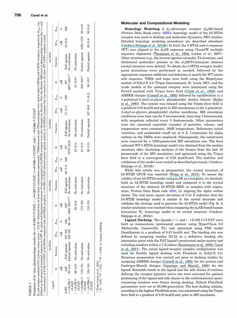
ABLE
1Sum
maryof
invitroph
armacolog
yresu
ltsfor(+)-6-OH-7-C
l-PAT,(2
)-6-OH-7-C
l-PAT,(+)-6-OMe-7-Cl-PAT,an
d(2
)-6-OMe-7-Cl-PAT
Kian
dreceptor-m
ediatedPLCactivity
values
(mea
n6
S.E.M
.)ob
tained
from
atleas
tthreeinde
pende
ntex
perimen
ts.[
3H]K
etan
serinan
d[3H]m
esulerginewereusedto
labe
l5-H
T2A
and5-HT2C
receptors,resp
ective
ly.E
maxwas
defined
asthepe
rcen
tof
max
imal
resp
onse
to5-HT.K
iva
lues
andfunctional
outcom
esof
each
compo
undat
h5-HT2B
receptorswerealso
obtained
(see
Results).N
otethedistinct
differen
cein
pharmacolog
ybe
twee
nthe(+)-an
d(2
)-en
antiom
ersof
6-OH-7-C
l-PATat
h5-HT2A
compa
redwithm5-HT2A
receptors.
(+)-6-OH-7-C
l-PAT
show
edpa
rtialag
onistactivity
ath5
-HT2A
receptorsbu
twas
inactive
atallothe
rWT5-HT2receptorstested
.
Ligan
dh5-HT2A
m5-HT2A
h5-H
T2C
m5-HT2C
Ki
Fun
ction
EC50
Emax
Ki
Fun
ction
EC50
Emax
Ki
Fun
ction
EC50
Emax
Ki
Fun
ction
EC50
Emax
nM
nM
%nM
nM
%nM
nM
%nM
nM
%
5-HT
128a
Fullag
onist
127(24)
100
138a
Fullag
onist
56(7.5)
100
15a
Fullag
onist,
11(2.7)
100
50a
Fullag
onist
18(2.8)
100
(1)-6-OH-7-
Cl-PAT
80(4.4)
Partial
agon
ist
125(32)
36(10)
3005
(321
)***
Noactiva
tion
1079
(42.9)
Noactiva
tion
3651
(481
)**
Noactiva
tion
(2)-6-OH-7-
Cl-PAT
69(4.9)
Noactiva
tion
58(6.8)
Noactiva
tion
21(2.4)
Noactiva
tion
51(9.5)*
Noactiva
tion
(1)-6-OMe-7-
Cl-PAT
176(18)
Noactiva
tion
173(29)
Noactiva
tion
47(5.3)
Noactiva
tion
243(22)**
Partial
agon
ist
1359
(178
)59
(4.0)
(2)-6-OMe-7-
Cl-PAT
134(18)
Noactiva
tion
119(12)
Noactiva
tion
30(3.3)
Partial
agon
ist
127(8.6)
71(8.2)
98(15)*
Partial
agon
ist
29(5.4)
80(5.4)
aFrom
Can
alet
al.,20
13.
*P,
0.05
;**P
,0.01
;***P,
0.00
1vs.human
ortholog
.
708 Canal et al.
Molecular Dynamics. The selected high-score pose of the dockedligand was subjected to an MD simulation run for 500 picoseconds,with other parameters the same as above, to allow adjustment of thepositions of side chains and helices. The final structure of the liganddocked at the receptormodel was obtained from the average of the last10 picoseconds of the MD simulation.
ResultsAffinity and Function of 6,7-Substituted PATs at
Mouse and Human WT 5-HT2A Receptors. Four PATcompounds were tested for affinity and function at each ofthem5-HT2 and h5-HT2 receptor subtypes, excludingm5-HT2B(which was not procured). 5-HT was used as a positive control,full agonist for assessing the functional efficacies and poten-cies of PAT compounds. Representative binding curves for(2)- and (1)-6-OH-7-Cl-PAT at m5-HT2A and h5-HT2A areshown in Fig. 2. A summary of all Ki results (mean 6 S.E.M.),excluding h5-HT2B data, is shown in Table 1. The mean 6S.E.M. Ki of (2)-6-OH-7-Cl-PAT at h5-HT2B receptors was102.4 6 2.6 nM, whereas (1)-6-OH-7-Cl-PAT had a Ki of1018 6 144.8 nM. The mean 6 S.E.M. Ki of (2)-6-OMe-7-Cl-PAT at h5-HT2B receptors was 29.3 6 4.8 nM, whereas (1)-6-OMe-7-Cl-PAT had a Ki of 72.7 6 10.3 nM.ANOVA of Ki values of (2)-6-OH-7-Cl-PAT at each receptor
revealed a significant main effect of receptor subtype (F4,16 518.71; P , 0.0001). The only post-test comparisons that didnot reach a significant difference were the affinities at m5-HT2A versus h5-HT2A receptors, m5-HT2C versus h5-HT2Areceptors, and m5-HT2A versus m5-HT2C receptors; similarresults were obtained for (1)-6-OH-7-Cl-PAT (F4,12 5 51.46;P , 0.0001). Only h5-HT2B versus h5-HT2C and m5-HT2Aversus m5-HT2C Ki values did not reach a significant differ-ence. Unpaired t tests comparing Ki values of the (2)- versus(1)-enantiomers at each of the 5-HT2 subtypes revealed thatthe h5-HT2A receptor was the only receptor where the Ki
values were not significantly different. Regarding functionaleffects, the (1)-6-OH-7-Cl-PAT compound was a partial agonistat h5-HT2A receptors only (Table 1). Neither 6-OH-7-Cl-PATenantiomer showed activity up to 10 mM at all other 5-HT2receptors screened, including m5-HT2A.
Separate ANOVAs of Ki values of (2)-6-OMe-7-Cl-PAT and(1)-6-OMe-7-Cl-PAT at each receptor revealed a significantmain effect of receptor subtype (F4,15 5 20.40; P , 0.0001 andF4,11 5 17.43; P , 0.0001, respectively). Post-test comparisonsthat did not reach a significant difference were the same acrossthe two enantiomers: m5-HT2A versus h5-HT2A receptors, h5-HT2B versus h5-HT2C receptors, h5-HT2A versus m5-HT2Creceptors, and m5-HT2A versus m5-HT2C. Unpaired t testscomparingKi values of the (2)- versus (1)-enantiomers at eachof the 5-HT2 subtypes revealed that the Ki values of theenantiomers were statistically different only at h5-HT2B re-ceptors and m5-HT2C receptors (P , 0.05). Regarding func-tional effects, (2)-6-OMe-7-Cl-PAT was a partial agonist at bothm5-HT2C and h5-HT2C receptors yet was inactive up to 10 mMat all other 5-HT2 receptors (Table 1). (1)-6-OMe-7-Cl-PAT wasa nonpotent, partial agonist at m5-HT2C receptors and wasinactive up to 10 mM at all other 5-HT2 receptors (Table 1).Bmax values for [3H]ketanserin-labeled h5-HT2A and
m5-HT2A receptors were obtained from HEK293 cells trans-fected separately from cells used for competition binding andfunctional assays, yet transfection procedures for all assayswere the same. m5-HT2A and h5-HT2A receptor Bmax valuesvaried across transfections; the mean 6 S.E.M. values were1311 6 137.9 and 1046 6 595.8 fmol/mg, respectively, whichwere not statistically different (P . 0.05).Affinity and Function of (1)- and (2)-6-OH-7-Cl-PATs
at Mouse and Human 5.46 Point-Mutated 5-HT2AReceptors. TheKD value of [3H]ketanserin atmutantm5-HT2AA5.46S and h5-HT2A S5.46A receptors was not statisticallydifferent. The mean 6 S.E.M. KD values of [3H]ketanserinat m5-HT2A A5.46S and h5-HT2A S5.46A receptors were1.2 6 0.3 and 1.6 6 0.4 nM, respectively, closely matchingmany reported literature values (Almaula et al., 1996a; Bradenand Nichols, 2007) and not statistically different from 5-HT2AWT receptors (P . 0.05). The KD value of [3H]ketanserin atm5-HT2A WT and h5-HT2A WT receptors was 1.3 6 0.2 and1.6 6 0.5, respectively. Figure 2 shows representativecompetition binding curves for (2)- and (1)-6-OH-7-Cl-PATat each of the [3H]ketanserin-labeled mutant receptors—only these two enantiomers were screened at the mutant
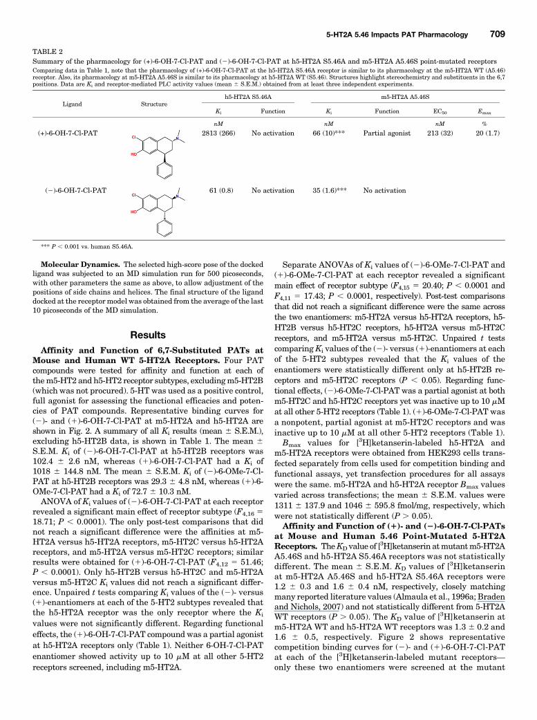
TABLE 2Summary of the pharmacology for (+)-6-OH-7-Cl-PAT and (2)-6-OH-7-Cl-PAT at h5-HT2A S5.46A and m5-HT2A A5.46S point-mutated receptorsComparing data in Table 1, note that the pharmacology of (+)-6-OH-7-Cl-PAT at the h5-HT2A S5.46A receptor is similar to its pharmacology at the m5-HT2A WT (A5.46)receptor. Also, its pharmacology at m5-HT2A A5.46S is similar to its pharmacology at h5-HT2A WT (S5.46). Structures highlight stereochemistry and substituents in the 6,7positions. Data are Ki and receptor-mediated PLC activity values (mean 6 S.E.M.) obtained from at least three independent experiments.
Ligand Structureh5-HT2A S5.46A m5-HT2A A5.46S
Ki Function Ki Function EC50 Emax
nM nM nM %
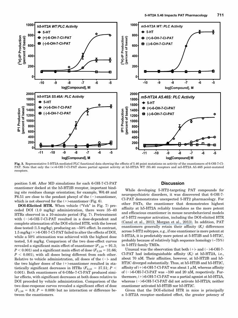
(+)-6-OH-7-Cl-PAT 2813 (266) No activation 66 (10)*** Partial agonist 213 (32) 20 (1.7)
(2)-6-OH-7-Cl-PAT 61 (0.8) No activation 35 (1.6)*** No activation
*** P , 0.001 vs. human S5.46A.
5-HT2A 5.46 Impacts PAT Pharmacology 709
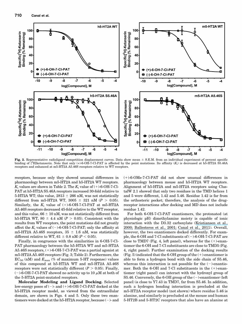
receptors, because only they showed unusual differences inpharmacology between m5-HT2A and h5-HT2A WT receptors.Ki values are shown in Table 2. The Ki value of (1)-6-OH-7-Cl-PAT at h5-HT2A S5.46A receptors increased 30-fold relative to5-HT2A WT; this value, 2813 6 266 nM, was not statisticallydifferent from m5-HT2A WT, 3005 6 321 nM (P . 0.05).Similarly, the Ki value of (1)-6-OH-7-Cl-PAT at m5-HT2AA5.46S receptors decreased 45-fold relative to theWT receptor,and this value, 666 10 nM, was not statistically different fromh5-HT2A WT, 80 6 4.4 nM (P . 0.05). Consistent with theresults fromWT receptors, the point mutations did not greatlyaffect the Ki values of (2)-6-OH-7-Cl-PAT; only the affinity atm5-HT2A A5.46S receptors, 35 6 1.6 nM, was statisticallydifferent relative to WT, 61 6 0.8 nM (P , 0.05).Finally, in congruence with the similarities in 6-OH-7-Cl-
PAT pharmacology between the h5-HT2A WT and m5-HT2AA5.46S receptors, (1)-6-OH-7-Cl-PAT was a partial agonist atm5-HT2AA5.46S receptors (Fig. 3; Table 2). Furthermore, theEC50 (nM) and Emax (% of maximum 5-HT response) valuesof this compound at h5-HT2A WT and m5-HT2A A5.46Sreceptors were not statistically different (P . 0.05). Finally,(2)-6-OH-7-Cl-PAT showed no activity up to 10 mM at both ofthe 5-HT2A point-mutated receptors.Molecular Modeling and Ligand Docking. Selected
low-energy poses of (2)- and (1)-6-OH-7-Cl-PAT docked at theh5-HT2A receptor model, as viewed from the extracellulardomain, are shown in Figs. 4 and 5. Only these two enan-tiomerswere docked at the h5-HT2A receptor, because (2)- and
(1)-6-OMe-7-Cl-PAT did not show unusual differences inpharmacology between mouse and h5-HT2A WT receptors.Alignment of h5-HT2A and m5-HT2A receptors using Clus-talW 2.1 showed that only two residues in the TMD helices 1and 5 were different, 1.42 and 5.46. Residue 1.42 is far fromthe orthosteric pocket; therefore, the analysis of the drug-receptor interactions after docking and MD does not includeresidue 1.42.For both 6-OH-7-Cl-PAT enantiomers, the protonated (at
physiologic pH) dimethylamine moiety is capable of ionicinteraction with the D3.32 carboxylate (Kristiansen et al.,2000; Ballesteros et al., 2001; Canal et al., 2011). Overall,however, the two enantiomers docked differently. For exam-ple, the 6-OH and 7-Cl substituents of (2)-6-OH-7-Cl-PAT areclose to TMD7 (Fig. 4, left panel), whereas for the (1)-enan-tiomer the 6-OH and 7-Cl substituents are close to TMD5 (Fig.4, right panel). Further examination of the docking results(Fig. 5) indicated that the 6-OH group of the (1)-enantiomer isable to form a hydrogen bond with the side chain of S5.46,whereas this interaction is not possible for the (2)-enantio-mer. Both the 6-OH and 7-Cl substituents in the (1)-enan-tiomer (right panel) can interact with the hydroxyl group ofS5.46. Conversely, the 6-OH group of the (2)-enantiomer (leftpanel) is close to Y7.43 in TMD7, far from S5.46. In addition,such a hydrogen bonding interaction is precluded at them5-HT2A receptor model (not shown) where residue 5.46 isalanine, and similarly is precluded at the mouse and human5-HT2B and 5-HT2C receptors that also have an alanine at
Fig. 2. Representative radioligand competition displacement curves. Data show mean 6 S.E.M. from an individual experiment of percent specificbinding of [3H]ketanserin. Note that only (+)-6-OH-7-Cl-PAT is affected by the point mutations. Its affinity (Ki) is decreased at h5-HT2A S5.46Areceptors and enhanced at m5-HT2A A5.46S receptors relative to WT receptors.
710 Canal et al.
position 5.46. After MD simulations for each 6-OH-7-Cl-PATenantiomer docked at the h5-HT2B receptor, important bind-ing site residues change orientation; for example, W6.48 andF6.51 are close to the pendant phenyl of the (2)-enantiomer,which is not observed for the (1)-enantiomer (Fig. 6).DOI-Elicited HTR. When vehicle (“Veh” in Fig. 7) pre-
ceded DOI (1.0 mg/kg) administration, there were 35–40HTRs observed in a 10-minute period (Fig. 7). Pretreatmentwith (2)-6-OH-7-Cl-PAT resulted in a dose-dependent andcomplete attenuation of the DOI-elicited HTR, with the lowestdose tested (1.5 mg/kg), producing an ∼50% effect. In contrast,1.5 mg/kg (1)-6-OH-7-Cl-PAT failed to alter the effects of DOI,while a 50% attenuation was achieved with the highest dosetested, 5.6 mg/kg. Comparison of the two dose-effect curvesrevealed a significant main effect of enantiomer (F1,28 5 91.3;P, 0.001) and a significant main effect of dose (F2,28 5 31.66;P , 0.001), with all doses being different from each other.Relative to vehicle administration, all doses of the (2)- andthe two higher doses of the (1)-enantiomer resulted in sta-tistically significant decreases in HTRs (F6,41 5 37.51; P ,0.001). Both enantiomers of 6-OMe-7-Cl-PAT produced simi-lar effects, with significant decreases at both doses relative toDOI preceded by vehicle administration. Comparison of thetwo dose-response curves revealed a significant effect of dose(F1,16 5 8.9; P 5 0.009) but no interaction or difference be-tween the enantiomers.
DiscussionWhile developing 5-HT2-targeting PAT compounds for
neuropsychiatric disorders, it was discovered that 6-OH-7-Cl-PAT demonstrates unexpected 5-HT2 pharmacology. Forother PATs, the enantiomer that demonstrates highestaffinity at h5-HT2A reliably translates as the more potentand efficacious enantiomer in mouse neurobehavioral modelsof 5-HT2 receptor activation, including the DOI-elicited HTR(Canal et al., 2013; Morgan et al., 2013). In addition, PATenantiomers generally retain their affinity (Ki) differencesacross 5-HT2 subtypes, e.g., if one enantiomer ismore potent at5-HT2A, it is predictably more potent at 5-HT2B and 5-HT2C,probably because of relatively high sequence homology (∼75%)in 5-HT2 family TMDs.Unusual was the observation that both (1)- and (2)-6-OH-7-
Cl-PAT had indistinguishable affinity (Ki) at h5-HT2A, i.e.,about 70 nM. Their affinities, however, at h5-HT2B and h5-HT2C diverged substantially. Thus, at h5-HT2B and h5-HT2C,affinity of (1)-6-OH-7-Cl-PAT was about 1 mM, whereas affinityof (2)-6-OH-7-Cl-PAT was ~100 and 20 nM, respectively. Fur-thermore, (1)-6-OH-7-Cl-PAT was a partial agonist at h5-HT2A,whereas (2)-6-OH-7-Cl-PAT did not activate h5-HT2A; neitherenantiomer activated h5-HT2B nor h5-HT2C.Given that the DOI-elicited HTR in mice is principally
a 5-HT2A receptor–mediated effect, the greater potency of
Fig. 3. Representative 5-HT2A-mediated PLC functional data showing the effects of 5.46 point mutations on activity of the enantiomers of 6-OH-7-Cl-PAT. Note that only the (+)-6-OH-7-Cl-PAT shows partial agonist activity at h5-HT2A WT (S5.46) receptors and m5-HT2A A5.46S point-mutatedreceptors.
5-HT2A 5.46 Impacts PAT Pharmacology 711

(2)-6-OH-7-Cl-PAT over (1)-6-OH-7-Cl-PAT in this assay wasunexpected in view of their similar affinities at h5-HT2A.These results initially suggested that other receptors, including5-HT2C, could have played a modulating role in the behavior(Canal et al., 2010, 2013) and/or that the 5-HT2A partialagonist activity of (1)-6-OH-7-Cl-PAT negatively impacted itsefficacy to attenuate the DOI-elicited HTR. Results frombinding and functional assays using the m5-HT2A andm5-HT2C, however, were parsimonious with another hypothe-sis that accounted for the behavioral data of 6-OH-7-Cl-PAT,i.e., the (1), but not the (2)-, enantiomer has different
pharmacology at m5-HT2A compared with h5-HT2A (seeTable 1). Affinity (Ki) of (1)-6-OH-7-Cl-PAT was nearly 40times lower at m5-HT2A compared with h5-HT2A, likelyaccounting for its lower potency versus (2)-6-OH-7-Cl-PATfor attenuating the DOI-elicited HTR. In contrast, potencies of(2)- and (1)-6-OMe-7-Cl-PAT for attenuating the DOI-elicitedHTR were not different, probably caused by their indistin-guishable affinities and lack of activity (e.g., neutral antag-onism) at m5-HT2A receptors.With the exception of unrestricted rotation of the C(4)
phenyl group and limited boat/chair conformational flexibility
Fig. 4. (2)-6-OH-7-Cl-PAT (left) and (+)-6-OH-7-Cl-PAT (right) docked at the h5-HT2A receptor, built by homology to the b2AR structure (seeMaterialsand Methods). The view is extracellular looking intracellular. TMDs are spectrum color-coded, from TMD1 (blue) counterclockwise to TMD7 (red faded).Important residues in the orthosteric pocket are labeled using Ballesteros nomenclature. Note that the 6-OH moiety of (2)-6-OH-7-Cl-PAT (left panel)interacts closely with residues in TMD7, far from S5.46, whereas the 6-OH group of (+)-6-OH-7-Cl-PAT (right panel) interacts closely with residues inTMD5, including S5.46.
Fig. 5. An alternative viewpoint of (2)-6-OH-7-Cl-PAT (left) and (+)-6-OH-7-Cl-PAT (right) docked at the h5-HT2A receptor. TMDs are spectrum color-coded, from TMD1 (blue) to TMD7 (red faded). Important residues in the orthosteric pocket are labeled using Ballesteros nomenclature.
712 Canal et al.

of the cyclohexyl moiety, the PAT molecular scaffold isrelatively rigid. Moreover, PATs are chiral and can serve astemplates to define the three-dimensional arrangement of5-HT2 receptor amino acids involved in ligand binding, lead-ing to inferences about receptor structure and conformationalchanges required for activation. Thus, the PATs are especiallywell suited for ligand docking studies at 5-HT2 receptormolecular models (Córdova-Sintjago et al., 2012a,b).The m5-HT2A and h5-HT2A vary at only one amino acid,
residue 5.46, in their putative orthosteric binding pockets,where there is serine in the human receptor and alanine in
the mouse receptor. Results from molecular docking studiesindicated that the 6-OH substituent of (1)-6-OH-7-Cl-PATcould form a hydrogen bond with the side chain of S5.46 whendocked at the h5-HT2A receptor, similar to the hydrogen bondinteraction observed between the catechol para-hydroxylgroup of norepinephrine and residue S5.46 of bAR (Straderet al., 1989). An equivalent hydrogen bonding interaction isnot possible for (1)-6-OH-7-Cl-PAT at m5-HT2A (A5.46),likely accounting for its nearly 40-fold lower affinity and lackof partial agonist functional activity at the mouse comparedwith the human receptor, as well as its relatively low potencyin the mouse DOI-elicited HTR assay. In contrast, (2)-6-OH-7-Cl-PAT docked differently than the (1)-enantiomer at m5-HT2Aand h5-HT2A, having little interaction with TMD5, insteaddocking close to TMD7, with the 6-OH and 7-Cl substituentsclose to residue Y7.43. Thus, a single amino acid at position5.46 likely accounts for the differences in (1)-6-OH-7-Cl-PATpharmacology at m5-HT2A versus h5-HT2A and for therelatively low potency of (1)-6-OH-7-Cl-PAT in the mouseDOI-elicited HTR assay. Additionally, the low affinity of (1)-6-OH-7-Cl-PAT at 5-HT2B and 5-HT2Cmay be related to thepresence of alanine at residue 5.46 in these receptors.The role of residue 5.46 in the observed differences in PAT
5-HT2 pharmacology was confirmed by assessing affinity andfunction at the m5-HT2A A5.46S and h5-HT2A S5.46A point-mutated receptors. The mutations impacted the pharmacol-ogy of (1)-6-OH-7-Cl-PAT but did not alter the pharmacologyof (2)-6-OH-7-Cl-PAT. For (1)-6-OH-7-Cl-PAT, the h5-HT2AS5.46A mutation essentially rendered a receptor that wasindistinguishable from the m5-HT2A WT receptor; affinity(Ki) of (1)-6-OH-7-Cl-PAT was about 3 mM at the h5-HT2AS5.46A receptor, similar to its affinity at the m5-HT2A WTreceptor. The converse was also true. Affinity (Ki) of (1)-6-OH-7-Cl-PAT was about 70 nM at the m5-HT2A A5.46S receptor,
Fig. 6. Superimposition of the b2AR-based h5-HT2B homology model (green) on the 4IB4 crystal structure of the chimeric h5-HT2B–BRIL in complexwith ergotamine (aqua blue) (Wang et al., 2013). (Left) View from the extracellular domain; (right) transmembrane side view. The h5-HT2B homologymodel was generated as described for the h5-HT2A homology model. Root mean square difference = 3.34 Å.
Fig. 7. HTRs in C57Bl/6J mice. Shown are the effects of DOI whenpreceded by vehicle (Veh) administration (left panel), various doses of6-OH-7-Cl-PAT (center panel), and two doses of 6-OMe-7-Cl-PAT (rightpanel). Statistically significant differences are described in text.
5-HT2A 5.46 Impacts PAT Pharmacology 713

similar to its affinity at the h5-HT2A WT receptor. Thus, theinteraction of the (1)-6-OH-7-Cl-PAT 6-OH moiety with S5.46was crucial for its binding, whereas this amino acid hardlyimpacted binding of (2)-6-OH-7-Cl-PAT. This observation isunusual compared with most other 5-HT2A-targeting com-pounds reported (Almaula et al., 1996a; Braden and Nichols,2007; Shan et al., 2012), including DOI. For example, of morethan 20 compounds tested, only the ergot derivatives, e.g.,ergonovine, showed major losses in affinity at the humanS5.46A 5-HT2A receptor (Almaula et al., 1996a; Braden andNichols, 2007).In addition to binding, the interaction of the (1)-6-OH-7-Cl-
PAT 6-OHmoiety with S5.46 also impacted its function at the5-HT2A receptor. The (1)-6-OH-7-Cl-PAT enantiomer hadnil agonist activity at the m5-HT2A WT receptor (A5.46); how-ever, it demonstrated partial agonist activity at the m5-HT2AA5.46S point-mutated receptor, similar to its partial agonistactivity at the h5-HT2A receptor (S5.46). Similarly, activationof bARs by catecholamines involves hydrogen bonding be-tween the catechol OH2 group in the para position and theOH2 in S5.46 (Strader et al., 1989; Warne and Tate, 2013).The similar Bmax values for m5-HT2A and h5-HT2A WT re-ceptors suggested that the partial agonist activity of (1)-6-OH-7-Cl-PAT at h5-HT2A but not m5-HT2A WT receptorswas not due to receptor reserve.These results add to the growing literature that crucial
points of interaction between a ligand and the 5-HT2A re-ceptor are ligand-dependent, even within a restricted ortho-steric binding pocket, and these unique interactions couldcontribute to different physiologic outcomes, e.g., hallucino-genic or not (Almaula et al., 1996b; Weinstein, 2005). For ex-ample, results here with (1)-6-OH-7-Cl-PAT are the oppositeof results reported recently by another group testing theh5-HT2C-preferring ligand (R)-9-ethyl-1,3,4,10b-tetrahydro-7-trifluoromethylpyrazino[2,1-a]-isoindol-6(2H)-one (Milleret al., 2009). This ligand exhibits low affinity and efficacy ath5-HT2A (S5.46) yet is a potent and efficacious agonist at rat5-HT2A (A5.46) and shows 5-HT2A agonist activity in vivo inrats. The in vitro WT receptor pharmacology of the ligand wasreversed at the h5-HT2A S5.46A and rat 5-HT2A A5.46Spoint-mutated receptors. Thus, the interaction of this com-pound with S5.46 is detrimental to binding affinity andfunction, putatively due to a reduction in binding pocket sizeand negative steric interaction between ligand and receptorwhen serine exists instead of alanine at position 5.46 (Milleret al., 2009), similar to results from certain indole 5-HT2Aagonists (Ebersole et al., 2003).The complete set of data here converge on evidence that the
difference in residue 5.46 between m5-HT2A and h5-HT2Aaccounts for the difference in behavioral potencies of the6-OH-7-Cl-PAT enantiomers to attenuate the DOI-elicitedHTR. The behavioral data, however, are not completelyaccounted for by ligand interaction with 5-HT2A, as (1)-6-OH-7-Cl-PAT, with essentially nil affinity at the m5-HT2Aand m5-HT2C receptor subtypes, also attenuated the DOI-elicited HTR at higher but behaviorally relevant doses,comparable to the 6-OMe-7-Cl-PAT enantiomers. Thus, othermechanisms that modulate the effects of DOI may have comeinto play. For example, a-adrenergic receptors may modulateactivity of the 5-HT2 circuitry or be direct targets of DOI(Darmani, 1993; Ray, 2010). Relevant here is that an early (1993)NovaScreen of racemic 6-OH-7-Cl-PAT reported appreciable
(Ki ∼70 nM) at rat brain a1-adrenergic receptors yet negligibleaffinity for all other aminergic neurotransmitter receptorsand two dozen other central nervous system receptor sites,with the known exceptions of histamine H1 (Bucholtz et al.,1999) and 5-HT2 receptors. Pharmacological studies are underway to assess the role of specific a1-adrenergic receptor sub-types in regulating the DOI-elicited HTR, and molecularmodeling studies are in progress to assess interactions between6-OH-7-Cl-PAT enantiomers and a1-adrenergic receptor TMDamino acids. Regarding known affinity of PAT ligands for H1
receptors, it is also acknowledged that H1 receptor actions donot seem to affect the DOI-elicited HTR (Canal and Morgan,2012).Finally, we are unaware of any compound, other than (1)-6-
OH-7-Cl-PAT reported here, that possesses appreciable po-tency to bind and activate h5-HT2A together with negligibleaffinity and no ability to activate h5-HT2B or h5-HT2C. Giventhat (1)-6-OH-7-Cl-PAT penetrates mammalian brain (i.e.,modulates the DOI-elicited HTR), it could be a useful tool tostudy the neurobehavioral effects of specific 5-HT2A receptorpartial agonism in humans without the confound of 5-HT2Band/or 5-HT2C receptor binding and activation. Notably,partial agonist activity at 5-HT2A is possessed by classichallucinogens, including LSD (Marek and Aghajanian, 1996;Egan et al., 1998; Ebersole et al., 2003), yet these drugs alsobind and activate 5-HT2B and 5-HT2C (Porter et al., 1999),posing a challenge to distinguish specific receptor activationeffects without using selective antagonists that may possessbehavioral effects of their own.
Acknowledgments
Ki determinations of racemic 6-OH-7-Cl-PAT were provided byNovaScreen Biosciences, Calipur Discovery Alliances and Services.
Authorship Contributions
Participated in research design: Canal, Cordova-Sintjago, Kim,Booth.
Conducted experiments: Canal, Cordova-Sintjago, Kim, Liu,Morgan.
Contributed new reagents or analytic tools: Kim.Performed data analysis: Canal, Cordova-Sintjago, Liu, Morgan.Wrote or contributed to the writing of the manuscript: Canal,
Cordova-Sintjago, Liu, Morgan, Booth.
References
Abbas A and Roth BL (2008) Pimavanserin tartrate: a 5-HT2A inverse agonist withpotential for treating various neuropsychiatric disorders. Expert Opin Pharmac-other 9:3251–3259.
Almaula N, Ebersole BJ, Ballesteros JA, Weinstein H, and Sealfon SC (1996a)Contribution of a helix 5 locus to selectivity of hallucinogenic and non-hallucinogenic ligands for the human 5-hydroxytryptamine2A and 5-hydroxy-tryptamine2C receptors: direct and indirect effects on ligand affinity mediated bythe same locus. Mol Pharmacol 50:34–42.
Almaula N, Ebersole BJ, Zhang D, Weinstein H, and Sealfon SC (1996b) Mappingthe binding site pocket of the serotonin 5-hydroxytryptamine2A receptor.Ser3.36(159) provides a second interaction site for the protonated amine of se-rotonin but not of lysergic acid diethylamide or bufotenin. J Biol Chem 271:14672–14675.
Armbruster BN, Li X, Pausch MH, Herlitze S, and Roth BL (2007) Evolving the lockto fit the key to create a family of G protein-coupled receptors potently activated byan inert ligand. Proc Natl Acad Sci USA 104:5163–5168.
Ballesteros JA, Jensen AD, Liapakis G, Rasmussen SG, Shi L, Gether U, and JavitchJA (2001) Activation of the beta 2-adrenergic receptor involves disruption of anionic lock between the cytoplasmic ends of transmembrane segments 3 and 6. JBiol Chem 276:29171–29177.
Ballesteros JA and Weinstein H (1995) Integrated methods for the construction ofthree-dimensional models and computational probing of structure-function rela-tions in G protein-coupled receptors, in Receptor Molecular Biology (Sealfon SC ed)pp 366–428, Academic Press, San Diego, CA.
Booth RG, Fang L, Huang Y, Wilczynski A, and Sivendran S (2009) (1R, 3S)-(-)-trans-PAT: a novel full-efficacy serotonin 5-HT2C receptor agonist with
714 Canal et al.
5-HT2A and 5-HT2B receptor inverse agonist/antagonist activity. Eur J Phar-macol 615:1–9.
Braden MR and Nichols DE (2007) Assessment of the roles of serines 5.43(239) and5.46(242) for binding and potency of agonist ligands at the human serotonin5-HT2A receptor. Mol Pharmacol 72:1200–1209.
Bucholtz EC, Brown RL, Tropsha A, Booth RG, and Wyrick SD (1999) Synthesis,evaluation, and comparative molecular field analysis of 1-phenyl-3-amino-1,2,3,4-tetrahydronaphthalenes as ligands for histamine H(1) receptors. J Med Chem 42:3041–3054.
Canal CE, Booth RG, and Morgan D (2013) Support for 5-HT2C receptor functionalselectivity in vivo utilizing structurally diverse, selective 5-HT2C receptor ligandsand the 2,5-dimethoxy-4-iodoamphetamine elicited head-twitch response model.Neuropharmacology 70:112–121.
Canal CE, Cordova-Sintjago TC, Villa NY, Fang LJ, and Booth RG (2011) Drugdiscovery targeting human 5-HT(2C) receptors: residues S3.36 and Y7.43 impactligand-binding pocket structure via hydrogen bond formation. Eur J Pharmacol673:1–12.
Canal CE and Morgan D (2012) Head-twitch response in rodents induced by thehallucinogen 2,5-dimethoxy-4-iodoamphetamine: a comprehensive history,a re-evaluation of mechanisms, and its utility as a model. Drug Test Anal 4:556–576.
Canal CE, Olaghere da Silva UB, Gresch PJ, Watt EE, Sanders-Bush E, and AireyDC (2010) The serotonin 2C receptor potently modulates the head-twitch responsein mice induced by a phenethylamine hallucinogen. Psychopharmacology (Berl)209:163–174.
Cheng Y and Prusoff WH (1973) Relationship between the inhibition constant (K1)and the concentration of inhibitor which causes 50 per cent inhibition (I50) of anenzymatic reaction. Biochem Pharmacol 22:3099–3108.
Clark M, Cramer RD, III, and van Opdenhosch N (1989) Validation of the generalpurpose Tripos 5.2 force field. J Comput Chem 10:982–1012.
Córdova-Sintjago T, Sakhuja R, Kondabolu K, Canal CE, and Booth RG (2012a)Molecular determinants for ligand binding at serotonin 5-HT2A and 5-HT2CGPCRs: experimental affinity results analyzed by molecular modeling and liganddocking studies. Int J Quantum Chem 112:3807–3814.
Córdova-Sintjago T, Villa N, Canal C, and Booth R (2012b) Human serotonin 5-HT2C G protein-coupled receptor homology model from the beta(2) adrenoceptorstructure: ligand docking and mutagenesis studies. Int J Quantum Chem 112:140–149.
Cordova-Sintjago TC, Fang L, Bruysters M, Leurs R, and Booth RG (2012c) Molec-ular determinants of ligand binding at the human histamine H1 receptor: site-directed mutagenesis results analyzed with ligand docking and molecular dy-namics studies at H1 homology and crystal structure models. J Chem Pharm Res 4:2937–2951.
Cornell WD, Cieplak P, Bayly CI, Gould IR, Merz KM, Ferguson DM, Spellmeyer DC,Fox T, Caldwell JW, and Kollman PA (1995) A second-generation force field for thesimulation of proteins, nucleic acids, and organic molecules. J Am Chem Soc 117:5179–5197.
Cummings DF, Ericksen SS, Goetz A, and Schetz JA (2010) Transmembrane seg-ment five serines of the D4 dopamine receptor uniquely influence the interactionsof dopamine, norepinephrine, and Ro10-4548. J Pharmacol Exp Ther 333:682–695.
Cunningham KA, Anastasio NC, Fox RG, Stutz SJ, Bubar MJ, Swinford SE, WatsonCS, Gilbertson SR, Rice KC, and Rosenzweig-Lipson S, et al. (2013) Synergismbetween a serotonin 5-HT2A receptor (5-HT2AR) antagonist and 5-HT2CR agonistsuggests new pharmacotherapeutics for cocaine addiction. ACS Chem Neurosci 4:110–121.
Darmani NA (1993) Role of the inhibitory adrenergic alpha 2 and serotonergic5-HT1A components of cocaine’s actions on the DOI-induced head-twitch re-sponse in 5-HT2-receptor supersensitive mice. Pharmacol Biochem Behav 45:269–274.
Dougherty JP and Aloyo VJ (2011) Pharmacological and behavioral characterizationof the 5-HT2A receptor in C57BL/6N mice. Psychopharmacology (Berl) 215:581–593.
Dracheva S, Patel N, Woo DA, Marcus SM, Siever LJ, and Haroutunian V (2008)Increased serotonin 2C receptor mRNA editing: a possible risk factor for suicide.Mol Psychiatry 13:1001–1010.
Ebersole BJ, Visiers I, Weinstein H, and Sealfon SC (2003) Molecular basis ofpartial agonism: orientation of indoleamine ligands in the binding pocket of thehuman serotonin 5-HT2A receptor determines relative efficacy. Mol Pharmacol63:36–43.
Egan CT, Herrick-Davis K, Miller K, Glennon RA, and Teitler M (1998) Agonistactivity of LSD and lisuride at cloned 5HT2A and 5HT2C receptors. Psychophar-macology (Berl) 136:409–414.
Gasteiger J and Marsili M (1980) Iterative partial equalization of orbital electro-negativity—a rapid access to atomic charges. Tetrahedron 36:3219–3228.
Glennon RA (1992) Animal models for assessing classical hallucinogens, in AnimalModels for the Assessment of Psychoactive Drugs (Boulton AA, Baker GB, and WuPH eds) pp 345–381, Humana Press, Clifton, NJ.
Halberstadt AL and Geyer MA (2013) Effects of the hallucinogen 2,5-dimethoxy-4-iodophenethylamine (2C-I) and superpotent N-benzyl derivatives on the headtwitch response. Neuropharmacology pii: S0028-3908(13)00392-4. DOI: 10.1016/j.neuropharm.2013.08.025.
Heller H, Schaefer M, and Schulten K (1993) Molecular dynamics simulation ofa bilayer of 200 lipids in the gel and in the liquid crystal phases. J Phys Chem 97:8343–8360.
Higgins GA, Sellers EM, and Fletcher PJ (2013) From obesity to substance abuse:therapeutic opportunities for 5-HT2C receptor agonists. Trends Pharmacol Sci pii:S0165-6147(13)00150-8. DOI: 10.1016/j.tips.2013.08.001.
Istyastono EP, Nijmeijer S, Lim HD, van de Stolpe A, Roumen L, Kooistra AJ,Vischer HF, de Esch IJ, Leurs R, and de Graaf C (2011) Molecular determinants
of ligand binding modes in the histamine H(4) receptor: linking ligand-based three-dimensional quantitative structure-activity relationship (3D-QSAR) models to insilico guided receptor mutagenesis studies. J Med Chem 54:8136–8147.
Jensen AA, Plath N, Pedersen MH, Isberg V, Krall J, Wellendorph P, Stensbøl TB,Gloriam DE, Krogsgaard-Larsen P, and Frølund B (2013) Design, synthesis, andpharmacological characterization of N- and O-substituted 5,6,7,8-tetrahydro-4H-isoxazolo[4,5-d]azepin-3-ol analogues: novel 5-HT(2A)/5-HT(2C) receptor agonistswith pro-cognitive properties. J Med Chem 56:1211–1227.
Johnson MP, Loncharich RJ, Baez M, and Nelson DL (1994) Species variations intransmembrane region V of the 5-hydroxytryptamine type 2A receptor alter thestructure-activity relationship of certain ergolines and tryptamines. Mol Phar-macol 45:277–286.
Kao HT, Adham N, Olsen MA, Weinshank RL, Branchek TA, and Hartig PR (1992)Site-directed mutagenesis of a single residue changes the binding properties of theserotonin 5-HT2 receptor from a human to a rat pharmacology. FEBS Lett 307:324–328.
Kristiansen K, Kroeze WK, Willins DL, Gelber EI, Savage JE, Glennon RA, and RothBL (2000) A highly conserved aspartic acid (Asp-155) anchors the terminal aminemoiety of tryptamines and is involved in membrane targeting of the 5-HT(2A)serotonin receptor but does not participate in activation via a “salt-bridge dis-ruption” mechanism. J Pharmacol Exp Ther 293:735–746.
Larkin MA, Blackshields G, Brown NP, Chenna R, McGettigan PA, McWilliam H,Valentin F, Wallace IM, Wilm A, and Lopez R, et al. (2007) Clustal W and Clustal Xversion 2.0. Bioinformatics 23:2947–2948.
Marek GJ and Aghajanian GK (1996) LSD and the phenethylamine hallucinogenDOI are potent partial agonists at 5-HT2A receptors on interneurons in rat piri-form cortex. J Pharmacol Exp Ther 278:1373–1382.
Miller KJ, Wu GY, Varnes JG, Levesque P, Li J, Li D, Robl JA, Rossi KA, and WackerDA (2009) Position 5.46 of the serotonin 5-HT2A receptor contributes to a species-dependent variation for the 5-HT2C agonist (R)-9-ethyl-1,3,4,10b-tetrahydro-7-trifluoromethylpyrazino[2,1-a]isoindol-6(2H)-one: impact on selectivity and toxi-cological evaluation. Mol Pharmacol 76:1211–1219.
Morgan D, Kondabolu K, Kuipers A, Sakhuja R, Robertson KL, Rowland NE,and Booth RG (2013) Molecular and behavioral pharmacology of two novel orally-active 5HT2 modulators: potential utility as antipsychotic medications. Neuro-pharmacology 72:274–281.
Nelson DL, Lucaites VL, Audia JE, Nissen JS, and Wainscott DB (1993) Speciesdifferences in the pharmacology of the 5-hydroxytryptamine2 receptor: structurallyspecific differentiation by ergolines and tryptamines. J Pharmacol Exp Ther 265:1272–1279.
Nichols CD (2009) Serotonin 5-HT(2A) receptor function as a contributing factor toboth neuropsychiatric and cardiovascular diseases. Cardiovasc Psychiatry Neurol2009:475108.
Porter RH, Benwell KR, Lamb H, Malcolm CS, Allen NH, Revell DF, Adams DR,and Sheardown MJ (1999) Functional characterization of agonists at recombinanthuman 5-HT2A, 5-HT2B and 5-HT2C receptors in CHO-K1 cells. Br J Pharmacol128:13–20.
Ray TS (2010) Psychedelics and the human receptorome. PLoS ONE 5:e9019.Rowland NE, Crump EM, Nguyen N, Robertson K, Sun Z, and Booth RG (2008) Effectof (-)-trans-PAT, a novel 5-HT2C receptor agonist, on intake of palatable food inmice. Pharmacol Biochem Behav 91:176–180.
Sewell RA, Halpern JH, and Pope HG, Jr (2006) Response of cluster headache topsilocybin and LSD. Neurology 66:1920–1922.
Shan J, Khelashvili G, Mondal S, Mehler EL, and Weinstein H (2012) Ligand-dependent conformations and dynamics of the serotonin 5-HT(2A) receptor de-termine its activation and membrane-driven oligomerization properties. PLOSComput Biol 8:e1002473.
Shapiro DA, Kristiansen K, Kroeze WK, and Roth BL (2000) Differential modes ofagonist binding to 5-hydroxytryptamine(2A) serotonin receptors revealed by mu-tation and molecular modeling of conserved residues in transmembrane region 5.Mol Pharmacol 58:877–886.
Smith SR, Weissman NJ, Anderson CM, Sanchez M, Chuang E, Stubbe S, Bays H,and Shanahan WR; Behavioral Modification and Lorcaserin for Overweight andObesity Management (BLOOM) Study Group (2010) Multicenter, placebo-controlled trial of lorcaserin for weight management. N Engl J Med 363:245–256.
Strader CD, Candelore MR, Hill WS, Sigal IS, and Dixon RA (1989) Identification oftwo serine residues involved in agonist activation of the beta-adrenergic receptor. JBiol Chem 264:13572–13578.
Sun J, Kuo PH, Riley BP, Kendler KS, and Zhao Z (2008) Candidate genes forschizophrenia: a survey of association studies and gene ranking. Am J Med Genet BNeuropsychiatr Genet 147B:1173–1181.
Suratman S, Leach K, Sexton P, Felder C, Loiacono R, and Christopoulos A (2011)Impact of species variability and ‘probe-dependence’ on the detection and in vivovalidation of allosteric modulation at the M4 muscarinic acetylcholine receptor. BrJ Pharmacol 162:1659–1670.
Thanseem I, Anitha A, Nakamura K, Suda S, Iwata K, Matsuzaki H, Ohtsubo M,Ueki T, Katayama T, and Iwata Y, et al. (2012) Elevated transcription factorspecificity protein 1 in autistic brains alters the expression of autism candidategenes. Biol Psychiatry 71:410–418.
Thompson JD, Higgins DG, and Gibson TJ (1994) CLUSTAL W: improving thesensitivity of progressive multiple sequence alignment through sequence weight-ing, position-specific gap penalties and weight matrix choice. Nucleic Acids Res 22:4673–4680.
van Rhee AM and Jacobson KA (1996) Molecular architecture of G protein-coupledreceptors. Drug Dev Res 37:1–38.
Vincek AS and Booth RG (2009) New approach to 4-phenyl-b-aminotetralin from 4-(3-halophenyl)tetralen-2-ol phenylacetate. Tetrahedron Lett 50:5107–5109.
5-HT2A 5.46 Impacts PAT Pharmacology 715

Wainscott DB, Lucaites VL, Kursar JD, Baez M, and Nelson DL (1996) Pharmaco-logic characterization of the human 5-hydroxytryptamine2B receptor: evidence forspecies differences. J Pharmacol Exp Ther 276:720–727.
Wang C, Jiang Y, Ma JM, Wu HX, Wacker D, Katritch V, Han GW, Liu W, Huang XP,and Vardy E, et al. (2013) Structural basis for molecular recognition at serotoninreceptors. Science 340:610–614.
Warne T and Tate CG (2013) The importance of interactions with helix 5 in de-termining the efficacy of b-adrenoceptor ligands. Biochem Soc Trans 41:159–165.
Weinstein H (2005) Hallucinogen actions on 5-HT receptors reveal distinct mech-anisms of activation and signaling by G protein-coupled receptors. AAPS J 7:E871–E884.
Address correspondence to: Clinton E. Canal, Northeastern University,Center for Drug Discovery, 360 Huntington Avenue, Mugar Life SciencesBldg., Rm. 211A, Boston, MA 02115-5000. E-mail: [email protected]
716 Canal et al.