P670RS(-G) / P671RS(-G) - e-Weekly Newsletter - novatech.co.uk

BioMed CentralBMC Structural Biology
ss
Open AcceResearch articleMolecular models for intrastrand DNA G-quadruplexesFederico Fogolari*1,2, Haritha Haridas1, Alessandra Corazza1,2, Paolo Viglino1,2, Davide Corà3, Michele Caselle3, Gennaro Esposito1,2 and Luigi E Xodo1Address: 1Dipartimento di Scienze e Tecnologie Biomediche, Università di Udine, Piazzale Kolbe 4 - 33100 Udine, Italy, 2Istituto Nazionale Biostrutture e Biosistemi, Viale Medaglie d'Oro 305, 00136 Roma, Italy and 3Dipartimento di Fisica Teorica Università di Torino, Via P. Giuria 1 10125 Torino, Italy
Email: Federico Fogolari* - [email protected]; Haritha Haridas - [email protected]; Alessandra Corazza - [email protected]; Paolo Viglino - [email protected]; Davide Corà - [email protected]; Michele Caselle - [email protected]; Gennaro Esposito - [email protected]; Luigi E Xodo - [email protected]
* Corresponding author
AbstractBackground: Independent surveys of human gene promoter regions have demonstrated anoverrepresentation of G3Xn1G3Xn2G3Xn3G3 motifs which are known to be capable of formingintrastrand quadruple helix structures. In spite of the widely recognized importance of G-quadruplex structures in gene regulation and growing interest around this unusual DNA structure,there are at present only few such structures available in the Nucleic Acid Database. In the presentwork we generate by molecular modeling feasible G-quadruplex structures which may be useful forinterpretation of experimental data.
Results: We have used all quadruplex DNA structures deposited in the Nucleic Acid Database inorder to select a list of fragments entailing a strand of three adjacent G's paired with another strandof three adjacent G's separated by a loop of one to four residues. These fragments were furtherclustered and representative fragments were finally selected. Further fragments were generated byassemblying the two strands of each fragment with loops from different fragments whenever theanchor G's were superimposable. The fragments were used to assemble G quadruplex based on asuperimposability criterion.
Conclusion: Molecular models have been generated for a large number of G3Xn1G3Xn2G3Xn3G3sequences. For a given sequence not all topologies are possible with the available repertoire offragments due to steric hindrance and low superimposability. Since all molecular models aregenerated by fragments coming from observed quadruplex structures, molecular models are inprinciple reliable and may be used for interpretation of experimental data. Some examples ofapplications are given.
BackgroundIt is generally recognized that in addition to the canonicalWatson-Crick double-stranded conformation, DNA can
assume a variety of secondary structures including triplex[1-3], cruciform [4], quadruplex [5-7] and Z-DNA [8].Quadruplex DNA, also called G4-DNA, is stabilized by G-
Published: 7 October 2009
BMC Structural Biology 2009, 9:64 doi:10.1186/1472-6807-9-64
Received: 8 June 2009Accepted: 7 October 2009
This article is available from: http://www.biomedcentral.com/1472-6807/9/64
© 2009 Fogolari et al; licensee BioMed Central Ltd. This is an Open Access article distributed under the terms of the Creative Commons Attribution License (http://creativecommons.org/licenses/by/2.0), which permits unrestricted use, distribution, and reproduction in any medium, provided the original work is properly cited.
Page 1 of 20(page number not for citation purposes)

BMC Structural Biology 2009, 9:64 http://www.biomedcentral.com/1472-6807/9/64
quartets, planar arrays of four guanines paired by Hoogs-teen hydrogen bonding, and monovalent alkali cation, K+or Na+, located in the central cavity of the structure. G-quartets can stabilize a variety of quadruplex structureswhich can be intermolecular or intramolecular, in whichsingle-stranded DNA is folded to provide the four strandsof the guanine scaffold. In the human genome the sitesthat can potentially form G4-DNA are estimated to bemore than 300.000. They are not randomly distributed,but located preferentially in repetitive genomic sequencessuch as the telomeres, ribosomal DNA and the immu-noglobulin heavy-chain switch regions [7]. Moreover, G-rich sequences have been found with a high frequency inthe control regions of proto-oncogenes, either upstreamor downstream the transcription start site (TSS) [9]. Whilethe formation of G4-DNA structures in the 5' overhang ofthe telomeres has the function of reducing the effect ofendogenous nucleases and stabilizing the chromosomes,the possible role of G4-DNA in the promoter of proto-oncogenes is still a matter of debate. The observation thatsome common transcription factors including SP1 (bind-ing site: RGGCGKR), KLF (binding site: GGGGTGGGG),and MAZ (binding site: GGGAGGG), recognize regionscomposed by runs of guanines, potentially capable toextrude G4-DNA, raises the hypothesis that this unusualstructure may be somehow involved in transcription reg-ulation. Hurley and co-workers reported that a G-rich ele-ment (-142 to -115 bp) upstream of the major P1promoter folds into a stable G-quadruplex [10]. As G > Apoint mutations abrogating the capacity of the promoterto form a quadruplex enhance transcription, while por-phyrinic ligands that stabilize G4-DNA reduce transcrip-tion, it was concluded that quadruplex DNA shouldbehave as a repressor. Such mechanism has been hypoth-esized also for other proto-oncogenes including KRAS[11-13], CKIT [14], VEGF [15], CMYB [16], Rb [17] andBCL-2 [18,19]. Nucleic acids structures are difficult toprobe in vivo, and the main evidence that G4-DNA existsin cells is that antibodies raised against G-quadruplexDNA label the macronuclei of a ciliate [20]. Furthermore,the observation that several prokaryotic and eukaryoticproteins recognize and bind to quadruplex DNA [21] alsosupports indirectly that it exists in vivo. Some of these pro-teins, hnRNP A1 [22], POT-1 [23] and human Wernersyndrome helicase [24] have also resolvase activity againstthis structure.
Given its biological importance, G-quadruplex structureshave become target for several drug design studies (see e.g.[6,25-28]). Many efforts have been made to resolve bycrystallography or NMR the structure of quadruplex DNA.However, so far a limited number of structures has beenresolved, mainly because G-rich sequences at high con-centrations tend to assume a variety of inter-molecularand intra-molecular structures. So, molecular modeling
can be very helpful to get insight into putative G4-DNAstructures formed by biological relevant sites.
In particular, there is a widespread interest in sequencespossessing the motif G3+Xn1G3+Xn2G3+Xn3G3+, where G3+indicates 3 or more G's and n1, n2 and n3 are numbersgreater than one. These sequences have been demon-strated to be able to form intrastrand G-quadruplexes[5,25,29-39].
Structure determination of intrastrand G-quadruplex hasbeen elusive, because of the observed conformationalequilibria which are detrimental for both NMR and X-raycrystallographic studies. Indeed, base modifications havebeen used to stabilize a particular conformation and morein general it has been reported that only one out of severaltens of starting G-quadruplex putative sequences are ame-nable to structural study [5]. To the best of our knowledgethere are only thirteen intrastrand G-quadruplex struc-tures solved which do not contain modified bases.
When this figure is compared with the number of poten-tial G-quadruplexes identified around the TSS of genesand involved in gene regulation by independent studies[40-48] the enormous gap between sequence and struc-ture studies is apparent.
Besides the possibility that the same sequence couldadopt more conformations, which could prevent structureresolution, the high concentration typically required forstructural methods could favor intermolecular assemblyover intramolecular formation of G-quadruplexes. Inter-molecular G-quadruplexes (dimers or tetramers) areroughly ten times more represented in the Nucleic AcidDatabase (NDB) [49] or Protein Data Bank (PDB) [50]than intramolecular G-quadruplexes.
However, the 3D structure of nucleic acids can be inferredfrom sequence and indeed a pipeline of RNA secondarystructure prediction and structure reconstruction has beenrecently shown to predict RNA structures with high accu-racy [51-53]. The quality of the putative models relies onthe quality of RNA secondary structure prediction.
For G-quadruplexes the complexity of possible topologiesand the limited repertoire of structures solved makes thistask much more difficult. The MC-Fold and MC-sym pre-diction pipeline proceeds from a single sequence to a sin-gle structural model determined according to restraintsderived from structural prediction [51].
In this work we proceed in a different way, i.e. we simplyexplore what conformations could be assembled by therepertoire of observed fragments in a dataset of quadru-plex structures. The rationale behind this study is that the
Page 2 of 20(page number not for citation purposes)
BMC Structural Biology 2009, 9:64 http://www.biomedcentral.com/1472-6807/9/64
latter dataset entails the most stable structural features ofG-quadruplexes. It is reasonable to expect that a predictivemodel incorporating features found in this dataset shouldbe stable. We assemble novel quadruplex structures byassembling combinatorially all fragments encoding forstrands participating in the G-quadruplex stems and loopsconnecting two strands of the G-quadruplex. The set ofpredictive models is instructive in that it highlights thosetopologies and loop lengths which can be combined toassemble a model together with their frequencies.
The method is inspired by the program MC-Sym [51-53]which, combined with the secondary stucture predictionprogram MC-Fold was able to accurately predict RNAstructure starting from a dataset of fragments. The pro-gram assembles the fragments in a hierarchical manner,subject to constraints and retaining all or only the bestfragments generated at each step [51]. The program hasmany options to control the number of fragments kept ateach step of the building procedure and is designed toachieve accuracy and efficiency.
No energy or scoring function is used on the contrary herebecause the constraints imposed by the quadruplex struc-ture are sufficient to efficiently counterbalance thenumber of conformations assembled combinatoriallyfrom the starting fragments.
We determine a library of 4418 structures (andsequences), further refined by energy minimization,which cover more than half of the possible topologies.The structures are grouped together according to uniqueglycosidic bond conformation, topology and loop lengthand for each group the most representative structure ischosen. This clustering procedure results in a set of 116representative G-quadruplex structures which can be used,in the absence of other structural information, to interpretdata like those coming from UV, CD or FRET experimentswhich provide only partial structural information. Exam-ples of possible applications are given.
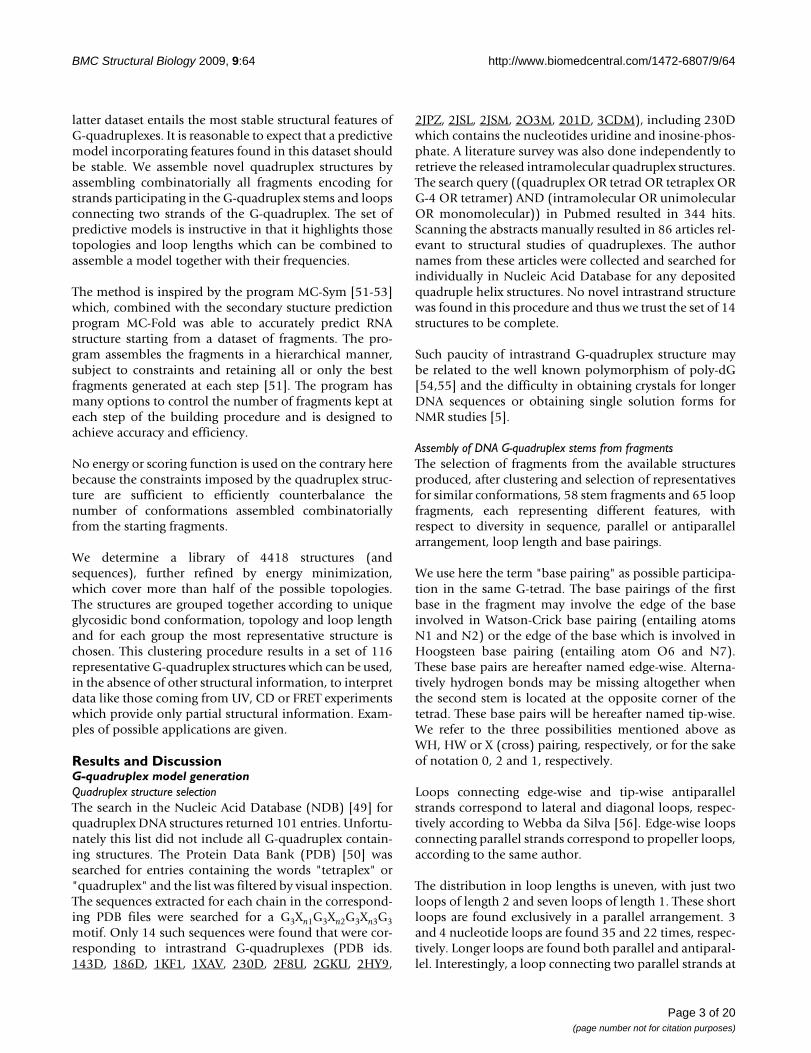
Results and DiscussionG-quadruplex model generationQuadruplex structure selectionThe search in the Nucleic Acid Database (NDB) [49] forquadruplex DNA structures returned 101 entries. Unfortu-nately this list did not include all G-quadruplex contain-ing structures. The Protein Data Bank (PDB) [50] wassearched for entries containing the words "tetraplex" or"quadruplex" and the list was filtered by visual inspection.The sequences extracted for each chain in the correspond-ing PDB files were searched for a G3Xn1G3Xn2G3Xn3G3motif. Only 14 such sequences were found that were cor-responding to intrastrand G-quadruplexes (PDB ids.143D, 186D, 1KF1, 1XAV, 230D, 2F8U, 2GKU, 2HY9,
2JPZ, 2JSL, 2JSM, 2O3M, 201D, 3CDM), including 230Dwhich contains the nucleotides uridine and inosine-phos-phate. A literature survey was also done independently toretrieve the released intramolecular quadruplex structures.The search query ((quadruplex OR tetrad OR tetraplex ORG-4 OR tetramer) AND (intramolecular OR unimolecularOR monomolecular)) in Pubmed resulted in 344 hits.Scanning the abstracts manually resulted in 86 articles rel-evant to structural studies of quadruplexes. The authornames from these articles were collected and searched forindividually in Nucleic Acid Database for any depositedquadruple helix structures. No novel intrastrand structurewas found in this procedure and thus we trust the set of 14structures to be complete.
Such paucity of intrastrand G-quadruplex structure maybe related to the well known polymorphism of poly-dG[54,55] and the difficulty in obtaining crystals for longerDNA sequences or obtaining single solution forms forNMR studies [5].
Assembly of DNA G-quadruplex stems from fragmentsThe selection of fragments from the available structuresproduced, after clustering and selection of representativesfor similar conformations, 58 stem fragments and 65 loopfragments, each representing different features, withrespect to diversity in sequence, parallel or antiparallelarrangement, loop length and base pairings.
We use here the term "base pairing" as possible participa-tion in the same G-tetrad. The base pairings of the firstbase in the fragment may involve the edge of the baseinvolved in Watson-Crick base pairing (entailing atomsN1 and N2) or the edge of the base which is involved inHoogsteen base pairing (entailing atom O6 and N7).These base pairs are hereafter named edge-wise. Alterna-tively hydrogen bonds may be missing altogether whenthe second stem is located at the opposite corner of thetetrad. These base pairs will be hereafter named tip-wise.We refer to the three possibilities mentioned above asWH, HW or X (cross) pairing, respectively, or for the sakeof notation 0, 2 and 1, respectively.
Loops connecting edge-wise and tip-wise antiparallelstrands correspond to lateral and diagonal loops, respec-tively according to Webba da Silva [56]. Edge-wise loopsconnecting parallel strands correspond to propeller loops,according to the same author.
The distribution in loop lengths is uneven, with just twoloops of length 2 and seven loops of length 1. These shortloops are found exclusively in a parallel arrangement. 3and 4 nucleotide loops are found 35 and 22 times, respec-tively. Longer loops are found both parallel and antiparal-lel. Interestingly, a loop connecting two parallel strands at
Page 3 of 20(page number not for citation purposes)

BMC Structural Biology 2009, 9:64 http://www.biomedcentral.com/1472-6807/9/64
the opposite corners of a tetrad is also present. Bases inthis loop, however, participate the G-tetrads and thereforewill be discarded, for steric reasons, in the followingassembly of G-quadruplexes.
The features of the selected fragments are reported inTable 1.
The stems of the fragments were used to build up the four-strand G-quadruplex stems. With the loose requirementsof no more than 0.8 Å RMSD between the superimposing
fragments and no overlap below 0.5 times the sum of vander Waals radii (see Methods) 646 G-quadruplexes werebuilt whose tetrad planes were rebuilt using the frameprovided by the first three G strand in the sequence. In thisstep it was checked that the model G-tetrad could be wellplaced on the C1' anchor points. Models which exhibitedan RMSD larger than 3.0 Å were discarded, leaving a set of509 G-quadruplex stem models.
Rebuilding the G-tetrad was necessary because, due to thetolerant cutoff used for fragment assembly, base pairingwas not always consistent with the hydrogen bonding pat-tern of a G-tetrad. For this reason the four G's constitutingthe G-tetrad were replaced by a standard G-tetrad by firstsuperimposing the first G (numbered 1 in Figure 1) on theG of the first strand in the molecule in order to determinethe orientation of the G-tetrad and then superimposingthe C1' atoms of the tetrad with those of the G-quadru-plex.
Assembly of DNA G-quadruplex from G-quadruplex stems and loopsThe strands of the 509 G-quadruplex stems determined asdescribed above were connected using the loops of thefragments selected from the NDB and PDB quadruplexdataset. 65 non-redundant loops were used resulting in509 × 65 × 65 × 65 possible combinations. Many of thesewere ruled out by steric hindrance or poor superpositionof the anchor G's preceding and following the loop. Nev-ertheless 4418 molecular models have been generatedreflecting a variety of parallel/antiparallel dispositions,loop lengths, syn/anti glycosidic bond angles. Many ofthese models still suffered from long bonds resulting frommerging fragments and steric hindrances and for this rea-son they were refined by energy minimization.
Molecular mechanics refinementAll 4418 model were subjected to 300 steps of molecularmechanics minimization keeping the G-tetrads fixed. Atthe end the energy distribution of the models was quitehomogeneous with energies ranging mostly between 700and 1500 kcal/mol. Only four models were clearly sepa-rated from the remaining ones at much higher energy(two at ca. 13000 kcal/mol and two at ca. 63000 kcal/mol) pointing out serious steric hindrance. Visual exami-nation shows that the rebuilt G-tetrads are too close forthese four models. The latter models have not been con-sidered in the following clustering procedure.
Clustering of structural modelsAll energy minimized models are available, together withsequences and a table of energies and topologies, from theauthors. However, for more convenient usage, the modelswere clustered according to unique glycosidic bond con-formation, topology and loop length. The models sharingthe same glycosidic bond conformations, topology and
Table 1: Non-redundant features of the fragments selected from the database.
syn/anti a/p loop length pairing counts
a s a a 4 111 13
a a a p 3 000 10
s a a a 3 200 7
s s a a 3 002 6
s a a p 3 200 6
a a a p 1 000 6
a s a a 4 020 5
s s a a 3 220 2
a s a a 3 020 2
a a a p 4 111 2
s s a a 4 111 1
s a s a 3 111 1
s a a p 2 200 1
s a a p 1 200 1
s a a a 4 200 1
s a a a 4 111 1
a s a a 4 202 1
a s a a 3 202 1
a a a p 2 000 1
syn/anti indicates the conformation at the glycosidic bond of the first three G's, a/p indicates antiparallel/parallel arrangement, 0, 1 and 2 pairings are described in the text and the number of fragments different in sequence or conformation is given in the column "counts".
Page 4 of 20(page number not for citation purposes)
BMC Structural Biology 2009, 9:64 http://www.biomedcentral.com/1472-6807/9/64
loop lengths were pairwise compared and for each modela threshold RMSD was chosen and a weight was assignedbased on the RMSD with all other structures. The modelwith largest weight was chosen as representative of allmodels with RMSD lower than threshold. The procedurewas repeated, increasing the threshold RMSD, until a sin-gle model was left. The energies of the resulting modelsrange between 690 and 1656 kcal/mol, a range compara-ble with that found for experimental structures subjectedto the same minimization procedure (590 to 913 kcal/mol), taking into account that experimental structures aretypically already refined. The most representative struc-ture for each of the 116 clusters (see Table 2) is provided[see Additional file 1].
Analysis of G-quadruplex modelsComparison with experimental structuresAn obvious test for the methodology is to check whetherit is able to recover the observed intrastrand G-quadru-plexes from the fragments which are not taken from that
intrastrand G-quadruplexes. Due to sequence diversity itwill be in general hard to recover exactly the samesequence. For instance, for loops of length 2 there are onlytwo fragments with different sequence. When one of thetwo loops is excluded from the list of fragments there willbe no possibility to obtain a loop of length 2 with thesame sequence. Nevertheless we will consider here thetopologies which are generated from assembly of frag-ments with the same loop lengths.
In order to test the overall reliability of the method weconsidered the set of intrastrand structures with three Gstem strands with loops of length 3. In the following weindicate the overall topology of a model by noting thesequence of loops as lateral (l), propeller (p) or diagonal(d). The clockwise (+) or anti-clockwise (-) rotation of lat-eral and propeller loops is with respect to a commonframe of reference (see [56]). It was not possible to extendthis analysis to the other structures because they containloops of length 2 for which only two fragments are presentin the dataset.
The structures with pdb id. 1KF1, 2GKU, 2HY9, 2JPZ,2JSL, 2JSM and 3CDM all contain the core sequenceGGGTTAGGGTTAGGGTTAGGG and adopt three differ-ent topologies: namely -p-p-p, -l-l-p, -p-l-l.
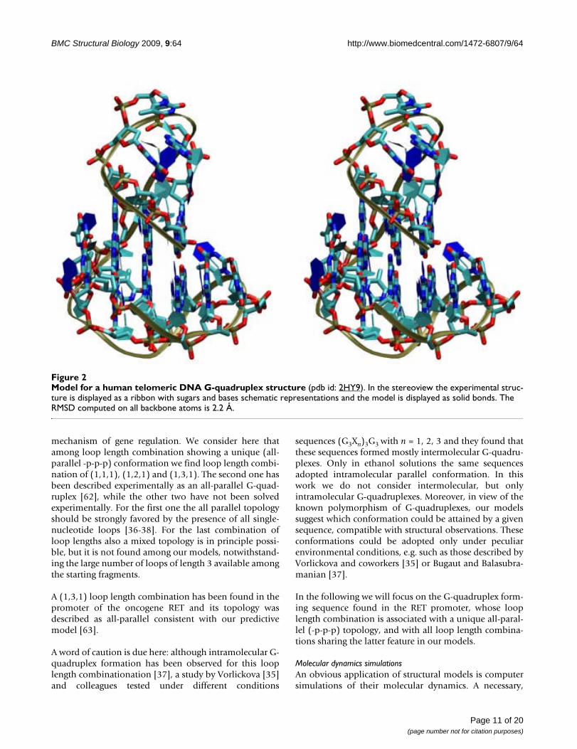
We considered for each topology all built models whichdo not contain any fragment derived from structures withthe same topology. Moreover, due to the fragment cluster-ing procedure adopted, no fragment is present in the data-set closely related to those present in those structures. Allthree topologies are actually represented several times inthe built models with RMSD over sugars and phosphatesfrom the original PDB structures between 2.0 and 3.0 Å.An example with the real structure (pdb id: 2hy9) and themodel assembled from fragments is reported in Figure 2.Although not all loops are similar to the real ones, by con-struction, the topology and overall conformation is repro-duced well by the model. The RMSD computed on allbackbone atoms is 2.2 Å.
It must be however noted that not only the observedtopologies for the sequence G3N3G3N3G3N3G3 are foundin the models but also others, although the observedtopology is among the most represented ones.
Topologies and loop lengthsAnother test for the methodology is to check whether therelationship between loop lengths and topology matchesthe available experimental evidences.
Overall, the topologies of the models generated using allthe available fragments are not evenly distributed (Table3). Only 14 out of 26 possible looping topologies are
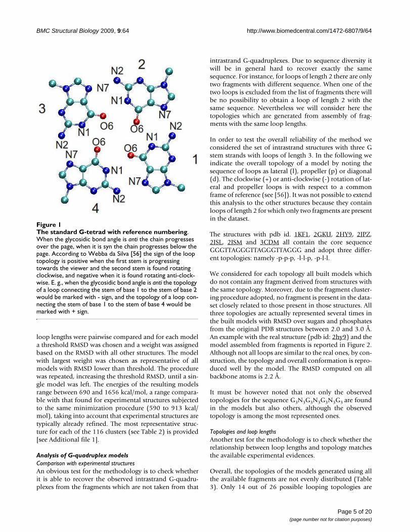
The standard G-tetrad with reference numberingFigure 1The standard G-tetrad with reference numbering. When the glycosidic bond angle is anti the chain progresses over the page, when it is syn the chain progresses below the page. According to Webba da Silva [56] the sign of the loop topology is positive when the first stem is progressing towards the viewer and the second stem is found rotating clockwise, and negative when it is found rotating anti-clock-wise. E. g., when the glycosidic bond angle is anti the topology of a loop connecting the stem of base 1 to the stem of base 2 would be marked with - sign, and the topology of a loop con-necting the stem of base 1 to the stem of base 4 would be marked with + sign.
Page 5 of 20(page number not for citation purposes)

BMC Structural Biology 2009, 9:64 http://www.biomedcentral.com/1472-6807/9/64
found. There is a clear predominance of the -p-p-p topol-ogy that is found in ca. one third of all models.
This finding is consistent with the observation that thenumber of parallel fragments which are actually used forassemblying the stems of G-quadruplex is larger than thenumber of antiparallel fragments, although the startingfragments (i.e. strand-loop-strand fragments) do notshow such parallel predominance. This might reflect ageneral more regular arrangement of parallel versusantiparallel strands, at least in the selected dataset. Theadoption of a -p-p-p topology leads to right-handednessof the polynucleotide chain in the G-quadruplex.
Other well represented topologies are the mixed topolo-gies +l+p+p, -l-l-p, -p-p-l, -pd+p, +l+p+l d+pd and the all-antiparallel +l+l+l.
Some care must be taken when considering potential G-quadruplexes involving loops of 4 residues because nopropeller-like loop is found in the starting dataset of frag-ments. There are therefore no all parallel topologiesinvolving loops of 4 residues, although it has been shownthat the G-rich sequence in the human VEGF gene pro-moter adopts an all-parallel structure involving a loop of4 residues [15,57]. Most frequently diagonal fragmentsare found.
Only 47 possible combinations of loop lengths are foundout of 64 possible. Of these some are more largely repre-sented as a consequence of the uneven distribution of thenumber of starting loops. In general there is no direct rela-tionship between loop lengths and topology, althoughsequences with loops as short as 1 or 2 nucleotides are, asexpected from the starting fragments, found in all-paralleltopology. For longer loop lengths typically many differenttopologies are found.
It is interesting to note that some of the combination ofloop lengths are found with unique topology, amongthese the most widely represented are 4, 3 and 4 (122models), 1, 3 and 1 (58 models), 1, 1 and 3 (53 models)(Table 2).
The effect of loop length on G-quadruplex topology and/or stability has been studied by many authors [34,35,37-39,58-61] under different conditions. Not all studies how-ever address the formation of intramolecular G-quadru-plexes. The two recent papers by Bugaut andBalasubramanian [37] and Smargiasso et al. [38] investi-gate systematically the effect of loop length, with rand-omized sequences, on G-quadruplex stability andtopology. In both studies intramolecular vs. intermolecu-lar G-quadruplex formation is experimentally addressed.Notwithstanding different experimental conditions, these
studies provide, among other results, a general conclusionwhich is well in line with previous evidences: in generalshort loops (and in particular the presence of loops oflength one) strongly favor parallel arrangement of thestrands while for longer loops antiparallel and mixedarrangements are observed. The topology of the modelsbuilt here appears consistent with experimental evi-dences.
Possible applicationsThe present study constitutes a proof of principle, obvi-ously physical or statistical effective energy functionsshould more accurately measure the stability of the pre-dictive models. Moreover the limited diversity insequence does not allow to build models for all possiblesequences. A third limitation of the present approach isthat no consideration of flanking residues which areknown to be important for the stability of G-quadruplexis taken into account. In addition to these problems thestarting fragments are in limited number as exemplifiedby the lack of parallel propeller loops of length 4.
It is worth however to explore how structural predictionscould complement experimental and bioinformaticsapproaches.
It must be clear that the actual structure adopted by a DNAsequence depends on many factors including flanking andloop sequences and environmental conditions. The mod-els built from experimental fragments constitute howevera set of structures whose features are consistent withexperimental structures. Due to the limited number ofstructures solved so far, the set is not expected to cover allpossible structures. However, even in the presence of pol-ymorphism the models proposed here constitute struc-tural working hypotheses that can complementexperimental techniques.
The aim of the following subsection is to show, by select-ing a few possible applications that inferences based onthe built models are consistent with experimental evi-dence and thus provide an overall test of reliability for theproposed models.
It is well known that potential G-quadruplex sequencesplay a regulatory role but the nature of such role is differ-ent according to the position of the sequence with respectto the TSS and the strand where it is found [43].
The models provided by the present study could be usedstraightforwardly as starting models for moleculardynamics simulations or docking studies. Another possi-bility is to use the topology information provided here tocomplement other studies. The same topology could berequired by different DNA quadruplex sharing a common
Page 6 of 20(page number not for citation purposes)

BMC Structural Biology 2009, 9:64 http://www.biomedcentral.com/1472-6807/9/64
Table 2: Features of modeled intrastrand G-quadruplexes.
syn/anti loop topology strand polarity loop 1 loop 2 loop 3 counts
a a a -p-p-p ppp 1 1 1 10
a a a -p-p-p ppp 1 1 2 6
a a a -p-p-p ppp 1 1 3 53
a a a -p-p-p ppp 1 2 1 8
a a a -p-p-p ppp 1 2 2 2
a a a -p-p-p ppp 1 2 3 20
a a a -p-p-l ppa 1 2 3 6
a a a -p-p-l ppa 1 2 4 2
a a a -p-p-p ppp 1 3 1 58
a a a -p-p-p ppp 1 3 2 8
a a a -p-p-p ppp 1 3 3 237
a a a -p-p-l ppa 1 3 3 34
a a a -p-l-l pap 1 3 3 3
a a a -p-p-l ppa 1 3 4 12
a a a -pd+p paa 1 4 1 3
a a a -pd+p paa 1 4 2 10
a a a -pd+l ppa 1 4 3 10
a a a -p-l-l pap 1 4 3 1
a a a -pd+p paa 1 4 3 35
a a a -p-p-p ppp 2 1 1 4
a a a -p-p-p ppp 2 1 2 2
a a a -p-p-p ppp 2 1 3 21
a a a -p-p-p ppp 2 2 1 3
a a a -p-p-p ppp 2 2 2 1
a a a -p-p-p ppp 2 2 3 11
a a a -p-p-l ppa 2 2 3 3
a a a -p-p-l ppa 2 2 4 1
a a a -p-p-p ppp 2 3 1 23
a a a -p-p-p ppp 2 3 2 3
Page 7 of 20(page number not for citation purposes)

BMC Structural Biology 2009, 9:64 http://www.biomedcentral.com/1472-6807/9/64
a a a -p-p-l ppa 2 3 3 17
a a a -p-l-l pap 2 3 3 2
a a a -p-p-p ppp 2 3 3 74
a a a -p-p-l ppa 2 3 4 5
a a a -pd+p paa 2 4 1 13
a a a -pd+p paa 2 4 2 3
a a a -pd+p paa 2 4 3 16
a a a -pd+l ppa 2 4 3 1
a a a -p-l-l pap 2 4 3 1
a a a -p-p-p ppp 3 1 1 28
s a s -p-p-p ppp 3 1 1 2
s s a +l+p+p aaa 3 1 1 34
s a a -p-p-p ppp 3 1 1 6
a a a -p-p-p ppp 3 1 2 10
s a a -p-p-p ppp 3 1 2 6
s s a +l+p+p aaa 3 1 2 7
s s a +l+p+p aaa 3 1 3 120
s a a -p-p-l ppa 3 1 3 12
a a a -p-p-p ppp 3 1 3 131
s s a +l+p+l paa 3 1 3 27
s a a -p-p-p ppp 3 1 3 49
a a a -p-p-l ppa 3 1 3 4
a a a -p-p-l ppa 3 1 4 1
s a a -p-p-l ppa 3 1 4 3
s s a +l+p+l paa 3 1 4 6
s s a +l+p+p aaa 3 2 1 10
s a a -p-p-p ppp 3 2 1 6
a a a -p-p-p ppp 3 2 1 9
s a a -p-p-p ppp 3 2 2 2
a a a -p-p-p ppp 3 2 2 3
Table 2: Features of modeled intrastrand G-quadruplexes. (Continued)
Page 8 of 20(page number not for citation purposes)

BMC Structural Biology 2009, 9:64 http://www.biomedcentral.com/1472-6807/9/64
s s a +l+p+p aaa 3 2 2 3
s a a -p-p-p ppp 3 2 3 22
a a a -p-p-p ppp 3 2 3 33
s s a +l+p+p aaa 3 2 3 46
s a a -p-p-l ppa 3 2 3 6
a a a -p-p-l ppa 3 2 3 9
s s a +l+p+l paa 3 2 3 9
s a a -p-p-l ppa 3 2 4 2
a a a -p-p-l ppa 3 2 4 3
s s a +l+p+l paa 3 2 4 3
s a a -l-l-p app 3 3 1 102
s s a +l+p+p aaa 3 3 1 102
a a a -p-p-p ppp 3 3 1 125
s a a -p-p-p ppp 3 3 1 80
a s a -l-l-p app 3 3 2 2
s a a -l-l-p app 3 3 2 70
s a a -p-p-p ppp 3 3 2 8
a a a -p-p-p ppp 3 3 2 9
s s a +l+p+p aaa 3 3 2 9
s a a -l-l-l apa 3 3 3 10
s s a +l+l+l apa 3 3 3 167
s a a -p-p-p ppp 3 3 3 241
a a a -p-l-l pap 3 3 3 30
s s a +l+p+p aaa 3 3 3 367
s a a -l-l-p app 3 3 3 385
a s a -ld+l aap 3 3 3 3
a a a -p-p-p ppp 3 3 3 450
s a a -p-p-l ppa 3 3 3 66
a a a -p-p-l ppa 3 3 3 68
a s a -l-l-p app 3 3 3 8
Table 2: Features of modeled intrastrand G-quadruplexes. (Continued)
Page 9 of 20(page number not for citation purposes)

BMC Structural Biology 2009, 9:64 http://www.biomedcentral.com/1472-6807/9/64
s a a -p-l-l pap 3 3 3 91
s s a +l+p+l paa 3 3 3 95
s a a -p-p-l ppa 3 3 4 12
s s a +l+p+l paa 3 3 4 15
a a a -p-p-l ppa 3 3 4 19
s a a -l-l-l apa 3 3 4 19
s s a +ld-p ppa 3 4 1 24
s a a -pd+p paa 3 4 1 6
a a a -pd+p paa 3 4 1 9
a a a -pd+p paa 3 4 2 15
s s a +ld-p ppa 3 4 2 3
s a a -pd+p paa 3 4 2 5
s a a -pd+l ppa 3 4 3 12
a a a -pd+l ppa 3 4 3 13
s a a -p-l-l pap 3 4 3 14
a a a -p-l-l pap 3 4 3 3
s s a +ld-l paa 3 4 3 3
s a a -pd+p paa 3 4 3 44
s s a +ld-p ppa 3 4 3 51
a a a -pd+p paa 3 4 3 80
s s a +l+l+l apa 3 4 3 82
s a a -l-l-p app 4 3 1 2
s a a -l-l-p app 4 3 2 8
a s a -ld+l aap 4 3 3 2
s a a -l-l-p app 4 3 3 33
a s a d+pd aap 4 3 4 114
s a a d+pd aap 4 3 4 8
The notation here follows Webba da Silva [56]. syn/anti indicates the conformation at the glycosidic bond of the first three G's. The loop topology is indicated by letters p (parallel), l (lateral) and d (diagonal) preceded by + or - sign to indicate clockwise or anti clockwise rotation when the first strand is progressing towards the viewer (see Figure 1). Similarly, the parallel or antiparallel (a/p) strand polarity in column 2 is with reference to the first strand and the order is according to the position in the quadruplex (rotating anti-clockwise with the first strand progressing towards the viewer), and in general not according to sequence order. The next three fields indicate loop lengths and the last field indicate the number of built models found with these features.
Table 2: Features of modeled intrastrand G-quadruplexes. (Continued)
Page 10 of 20(page number not for citation purposes)
BMC Structural Biology 2009, 9:64 http://www.biomedcentral.com/1472-6807/9/64
mechanism of gene regulation. We consider here thatamong loop length combination showing a unique (all-parallel -p-p-p) conformation we find loop length combi-nation of (1,1,1), (1,2,1) and (1,3,1). The second one hasbeen described experimentally as an all-parallel G-quad-ruplex [62], while the other two have not been solvedexperimentally. For the first one the all parallel topologyshould be strongly favored by the presence of all single-nucleotide loops [36-38]. For the last combination ofloop lengths also a mixed topology is in principle possi-ble, but it is not found among our models, notwithstand-ing the large number of loops of length 3 available amongthe starting fragments.
A (1,3,1) loop length combination has been found in thepromoter of the oncogene RET and its topology wasdescribed as all-parallel consistent with our predictivemodel [63].
A word of caution is due here: although intramolecular G-quadruplex formation has been observed for this looplength combinationation [37], a study by Vorlickova [35]and colleagues tested under different conditions
sequences (G3Xn)3G3 with n = 1, 2, 3 and they found thatthese sequences formed mostly intermolecular G-quadru-plexes. Only in ethanol solutions the same sequencesadopted intramolecular parallel conformation. In thiswork we do not consider intermolecular, but onlyintramolecular G-quadruplexes. Moreover, in view of theknown polymorphism of G-quadruplexes, our modelssuggest which conformation could be attained by a givensequence, compatible with structural observations. Theseconformations could be adopted only under peculiarenvironmental conditions, e.g. such as those described byVorlickova and coworkers [35] or Bugaut and Balasubra-manian [37].
In the following we will focus on the G-quadruplex form-ing sequence found in the RET promoter, whose looplength combination is associated with a unique all-paral-lel (-p-p-p) topology, and with all loop length combina-tions sharing the latter feature in our models.
Molecular dynamics simulationsAn obvious application of structural models is computersimulations of their molecular dynamics. A necessary,
Model for a human telomeric DNA G-quadruplex structure (pdb id: 2HY9)Figure 2Model for a human telomeric DNA G-quadruplex structure (pdb id: 2HY9). In the stereoview the experimental struc-ture is displayed as a ribbon with sugars and bases schematic representations and the model is displayed as solid bonds. The RMSD computed on all backbone atoms is 2.2 Å.
Page 11 of 20(page number not for citation purposes)

BMC Structural Biology 2009, 9:64 http://www.biomedcentral.com/1472-6807/9/64
albeit not sufficient, condition for a model to be accurateis that the structure is stable during a molecular dynamicssimulation for a time sufficient in principle to developmajor conformational rearrangements. The benefits andlimits of molecular dynamics simulations of G-quadru-plexes have been reviewed by Spooner and Spackova [64].In the study by Hazel et al. [58] molecular dynamics sim-ulations complemented experiments and model buildingwas performed in order to provide starting models. Weconsider here as an example the sequence GGGCG-GGGCGGGGCGGG that is found in the promoter of theoncogene RET, which adopts an all-parallel topology [63].
The most representative predictive model for the uniqueall-parallel (-p-p-p) topology for loop length combina-tion (1,3,1) was taken and the sequence was mutated to
the target sequence. Two potassium ions were added atthe centre of the O6 atoms of adjacent tetrads and coun-terions were further added to make the system neutral.The system was solvated in a box of water extending atleast 12 Å away from each heavy atom of the solute. Thepreparation of the system is essentially as previouslydescribed for a different system [65]. The forcefieldemployed is CHARMM version 31 [66,67].
Molecular dynamics simulation was run for 20 ns in orderto check for any major conformational change whichcould indicate bad quality of the starting model or wrongtopology [58,64].
After few hundred picoseconds one of the two potassiumions at the centre of adjacent tetrads goes in solutionwhile the other is firmly retained. Loss of ions from thecentral channel has been observed before in moleculardynamics simulations and it has been ascribed to force-field inaccuracies [64]. During the simulation the G-quad-ruplex structure is mantained. The average RMSD from thestarting structure is 1.0 Å. Larger fluctuations are observedat the three residue loop both for the backbone and forthe base moieties similar to other molecular dynamicssimulation studies [58,64]. No loop residue is involved inintramolecular hydrogen bonds. This example proves (atleast on the timescale of 20 ns) that the model quality issuitable for molecular dynamics simulations because oth-erwise large changes in the G-quadruplex structure wouldbe expected [58,64].
Docking studiesPredictive models of G4 may be employed for dockingstudies (see e.g. [68,69]). As an example we consideredthe model for the sequence GGGCGGGGCGGGGCGGGthat is found in the promoter of the oncogene RET, whichadopts an all-parallel topology [63], as in the previous sec-tion.
This sequence has been shown to be stabilized by the cat-ionic porphyrin TMPyP4 (5,10,15,20-tetrakis(1-methyl-pyridin-1-ium-4-yl)-21,22-dihydroporphyrin) and it wassuggested that the binding involves stacking rather thanintercalation [63].
Two G-quadruplex-TmPyP4 complexes have been struc-turally characterised by NMR (pdb id. 2A5R, [70]) and byX-ray crystallography (pdb id. 2HRI, [71]). The two com-plexes show remarkable differences. In the NMR structurethe porphyrin is stacking over the first tetrad and is cov-ered by the two residues 5' to the G-quadruplex. In thecrystal structure one porphyrin is stacked over a base pairover the tetrads and the other is contacting a grove withelectrostatic interaction with a phosphate and a stackinginteraction with a base in the loop.
Table 3: Topology distribution of model G-quadruplexes.
topology counts
-p-p-p 1764
+l+p+p 698
-l-l-p 610
-p-p-l 285
+l+l+l 249
-pd+p 239
+l+p+l 155
-p-l-l 145
d+pd 122
+ld-p 78
-pd+l 36
-l-l-l 29
-ld+l 5
+ld-l 3
total number of topologies total number of models
14 4418
The distribution of topologies (independent of glycosidic bond conformation and loop lengths) of all 4418 models is reported. The notation here follows Webba da Silva [56]. p, l and d stand for propeller-like, lateral or diagonal loop. Theand + signs refer to anti-clockwise or clockwise rotation of the loop around the G-quadruplex stem, respectively, when the first strand is progressing towards the viewer (see Figure 1).
Page 12 of 20(page number not for citation purposes)

BMC Structural Biology 2009, 9:64 http://www.biomedcentral.com/1472-6807/9/64
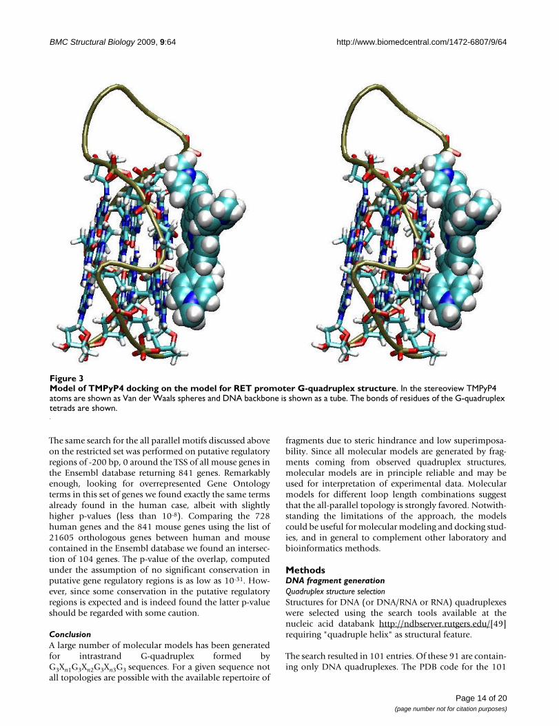
The ligand structure was taken from the Hic-Up serverhttp://xray.bmc.uu.se/hicup/[72]. Based on the SMILESrepresentation of the compound available from the data-base PubChem (CID: 4234) [73] partial charges havebeen assigned by the program Babel [74] implementingthe Gasteiger and Marsili method [75]. The structure ofthe model G-quadruplex with partial charges has beenobtained as decribed in the previous section. The programDock6.3 [76] has been used for generating, scoring andclustering TMPyP4-G-quadruplex complexes following astandard protocol and using the AMBER forcefield for esti-mating the energy of van der Waals intermolecular con-tacts. 20000 poses were generated and after clustering at2.0 Å RMSD the best 10 were retained, showing all largenegative interaction energies. Consistent with the abovecited previous studies, in nine out of ten complexes thearrangement of the prophyrin is parallel and stackingonto the tetrads, although stacking involves only half ofthe tetrad (Figure 3). In the remaining complex the por-phyrin is contacting the G-quadruplex in the groove anddisplays electrostatic interaction between the pyrimidin-ium and the phosphate.
Overall these results are consistent with what could beexpected based on previous structural characterizationand thus show that the models can provide a starting tar-get for use in docking studies.
Cancer genesPotential G-quadruplex forming sequences have beenfound in a number of cancer genes [33] mostly sharing thefirst and last loops of length 1.
Consistent with earlier studies [33,37,38], sequences hav-ing the second loop of length 1 to 3 can adopt in our mod-els only an all-parallel -p-p-p topology, while any othertopology would be not consistent with the available set ofexperimental structures. We further explore whether thepresence of sequences for which a unique all-paralleltopology is found could be a distinctive feature of cancergenes. Following previous analyses we have limited oursearch to a putative regulatory region of -200 bp, 0 bparound the TSS of all genes in the Ensembl database. Wefirst aligned the sequences:
G3N1G3N1G3N1G3,
G3N1G3N2G3N1G3,
G3N1G3N3G3N1G3
on the putative regulatory regions and the search returned728 unique genes containing at least one of thesequences.
Before any further consideration it should be consideredthat the pattern G3N1G3 is shared with consensus motifG3CG3 of SP1 binding site, which is rather common inhuman genes in the region within 200 bp upstream of theTSS [77]. Inclusion of the G3CG3 consensus sequence forSP1 binding site in many of the potential G-quadruplexsequences is likely to add noise to any statistical analysisand to reduce the calculated significance values.
The Ensembl gene names were translated, where possible,onto HNGC gene names and the overlap between the setof the resulting 686 genes and the Census set of 385 can-cer genes available at the http://www.sanger.ac.uk/genetics/CGP/Census/[78,79] was determined. The overlap setcontained 23 genes, a number higher than that expectedby chance, i.e. 14.5.
The probability that 686 genes chosen randomly out of19589 genes with HGNC name could have an overlap of23 or more with the Census set (385 genes) was calculatedusing the hypergeometric distribution and the resulting p-value was 0.01. This result, based on putative adoption ofa common structure, suggests that G-quadruplex gene reg-ulation may be a common feature of cancer genes.
The above set of loop length combinations is howeveronly a restricted set of the larger set of all loop length com-binations which are associated in the predicted models toa unique all-parallel (-p-p-p) topology (see Table 2).
The same analysis has been repeated considering the latterset. If the topology is an important feature shared by G-quadruplex sequences found in many oncogenes wewould expect also for the larger set of loop combinationsa higher number of hits in oncongenes than expected bychance. Indeed this is the case. The overlap between the1607 genes, containing at least one of the selected loopcombinations, and the Census set consists of 47 genes(Table 4), higher than the expected 32, and correspondingto a p-value of 0.003. These results are consistent with theknown importance of all-parallel topology for G-quadru-plex forming sequences in the regulation of proto-onco-genes [33].
Developmental genesThe restricted set of genes that contain a potential all-par-allel quadruplex helix has been screened for overrepresen-tation in Gene Ontology annotation. The general terms"developmental process", "system development", "ana-tomical structure development", "multicellular organis-mal development" are found with the lowest p-values(less than 10-10). The same analysis on the larger set ofgenes containing a potential all-parallel quadruplex helixgives essentially the same results with even lower p-values(ranging from 10-12 to 10-17).
Page 13 of 20(page number not for citation purposes)
BMC Structural Biology 2009, 9:64 http://www.biomedcentral.com/1472-6807/9/64
The same search for the all parallel motifs discussed aboveon the restricted set was performed on putative regulatoryregions of -200 bp, 0 around the TSS of all mouse genes inthe Ensembl database returning 841 genes. Remarkablyenough, looking for overrepresented Gene Ontologyterms in this set of genes we found exactly the same termsalready found in the human case, albeit with slightlyhigher p-values (less than 10-8). Comparing the 728human genes and the 841 mouse genes using the list of21605 orthologous genes between human and mousecontained in the Ensembl database we found an intersec-tion of 104 genes. The p-value of the overlap, computedunder the assumption of no significant conservation inputative gene regulatory regions is as low as 10-31. How-ever, since some conservation in the putative regulatoryregions is expected and is indeed found the latter p-valueshould be regarded with some caution.
ConclusionA large number of molecular models has been generatedfor intrastrand G-quadruplex formed byG3Xn1G3Xn2G3Xn3G3 sequences. For a given sequence notall topologies are possible with the available repertoire of
fragments due to steric hindrance and low superimposa-bility. Since all molecular models are generated by frag-ments coming from observed quadruplex structures,molecular models are in principle reliable and may beused for interpretation of experimental data. Molecularmodels for different loop length combinations suggestthat the all-parallel topology is strongly favored. Notwith-standing the limitations of the approach, the modelscould be useful for molecular modeling and docking stud-ies, and in general to complement other laboratory andbioinformatics methods.
MethodsDNA fragment generationQuadruplex structure selectionStructures for DNA (or DNA/RNA or RNA) quadruplexeswere selected using the search tools available at thenucleic acid databank http://ndbserver.rutgers.edu/[49]requiring "quadruple helix" as structural feature.
The search resulted in 101 entries. Of these 91 are contain-ing only DNA quadruplexes. The PDB code for the 101
Model of TMPyP4 docking on the model for RET promoter G-quadruplex structureFigure 3Model of TMPyP4 docking on the model for RET promoter G-quadruplex structure. In the stereoview TMPyP4 atoms are shown as Van der Waals spheres and DNA backbone is shown as a tube. The bonds of residues of the G-quadruplex tetrads are shown.
Page 14 of 20(page number not for citation purposes)
BMC Structural Biology 2009, 9:64 http://www.biomedcentral.com/1472-6807/9/64
entries was used for retrieving the relevant structures fromthe Protein Data Bank http://www.rcsb.org/[50].
This search was apparently missing some of the G-quad-ruplexes in the PDB. For this reasons we selected all struc-tures in the PDB containg anywhere the words quadruplexor tetraplex and we hand filtered those that could containa genuine DNA G-quadruplex. The latter step retrievedadditional 51 structures. The selected structures weresearched for the presence of strands with three adjacentG's paired with another strand with three adjacent G'swith either parallel or antiparallel linear arrangement, andwith loop connection of one to four nucleotides. The looplenghts considered here are somewhat shorter than thelimit of seven used e.g. by Chowdhury and coworkers [47]
Since the quadruplex are assembled from these fragmentswe found useful to reference these fragments and theirpairs of bases defining the G-tetrad plane to a standard G-tetrad. The parallel or antiparallel orientation of the twoG-strand is an obviously important feature of the frag-ment. Referencing serves the purpose of detecting andstoring fragments that can be used for building the G-quadruplex and is not meant as a definition for classifica-tion. A standard for classification and notation of G-quad-ruplexes has been proposed by Webba da Silva [56]. Weconform here to that proposed standard, although wereport also a local description of the structure (vide infra)
A standard G-tetrad was generated by rotation and trans-lation of a G base (taken from the standard fragments ofthe X3DNA program [80]). The best hydrogen bondinggeometry was obtained by rotating repeatedly the G baseof 90 degrees and traslating by -.70 and 7.10 Å along thex and y axis respectively, with reference to the coordinatesused in the X3DNA base coordinates.
The first G base of each fragment with sequence G3Xn1G3was superimposed to base 1 in the model G-tetrad. Thenthe first (last) G base of the second run of three G's in thefragment was superimposed in turn onto the other basesin the G-tetrad in order to find the first base pair of theparallel (antiparallel) three G's pair. The same procedurewith due modifications was repeated for the second andthird G pairs. The fragment was accepted as good if theRMSD in all three superpositions was less than 1.0 Å. Thetolerant threshold was dictated by the large conforma-tional heterogeneity observed in G-tetrads.
The first (last) G base could be in anyone of the threeother positions of the G-tetrad (Figure 1). The second andthird G's could be found over or below the plane definedby the G-tetrad used for superposition of the first Gdepending on the torsion angle at the glycosidic bond.
For each fragment the loop and the "stem" constituted bytwo strands of 3 adjacent G's were stored. The 147 frag-ments (with the 147 loops and 147 stems) obtained inthis way are redundant because the same structure mayhave been resolved by different groups and techniquesand because a single PDB entry may contain the samestructure more than once.
In order to remove redundancy we performed clustering.All fragments identical in sequence were compared andrepresentatives were selected in such a way that none cho-sen conformation has less than 0.8 Å RMSD on all heavyatoms. This led to 68 unique complete fragments, 67loops and 57 stems.
DNA G-quadruplex assembly from fragmentsThe 57 stems were used to assemble the quadruple helix.Three "stems" were assembled together by superimposingthe second three G's with the first three G's of the nextstem with less than 0.8 Å RMSD and with no overlap ofheavy atoms at more than 0.5 times the sum of their vander Waals radii. This procedure led to 646 quadruplexstem models. The stems were therefore further modifiedby substituting the model G-tetrad for each G-tetrad. Themodel G-tetrad was set in place by superimposing first theG of the first strand and then superimposing the four C1'atoms to the closest ones in the G-tetrad. If the RMSD waslarger than 3.0 Å the model was not taken into account. Atthe end of this step 509 models were retained. The confor-mation of the glycosidic bond angle is thus determined bythe first strand of the quadruplex.
Finally loops were added to the quadruplexes wheneverthe superposition of the sugars linked to the G's precedingand following the loop gave an RMSD less than 0.8 Å andwith no overlap of heavy atoms at more than 0.5 times thesum of their van der Waals radii. Although the combina-torial number of possible models is extremely large, inpractice this computation may be performed on a PC. Thislast step generated 4418 models.
Molecular mechanics refinementDue to the rather tolerant cutoff on RMSD's the bondsconnecting stems and loops were in many instances large.The refinement was performed by first substituting the G-tetrads with the regular G-tetrad generated by optimalrototraslation of G, as described above, and then keepingthe G-tetrads fixed and performing energy minimization.For this purpose the program NAMD [81] was usedemploying a dielectric constant of 10 and the forcefieldCHARMM version 31 [66,67]. 300 steps of conjugate gra-dients minimization were performed keeping the baseatoms of the tetrads fixed.
Page 15 of 20(page number not for citation purposes)

BMC Structural Biology 2009, 9:64 http://www.biomedcentral.com/1472-6807/9/64
Table 4: Human cancer genes containing potential all-parallel G-quadruplexes.
Gene symbol Gene name
AKT1 v-akt murine thymoma viral oncogene homolog 1
ASPSCR1 alveolar soft part sarcoma chromosome region, candidate 1
ATF1 activating transcription factor 1
BCL3 B-cell CLL/lymphoma 3
BRCA2 familial breast/ovarian cancer gene 2
CARD11 caspase recruitment domain family, member 11
CDH11 cadherin 11, type 2, OB-cadherin (osteoblast)
CLTCL1 clathrin, heavy polypeptide-like 1
ELN elastin
EPS15 epidermal growth factor receptor pathway substrate 15 (AF1p)
ERCC2 excision repair cross-complementing rodent repair deficiency complementation group 2 (xeroderma pigmentosum D)
ETV6 ets variant gene 6 (TEL oncogene)
FGFR3 fibroblast growth factor receptor 3
FNBP1 formin binding protein 1 (FBP17)
FOXP1 forkhead box P1
FSTL3 follistatin-like 3 (secreted glycoprotein)
GATA1 GATA binding protein 1 (globin transcription factor 1)
HIP1 huntingtin interacting protein 1
HOXA11 homeo box A11
HOXA13 homeo box A13
HOXA9 homeo box A9
IGK@ immunoglobulin kappa locus
IRF4 interferon regulatory factor 4
JAZF1 juxtaposed with another zinc finger gene 1
LHFP lipoma HMGIC fusion partner
MLLT6 myeloid/lymphoid or mixed-lineage leukemia (trithorax homolog, Drosophila); translocated to, 6 (AF17)
MSI2 musashi homolog 2 (Drosophila)
MSN moesin
Page 16 of 20(page number not for citation purposes)

BMC Structural Biology 2009, 9:64 http://www.biomedcentral.com/1472-6807/9/64
Clustering of structural modelsAll energy minimized models sharing the same glycosidicbond conformations, topology and loop lengths wereclustered in separate groups. All models within a singlegroup were pairwise compared. A threshold RMSD t waschosen and a weight wi was assigned to each model i basedon the RMSDs lower than t with all other structures:
The model with the largest weight was chosen as repre-sentative of all models with RMSD lower than threshold.The procedure was repeated doubling progressively thethreshold starting from 0.4 Å until a single model was left.
Genomic searches and analysisAll regions 200 bp upstream the ranscription start site(TSS) of all human genes for all transcripts have beendownloaded from Biomart site http://www.biomart.org/.The database and the dataset were ENSEMBL 53 GENESand NCBI36i respectively. The search for potential G-quadruplex sequences with proper loop lengths was per-formed using the program glsearch in the fasta35.1 soft-ware package ftp://ftp.ebi.ac.uk/pub/software/unix/fasta/fasta3/. The same analysis was repeated for all mousegenes using the same database and the dataset NCBI37.
The list of orthologous genes was obtained from theBiomart site selecting only protein-coding genes.
The Census set of 385 cancer genes was downloaded fromthe http://www.sanger.ac.uk/genetics/CGP/Census/[78,79].
wrmsdij
ti
j
= ∑ cos( )p2
MUC1 mucin 1, transmembrane
MYCL1 v-myc myelocytomatosis viral oncogene homolog 1, lung carcinoma derived (avian)
MYCN v-myc myelocytomatosis viral related oncogene, neuroblastoma derived (avian)
MYC v-myc myelocytomatosis viral oncogene homolog (avian)
PIM1 pim-1 oncogene
POU2AF1 POU domain, class 2, associating factor 1 (OBF1)
PTEN phosphatase and tensin homolog gene
RANBP17 RAN binding protein 17
RAP1GDS1 RAP1, GTP-GDP dissociation stimulator 1
RET ret proto-oncogene
SEPT6 septin 6
SFRS3 splicing factor, arginine/serine-rich 3
SS18L1 synovial sarcoma translocation gene on chromosome 18-like 1
TAF15 TAF15 RNA polymerase II, TATA box binding protein (TBP)-associated factor, 68 kDa
TCF12 transcription factor 12 (HTF4, helix-loop-helix transcription factors 4)
TMPRSS2 transmembrane protease, serine 2
TRIM33 tripartite motif-containing 33 (PTC7, TIF1G)
TSHR thyroid stimulating hormone receptor
ZNFN1A1 zinc finger protein, subfamily 1A, 1 (Ikaros)
Table 4: Human cancer genes containing potential all-parallel G-quadruplexes. (Continued)
Page 17 of 20(page number not for citation purposes)

BMC Structural Biology 2009, 9:64 http://www.biomedcentral.com/1472-6807/9/64
In order to evaluate the significance of the overlap of kgenes between two given sets of n and m genes both takenfrom the same set of N genes we estimated the probability(p-value) that an equal or larger overlap set could beobtained by chance.
This probability is computed using the hypergeometricdistribution:
Authors' contributionsFF and HH carried out the implementation and computa-tional analysis. AC, GE, PV carried out structural analysis.MC and DC wrote most programs for genomic analysisand participated designing genomic analysis. LEX partici-pated in designing the study and preparing the manu-script. All authors read and approved the finalmanuscript.
Additional material
AcknowledgementsThis work has been supported by Ministero dell'Universita' e della Ricerca (FIRB RBNE03B8KK (FF), FIRB RBRN07BMCT (GE), PRIN 2007M3E2T2 003 (FF) and Borse giovani ricercatori indiani (HH)).
References1. Moser H, Dervan P: Sequence-specific cleavage of double heli-
cal DNA by triple helix formation. Science 1987, 238:456-450.2. Cooney M, Czernuszewicz G, Postel E, Flint S, Hogan M: Site-spe-
cific oligonucleotide binding represses transcription of thehuman c-myc gene in vitro. Science 1988, 241:456-459.
3. Paramasivam M, Cogoi S, Filichev V, Bomholt N, Pedersen E, Xodo L:Purine twisted-intercalating nucleic acids: a new class of anti-gene molecules resistant to potassium-induced aggregation.Nucleic Acids Res 2008, 36:3494-3507.
4. Timsit Y, Moras D: Cruciform structures and functions. Q RevBiophys 1996, 29:279-307.
5. Burge S, Parkinson GN, Hazel P, Todd AK, Neidle S: QuadruplexDNA: sequence, topology and structure. Nucl. Acids Res 2006,34:5402-5415.
6. Han H, Hurley L: G-quadruplex DNA: a potential target foranti-cancer drug design. Trends Pharmacol Sci 2000, 21:136-142.
7. Eddy J, Maizels N: Gene function correlates with potential forG4 DNA formation in the human genome. Nucleic Acids Res2006, 34:3887-3896.
8. Wang G, Vasquez K: Z-DNA, an active element in the genome.Front Biosci 2007, 12:4424-4438.
9. Maizels N: Dynamic roles for G4 DNA in the biology ofeukaryotic cells. Nat Struct Mol Biol 2006, 13:1055-1059.
10. Siddiqui-Jain A, Grand C, Bearss D, Hurley L: Direct evidence fora G-quadruplex in a promoter region and its targeting witha small molecule to repress c-MYC transcription. Proc NatlAcad Sci USA 2002, 99:11593-11598.
11. Cogoi S, Xodo L: G-quadruplex formation within the pro-moter of the KRAS proto-oncogene and its effect on tran-scription. Nucleic Acids Res 2006, 34:2536-2549.
12. Cogoi S, Paramasivam M, Spolaore B, Xodo L: Structural polymor-phism within a regulatory element of the human KRAS pro-moter: formation of G4-DNA recognized by nuclearproteins. Nucleic Acids Res 2008, 36:3765-3680.
13. Rankin S, Reszka A, Huppert J, Zloh M, Parkinson G, Todd A, LadameS, Balasubramanian S, Neidle S: Putative DNA quadruplex forma-tion within the human c-kit oncogene. J Am Chem Soc 2005,127:10584-10589.
14. Bejugam M, Sewitz S, Shirude P, Rodriguez R, Shahid R, Balasubrama-nian S: Trisubstituted isoalloxazines as a new class of G-quad-ruplex binding ligands: small molecule regulation of c-kitoncogene expression. J Am Chem Soc 2007, 129:12926-12927.
15. Sun D, Guo K, Rusche J, Hurley L: Facilitation of a structuraltransition in the polypurine/polypyrimidine tract within theproximal promoter region of the human VEGF gene by thepresence of potassium and G-quadruplex-interactive agents.Nucleic Acids Res 2005, 33:6070-6080.
16. Palumbo S, Memmott R, Uribe D, Krotova-Khan Y, Hurley L, Ebbing-haus S: A novel G-quadruplex-forming GGA repeat region inthe c-myb promoter is a critical regulator of promoter activ-ity. Nucleic Acids Res 2008, 36:1755-1769.
17. Xu Y, Sugiyama H: Formation of the G-quadruplex and i-motifstructures in retinoblastoma susceptibility genes (Rb).Nucleic Acids Res 2006, 34:949-954.
18. Dexheimer T, Sun D, Hurley L: Deconvoluting the structural anddrug-recognition complexity of the G-quadruplex-formingregion upstream of the bcl-2 P1 promoter. J Am Chem Soc2006, 128:5404-5415.
19. Dai J, Dexheimer T, Chen D, Carver M, Ambrus A, Jones R, Yang D:An intramolecular G-quadruplex structure with mixed par-allel/antiparallel G-strands formed in the human BCL-2 pro-moter region in solution. J Am Chem Soc 2006, 128:1096-1098.
20. Schaffitzel C, Berger I, Postberg J, Hanes J, Lipps H, Pluckthun A: Invitro generated antibodies specific for telomeric guanine-quadruplex DNA react with Stylonychia lemnae macronu-clei. Proc Natl Acad Sci USA 2001, 98:8572-8577.
21. Fry M: Tetraplex DNA and its interacting proteins. Front Biosci2007, 12:4336-4351.
22. Paramasivam M, Membrino A, Cogoi S, Fukuda H, Nakagama H, XodoL: Protein hnRNP A1 and its derivative Up1 unfold quadru-plex DNA in the human KRAS promoter: implications fortranscription. Nucleic Acids Res 2009, 37:2841-2853.
23. Zaug A, Podell E, Cech T: Human POT1 disrupts telomeric G-quadruplexes allowing telomerase extension in vitro. ProcNatl Acad Sci USA 2005, 102:10864-10869.
24. Fry M, Loeb L: Human werner syndrome DNA helicaseunwinds tetrahelical structures of the fragile X syndromerepeat sequence d(CGG)n. J Biol Chem 1999, 274:12797-12802.
25. Neidle S, Parkinson GN: Quadruplex DNA crystal structuresand drug design. Biochimie 2008, 90:1184-1196.
Additional file 1Models for intrastrand G-quadruplexes. The name of the file contains all topology information. Each field is separated by the underscore charac-ter. The notation here follows Webba da Silva [56]. The first field indi-cates the glycosidic bond conformation in the first G-quadruplex strand a stands for anti and s stands for syn. The second field indicates the loop topology by letters p (parallel), l (lateral) and d (diagonal) preceded by + or - sign to indicate clockwise or anti clockwise rotation when the first strand is progressing towards the viewer. Similarly, the third field indi-cates the parallel or antiparallel (a/p) strand polarity with reference to the first strand. The order is according to the position in the quadruplex (rotating anti-clockwise with the first strand progressing towards the viewer), and in general not according to sequence order. The next three fields indicate loop lengths. The .nrg files contain the energy as ouput by the program NAMD [81]. The total energy is reported in the twelfth field. The .fas files contain the sequence of the representative model.Click here for file[http://www.biomedcentral.com/content/supplementary/1472-6807-9-64-S1.TGZ]
p k f N n m k
m
l
N m
n lN
nl k n ml
( ) ( , , , ),min( , )
= =
⎛
⎝⎜
⎞
⎠⎟
−−
⎛
⎝⎜
⎞
⎠⎟
⎛
⎝⎜
⎞
⎠⎟=
∑==
∑k n m,min( , )
Page 18 of 20(page number not for citation purposes)

BMC Structural Biology 2009, 9:64 http://www.biomedcentral.com/1472-6807/9/64
26. Franceschin M: G-quadruplex DNA structures and organicchemistry: more than one connection. Eur J Org Chem 2009,14:2225-2238.
27. Monchaud D, Teulade-Fichou MP: A hitchhiker's guide to G-quadruplex ligands. Org Biomol Chem 2008, 6:627-636.
28. De Cian A, Lacroix L, Douarre C, Temime-Smaali N, Trentesaux C,Riou JF, Mergny JL: Targeting telomeres and telomerase. Bio-chimie 2008, 90:131-155.
29. Gilbert DE, Feigon J: Multistranded DNA structures. Curr OpinStruct Biol 1999, 9:305.
30. Simonsson T: G-quadruplex DNA structures-variations on atheme. Biol Chem 2001, 382:621-628.
31. Parkinson GN: Fundamentals of quadruplex structures. InQuadruplex nucleic acids Edited by: Neidle S, Balasubramanian S. Cam-bridge, UK: RSC Publishing; 2006:1-30.
32. Patel DJ, Phan AT, Kuryavyi V: Human telomere, oncogenic pro-moter and 5'-UTR G-quadruplexes: diverse higher orderDNA and RNA targets for cancer therapeutics. Nucl Acids Res2007, 35:7429-7455.
33. Qin Y, Hurley LH: Structures, folding patterns, and functions ofintramolecular DNA G-quadruplexes found in eukaryoticpromoter regions. Biochimie 2008, 90:1149-1171.
34. Risitano A, Fox KR: Stability of Intramolecular DNA Quadru-plexes: Comparison with DNA Duplexes. Biochemistry 2003,42:6507-6513.
35. Vorlickova M, Bednarova K, Kejnovska I, Kypr J: Intramolecularand intermolecular guanine quadruplexes of DNA in aque-ous salt and ethanol solutions. Biopolymers 2007, 86:1-10.
36. Rachwal PA, Brown T, Fox KR: Sequence effects of single baseloops in intramolecular DNA quadruplex DNA. FEBS Lett2007, 581:1657-1660.
37. Bugaut A, Balasubramanian S: A sequence-independent study ofthe in uence of short loop lengths on the stability and topol-ogy of intramolecular DNA G-quadruplexes. Biochemistry2008, 47:689-697.
38. Smargiasso N, Rosu F, Hsia W, Colson P, Baker ES, Bowers MT, DePauw E, Gabelica V: G-quadruplex DNA assemblies: looplength, cation identity, and multimer formation. J Am ChemSoc 2008, 130:10208-10216.
39. Kumar N, Maiti S: A thermodynamic overview of naturallyoccurring intramolecular DNA quadruplexes. Nucleic Acids Res2008, 36:5610-5622.
40. Huppert JL, Balasubramanian S: Prevalence of quadruplexes inthe human genome. Nucl Acids Res 2005, 33:2908-2916.
41. Todd AK, Johnston M, Neidle S: Highly prevalent putative quad-ruplex sequence motifs in human DNA. Nucl Acids Res 2005,28:2901-2907.
42. Rawal P, Kummarasetti VBR, Ravindran J, Kumar N, Halder K, SharmaR, Mukerji M, Das SK, Chowdhury S: Genome-wide prediction ofG4 DNA as regulatory motifs: role in Escherichia coli globalregulation. Genome Res 2006, 16:644-655.
43. Du Z, Kong P, Gao Y, Li N: Genome-wide analysis reveals regu-latory role of G4 DNA in gene transcription. Genome Res 2008,18:233-241.
44. Huppert JL, Balasubramanian S: G-quadruplexes in promotersthroughout the human genome. Nucl Acids Res 2007,35:406-413.
45. Eddy J, Maizels N: Conserved elements with potential to formpolymorphic G-quadruplex structures in the first intron ofhuman genes. Nucl Acids Res 2008, 36:1321-1333.
46. Hershman SG, Chen Q, Lee JY, Kozak ML, Yue P, Wang LS, JohnsonFB: Genomic distribution and functional analyses of potentialG-quadruplex-forming sequences in Saccharomyces cerevi-siae. Nucl Acids Res 2008, 36:144-146.
47. Yadav VK, Kappukalayil A, Mani P, Kulshrestha R, Chowdhury S:QuadBase: genome-wide database of G4 DNA-occurrenceand conservation in human, chimpanzee, mouse and rat pro-moters and 146 microbes. Nucl Acids Res 2008, 36:381-385.
48. Zhang R, Lin Y, Zhang CT: Greglist: a database listing potentialG-quadruplex regulated genes. Nucl Acids Res 2008,36:D372-D376.
49. Berman HM, Olson WK, Beveridge D, Westbrook J, Gelbin A,Demeny T, Hsieh SH, Srinivasan AR, Schneider B: The Nucleic AcidDatabase: A Comprehensive Relational Database of Three-Dimensional Structures of Nucleic Acids. Biophys J 1992,63:751-759.
50. Berman HM, Westbrook J, Feng Z, Gilliland G, Bhat TN, Weissig H,Shindyalov S, Bourne PE: The Protein Data Bank. Nucl Acids Res2000, 28:235-242.
51. Parisien M, Major F: The MC-Fold and MC-Sym pipeline infersRNA structure from sequence data. Nature 2008, 452:51-55.
52. Major F, Turcotte F, Gutheret D, Lapalme G, Fillion E, Cedergren R:The combination of symbolic and numerical computationfor three-dimensional modeling of RNA. Science 1991,253:1255-1260.
53. Major F: Building three-dimensional ribonucleic acid struc-tures. Comput Sci Eng 2003, 5:44-53.
54. Guschlbauer W, Chantot JF, Thiele D: Four-stranded nucleic acidstructures 25 years later: from guanosine gels to telomerDNA. J Biomol Struct Dyn 1990, 8:491-511.
55. Davies JT: G-Quartets 40 Years Later: From 5 -GMP to Molec-ular Biology and Supramolecular Chemistry. Angew Chem2004, 43:668-698.
56. Webba da Silva M: Geometric formalism for DNA quadruplexfolding. Chemistry 2007, 13:9738-9745.
57. Sun D, Guo K, Rusche J, Hurley L: The proximal promoter regionof the human vascular endothelial growth factor gene has aG-quadruplex structure that can be targeted by G-quadru-plex-interactive agents. Mol Cancer Ther 2008, 7:880-889.
58. Hazel P, Huppert JL, Balasubramanian S, Neidle S: Loop-lengthdependent folding of G-quadruplexes. J Am Chem Soc 2004,126:16405-16415.
59. Rachwal PA, Findlow IS, Werner JM, Brown T, Fox KR: Intramo-lecular DNA quadruplexes with different arrangements ofshort and long loops. Nucl Acids Res 2007, 35:4214-4222.
60. Kumar N, Sahoo B, Maiti S, Maiti S: Effect of loop length variationon quadruplex-Watson Crick duplex competition. NucleicAcids Res 2008, 36:4433-4442.
61. Arora A, Maiti S: Stability and molecular recognition of quad-ruplexes with different loop length in the absence and pres-ence of molecular crowding agents. J Phys Chem B 2009,113:8784-8792.
62. Ambrus A, Chen D, Dai J, Jones RA, Yang D: Solution structure ofthe biologically relevant G-quadruplex element in thehuman c-MYK promoter. Implications for G-quadruplex sta-bilization. Biochemistry 2005, 44:2048-2058.
63. Guo K, Pourpak A, Beetz-Rogers K, Gokhale V, Sun D, Hurley LH:Formation of pseudo-symmetrical G-quadruplex and i-motifstructures in the proximal promoter region of the REToncogene. J Am Chem Soc 2007, 129:10220-10228.
64. Spooner J, Spackova N: Molecular dynamics simulations andtheir application to four-stranded DNA. Methods 2007,43:278-290.
65. Fogolari F, Corazza A, Viglino P, Zuccato P, Pieri L, Faccioli P, BellottiV, Esposito G: Molecular dynamics simulation suggests possi-ble interaction patterns at early steps of 2-microglobulinaggregation. Biophys J 2007, 92:1673-1681.
66. Mackerell AD, Banavali N: All-atom empirical force field fornucleic acids: II. Application to molecular dynamics simula-tions of DNA and RNA in solution. J Comp Chem 2000,21:105-120.
67. Foloppe N, Mackerell AD: All-atom empirical force field fornucleic acids: I. Parameter optimization based on small mol-ecule and condensed phase macromolecular target data. JComp Chem 2000, 21:86-104.
68. Redman JE, Granadino-Roldán JM, Schouten JA, S L, Reszka AP, Nei-dle S, Balasubramanian S: Recognition and discrimination ofDNA quadruplexes by acridine-peptide conjugates. Org Bio-mol Chem 2009, 7:76-84.
69. Foloppe N, Mackerell AD: Molecular docking study of binding ofTMPyP4 to a bimolecular human telomeric G-quadruplex.Nucleic Acids Symp Ser (Oxf) 2008, 52:173-174.
70. Phan AT, Kuryavyi V, Gaw HY, Patel DJ: Small-molecule interac-tion with a five-guanine-tract G-quadruplex structure fromthe human MYC promoter. Nat Chem Biol 2005, 1:167-173.
71. Parkinson GN, Ghosh R, Neidle S: Structural basis for binding ofporphyrin to human telomeres. Biochemistry 2007,46:2390-2397.
72. Kleywegt GJ: Crystallographic refinement of ligand com-plexes. Acta Cryst D 2000, 63:94-100.
73. Sayers E, Barrett T, Benson D, Bryant S, Canese K, Chetvernin V,Church D, DiCuccio M, Edgar R, Federhen S, Feolo M, Geer L, Helm-
Page 19 of 20(page number not for citation purposes)

BMC Structural Biology 2009, 9:64 http://www.biomedcentral.com/1472-6807/9/64
Publish with BioMed Central and every scientist can read your work free of charge
"BioMed Central will be the most significant development for disseminating the results of biomedical research in our lifetime."
Sir Paul Nurse, Cancer Research UK
Your research papers will be:
available free of charge to the entire biomedical community
peer reviewed and published immediately upon acceptance
cited in PubMed and archived on PubMed Central
yours — you keep the copyright
Submit your manuscript here:http://www.biomedcentral.com/info/publishing_adv.asp
BioMedcentral
berg W, Kapustin Y, Landsman D, Lipman D, Madden T, Maglott D,Miller V, Mizrachi I, Ostell J, Pruitt K, Schuler G, Sequeira E, Sherry S,Shumway M, Sirotkin K, Souvorov A, Starchenko G, Tatusova T,Wagner L, Yaschenko E, Ye J: Database resources of theNational Center for Biotechnology Information. Nucleic AcidsRes 2009, 37:D5-D15.
74. Guha R, Howard M, Hutchison G, Murray-Rust P, Rzepa H, SteinbeckC, Wegner J, EL W: The Blue Obelisk - interoperability inchemical informatics. J Chem Inf Model 2006, 46:991-998.
75. Gasteiger J, Marsili M: A new model for calculating atomiccharges in molecules. Tetrahedron Lett 1978, 34:3181-3184.
76. Lang P, Brozell S, Mukherjee S, Pettersen E, Meng E, Thomas V, RizzoR, Case D, James T, Kuntz I: DOCK 6: Combining Techniques toModel RNA-Small Molecule Complexes. RNA 2009,15:991-998.
77. Todd AK, Neidle S: The relationship of potential G-quadruplexsequences in cis-upstream regions of the human genome toSP1-binding elements. Nucl Acids Res 2008, 36:2700-2704.
78. Futreal PA, Coin L, Marshall M, Down T, Hubbard T, Wooster R,Rahman N, Stratton MR: A census of the human cancer genes.Nature Rev Cancer 2004, 4:177-183.
79. Stratton MR, J CP, Futreal PA: The cancer genome. Nature 2009,458:719-724.
80. Olson WK, Bansal M, Burley SK, Dickerson RE, Gerstein M, HarveySC, Heinemann U, Lu XJ, Neidle S, Shakked Z, Sklenar H, Suzuki M,Tung CS, Westhof E, Wolberger C, Berman HM: A standard refer-ence frame for the description of nucleic acid base-pairgeometry. J Mol Biol 2001, 313:229-237.
81. Kale L, Skeel R, Bhandarkar M, Brunner R, Gursoy A, Krawetz N, Phil-lips J, Shinozaki A, Varadarajan K, Schulten K: NAMD2: greaterscalability for parallel molecular dynamics. J Comp Phys 1999,151:283-312.
Page 20 of 20(page number not for citation purposes)
Copyright © 2022 FDOKUMEN