Molecular Mechanisms of Maternal Diabetes Effects on Fetal and ...
10
children Review Molecular Mechanisms of Maternal Diabetes Effects on Fetal and Neonatal Surfactant Hilal Yildiz Atar 1, *, John E. Baatz 2 and Rita M. Ryan 1 Citation: Yildiz Atar, H.; Baatz, J.E.; Ryan, R.M. Molecular Mechanisms of Maternal Diabetes Effects on Fetal and Neonatal Surfactant. Children 2021, 8, 281. https://doi.org/ 10.3390/children8040281 Academic Editor: Roberto W. Dal Negro Received: 15 February 2021 Accepted: 2 April 2021 Published: 6 April 2021 Publisher’s Note: MDPI stays neutral with regard to jurisdictional claims in published maps and institutional affil- iations. Copyright: © 2021 by the authors. Licensee MDPI, Basel, Switzerland. This article is an open access article distributed under the terms and conditions of the Creative Commons Attribution (CC BY) license (https:// creativecommons.org/licenses/by/ 4.0/). 1 Departments of Pediatrics (Neonatology), UH Rainbow Babies and Children’s Hospital, Case Western Reserve University, Cleveland, OH 44106, USA; [email protected] 2 Departments of Pediatrics (Neonatology), Medical University of South Carolina, Charleston, SC 29425, USA; [email protected] * Correspondence: [email protected] Abstract: Respiratory distress is a significant contributor to newborn morbidity and mortality. An association between infants of diabetic mothers (IDMs) and respiratory distress syndrome (RDS) has been well recognized for decades. As obesity and diabetes prevalence have increased over the past several decades, more women are overweight and diabetic in the first trimester, and many more pregnant women are diagnosed with gestational diabetes. Glycemic control during pregnancy can be challenging due to the maternal need for higher caloric intake and higher insulin resistance. Surfactant is a complex molecule at the alveolar air–liquid interface that reduces surface tension. Impaired surfactant synthesis is the primary etiology of RDS. In vitro cell line studies, in vivo animal studies with diabetic rat offspring, and clinical studies suggest hyperglycemia and hyperinsulinemia can disrupt surfactant lipid and protein synthesis, causing delayed maturation in surfactant in IDMs. A better understanding of the molecular mechanisms responsible for surfactant dysfunction in IDMs may improve clinical strategies to prevent diabetes-related complications and improve neonatal outcomes. Keywords: surfactant protein; surfactant lipids; infants of a diabetic mother; respiratory distress syndrome; lung development; hyperglycemia 1. Diabetes and Maternal/Fetal Health The prevalence of type 1 diabetes mellitus (DM), type 2 DM, and gestational DM (GDM) have increased dramatically in the last 20 years [1,2]. As its prevalence continues to grow, diabetes is becoming a significant health problem and one of the most common diseases that adversely affects maternal and fetal health. According to the most recent International Diabetes Federation data in 2019 [3], one in six live births is affected by hyperglycemia during pregnancy. Among those pregnancies, 84% of women have GDM. Thus, annually 20 million babies are exposed to hyperglycemia in utero. Glucose homeostasis affects fetal growth and development throughout pregnancy [4]. Insulin resistance (IR) in the mother increases physiologically during pregnancy, especially in the last trimester [5]. Increased IR results in maternal energy coming more from fat metabolism, sparing carbohydrate usage by the rapidly growing fetus [5]. During preg- nancy, this physiologic state of IR increases the risk in women who already have insulin resistance to have more imbalanced glucose homeostasis, leading to GDM or worsening pregestational diabetes (PGD) [6]. Studies have shown that women diagnosed with GDM already have some degree of IR [7]. Up to 60% of those women develop type 2 DM later in life [8]. O’Sullivan described the necessity of glucose tolerance testing during pregnancy in the 1960s, and also developed the first diagnostic criteria for GDM in 1964 [9]. The diagnostic criteria have been revised multiple times since then. Most recently, the American Diabetes Association recommends using either the one- or two-step approach at 24–28 weeks of Children 2021, 8, 281. https://doi.org/10.3390/children8040281 https://www.mdpi.com/journal/children
-
Upload
khangminh22 -
Category
Documents
-
view
2 -
download
0
Transcript of Molecular Mechanisms of Maternal Diabetes Effects on Fetal and ...
children
Review
Molecular Mechanisms of Maternal Diabetes Effects on Fetaland Neonatal Surfactant
Hilal Yildiz Atar 1,*, John E. Baatz 2 and Rita M. Ryan 1
�����������������
Citation: Yildiz Atar, H.; Baatz, J.E.;
Ryan, R.M. Molecular Mechanisms of
Maternal Diabetes Effects on Fetal
and Neonatal Surfactant. Children
2021, 8, 281. https://doi.org/
10.3390/children8040281
Academic Editor: Roberto W.
Dal Negro
Received: 15 February 2021
Accepted: 2 April 2021
Published: 6 April 2021
Publisher’s Note: MDPI stays neutral
with regard to jurisdictional claims in
published maps and institutional affil-
iations.
Copyright: © 2021 by the authors.
Licensee MDPI, Basel, Switzerland.
This article is an open access article
distributed under the terms and
conditions of the Creative Commons
Attribution (CC BY) license (https://
creativecommons.org/licenses/by/
4.0/).
1 Departments of Pediatrics (Neonatology), UH Rainbow Babies and Children’s Hospital,Case Western Reserve University, Cleveland, OH 44106, USA; [email protected]
2 Departments of Pediatrics (Neonatology), Medical University of South Carolina, Charleston, SC 29425, USA;[email protected]
* Correspondence: [email protected]
Abstract: Respiratory distress is a significant contributor to newborn morbidity and mortality. Anassociation between infants of diabetic mothers (IDMs) and respiratory distress syndrome (RDS)has been well recognized for decades. As obesity and diabetes prevalence have increased over thepast several decades, more women are overweight and diabetic in the first trimester, and manymore pregnant women are diagnosed with gestational diabetes. Glycemic control during pregnancycan be challenging due to the maternal need for higher caloric intake and higher insulin resistance.Surfactant is a complex molecule at the alveolar air–liquid interface that reduces surface tension.Impaired surfactant synthesis is the primary etiology of RDS. In vitro cell line studies, in vivo animalstudies with diabetic rat offspring, and clinical studies suggest hyperglycemia and hyperinsulinemiacan disrupt surfactant lipid and protein synthesis, causing delayed maturation in surfactant in IDMs.A better understanding of the molecular mechanisms responsible for surfactant dysfunction in IDMsmay improve clinical strategies to prevent diabetes-related complications and improve neonataloutcomes.
Keywords: surfactant protein; surfactant lipids; infants of a diabetic mother; respiratory distresssyndrome; lung development; hyperglycemia
1. Diabetes and Maternal/Fetal Health
The prevalence of type 1 diabetes mellitus (DM), type 2 DM, and gestational DM(GDM) have increased dramatically in the last 20 years [1,2]. As its prevalence continuesto grow, diabetes is becoming a significant health problem and one of the most commondiseases that adversely affects maternal and fetal health. According to the most recentInternational Diabetes Federation data in 2019 [3], one in six live births is affected byhyperglycemia during pregnancy. Among those pregnancies, 84% of women have GDM.Thus, annually 20 million babies are exposed to hyperglycemia in utero.
Glucose homeostasis affects fetal growth and development throughout pregnancy [4].Insulin resistance (IR) in the mother increases physiologically during pregnancy, especiallyin the last trimester [5]. Increased IR results in maternal energy coming more from fatmetabolism, sparing carbohydrate usage by the rapidly growing fetus [5]. During preg-nancy, this physiologic state of IR increases the risk in women who already have insulinresistance to have more imbalanced glucose homeostasis, leading to GDM or worseningpregestational diabetes (PGD) [6]. Studies have shown that women diagnosed with GDMalready have some degree of IR [7]. Up to 60% of those women develop type 2 DM later inlife [8].
O’Sullivan described the necessity of glucose tolerance testing during pregnancy in the1960s, and also developed the first diagnostic criteria for GDM in 1964 [9]. The diagnosticcriteria have been revised multiple times since then. Most recently, the American DiabetesAssociation recommends using either the one- or two-step approach at 24–28 weeks of
Children 2021, 8, 281. https://doi.org/10.3390/children8040281 https://www.mdpi.com/journal/children
Children 2021, 8, 281 2 of 10
gestation to test mothers, who do not previously have diabetes to assess for the presence ofGDM [10]. Clinical studies using maternal glycosylated hemoglobin (HbA1C) as a diagnos-tic tool for GDM are controversial [11–13]. HbA1C is significantly lower in pregnancy thanin nonpregnant women, and fasting glucose is lower in early pregnancy [14]. Establishingdiagnostic criteria for HbA1C during pregnancy might reduce the need for an oral glucosetolerance test (OGTT) among pregnant women and perhaps be easier to test as it can be aone-step test, requiring less time than oral OGTT. Randomized controlled trials in largerpopulations could improve our understanding of the role of HbA1C during pregnancy. Aswe do not have a clear answer on how to use HbA1C during pregnancy, OGTT remains thepreferred diagnostic test for GDM.
There is a strong association between impaired glucose tolerance and diabetes dur-ing pregnancy with multiple fetal congenital anomalies, indicating that maternal hyper-glycemia may be a significant teratogen to the growing fetus [15,16]. Maternal HbA1Ccorrelation with congenital malformations was found in an analysis of seven cohort studiesfrom 1997 pregnancies [17]. These pregnancies resulted in 117 live births with congenitalanomalies, and maternal HbA1C ≥14% resulted in a 20% congenital malformation rate,while an HbA1C of 7.6% had a congenital malformation rate of approximately 4%. Theother most common problems in infants of diabetic mothers (IDMs) include, but are notlimited to, hypoglycemia, hypocalcemia, hyperbilirubinemia, hyperinsulinism, macroso-mia, respiratory distress syndrome (RDS), preterm delivery, cardiac anomalies, includingdiabetic cardiomyopathy, caudal regression syndrome, and small colon syndrome [18,19].
2. Infants of a Diabetic Mother (IDMs) and RDS
Respiratory distress syndrome (RDS) is a common cause of respiratory distress affect-ing newborns. RDS occurs secondary to surfactant deficiency due to inadequate productionof surfactant. Avery and Mead first discovered the link between surfactant deficiency andclinical RDS (called hyaline membrane disease at that time) in the 1950s [20]. In 1959 itwas first described by Gellis and Hsia that IDMs had increased mortality and morbiditydue to RDS [21]. Since that time, the effects of maternal diabetes on the fetus have becomean extensive research area of interest. A retrospective analysis by Robert, et al. showedthat after controlling for other confounder factors, including gestational age (GA) anddelivery route, IDMs have a 5.6 times greater risk of developing RDS than those infants ofnondiabetic gestation [22].
In a recent prospective study of late preterm infants born to a mother with GDM [23],the authors noted that GDM mothers were statistically older and had higher body massindex at the time of the delivery. Severe RDS in this study was defined as clinical signs ofearly respiratory distress occurring within the first two hours following birth, with consis-tent radiologic features and oxygen dependence requiring invasive and/or noninvasivemechanical ventilation with a fraction of inspired oxygen (FIO2) >0.25 for a minimumof 24 h and admission to a neonatal intensive care unit (NICU). GDM was found to be asignificant risk factor for severe RDS. UK’s Confidential Enquiry into Maternal and ChildHealth (CEMACH) study between 2002 and 2007 was the largest study to date to investi-gate outcomes of pregnant women with type 1 and type 2 DM [24]. In this study, neonataloutcomes of macrosomia, RDS, and shoulder dystocia were not significantly differentbetween maternal type 1 vs. type 2 DM. The most crucial factor for adverse neonatal effectswas thought to be high maternal glucose concentration [24]. A systematic meta-analysis ofstudies performed throughout 1987–2008 compared fetal outcomes between type 1 andtype 2 DM (total of 3781 and 7966 pregnancies, respectively) and did not reveal any statisti-cally significant difference in RDS [25]. These results suggest that the type of diabetes doesnot influence RDS outcome.
A small prospective study with 18 type 1 DM pregnant women was designed to showimprovement in maternal euglycemia with continuous subcutaneous glucose monitoringand continuous insulin administration [26]. Glucose monitoring was performed at twodifferent occasions where diabetic women are prone to be hyperglycemic: 72 h after
Children 2021, 8, 281 3 of 10
betamethasone administration and during labor. Infants were observed for hypoglycemiaand RDS as primary outcomes; none of them had hypoglycemia or RDS. Even thoughpreterm infants <34 weeks GA are more at risk of having RDS, preterm infants in this studydid not show RDS, suggesting better glycemic control in diabetic pregnancies can improvethe neonatal outcome. Unfortunately, this was a small study without a proper controlgroup, and this question would benefit from a larger randomized controlled trial (RCT).
3. Delayed Lung Maturation in IDMs3.1. Effects on Surfactant Phospholipid Composition
Pulmonary surfactant is a complex molecule with a mixture of lipids (90%) and pro-tein (10%), produced by type II alveolar epithelial cells (AEC2s) [27]. The function ofpulmonary surfactant is to decrease the alveolar surface tension to increase lung compli-ance and prevent alveolar collapse at the end of expiration [27]. The primary surfactantlipid components are phosphatidylcholine (PC), phosphatidylglycerol (PG), and phos-phatidylinositol (PI). PC constitutes approximately 70% of the lipid portion of surfactant,and it exists primarily in an unsaturated form known as dipalmitoyl phosphatidylcholine(DPPC) [28]. Surfactant is packaged and stored in large intracellular inclusions namedlamellar bodies. Although other cells can secrete some surfactant components, the AEC2 isthe only cell that regulates the secretion and storage of functional surfactant [29].
AEC2s start producing surfactant at ~24 weeks of GA. Historically, it is known thatthere is a net efflux of lung fluid into the amniotic fluid (AF). Hence, AF has been utilizedto understand lung maturation. Fetal lung maturation (FLM) testing was first discoveredby Gluck in 1971 [30], used to predict the risk of RDS, and has been used clinically and inresearch for years. FLM testing estimates fetal lung maturity by measuring the presenceand/or concentration of the surfactant components in the AF obtained by amniocentesis.Due to the widespread use of early first-trimester ultrasound for pregnancy dating, FLMtesting is less needed in current practice to predict the risk of having RDS followingbirth [31]; however, it provides insight about in utero FLM. Significant components of FLMare the lecithin (PC)/sphingomyelin (L/S) ratio, presence of phosphatidylglycerol (PG),disaturated phosphatidylcholine (DSPC), and lamellar body count. It is well known thatthe phospholipid composition changes over gestation in all air-breathing species [32]. Asterm gestation approaches, PG increases, phosphatidylinositol (PI) decreases [33], and theL/S ratio increases [34].
Infants with RDS have an absent or very low level of PG [35–37]. Diabetic pregnancieswere associated with delayed PG production compared with the nondiabetic controlgroup [38]. A case-control study comparing AF surfactant phospholipids between controlgroups and diabetic pregnancies examined 981 amniocenteses performed for FLM [39]. Ofthese pregnancies, 372 of the women had diabetes (74% GDM; 26% PGD). Their AF didnot show any difference in L/S ratio but did show delayed PG production by 7–10 days indiabetic pregnancies regardless of the type of diabetes. Although GDM patients showedmore significant delay than PGD patients, the difference was not statistically significant.The PI level difference was substantial: PI level in AF from PGD pregnancies peakedto higher levels in an earlier GA than in nondiabetic control groups; however, GDMpregnancies failed to show this effect. As in other studies, patients with diabetes had asignificantly higher C-section delivery rate; their babies had a higher NICU admission rateand a significantly higher birth weight of approximately 400 g. Hallman et al. noted similardata with diabetic vs. nondiabetic pregnancies: in AF from diabetic patients, PG was absentor significantly lower, and PI remained high with no difference in L/S between the twogroups [40]. Thus, maternal diabetes is associated with a delay in the changeover from PI toPG in surfactant phospholipid composition (Figure 1). The effect of glycemic control (goodvs. poor; good glycemic control defined as HbA1C < 5.8) on FLM was prospectively studiedin 621 pregnant diabetic women (511 GDM; 110 PGD women) [41]. A significant differencein levels of PG in near-term AF samples was observed in poor-glycemic-control pregnanciescompared to good-glycemic-control pregnancies. Consistent with these findings and those
Children 2021, 8, 281 4 of 10
of previous studies, there was no significant difference in surfactant components basedon the types of maternal diabetes. The exact mechanism for the evolution from less PI insurfactant and higher PG is not known.
Children 2021, 8, x FOR PEER REVIEW 4 of 11
pregnancies compared to good-glycemic-control pregnancies. Consistent with these find-ings and those of previous studies, there was no significant difference in surfactant com-ponents based on the types of maternal diabetes. The exact mechanism for the evolution from less PI in surfactant and higher PG is not known.
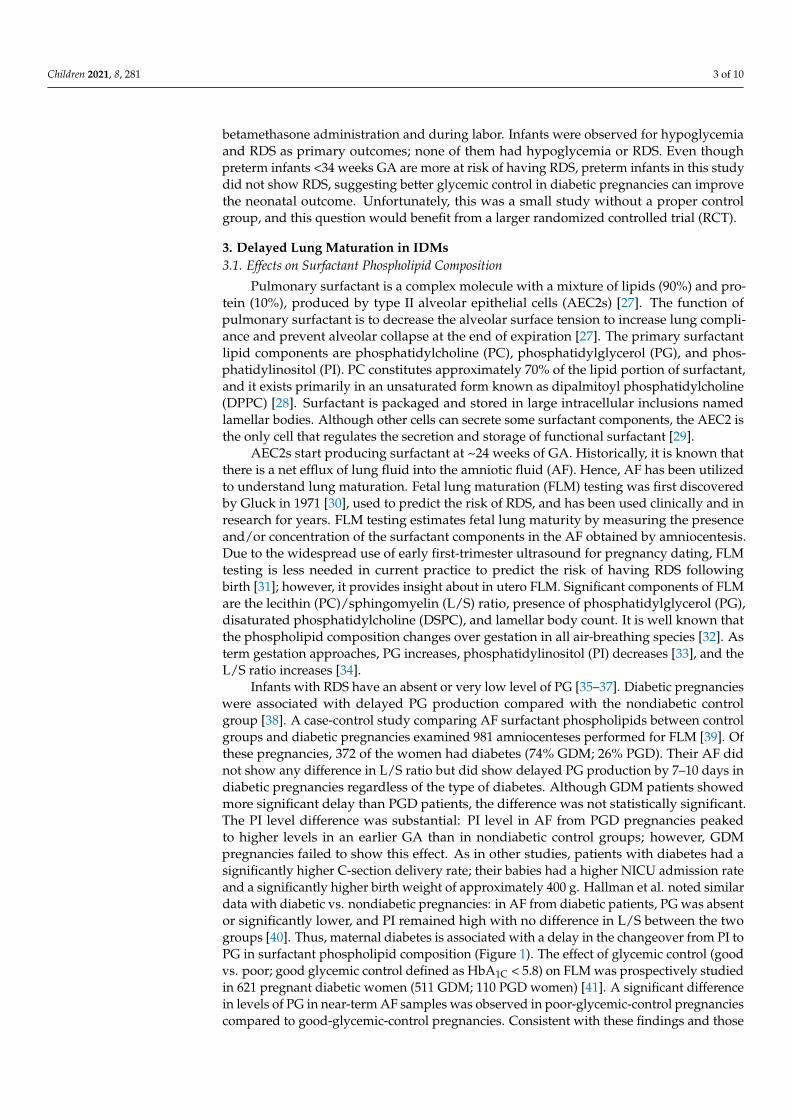
Figure 1. Comparison of alveolus between healthy lungs and infant of a diabetic mother (IDM) lung. Changes in IDM type II alveolar epithelial cell level magnified on the right. Fetal hyperglycemia and hyperinsulinemia secondary to maternal hyperglycemia affect surfactant production via different mechanisms. Hyperglycemia and hyperinsulinism are illustrated in the capillary. (1) Decreased level of surfactant proteins (SP-A, -B, -C, and -D) [42–46]. (2) Decreased level of SP-B causes abnormal tubular myelin formation. (3) Excess glucose inhibits phosphorylase A activity [47]. (4) Glucose decreases insulin receptor tyrosine kinase (TK) activity level [48]. (5) Glycogen storage is decreased due to decreased glycogen phosphory-lase A activity. (6) Alterations in surfactant phospholipid synthesis affected by inadequate glucose resulting in decreased phosphatidyl glycerol (PG) and increased phosphatidyl inositol (PI) [38–40]. (7) These changes contribute to increased respiratory distress syndrome secondary to surfactant deficiency in IDMs.
Glucose is an essential substrate for surfactant lipid synthesis. Insulin not only regu-lates glucose uptake to cells but also regulates surfactant synthesis [49]. The effect of dif-ferent insulin and glucose concentrations on glucose uptake, glucose metabolism, and sur-factant synthesis were examined in AEC2 cultures by Engle et al. [50]. AEC2s derived from fetal rat lung at 19 days of gestation (term = 21) were cultured in different insulin and glucose concentrations. The addition of 10 units/mL of insulin caused a 35% increase in surfactant PC synthesis. However, 100 units/mL insulin reduced PC synthesis to below control levels. The exposure to insulin (3 h vs. 24 h) did not change the result. These results indicate that a physiological level of insulin plays a role as a stimulatory hormone in sur-factant synthesis, but a high insulin level can inhibit surfactant PC synthesis (Figure 2). The effect of hyperglycemia in surfactant lipid production was further studied in fetal rat lung excipients to investigate if there is a critical time period in pregnancy during which AEC2s are more sensitive to hyperglycemia [51]. Fetal rat lung explants from embryonic day 18–22 were exposed to high glucose (100 mM). Increased choline incorporation into PC and DSPC was observed on days 18–19 and in the high glucose exposure group and significantly decreased on days 20–22. The total amount of PC and DSPC significantly decreased on day 20 and did not show any difference on day 18–19. These results suggest that AEC2 from late preterm neonatal rat lung explants was more sensitive to hypergly-cemia and not only had impaired glucose utilization but also had less PC and DSPC.
Figure 1. Comparison of alveolus between healthy lungs and infant of a diabetic mother (IDM) lung. Changes in IDM typeII alveolar epithelial cell level magnified on the right. Fetal hyperglycemia and hyperinsulinemia secondary to maternalhyperglycemia affect surfactant production via different mechanisms. Hyperglycemia and hyperinsulinism are illustratedin the capillary. (1) Decreased level of surfactant proteins (SP-A, -B, -C, and -D) [42–46]. (2) Decreased level of SP-B causesabnormal tubular myelin formation. (3) Excess glucose inhibits phosphorylase A activity [47]. (4) Glucose decreases insulinreceptor tyrosine kinase (TK) activity level [48]. (5) Glycogen storage is decreased due to decreased glycogen phosphorylaseA activity. (6) Alterations in surfactant phospholipid synthesis affected by inadequate glucose resulting in decreasedphosphatidyl glycerol (PG) and increased phosphatidyl inositol (PI) [38–40]. (7) These changes contribute to increasedrespiratory distress syndrome secondary to surfactant deficiency in IDMs.
Glucose is an essential substrate for surfactant lipid synthesis. Insulin not only reg-ulates glucose uptake to cells but also regulates surfactant synthesis [49]. The effect ofdifferent insulin and glucose concentrations on glucose uptake, glucose metabolism, andsurfactant synthesis were examined in AEC2 cultures by Engle et al. [50]. AEC2s derivedfrom fetal rat lung at 19 days of gestation (term = 21) were cultured in different insulin andglucose concentrations. The addition of 10 units/mL of insulin caused a 35% increase insurfactant PC synthesis. However, 100 units/mL insulin reduced PC synthesis to belowcontrol levels. The exposure to insulin (3 h vs. 24 h) did not change the result. Theseresults indicate that a physiological level of insulin plays a role as a stimulatory hormone insurfactant synthesis, but a high insulin level can inhibit surfactant PC synthesis (Figure 2).The effect of hyperglycemia in surfactant lipid production was further studied in fetal ratlung excipients to investigate if there is a critical time period in pregnancy during whichAEC2s are more sensitive to hyperglycemia [51]. Fetal rat lung explants from embryonicday 18–22 were exposed to high glucose (100 mM). Increased choline incorporation into PCand DSPC was observed on days 18–19 and in the high glucose exposure group and signifi-cantly decreased on days 20–22. The total amount of PC and DSPC significantly decreasedon day 20 and did not show any difference on day 18–19. These results suggest that AEC2from late preterm neonatal rat lung explants was more sensitive to hyperglycemia and notonly had impaired glucose utilization but also had less PC and DSPC.
Children 2021, 8, 281 5 of 10
Children 2021, 8, x FOR PEER REVIEW 5 of 11
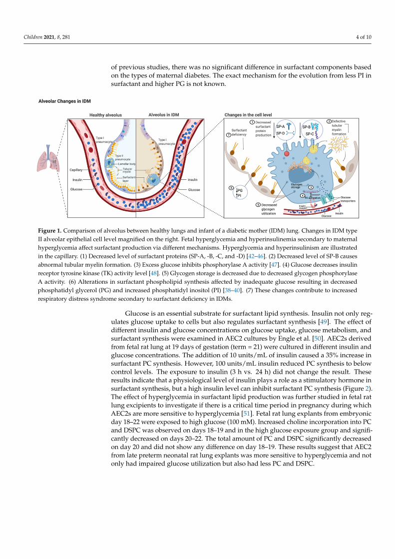
Figure 2. Metabolic changes associated with maternal diabetes effects on surfactant compositions, receptors, and enzymes summarized. Abbreviations: PG (phosphatidyl glycerol), PI (phosphatidyl inositol), PC (phosphatidyl choline), DSPC (de-saturated phosphatidyl choline), SP (surfactant protein), IGF-1R (insulin-like growth factor 1 receptor), and TK (tyrosine kinase) [38–40,42–48,50–54].
In addition to the glucose being an important substrate for surfactant lipid synthesis, glycogen storage in AEC2 is also crucial [55]. AEC2 glycogen increases with increased gestational age, followed by a rapid decline later in pregnancy, and correlates with the acceleration of PC and surfactant synthesis [56]. A rapid decrease in AEC2 glycogen after maternal betamethasone administration in animal studies suggests that glycogen degra-dation is pivotal for surfactant synthesis and positively correlates with lung maturation [57,58]. Glycogen phosphorylase, the enzyme that degrades glycogen, exists in two forms: “A” is the active (dephosphorylated form) and “B” is the inactive (phosphorylated form). An increase in glycogen phosphorylase A activity is related to increased degradation of glycogen storage. Pulmonary glycogen storage and degradation were studied in strepto-zotocin-induced diabetic rats (STZ-DB), a well-established model for diabetic pregnancy and their offspring [47]. STZ-DB fetuses failed to decline in their glycogen storage com-pared to the control group in late gestation on days 21–22 (term = 22). Glycogen phosphor-ylase A activity also was unable to increase and was significantly lower compared with nondiabetic control groups. These findings were consistent with glycogen underutiliza-tion, causing impaired surfactant synthesis leading to delayed lung maturation in diabetic pregnancies. Glucose inhibits phosphorylase A activity by increasing phosphorylate phosphatase, which decreases glycogen phosphorylase A activity [59]; hyperglycemia causing a marked reduction in glycogen phosphorylase A activity (Figure 2); however, the effect of hyperinsulinism on phosphorylase A activity is not apparent [47]. The animal model used in this study does not fully mimic IDM due to the lack of hyperinsulinism. It is unclear how this pathway works in humans, and additional research would improve our understanding of the association between glycogen degradation and RDS in IDMs.
In summary, maternal DM is a significant risk factor for altered surfactant lipid pro-duction in fetuses, and the type of DM does not seem to have a substantial effect on it.
Figure 2. Metabolic changes associated with maternal diabetes effects on surfactant compositions, receptors, and enzymessummarized. Abbreviations: PG (phosphatidyl glycerol), PI (phosphatidyl inositol), PC (phosphatidyl choline), DSPC(desaturated phosphatidyl choline), SP (surfactant protein), IGF-1R (insulin-like growth factor 1 receptor), and TK (tyrosinekinase) [38–40,42–48,50–54].
In addition to the glucose being an important substrate for surfactant lipid synthesis,glycogen storage in AEC2 is also crucial [55]. AEC2 glycogen increases with increasedgestational age, followed by a rapid decline later in pregnancy, and correlates with theacceleration of PC and surfactant synthesis [56]. A rapid decrease in AEC2 glycogenafter maternal betamethasone administration in animal studies suggests that glycogendegradation is pivotal for surfactant synthesis and positively correlates with lung matura-tion [57,58]. Glycogen phosphorylase, the enzyme that degrades glycogen, exists in twoforms: “A” is the active (dephosphorylated form) and “B” is the inactive (phosphorylatedform). An increase in glycogen phosphorylase A activity is related to increased degra-dation of glycogen storage. Pulmonary glycogen storage and degradation were studiedin streptozotocin-induced diabetic rats (STZ-DB), a well-established model for diabeticpregnancy and their offspring [47]. STZ-DB fetuses failed to decline in their glycogen stor-age compared to the control group in late gestation on days 21–22 (term = 22). Glycogenphosphorylase A activity also was unable to increase and was significantly lower comparedwith nondiabetic control groups. These findings were consistent with glycogen under-utilization, causing impaired surfactant synthesis leading to delayed lung maturation indiabetic pregnancies. Glucose inhibits phosphorylase A activity by increasing phosphory-late phosphatase, which decreases glycogen phosphorylase A activity [59]; hyperglycemiacausing a marked reduction in glycogen phosphorylase A activity (Figure 2); however, theeffect of hyperinsulinism on phosphorylase A activity is not apparent [47]. The animalmodel used in this study does not fully mimic IDM due to the lack of hyperinsulinism. It isunclear how this pathway works in humans, and additional research would improve ourunderstanding of the association between glycogen degradation and RDS in IDMs.
In summary, maternal DM is a significant risk factor for altered surfactant lipidproduction in fetuses, and the type of DM does not seem to have a substantial effect on it.
3.2. Effects on Surfactant Protein Composition
Although phospholipid is the major component of surfactant, the four surfactant-associated proteins play critical roles in surfactant synthesis and function [60]. These fourproteins are designated as surfactant protein A (SP-A), SP-B, SP-C, SP-D. These proteins
Children 2021, 8, 281 6 of 10
can be divided into two groups based on their structure and amino acid compositions: SP-Band SP-C are two very small (8 and 4 kDa) [28,61] hydrophobic proteins that are essentialfor normal surface tension-lowering ability of surfactant and packaging of lamellar bodies.SP-A and SP-D are larger (35 and 43 kDa) hydrophilic proteins [62,63] and part of theinnate immune system of the lung in addition to their roles in surfactant hemostasis [64].
The critical role of SP-B was solidified by recognition of a rare congenital SP-B defi-ciency in full-term infants who died shortly after birth due to severe respiratory failureresembling severe RDS [65]. SP-B knockout mice were created in 1996, and not only dothey lack SP-B, but their SP-C is abnormal [66]. As anticipated, the SP-B knockout mice dieshortly after birth due to respiratory failure [67]. SP-C knockout mice have a normal SP-Blevel, and they do not have significant respiratory distress, confirming the need for onlyone of the two hydrophobic proteins for normal surfactant function [68]. SP-C knockoutmice have unstable surfactant at lower lung volumes, suggesting an essential role of SP-Cat low lung volume in RDS [69]. SP-C-deficient animal models develop fibrosis later, andautosomal dominant SP-C mutations are also associated with fibrosis in humans [70].
McGillick et al. studied the effects of hyperglycemia in late-gestation fetal sheep [42].Control group lamb fetuses received infused saline between 130 and 140 days of gestation,and experimental group fetuses were infused glucose. Samples were obtained from animalson day 140 ± 1 (term 150 ± 1). Lung mRNA expression of glucose transporters SLC2A1and SLC2A4, and Insulin-Like Growth Factor-1 (IGF-1), and IGF-2 were not differentbetween the groups. However, decreased IGF-1 receptor (IGF-1R) mRNA expression wasseen in the lung of the glucose-infused fetus. Although the total number of SP-B-positivecells in the alveolar epithelium of the fetal lung was not different between saline- andglucose-infused fetuses, whole lung mRNA for all four surfactant proteins, measuredby quantitative polymerase chain reaction (qPCR), was significantly reduced. This studyprovided evidence for the direct effect of high glucose on depressing SP mRNA. A reductionin all four SPs is likely to impair surfactant ability to lower alveolar surface tension and thesmooth transition to extrauterine life, increasing the risk of having RDS in IDMs (Figure 1).
The effect of fetal hyperglycemia on SP-A, SP-A mRNA, SP-B, SP-B mRNA, SP-C, andSP-C mRNA levels was observed in STZ-DB offspring [43,44]: A significant reduction wasobserved in late gestation fetal days 18–21. Those offspring quickly recovered on neonataldays 1–2 with close to the expected levels of surfactant proteins and their associatedmRNAs. In this animal model, fetal rats were hyperglycemic with a low to normal level ofinsulin, so these results are suggestive of specifically a hyperglycemia inhibitory effect onprotein and mRNA, and not due to a hyperinsulinemia effect (Figure 2).
The effects of hyperglycemia on the production of SP-A, -B, and -C were furtherstudied ex vivo; STZ-DB fetal rat lungs were obtained on embryonic day 20 and culturedin different concentrations of glucose: 10, 25, 50, and 100 mM [45]. While SP-B and SP-CmRNA production was significantly reduced with increased glucose concentration at 100mM (<10%) compared to at 10 mM, SP-A mRNA was not significantly affected in variouslevels of glucose. These in vitro results suggested high glucose concentration inhibits the SP-B and SP-C synthesis, contradicting the previous in vivo SP-A study [43]. A separate studyusing the H441 cell line, a human pulmonary adenocarcinoma cell line shown to expressSP-A mRNA and SP-B mRNA, supports the hypothesis of the concentration-dependentinhibitory effect of high insulin level on surfactant protein gene expression [46].
As in in vitro studies using lung cell lines and in vivo animal studies, the level ofSP-A in AF is significantly reduced between 36 and 40 weeks of GA in human pregnanciescomplicated by diabetes compared to their GA-matched group [52]. These infants didnot develop RDS, but since SP-A does not exhibit surface tension-lowering properties asdetailed above, the lack of RDS is not an unexpected finding.
3.3. Effects on Receptors
Glucose is an essential substrate for surfactant phospholipid, and the insulin receptorregulates glucose uptake in cells [71]. Therefore, impaired glucose uptake, or insulin
Children 2021, 8, 281 7 of 10
receptor maladaptation can play a significant role in surfactant lipid production. Theinsulin receptor has a complex signaling pathway mechanism involving tyrosine kinase(TK) activation [72]. The combination of high glucose and high insulin resulted in asignificant reduction in insulin receptor TK activity and insulin receptor mRNA in fetal ratlung [48] (Figure 1). Downregulated receptors and TK activity resulted in significantly lowglucose uptake from the culture, suggesting clinically significant insulin receptor and TKactivity in glucose metabolism. After being exposed to hyperglycemia, in an immediateperiod of hypoglycemia, TK receptor activity remained diminished for up to 8 h andrecovered after a 12 h exposure [73]. The IDM physiology is similar to that shown in thisstudy: infants have sudden changes in their glucose levels from being hyperglycemic tobeing hypoglycemic after the umbilical cord is cut. Downregulated insulin TK activityreceptors may not provide enough glucose to AEC2, resulting in surfactant deficiency. Thisacute disruption to surfactant synthesis and later recovery of receptor activity in low tonormal glucose levels brings the question of whether or not a euglycemic state closer tobirth can increase substrate availability in AEC2s to overcome the defective surfactant andexplain the wide degree of RDS severity among IDMs.
IGF-1 regulates lung development via the IGF-1R. IGF-1R knockout mice exhibithypoplastic lungs and lung development arrested in the pseudoglandular stage [74]. Al-though an increased level of IGF-1 was seen in the cord blood sample from IDMs, it isnot known if IGF-1 expression is also high in the IDM lung [75]. Increased expression ofIGF-1 and IGF-1R from lung autopsy samples of babies who died from RDS and bron-chopulmonary dysplasia suggests balanced IGF-1/IGF-1R being very important for lungdevelopment and maturation [76].
In addition to the insulin receptor, there are other intracellular signaling mechanismsinvolved in surfactant synthesis, such as FOXA2. FOXA2, also known as hepatocytenuclear factor (HNF) 3B, is a member of the forkhead box (Fox) protein family and oneof the critical signaling molecules for surfactant synthesis regulation during fetal lungdevelopment [77]. Although FOXA2 plays a role in other organogenesis, such as in thepancreas, liver, and adipose tissue, the expression of FOXA2 is limited to AEC2 in the thirdtrimester of the pregnancy when lung development and surfactant synthesis peaks in fetallife. Maternal diabetes effects on FOXA2, SP-B, and SP-C synthesis were studied in theSTZ-DB rat models of preexisting diabetes and GDM [53,54]. Both groups of fetuses werederived from diabetic rats. In the diabetic offspring, fetal lung expressions of SP-B, SP-BmRNA, SP-C, SP-C mRNA, and FOXA2 in the nucleus (n-FOXA2) were significantly lowerin both PGD and GDM offspring, while phosphorylated FOXA2 (which decreases FOXA2transcription activity) in PGD, and nitrolyogenic FOXA2 (deactivated FOXA2) in GDMwas significantly higher (Figure 2).
Akt/mammalian target of rapamycin (mTOR) pathway is another insulin signalingmechanism. mTOR is a serine/threonine kinase that regulates cell growth and differ-entiation [78]. mTOR controls insulin signaling in cellular level. The importance of theAkt/mTOR pathway was studied in a transgenic mouse model [79]. In utero, Akt activationin lung epithelial cell showed impaired maturation of the lung epithelium, downregulationof SP-B, and increased glycogen storage. Inhibition of Akt/mTOR pathway with rapamycinimproved alveolar epithelial cell differentiation, decreased glycogen storage, and producednormal expression of SP-C. Downregulation of mTOR signaling mechanism possibly playsan important role in lung maturation and epithelial differentiation.
These findings suggest the importance of euglycemia and normal insulin level for theregulation of glucose uptake and surfactant synthesis.
4. Conclusions
Any variety of maternal diabetes (PGD type 1 or 2, or GDM) is a significant contributorto fetal health. Fetal hyperglycemia and hyperinsulinism secondary to maternal diabetesdisrupt normal surfactant synthesis and function, which leads to surfactant inadequacyand clinical RDS in neonates. As maternal diabetes increases the risk of RDS in near-term
Children 2021, 8, 281 8 of 10
infants, finding different methods to achieve better glycemic control is needed, as is furtherunderstanding of the molecular processes during GDM/PGD affecting the surfactantsystem. Identifying the critical factors in IDMs who do not show signs of RDS may openup other targets for the prevention and treatment of RDS in this population. As we learnmore about the increased incidence of long-term health problems related to IDMs, such ashypertension, insulin resistance, diabetes, obesity, and neurodevelopmental impairment,we need to pay more attention to maternal health to improve neonatal outcomes andlifelong health.
Author Contributions: R.M.R. provided leadership and supervision for the manuscript. H.Y.A.,J.E.B. and R.M.R. critically reviewed and revised the manuscript. All authors have read and agreedto the published version of the manuscript.
Funding: This research received no external funding.
Institutional Review Board Statement: Not applicable.
Informed Consent Statement: Not applicable.
Acknowledgments: Figures are created with BioRender.com.
Conflicts of Interest: The authors declare no conflict of interest.
References1. Dabelea, D.; Snell-Bergeon, J.K.; Hartsfield, C.L.; Bischoff, K.J.; Hamman, R.F.; McDuffie, R.S. Increasing prevalence of gestational
diabetes mellitus (GDM) over time and by birth cohort: Kaiser Permanente of Colorado GDM screening program. Diabetes Care2005, 28, 579–584. [CrossRef] [PubMed]
2. Mackin, S.T.; Nelson, S.M.; Kerssens, J.J.; Wood, R.; Wild, S.; Colhoun, H.M.; Leese, G.P.; Philip, S.; Lindsay, R.S. Diabetes andpregnancy: National trends over a 15 year period. Diabetologia 2018, 61, 1081–1088. [CrossRef] [PubMed]
3. International Diabetes Federation. IDF Diabetes Atlas, 9th ed.; Atlas De La Diabetes De La Fid: Brussels, Belgium, 2019.4. Barbour, L.A.; McCurdy, C.E.; Hernandez, T.L.; Kirwan, J.P.; Catalano, P.M.; Friedman, J.E. Cellular mechanisms for insulin
resistance in normal pregnancy and gestational diabetes. Diabetes Care 2007, 30, S112–S119. [CrossRef] [PubMed]5. Sonagra, A.D. Normal Pregnancy- A State of Insulin Resistance. J. Clin. Diagn. Res. 2014, 8, CC01–CC03. [CrossRef]6. Kitzmiller, J.L.; Block, J.M.; Brown, F.M.; Catalano, P.M.; Conway, D.L.; Coustan, D.R.; Gunderson, E.P.; Herman, W.H.; Hoffman,
L.D.; Inturrisi, M.; et al. Managing preexisting diabetes for pregnancy: Summary of evidence and consensus recommendationsfor care. Diabetes Care 2008, 31, 1060–1079. [CrossRef] [PubMed]
7. Catalano, P.M.; Bernstein, I.M.; Wolfe, R.R.; Srikanta, S.; Tyzbir, E.; Sims, E.A. Subclinical abnormalities of glucose metabolism insubjects with previous gestational diabetes. Am. J. Obs. Gynecol. 1986, 155, 1255–1262. [CrossRef]
8. Noctor, E. Type 2 diabetes after gestational diabetes: The influence of changing diagnostic criteria. World J. Diabetes 2015, 6, 234.[CrossRef]
9. O’Sullivan, J.B.; Mahan, C.M. Criteria for the Oral Glucose Tolerance Test in Pregnancy. Diabetes 1964, 13, 278–285.10. Care, D.; Suppl, S.S. Summary of Revisions: Standards of Medical Care in Diabetes-2020. Diabetes Care 2020, 43, S98–S110.11. Agarwal, M.M.; Dhatt, G.S.; Punnose, J.; Koster, G. Gestational diabetes: A reappraisal of HBA1c as a screening test. Acta Obs.
Gynecol. Scand. 2005, 84, 1159–1163. [CrossRef]12. Jovanovic, L.; Savas, H.; Mehta, M.; Trujillo, A.; Pettitt, D.J. Frequent monitoring of A1C during pregnancy as a treatment tool to
guide therapy. Diabetes Care 2011, 34, 53–54. [CrossRef] [PubMed]13. Rajput, R.; Rajput, M.; Nanda, S. Utility of HbA 1c for diagnosis of gestational diabetes mellitus. Diabetes Res. Clin. Pr. 2012, 98,
104–107. [CrossRef] [PubMed]14. Hughes, R.C.E.; Rowan, J.; Florkowski, C.M. Is There a Role for HbA1c in Pregnancy? Curr. Diab. Rep. 2016, 16, 1–10. [CrossRef]15. Chen, C.P. Congenital malformations associated with maternal diabetes. Taiwan. J. Obs. Gynecol. 2005, 44, 1–7. [CrossRef]16. Becerra, J.E.; Khoury, M.J.; Cordero, J.F.; Erickson, J.D. Diabetes mellitus during pregnancy and the risks for specific birth defects:
A population-based case-control study. Pediatrics 1990, 85, 1–9.17. Guerin, A.; Nisenbaum, R.; Ray, J.G. Use of Maternal GHb Concentration to Estimate the Risk of Congenital Anomalies. Diabetes
Care 2007, 30, 1920–1925. [CrossRef] [PubMed]18. Ogata, E.S. Problems of the infant of the diabetic mother. Neoreviews 2010, 11, e627–e631. [CrossRef]19. Michael Weindling, A. Offspring of diabetic pregnancy: Short-term outcomes. Semin. Fetal Neonatal Med. 2009, 14, 111–118.
[CrossRef]20. Avery, M.E.; Mead, J. Surface Properties in Relation to Atelectasis and Hyaline Membrane Disease. Ama. J. Dis. Child. 1959, 97,
517–523. [CrossRef]21. Gellis, S.S.; Hsia, D.Y. The infant of diabetic mother. Am. J. Dis Child. 1959, 97, 1–41. [CrossRef]
Children 2021, 8, 281 9 of 10
22. Robert, M.F.; Neff, R.K.; Hubbell, J.P.; Taeusch, H.W.; Avery, M.E. Association between Maternal Diabetes and the Respiratory-Distress Syndrome in the Newborn. N. Engl. J. Med. 1976, 294, 357–360. [CrossRef] [PubMed]
23. Mortier, I.; Blanc, J.; Tosello, B.; Gire, C.; Bretelle, F.; Carcopino, X. Is gestational diabetes an independent risk factor of neonatalsevere respiratory distress syndrome after 34 weeks of gestation? A prospective study. Arch. Gynecol. Obs. 2017, 296, 1071–1077.[CrossRef]
24. Confidential Enquiry into Maternal and Child Health. Pregnancy in Women with Type 1 and Type 2 Diabetes in 2002–2003, England,Wales and Northern Ireland; CEMACH: London, UK, 2005.
25. Balsells, M.; García-Patterson, A.; Gich, I.; Corcoy, R. Maternal and fetal outcome in women with type 2 versus type 1 diabetesmellitus: A systematic review and metaanalysis. J. Clin. Endocrinol. Metab. 2009, 94, 4284–4291. [CrossRef] [PubMed]
26. Iafusco, D.; Stoppoloni, F.; Salvia, G.; Vernetti, G.; Passaro, P.; Petrovski, G.; Prisco, F. Use of real time continuous glucosemonitoring and intravenous insulin in type 1 diabetic mothers to prevent respiratory distress and hypoglycaemia in infants. BMCPregnancy Childbirth 2008, 8, 23. [CrossRef] [PubMed]
27. Veldhuizen, E.J.A.; Haagsman, H.P. Role of pulmonary surfactant components in surface film formation and dynamics. Biochim.Biophys. Acta Biomembr. 2000, 1467, 255–270. [CrossRef]
28. Griese, M. Pulmonary surfactant in health and human lung diseases: State of the art. Eur. Respir. J. 1999, 13, 1455–1476. [CrossRef][PubMed]
29. McGowan, S.E. The Formation of Pulmonary Alveoli. Lung Dev. Aging Environ. Second Ed. 2014, 65–84. [CrossRef]30. Gluck, L.; Kulovich, M.V.; Borer, R.C., Jr. Diagnosis of respiratory distress by amniocentesis. Am. J. Obs. Gynecol. 1971, 109,
440–445.31. Johnson, L.M.; Johnson, C.; Karger, A.B. End of the line for fetal lung maturity testing. Clin. Biochem. 2019, 71, 74–76. [CrossRef]32. Hodson, W.A. Lung Development in the Fetal Primate. Pediatr. Res. 1977, 11, 1015–1021.33. Hallman, M.; Kulovich, M.; Kirkpatrick, E.; Sugarman, R.G.; Gluck, L. Phosphatidylinositol and phosphatidylglycerol in amniotic
fluid: Indices of lung maturity. Am. J. Obs. Gynecol. 1976, 125, 613–617. [CrossRef]34. Gluck, L.; Kulovich, M.V. Lecithin/sphingomyelin ratios in amniotic fluid in normal and abnormal pregnancy. Am. J. Obs.
Gynecol. 1973, 115, 539–546. [CrossRef]35. Hallman, M.; Merritt, T.A.; Pohjavuori, M.; Gluck, L. Effect of surfactant substitution on lung effluent phospholipids in respiratory
distress syndrome: Evaluation of surfactant phospholipid turnover, pool size, and the relationship to severity of respiratoryfailure. Pediatr. Res. 1986, 20, 1228–1235. [CrossRef] [PubMed]
36. Hallman, M.; Feldman, B.H.; Kirkpatrick, E.; Gluck, L. Absence of phosphatidylglycerol (PG) in respiratory distress syndrome inthe newborn: Study of the minor surfactant phospholipids in newborns. Pediatr. Res. 1977, 11, 714–720. [CrossRef]
37. Obladen, M. Factors influencing surfactant composition in the newborn infant. Eur. J. Pediatr. 1978, 128, 129–143. [CrossRef][PubMed]
38. Piper, J.M.; Langer, O. Does maternal diabetes delay fetal pulmonary maturity? Am. J. Obs. Gynecol. 1993, 168, 783–786. [CrossRef]39. Moore, T.R. A comparison of amniotic fluid fetal pulmonary phospholipids in normal and diabetic pregnancy. Am. J. Obs. Gynecol.
2002, 186, 641–650. [CrossRef] [PubMed]40. Hallman, M.; Teramo, K. Amniotic fluid phospholipid profile as predictor of fetal maturity in diabetic pregnancies. Obs. Gynecol.
1979, 54, 703–707.41. Piper, J.M.; Xenakis, E.M.-J.; Langer, O. Delayed appearance of pulmonary maturation markers is associated with poor glucose
control in diabetic pregnancies. J. Matern. Fetal. Med. 1998, 7, 148–153.42. McGillick, E.V.; Morrison, J.L.; McMillen, I.C.; Orgeig, S. Intrafetal glucose infusion alters glucocorticoid signaling and reduces
surfactant protein mRNA expression in the lung of the late-gestation sheep fetus. Am. J. Physiol. Regul. Integr. Comp. Physiol. 2014,307, 538–545. [CrossRef]
43. Guttentag, S.H.; Phelps, D.S.; Stenzel, W.; Warshaw, J.B.; Floros, J. Surfactant protein A expression is delayed in fetuses ofstreptozotocin- treated rats. Am. J. Physiol. Lung Cell. Mol. Physiol. 1992, 262, 489–494. [CrossRef]
44. Guttentag, S.H.; Phelps, D.S.; Warshaw, J.B.; Floros, J. Delayed hydrophobic surfactant protein (SP-B, SP-C) expression in fetusesof streptozotocin-treated rats. Am. J. Respir. Cell Mol. Biol. 1992, 7, 190–197. [CrossRef]
45. Rayani, H.H.; Gewolb, I.H.; Floros, J. Glucose decreases steady state mRNA content of hydrophobic surfactant proteins B and Cin fetal rat lung explants. Exp. Lung Res. 1999, 25, 69–79. [CrossRef]
46. Miakotina, O.L.; Dekowski, S.A.; Snyder, J.M. Insulin inhibits surfactant protein A and B gene expression in the H441 cell line.Biochim. Biophys. Acta Gene Struct. Expr. 1998, 1442, 60–70. [CrossRef]
47. Gewolb, I.H.; Barrett, C.; Wilson, C.M.; Warshaw, J.B. Delay in pulmonary glycogen degradation in fetuses of streptozotocindiabetic rats. Pediatr. Res. 1982, 16, 869–873. [CrossRef] [PubMed]
48. Gewolb, I.H.; O’brien, J.; Palese, T.A.; Phillip, M. High Glucose and Insulin Decrease Fetal Lung Insulin Receptor mRNA andTyrosine Kinase Activity in Vitro. Biochem. Biophys. Res. Commun. 1994, 202, 694–700. [CrossRef]
49. Patel, D.M.; Rhodes, P.G. Effects of insulin and hydrocortisone on lung tissue phosphatidyl choline and disaturated phosphatidylcholine in fetal rabbits in vivo. Diabetologia 1984, 27, 478–481. [CrossRef] [PubMed]
50. Engle, M.J.; Langan, S.M.; Sanders, R.L. The effects of insulin and hyperglycemia on surfactant phospholipid synthesis inorganotypic cultures of type II pneumocytes. Biochim. Biophys. Acta (Bba)/Lipids Lipid Metab. 1983, 753, 6–13. [CrossRef]
Children 2021, 8, 281 10 of 10
51. Ira, H. Gewolb (1996) Effect of High Glucose on Fetal Lung Maturation at Different Times in Gestation. Exp. Lung Res. 1996, 22,201–211. [CrossRef]
52. Salisbury-Murphy, S.; Rubinstein, D.; Beck, J.C. Lipid metabolism in lung slices. Am. J. Physiol. 1966, 211, 988–992. [CrossRef]53. Maniscalco, W.M.; Wilson, C.M.; Gross, I.; Gobran, L.; Rooney, S.A.; Warshaw, J.B. Development of glycogen and phospholipid
metabolism in fetal and newborn rat lung. Biochim. Biophys. Acta (Bba)/Lipids Lipid Metab. 1978, 530, 333–346. [CrossRef]54. Kikkawa, Y.; Kaibara, M.; Motoyama, E.K.; Orzalesi, M.M.; Cook, C.D. Morphologic development of fetal rabbit lung and its
acceleration with cortisol. Am. J. Pathol. 1971, 64, 423–442. [PubMed]55. Gilden, C.; Sevanian, A.; Tierney, D.F.; Kaplan, S.A.; Barrett, C.T. Regulation of fetal lung phosphatidyl choline synthesis by
cortisol: Role of glycogen and glucose. Pediatr. Res. 1977, 11, 845–848. [CrossRef] [PubMed]56. Holmes, P.A.; Mansour, T.E. Glucose as a regulator of glycogen phosphorylase in rat diaphragm. Biochim. Biophys. Acta Gen. Subj.
1968, 156, 275–284. [CrossRef]57. Pérez-Gil, J. Structure of pulmonary surfactant membranes and films: The role of proteins and lipid-protein interactions. Biochim.
Biophys. Acta Biomembr. 2008, 1778, 1676–1695. [CrossRef]58. Akella, A.; Deshpande, S.B. Pulmonary surfactants and their role in pathophysiology of lung disorders. Indian J. Exp. Biol. 2013,
51, 5–22.59. Veldhuizen, R.; Nag, K.; Orgeig, S.; Possmayer, F. The role of lipids in pulmonary surfactant. Biochim. Biophys. Acta Mol. Basis Dis.
1998, 1408, 90–108. [CrossRef]60. Vieira, F.; Kung, J.W.; Bhatti, F. Structure, genetics and function of the pulmonary associated surfactant proteins A and D: The
extra-pulmonary role of these C type lectins. Ann. Anat. 2017, 211, 184–201. [CrossRef]61. Kingma, P.S.; Whitsett, J.A. In defense of the lung: Surfactant protein A and surfactant protein D. Curr. Opin. Pharm. 2006, 6,
277–283. [CrossRef]62. Nogee, L.M.; Garnier, G.; Singer, L.; Dietz, H.C.; Singer, L.; Murphy, A.M.; deMello, D.E.; Colten, H.R. A mutation in the surfactant
protein B gene responsible for fatal neonatal respiratory disease in multiple kindreds. J. Clin. Investig. 1994, 93, 1860–1863.[CrossRef]
63. Tokieda, K.; Whitsett, J.A.; Clark, J.C.; Weaver, T.E.; Ikeda, K.; McConnell, K.B.; Jobe, A.H.; Ikegami, M.; Iwamoto, H.S. Pulmonarydysfunction in neonatal SP-B-deficient mice. Am. J. Physiol. Lung Cell. Mol. Physiol. 1997, 273, 875–882. [CrossRef] [PubMed]
64. Clark, J.C.; Wert, S.E.; Bachurski, C.J.; Stahlman, M.T.; Stripp, B.R.; Weaver, T.E.; Whitsett, J.A. Targeted disruption of thesurfactant protein B gene disrupts surfactant homeostasis, causing respiratory failure in newborn mice. Proc. Natl. Acad. Sci. USA1995, 92, 7794–7798. [CrossRef] [PubMed]
65. Spragg, R.G.; Lewis, J.F. Pathology of the surfactant system of the mature lung: Second San Diego conference. Am. J. Respir. Crit.Care Med. 2001, 163, 280–282. [CrossRef] [PubMed]
66. Glasser, S.W.; Burhans, M.S.; Korfhagen, T.R.; Na, C.L.; Sly, P.D.; Ross, G.F.; Ikegami, M.; Whitsett, J.A. Altered stability ofpulmonary surfactant in SP-C-deficient mice. Proc. Natl. Acad. Sci. USA 2001, 98, 6366–6371. [CrossRef]
67. Nogee, L.M.; Dunbar, A.E.; Wert, S.E.; Askin, F.; Hamvas, A.; Whitsett, J.A. A Mutation in the Surfactant Protein C GeneAssociated with Familial Interstitial Lung Disease. N. Engl. J. Med. 2001, 344, 573–579. [CrossRef]
68. Snyder, J.M.; Kwun, J.E.; O’brien, J.A.; Rosenfeld, C.R.; Odom, M.J. The concentration of the 35-kDa surfactant apoprotein inamniotic fluid from normal and diabetic pregnancies. Pediatr. Res. 1988, 24, 728–734. [CrossRef]
69. Kahn, C.R. The molecular mechanism of insulin action. Annu. Rev. Med. 1985, 36, 429–451. [CrossRef]70. Czech, M.P. The nature and regulation of the insulin receptor: Structure and function. Annu. Rev. Physiol. 1985, 47, 357–381.
[CrossRef]71. Marwah, G.S.; O’Brien, J.; Gewolb, I.H. Effect of acute glucose depletion following glucose excess on surfactant phospholipid
synthesis in developing fetal lung. Exp. Lung Res. 1999, 25, 291–302.72. Epaud, R.; Aubey, F.; Xu, J.; Chaker, Z.; Clemessy, M.; Dautin, A.; Ahamed, K.; Bonora, M.; Hoyeau, N.; Flejou, J.-F.; et al.
Knockout of insulin-like growth factor-1 receptor impairs distal lung morphogenesis. PLoS ONE 2012, 7, e48071. [CrossRef][PubMed]
73. Hiden, U.; Glitzner, E.; Hartmann, M.; Desoye, G. Insulin and the IGF system in the human placenta of normal and diabeticpregnancies. J. Anat. 2009, 215, 60–68. [CrossRef]
74. Chetty, A.; Andersson, S.; Lassus, P.; Nielsen, H.C. Insulin-Like Growth Factor-1 (IGF-1) and IGF-1 Receptor (IGF-1R) Expressionin Human Lung in RDS and BPD. Pediatr. Pulmonol. 2004, 37, 128–136. [CrossRef] [PubMed]
75. Wan, H.; Xu, Y.; Ikegami, M.; Stahlman, M.T.; Kaestner, K.H.; Ang, S.L.; Whitsett, J.A. Foxa2 is required for transition to airbreathing at birth. Proc. Natl. Acad. Sci. USA 2004, 101, 14449–14454. [CrossRef]
76. Zhang, Q.-M.; Chai, X.-Q.; Deng, F.-T.; Quyang, W.; Song, T. The reduction in FOXA2 activity during lung development in fetusesfrom diabetic rat mothers is reversed by Akt inhibition. FEBS Open Bio. 2018, 8, 1594–1604. [CrossRef] [PubMed]
77. Zhang, Q.-M.; Ouyang, W.-X.; Chai, X.-Q.; Deng, F.-T. Expression of Lung Surfactant Proteins SP-B and SP-C and Their RegulatoryFactors in Fetal Lung of GDM Rats. Curr. Med. Sci. 2018, 38, 847–852. [CrossRef] [PubMed]
78. Yoon, M.S. The Role of Mammalian Target of Rapamycin (mTOR) in Insulin Signaling. Nutrients 2017, 9, 1176. [CrossRef]79. Ikeda, H.; Shiojima, I.; Oka, T.; Yoshida, M.; Maemura, K.; Walsh, K.; Igarashi, T.; Komuro, I. Increased Akt-mTOR signaling in
lung epithelium is associated with respiratory distress syndrome in mice. Mol. Cell Biol. 2011, 31, 1054–1065. [CrossRef]