Guideline Therapeutics and COVID-19 - WHO | World Health ...
Upload
kingabdulazizCategory
view
0download
0
RESEARCH ARTICLE
Molecular Interaction of a Kinase InhibitorMidostaurin with Anticancer Drug Targets,S100A8 and EGFR: Transcriptional Profilingand Molecular Docking Study for KidneyCancer TherapeuticsZeenat Mirza1, Hans-Juergen Schulten2, Hasan Ma Farsi3, Jaudah A. Al-Maghrabi4,5,Mamdooh A. Gari2, Adeel Ga Chaudhary1,2, Adel M. Abuzenadah1,2,6, Mohammed H. Al-Qahtani2*, Sajjad Karim2*
1 King Fahd Medical Research Center, King Abdulaziz University, PO BOX 80216, Jeddah, 21589, SaudiArabia, 2 Center of Excellence in Genomic Medicine Research, Faculty of Applied Medical Sciences, KingAbdulaziz University, PO BOX 80216, Jeddah, 21589, Saudi Arabia, 3 Department of Urology, Faculty ofMedicine, King Abdulaziz University Hospital, Jeddah, Saudi Arabia, 4 Department of Pathology, Faculty ofMedicine, King Abdulaziz University Hospital, Jeddah, Saudi Arabia, 5 Department of Pathology, King FaisalSpecialist Hospital and Research Center, Jeddah, Saudi Arabia, 6 KACST Technology Innovation Center inPersonalized Medicine at King Abdulaziz University, Jeddah, Saudi Arabia
* [email protected] (SK); [email protected] (MQ)
AbstractThe S100A8 and epidermal growth factor receptor (EGFR) proteins are proto-oncogenes
that are strongly expressed in a number of cancer types. EGFR promotes cellular prolifera-
tion, differentiation, migration and survival by activating molecular pathways. Involvement of
proinflammatory S100A8 in tumor cell differentiation and progression is largely unclear and
not studied in kidney cancer (KC). S100A8 and EGFR are potential therapeutic biomarkers
and anticancer drug targets for KC. In this study, we explored molecular mechanisms of in-
teraction profiles of both molecules with potential anticancer drugs. We undertook transcrip-
tional profiling in Saudi KCs using Affymetrix HuGene 1.0 ST arrays. We identified 1478
significantly expressed genes, including S100A8 and EGFR overexpression, using cut-off
p=value<0.05 and fold change�2. Additionally, we compared and confirmed our findings
with expression data available at NCBI’s GEO database. A significant number of genes as-
sociated with cancer showed involvement in cell cycle progression, DNA repair, tumor mor-
phology, tissue development, and cell survival. Atherosclerosis signaling, leukocyte
extravasation signaling, notch signaling, and IL-12 signaling were the most significantly dis-
rupted signaling pathways. The present study provides an initial transcriptional profiling of
Saudi KC patients. Our analysis suggests distinct transcriptomic signatures and pathways
underlying molecular mechanisms of KC progression. Molecular docking analysis revealed
that the kinase inhibitor "midostaurin" has amongst the selected drug targets, the best li-
gand properties to S100A8 and EGFR, with the implication that its binding inhibits down-
stream signaling in KC. This is the first structure-based docking study for the selected
PLOSONE | DOI:10.1371/journal.pone.0119765 March 19, 2015 1 / 17
a11111
OPEN ACCESS
Citation: Mirza Z, Schulten H-J, Farsi HM, Al-Maghrabi JA, Gari MA, Chaudhary AG, et al. (2015)Molecular Interaction of a Kinase Inhibitor Midostaurinwith Anticancer Drug Targets, S100A8 and EGFR:Transcriptional Profiling and Molecular Docking Studyfor Kidney Cancer Therapeutics. PLoS ONE 10(3):e0119765. doi:10.1371/journal.pone.0119765
Academic Editor: Jaydutt V. Vadgama, Charles R.Drew University of Medicine and Science, UNITEDSTATES
Received: September 9, 2014
Accepted: January 16, 2015
Published: March 19, 2015
Copyright: © 2015 Mirza et al. This is an openaccess article distributed under the terms of theCreative Commons Attribution License, which permitsunrestricted use, distribution, and reproduction in anymedium, provided the original author and source arecredited.
Data Availability Statement: All relevant data arewithin the paper and its Supporting Information files.
Funding: This work was supported by King AbdullahCity for Science and Technology, Riyadh, SaudiArabia (KACST, Strategic Project ID. 10-BIO1258-03and 10-BIO1073-03) and Deanship of Research,King Abdulaziz University (Project ID: HiCi-1434-117-2). The funders had no role in study design, datacollection and analysis, decision to publish, orpreparation of the manuscript.
protein targets and anticancer drug, and the results indicate S100A8 and EGFR as attrac-
tive anticancer targets and midostaurin with effective drug properties for therapeutic inter-
vention in KC.
IntroductionCancer is a global major health problem. Dysregulation in molecular signaling pathways is ahallmark of cancer initiation and progression [1–3]. Kidney cancer (KC) accounts for approxi-mately 1.5 percent of all cancer deaths. In particular, KC is common in obese male population[4]. Surgical tumor resection is the standard curative treatment. Metastatic KC is almost nonre-sponsive to conventional systemic treatments and nearly all patients die of metastasis. Lack ofpromising biomarkers for effective targeted chemotherapy poses a big challenge in KC man-agement. Better understanding of the molecular mechanisms effective in KC have widened thewindow for development of effective targeted therapies [5,6]. High-throughput microarrayplatforms are well suited for identification of the novel induced or suppressed disease-relatedculprit genes [7]. Molecules showing direct involvement in a biochemical or regulatory path-way leading to disease are potential anticancer target. Drug/molecule interaction involvingthese targets can either be investigated by co-crystallization or tested by docking simulation toindentify molecular interactions required for rational drug designing [8]. High-throughputdocking is the key entrance for drug discovery [9,10]. Transcriptomic profiling and functionalpathway analysis in KC have identified several significantly differentially expressed genes, in-cluding S100A8 and EGFR. We tried to evaluate their potential as KC drug target by in silicodocking with known protein kinase inhibitors. Overall, this study illustrates structure-basedvirtual screening and ligand-protein docking of anticancer drugs e.g. midostaurin, enzastaurin,and gefitinib, with anticancer targets, S100A8 and EGFR.
S100A8 is a small (10 kDa) proinflammatory protein of S100 family, which tends to formheterodimeric complexes with S100A9 (S100A8/A9) [9], that undergo conformational changesupon Ca2+ binding and function as intracellular Ca2+ sensors [10]. Under physiological condi-tions, these Ca2+ binding EF hand type proteins are constitutively expressed by myeloid cells[11–13]. However, under pathological conditions like inflammation and cancer, an increasedexpression of S100A8 is seen in epithelial cells [14,15]. Early stage death of S100A8 knock-outmice proves the essentiality of this gene for survival [16]. Enhanced level of S100A8 is found indifferent carcinomas including breast [15], prostate [17,18], lung [19], gastric [20], hepatic[21], pancreatic [22] and colorectal cancer [23,24]. A recent study shows cell growth-promoting activity and binding to receptor for advanced glycation endproducts (RAGE) at lowS100A8 concentrations [15]; however, its direct role in tumorogenesis is ambiguous and has tobe elucidated yet. It has been reported that primary tumors secrete soluble factors, which in-duce expression of S100A8 in the endothelial cells prior to tumor metastasis [19]. By activatingthe p38 MAPK pathway, it increases the motility of circulating cancer cells [25]. Therefore tar-geting S100A8 could be used to prevent the tumor cell migration and growth. Several lines ofevidence point to vital functions of S100A8 during tumorigenesis and, although it’s exact rolewithin the tumor microenvironment is still not clear, different tumor-promoting effects havebeen proposed. Though, research is limited on its expression pattern in cancer, its involvementin oncogenesis and its potential as therapeutic biomarker. To the best of our knowledge, nostudy has reported yet its expression pattern or its role in KC progression.
Midostaurin a Potential Kidney Cancer Drug against S100A8 and EGFR
PLOSONE | DOI:10.1371/journal.pone.0119765 March 19, 2015 2 / 17
Competing Interests: The authors have declaredthat no competing interests exist.

EGFR is one of the four member of the ErbB family of receptor tyrosine kinases. The recep-tor is overexpressed or mutated in many cancers, highlighting its role as therapeutic cancer bio-marker. It is involved in diverse cellular functions such as proliferation, angiogenesis andsuppression of cell death. Being a transmembrane protein, EGFR passes crucial signals fromepithelial cell surface to the intracellular domain for controlled cell proliferation, migrationand adhesion. Overexpressed EGFR transmits multiple signals to cell for accelerated growthand cellular longevity, and plays a key role in the carcinogenesis of different types of cancer[26–29]. Improved perception of the molecular signaling pathways has opened new strategiesand ways for cancer treatment. Thus, targeting EGFR to turn off its signal transduction is en-visaged to block growth and survival of cancer cells.
S100A8 and EGFR do play an important role in malignancy and thus considered as poten-tial drug targets for cancer therapeutics development [26–30]. The reason for choosing midos-taurin, enzastaurin, and gefitinib as inhibitor for S100A8 and EGFR is the recent researchshowing that EGFR-targeted therapies using kinase inhibitors are effective in many cancer pa-tients [31–36]. Gefitinib, an inhibitor of EGFR kinase function, was approved by the U.S. FDAfor lung cancer treatment [37]. S100A8 does not own any kinase domain or ATP binding pock-et, however it participates in protein phosphorylation [38] and is an upstream pathway mole-cule of EGFR. Thus, a drug capable of inhibiting both EGFR and S100A bears prospectivetherapeutic impact.
After the discovery of protein kinase activity in 1954, a lot of kinase inhibitors have beenidentified [39]. Because of high degree of similarity in their structure and function within the“kinome” [40], identification of effective protein kinase inhibitors (PKIs) is a great challenge.At present kinase inhibitors comprise more than 30% of drug-discovery programs resulting inapproval of dozens of kinase inhibitors as anti-cancer drugs or at least testing in clinical trials[41]. In this study, we determined by docking simulation the potential efficiency of three suchdrugs named midostaurin, enzastaurin, and gefitinib.
(i) Midostaurin (CID 104937; also known as PKC412 and benzoylstaurosporine) is a multi-target kinase inhibitor used for acute myeloid leukemia treatment. It is a semi-synthetic alka-loid derived from bacterial staurosporine used to treat patients with CD135 (FMS-like tyrosinekinase 3 receptor) mutations [42]. Midostaurin inhibits growth or induces apoptosis in severaltypes of cancer, blocks angiogenesis and sensitizes cancer cells to ionizing radiation, justifyingits use in cancer treatment [43]. Preclinical studies have shown that midostaurin works syner-gistically with chemotherapy agents, reinforcing each other's effect against cancer [44]. (ii)Enzastaurin is a synthetic acyclic bisindolylmaleimide showing antineoplastic activity. It is asmall oral serine/threonine kinase inhibitor of PKCβ and AKT pathways. It inhibits protein ki-nase Cβ that is known stimulator of neo-angiogenesis through induction of vascular endotheli-al growth factor (VEGF). Binding of protein kinase Cβ to the ATP-binding site of VEGFprobably decreases tumor blood supply and prevents growth. Apart from its anti—VEGF fac-tor effects, low concentration of enzastaurin suppresses proliferation in cancer cell lines [45].Loss of GSK3β and AKT phosphorylation has been reported in myeloma cells treatment withenzastaurin [46]. (iii) Gefitinib (Iressa) selectively targets the ATP cleft within EGFR tyrosinekinase domain [47]. It interrupts EGFR signaling of target cells and therefore is effective onlyin cancer cells with mutated and overactive EGFR.
Molecular docking forecasts an optimized conformation and relative orientation for boththe protein and ligand molecule. In this study we were aiming to evaluate the potential ofS100A8 and EGFR as KC drug target by in silicomolecular docking with the known protein ki-nase inhibitors midostaurin, enzastaurin, and gefitinib to test the effectiveness of these antican-cer drugs under these constellations.
Midostaurin a Potential Kidney Cancer Drug against S100A8 and EGFR
PLOSONE | DOI:10.1371/journal.pone.0119765 March 19, 2015 3 / 17
Materials and Methods
Patients and samplesThe study was performed on patients from Saudi Arabia diagnosed with renal cell carcinoma.The samples were collected from King Abdulaziz University Hospital, Bakhsh Hospital andKing Faisal Specialist Hospital & Research Center, Jeddah during 2010–2012. For gene expres-sion analysis, fresh tumor and normal tissue specimens were taken from surgical resections oftumor and normal kidney tissues, respectively and were immediately placed in RNALater(Invitrogen—Life Technologies, Grand Island, NY, USA). Out of 18 tumor specimen, only twopassed the criteria (RNA integrity number (RIN)>5) to be used for array expression analysis.One patient was 61 year old Saudi male, diagnosed with clear cell renal cell carcinoma of nucle-ar grade II and tumor size 4.5 x 3 x 4 cm. The second patient was 47 year old Saudi female, di-agnosed with chromophobe renal cell carcinoma of Fuhrman’s grade II.
Ethical approval. All patients included in the study provided written informed consent.The study was reviewed and approved by the Center of Excellence in Genomic Medicine Re-search (CEGMR) local ethical committee (approval number 08-CEGMR-02-ETH).
RNA extraction and array processingTotal RNA was extracted from freshly preserved kidney tissue specimens with the QiagenRNeasy Mini Kit (Qiagen, Hilden, Germany) including an on-column DNAse treatment ac-cording to manufacturer’s recommendations. Quality of the purified RNA was verified on anAgilent 2100 Bioanalyzer (Agilent Technologies, Palo Alto, CA). Mean value of RIN for pro-cessed samples was 8.0 only for two cancer samples and four normal kidney tissues. RNA con-centrations were determined using a NanoDrop ND-1000 spectrophotometer (NanoDropTechnologies, Wilmington, DE). RNA samples (250 ng) were processed according to the man-ufacturer’s recommendations (Life Technology, Grand Island, NY). After fragmentation andlabeling, the samples were hybridized at 45°C for 17 hours to Human Gene 1.0 ST GeneChiparrays (Affymetrix, Santa Clara, CA, USA). These arrays are conceptually based on the HumanGenome sequence assembly UCSC hg18, NCBI Build 36 and interrogated with a set of 764,885probes 28,869 annotated genes.
Gene Expression AnalysisWe conducted expression profiling of two renal cell carcinomas and four normal kidney tis-sues. Because of our limited number of samples, independent datasets available in public do-main were incorporated in the gene expression analysis process. We used selected KCexpression data from NCBI's GEO database (Accession no: GSE781, GSE7023, and GSE6344)for comparative analysis and confirmation. Sample size for GSE781, GSE7023, and GSE6344were 34, 47, and 40 respectively. Affymetrix. CEL files were imported to Partek Genomics Suiteversion 6.6 (Partek Inc., MO, USA). The data was normalized using RMA normalization. Anal-ysis of Variance (ANOVA) was applied on the complete data set and the list of differentially ex-pressed genes was then generated using false discovery rate (FDR) of 0.05 with 2-fold changecut off. Unsupervised two-dimensional average linkage hierarchical clustering was performedusing Spearman’s correlation as a similarity matrix.
Functional and Pathway analysisTo define biological networks, interaction and functional analysis among the differentially reg-ulated genes in KC, pathway analyses were performed using Ingenuity Pathways Analysis soft-ware (IPA) (Ingenuity Systems, Redwood City, CA). Statistically differentially expressed
Midostaurin a Potential Kidney Cancer Drug against S100A8 and EGFR
PLOSONE | DOI:10.1371/journal.pone.0119765 March 19, 2015 4 / 17
datasets with probesets ID, Gene symbol and Entrez gene ID as clone identifier, p-value andfold change values were uploaded into IPA. The functional/pathway analysis of IPA identifiesthe biological functions and/or diseases and pathways that are most significantly altered for thedifferentially expressed gene set. The significance of the connection between the expressiondata and the canonical pathways were calculated by ratio and/or Fisher’s exact test.
Molecular DockingWe performed molecular docking studies (i) to investigate the role of S100A8 as inflammatorymediators and to establish their involvement in inflammation-associated cancers at molecularlevel and (ii) to investigate the role of EGFR signaling in proliferation, angiogenesis and sup-pression of cell death causing cancer. The 3-D structures of identified cancer drug targets wereretrieved from Protein Data Bank (PDB ID: S100A8 dimer, 1MR8 and EGFR tyrosine kinasedomain, 2GS2). The molecular structures of inhibitors were retrieved from PubChem com-pound database (midostaurin with CID 104937; enzastaurin with CID 176167 and gefitinibwith CID 123631) (Fig. 1). The bound ligand was used as probe for the binding site grid gener-ation. Structure visualization and identification of drug binding site was done using PyMol[48] (Fig. 2). PyMOL was used to analyze and generate an illustration of whole protein-ligand complex.
Docking calculations were done with Molecular Docking Server [49]. Merck molecularforce field 94 (MMFF94) [50] was used for energy minimization of drug molecules used as li-gand: midostaurin, enzastaurin and gefitinib. Gasteiger partial charges were added to ligandatoms. Non-polar hydrogen atoms were combined, and rotatable bonds were defined. Molecu-lar docking of each ligand was performed individually with (S100A8)2 homo-dimmer (1MR8),and EGFR tyrosine kinase domain (2GS2) protein models, to predict the binding orientationand interaction. Essential hydrogen atoms, Kollman united atom type charges, and solvationparameters were added with the aid of AutoDock tools [51]. Autogrid program was used togenerate affinity (grid) maps of 20×20×20 Å grid points and 0.375 Å binding site grid genera-tion spacing [52]. AutoDock parameter set- and distance-dependent dielectric functions wereused in van der Waals and the electrostatic terms calculation, respectively. Docking simulationswere achieved using the Lamarckian genetic algorithm (LGA) and the Solis &Wets local searchmethod [53]. Orientation, initial position, and torsion angles of ligand molecules were set ran-domly. Each docking experiment was the resultant of 10 different runs that were set to ceaseafter a maximum of 250000 energy evaluations. Population size was set to 150. During thesearch, a translational step of 0.2 Å, and quaternion and torsion steps of 5 were applied in thecurrent series of docking analysis.
Availability of supporting dataThe data sets supporting the results of this study are available in the NCBI's Gene ExpressionOmnibus (GEO), identified as GSE781, GSE7023, and GSE6344 (http://www.ncbi.nlm.nih.gov/gds/?term=GSE781).
ResultsThe main focus of this study was to discover novel anticancer drug target by transcriptomicprofiling and to identify possible protein-drug interactions by molecular docking analysis. Weidentified S100A8 and EGFR as important proteins of KC and made an attempt to demonstratetheir anticancer drug target potential.
Midostaurin a Potential Kidney Cancer Drug against S100A8 and EGFR
PLOSONE | DOI:10.1371/journal.pone.0119765 March 19, 2015 5 / 17

Identification of differentially expressed genesWe profiled fresh kidney tissue specimens and compared them with normal control samples.Clear differences were also observed between the tumors and normal tissues revealing distinctexpression profiles for each tissue types. Comparison of the genome-wide expression of KC re-vealed 1478 differentially expressed genes, 943 up-regulated and 535 down-regulated, with a�2 fold change and false discovery rate of p< 0.05 (Fig. 3, Table 1, S1 Table). Identified differen-tially expressed genes in our dataset were compared with re-analyzed dataset (GSE-781, -6344and-7023) retrieved from GEO database. 876, 1200 and 1258 differentially expressed geneswere found significant for GSE781, GSE6344 and GSE7023 dataset respectively at cut off valueof fold change>2 and p value<0.05. As expected, there were hundreds of genes showing simi-lar expression pattern including S100A8 and EGFR, both gene were over-expressed in all data-set under study except GSE7023, where fold change were 1.948 for S100A8 and 1.893 forEGFR. Elevated expression of S100A8 and EGFR in our CEGMR dataset and GEO dataset isconfirming our finding (Table 2, S2 Table).
Pathways and networks underlying kidney cancerTo understand the mechanisms by which the genes alter a wide range of physiological process-es, we examined biofunctions, molecular network and pathways associated with KC. Interest-ingly, the biological process, cellular movement was significantly overrepresented in both
Fig 1. Two dimensional molecular structure of the three anti-cancer drugs: midostaurin, enzastaurinand gefitinib.
doi:10.1371/journal.pone.0119765.g001
Fig 2. Structure visualization of cancer signaling target proteins S100A8 (1MR8), and EGFR tyrosinekinase domain (2GS2) retrieved from Protein Data Bank. Surface representation of the two PDBstructures used for docking analysis. Figure made using PyMol. (1) MR8 chain A (green) + chain B (cyan) (2)2GS2 chain (yellow), drug binding cavity in magenta.
doi:10.1371/journal.pone.0119765.g002
Midostaurin a Potential Kidney Cancer Drug against S100A8 and EGFR
PLOSONE | DOI:10.1371/journal.pone.0119765 March 19, 2015 6 / 17

down-regulated and up-regulated gene lists pointing that the metastasis is probably linked to adifferent equilibrium of switching on and off. Functional analysis of KC-associated genesfound an over expression of genes involved in cell cycle progression, DNA repair, cell death,tumor morphology and tissue developments. Pathway analysis showed significant disruptionin certain signaling pathways including atherosclerosis signaling, LXR/RXR activation,
Fig 3. Hierarchical clustering and functional analysis of significantly differentially expressed genes inkidney cancer using Affymetrix Human ST 1.0 array and Partek Genomics suite (ver 6.6).
doi:10.1371/journal.pone.0119765.g003
Table 1. Differentially expressed significant genes in Kidney cancer.
Gene Symbol Gene_assignment Transcript ID RefSeq p-value Fold-Change
TOPBP1 topoisomerase (DNA) II binding protein 1 8090772 NM_007027 1.11E-06 2.05612
TDO2 tryptophan 2,3-dioxygenase 8097991 NM_005651 7.91E-06 3.30054
FOXM1 forkhead box M1 7960340 NM_202002 8.08E-06 2.89359
NPHS2 nephrosis 2, idiopathic, steroid-resistant (podocin) 7922627 NM_014625 1.56E-05 -41.0848
C3orf58 chromosome 3 open reading frame 58 8083223 NM_173552 1.65E-05 2.13922
UMOD uromodulin 7999936 NM_003361 1.66E-05 -150.524
ANKRD13A ankyrin repeat domain 13A 7958600 NM_033121 3.83E-05 2.78943
KCNJ1 potassium inwardly-rectifying channel, subfamily J, member 1 7952617 NM_153767 4.93E-05 -24.6069
ANKRD2 ankyrin repeat domain 2 (stretch responsive muscle) 7929653 NM_020349 4.95E-05 -3.11353
CALB1 calbindin 1, 28kDa 8151730 NM_004929 5.79E-05 -158.598
PRSS42 protease, serine, 42 8086683 NM_182702 5.84E-05 -2.01485
GLTPD2 glycolipid transfer protein domain containing 2 8003948 NM_001014985 5.90E-05 -2.68064
ESCO1 establishment of cohesion 1 homolog 1 (S. cerevisiae) 8022473 NM_052911 5.99E-05 2.10289
NTRK2 neurotrophic tyrosine kinase, receptor, type 2 8156134 NM_006180 6.46E-05 -2.76027
BLM Bloom syndrome, RecQ helicase-like 7986068 NM_000057 7.64E-05 2.14272
SLC12A3 solute carrier family 12 (sodium/chloride transporters), member 7995868 NM_000339 8.17E-05 -61.4283
ITGA6 integrin, alpha 6 8046380 NM_000210 8.51E-05 2.82412
SPN sialophorin 7994603 NM_001030288 0.00010918 2.01423
S100A8 S100 calcium binding protein A8 7920244 NM_002964 0.0159132 2.66364
EGFR epidermal growth factor receptor (erythroblastic leukemia viral (v) 8132860 NM_005228 0.0410945 3.40122
Negative fold change value indicates the downregulation.
doi:10.1371/journal.pone.0119765.t001
Midostaurin a Potential Kidney Cancer Drug against S100A8 and EGFR
PLOSONE | DOI:10.1371/journal.pone.0119765 March 19, 2015 7 / 17

leukocyte extravasation signaling, hepatic fibrosis / hepatic stellate cell activation, notch signal-ing, and IL-12 signaling and production in macrophages (Fig. 4, Table 3). Extensive pathwayanalysis of differentially regulated genes may provide novel hypotheses underlying tumor inva-sion and metastatic progression of KC.
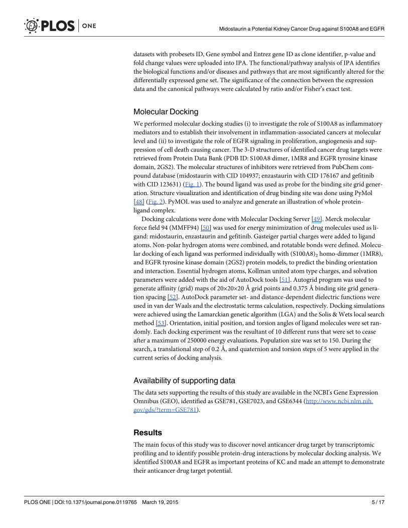
Docking studiesMolecular docking studies predicted potential interactions of our proposed protein drug targetwith the selected drug molecules. This is a structural modeling approach to study possiblebinding for cancer therapeutics. To understand the molecular interaction between drugs andS100A8, series of molecular docking analysis were performed using three dimensional struc-ture available (PDBID: 1MR8) with three anti-cancer drugs i.e. midostaurin, enzastaurin andgefitinib. The ligand binding site was a hinge region containing two EF-hand motifs. In similarway, we also simulated docking of the above-mentioned drugs with the tyrosine kinase domainof EGFR. Based on their size, stereochemistry and structural differences the ligands exhibitedvaried intensity in binding with the protein target molecules. The predicted parameters of esti-mated binding free energy, inhibition constant (Ki), total energy of vdW + Hbond + desolva-tion + electrostatic energy, total intermolecular energy and interacting surface area wereevaluated to estimate the favorable binding of ligand drug molecules to the target protein. Mo-lecular visualization was performed using PyMol. Complete interaction profile (H-bonds,polar, hydrophobic, pi-pi, cation-pi and other contacts), and hydrogen bonding (HB plot) in-teractions were studied (Figs. 5 and 6, Table 4).
The docking studies reveal the presence of one midostaurin molecule at the substrate-bind-ing site of S100A8 dimer (PDB code 1MR8). It is observed that the drug molecule is buried inthe substrate-binding hydrophobic channel of the S100A8 ligand binding domain. Most of theinteracting residues are hydrophobic in nature. Interaction and accessible surface area analysisreveal that the cavity involved in the binding site of drugs has a molecular accessible surfacearea of 123.017 Å2 and a solvent accessible surface area of 509.834 Å2. The conformations ofthe binding site in S100A8 as well as that in drugs were not altered upon binding as rigid dock-ing simulation was carried out. The binding cavity is the same for all three drugs.
We selected the crystal structure of the active EGFR kinase domain having a length of 330residues (PDB code 2GS2) for docking study. The drug binding cavity of the EGFR tyrosine ki-nase domain is more like a channel and is deeper as compared to the S100A8 case and has amolecular and solvent accessible surface area of 181.409 Å2 and 875.519 Å2 respectively. Thecavity is lined with mostly polar as well as few non-polar residues. Enzastaurin did not exhibitany binding with the selected tyrosine kinase domain. Gefitinib is a previously known inhibitorof EGFR and its co-crystal structure has been determined and presented in PDB (3UG2), how-ever docking studies revealed that midostaurin is the more suitable binding partner of EGFRthan gefitinib.
Table 2. Expression of S100A8 and EGFR in kidney cancer among Saudi patients (CEGMR dataset) and GEO dataset (GSE781, GSE6344 andGSE7023).
Gene Symbol CEGMR (own data)Sample size = 6
GSE781 Sample size = 34 GSE6344 Sample size = 40 GSE7023 Sample size =47
Fold Change p-value Fold Change p-value Fold Change p-value Fold Change p-value
S100A8 2.663 0.0159 2.759 0.0109 3.107 0.0019 1.948 0.0105
EGFR 3.401 0.0410 5.563 8.54E-05 3.32472 6.00E-08 1.893 0.0026
doi:10.1371/journal.pone.0119765.t002
Midostaurin a Potential Kidney Cancer Drug against S100A8 and EGFR
PLOSONE | DOI:10.1371/journal.pone.0119765 March 19, 2015 8 / 17

Fig 4. Leukocyte Extravasation Signaling: Transcriptomic signatures of kidney cancer showed a significant activation in leukocyte extravasationsignaling pathway. Red represents overexpression and green underexpression.
doi:10.1371/journal.pone.0119765.g004
Table 3. Canonical pathways predicted by Ingenuity Pathway Analysis for significant genes differentially expressed in kidney cancer.
Ingenuity Canonical Pathways -log (p-value)
Down-regulated
Upregulated Molecules
Atherosclerosis Signaling 3.14E00 5/136 (4%) 13/136(10%)
APOE,APOM,MSR1,PLA2R1,PLA2G7,SELPLG,COL1A2,APOC1,APOL1,COL1A1,IL18,ALB, LYZ,CCL2, S100A8,PDGFD,RBP4,COL3A1
LXR/RXR Activation 2.93E00 8/139 (6%) 10/139 (7%) KNG1,SCD,APOE,APOM,ECHS1,MSR1,AMBP,ABCG1,APOC1,APOL1,IL18,ALB,LYZ, LY96, CCL2, S100A8,HADH,RBP4
Leukocyte ExtravasationSignaling
2.65E00 4/205 (2%) 20/205(10%)
CLDN10,ARHGAP6,SPN,PIK3C2A,CLDN19,JAM2,ITGA6,RAPGEF4,SELPLG,PIK3R3,BTK,ROCK1,NCF1,WIPF1,ITGAM,CDH5,JAM3,CLDN16,RASGRP1,NCF2,PRKCH,RASSF5, ACTN1,CLDN3
Hepatic Fibrosis / HepaticStellate Cell Activation
2.52E00 3/153 (2%) 15/153(10%)
FN1,CXCL9,FGFR1,EGF,FGF1,BCL2,COL1A2,COL1A1,LY96,IGF1,CCL2,TGFB2,IL10RA, MYH9, EDNRA,ECE1,MYL3,COL3A1
Maturity Onset Diabetes ofYoung (MODY) Signaling
2.46E00 5/29 (17%) 1/29 (3%) HNF1B,PKLR,ALDOB,SLC2A2,CACNA1C, FABP1
Coagulation System 2.21E00 5/38 (13%) 2/38 (5%) F11,KNG1,PLG,SERPINA5,PROC,VWF, PLAUR
Valine Degradation I 2.21E00 6/35 (17%) 1/35 (3%) HIBCH,BCAT1,ECHS1,ABAT,ACADSB,EHHADH,ALDH6A1
Notch Signaling 2.07E00 1/42 (2%) 6/42 (14%) DLL1,ADAM17,DTX1,JAG2,MAML3,DLL4, HEY1
Intrinsic Prothrombin ActivationPathway
2.03E00 3/36 (8%) 3/36 (8%) F11,KNG1,COL1A2,COL1A1,PROC,COL3A1
IL-12 Signaling and Productionin Macrophages
1.88E00 5/154 (3%) 11/154 (7%) PPARG,APOE,APOM,PIK3C2A,MST1,APOL1,PIK3R3,APOC1,ALB,LYZ,IL18,TGFB2,MAP3K8, S100A8,PRKCH,RBP4
doi:10.1371/journal.pone.0119765.t003
Midostaurin a Potential Kidney Cancer Drug against S100A8 and EGFR
PLOSONE | DOI:10.1371/journal.pone.0119765 March 19, 2015 9 / 17
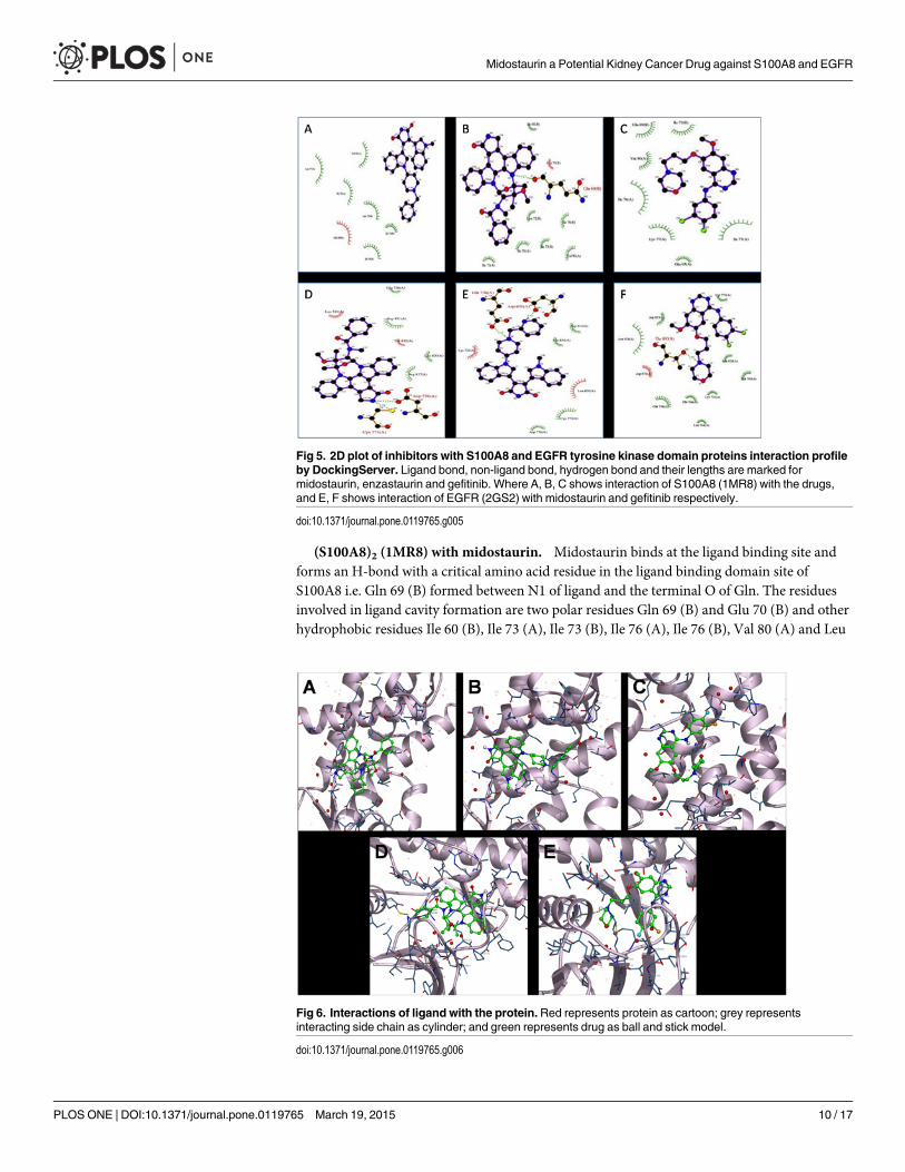
(S100A8)2 (1MR8) with midostaurin. Midostaurin binds at the ligand binding site andforms an H-bond with a critical amino acid residue in the ligand binding domain site ofS100A8 i.e. Gln 69 (B) formed between N1 of ligand and the terminal O of Gln. The residuesinvolved in ligand cavity formation are two polar residues Gln 69 (B) and Glu 70 (B) and otherhydrophobic residues Ile 60 (B), Ile 73 (A), Ile 73 (B), Ile 76 (A), Ile 76 (B), Val 80 (A) and Leu
Fig 5. 2D plot of inhibitors with S100A8 and EGFR tyrosine kinase domain proteins interaction profileby DockingServer. Ligand bond, non-ligand bond, hydrogen bond and their lengths are marked formidostaurin, enzastaurin and gefitinib. Where A, B, C shows interaction of S100A8 (1MR8) with the drugs,and E, F shows interaction of EGFR (2GS2) with midostaurin and gefitinib respectively.
doi:10.1371/journal.pone.0119765.g005
Fig 6. Interactions of ligand with the protein.Red represents protein as cartoon; grey representsinteracting side chain as cylinder; and green represents drug as ball and stick model.
doi:10.1371/journal.pone.0119765.g006
Midostaurin a Potential Kidney Cancer Drug against S100A8 and EGFR
PLOSONE | DOI:10.1371/journal.pone.0119765 March 19, 2015 10 / 17

72 (B) as shown in Fig. 2. More than 20 hydrophobic contacts and van der Waals interactionsare also noted. The inhibitor exhibits favorable binding characteristics with the predicted freebinding energy-9.77 kcal/mol and estimated inhibition constant, Ki of 68.48 nM. These resultsare promising and are superior to other docking results done in this particular study.
(S100A8)2 (1MR8) with enzastaurin. The drug binds to the protein molecule satisfactori-ly with an estimated free binding energy of-4.19 kcal/mol and Ki of 844.18 μM. It shows bind-ing with the S100A8 dimer but without the formation of neither any H-bonds nor polarcontacts. It displays five hydrophobic contacts mediated by aliphatic non-polar amino acidsLeu 72 (B), Ile 73 (B), Ile 76 (A), Ile 76 (B), and Val 80 (A). Other noticeable interactions arewith Lys 77 (A) and Gln 69 (B).
(S100A8)2 (1MR8) with gefitinib. Binding ability of gefitinib is good but there are noH-bonds formed and the free energy of bound structure is-4.21 kcal/mol. One noticeable polarinteraction is with Lys 77 of protein chain A. There are around ten hydrophobic interactionswith the drug binding site lining hydrophobic residues—Ile 73 (A), Ile 73 (B), Ile 76 (A) andVal 80 (A). Halogen atoms F and Cl of the drug show non-covalent interaction. For protein-ligand complexes, halogen bonds are energetically and geometrically comparably equal to thatof hydrogen bonding if the donor-acceptor directionality remains consistent. This intermolec-ular interaction has been shown to be stabilizing and a conformational determinant in com-plexes [54]. F displays a water mediated bond and Cl shows interaction with Gln 69 (A). Acouple of other weak interactions was noticed too.
EGFR kinase domain (2GS2) with midostaurin. Midostaurin being primarily a kinase in-hibitor docks well with the EGFR tyrosine kinase domain with free binding energy of-6.58kcal/mol and inhibition constant of 15.13 μM. Four H-bonds are possibly involved between theprotein and ligand molecule. Cys 773 Sγ shows H-bonding with N4 of drug at a interatomicdistance of 3.28 Å. In addition, atom 0δ2 of Cys 773 likely forms another H-bond with H atomof ligand. Similar pattern of H-bond is seen with Asp 776. Residues Arg 817, Thr 830 and Asp831 are involved in polar interactions with the ligand. Hydrophobic interactions are mediatedby Cys 773 and Leu 820. Other interactions with the drug are mediated through Lys 721, Glu738, Asp776, Arg 817, Leu 820, Thr 830 and Asp 831.
Table 4. Binding and interaction values for docking of S100A8 dimer and EGFR kinase domain with inhibitors (midostaurin, enzastaurin andgefitinib).
(S100A8)2 (1MR8) Midostaurin Enzastaurin Gefitinib
Est. Free Energy of Binding -9.77 kcal/mol -4.19 kcal/mol -4.21 kcal/mol
Est. Inhibition Constant, Ki 68.48 nM 844.18 μM 813.89 μM
vdW + Hbond + desolv Energy -7.65 kcal/mol -6.33 kcal/mol -6.11 kcal/mol
Electrostatic Energy -0.03 kcal/mol -0.02 kcal/mol -0.07 kcal/mol
Total Intermolec. Energy -7.68 kcal/mol -6.35 kcal/mol -6.18 kcal/mol
Interact. Surface 754.992 849.622 667.469
EGFR kinase domain (2GS2) Midostaurin Enzastaurin Gefitinib
Est. Free Energy of Binding -6.58 kcal/mol +7.10 kcal/mol -4.15 kcal/mol
Est. Inhibition Constant, Ki 15.13 μM ——— 905.30 μM
vdW + Hbond + desolv Energy -1.49 kcal/mol +0.44 kcal/mol -6.07 kcal/mol
Electrostatic Energy -0.16 kcal/mol -0.88 kcal/mol -0.17 kcal/mol
Total Intermolec. Energy -1.65 kcal/mol -0.44 kcal/mol -6.24 kcal/mol
Interact. Surface 911.021 895.39 795.106
doi:10.1371/journal.pone.0119765.t004
Midostaurin a Potential Kidney Cancer Drug against S100A8 and EGFR
PLOSONE | DOI:10.1371/journal.pone.0119765 March 19, 2015 11 / 17

EGFR kinase domain (2GS2) with enzastaurin. The docked structure shows positive freeenergy of binding implying that binding is not feasible as most of the decomposed interactionenergies have positive value. The result can be partly explained by the fact that enzastaurin ismainly a serine/threonine kinase inhibitor but we have docked it with EGFR tyrosine kinasedomain. Otherwise, enhanced docking properties can be possibly achieved by increasing thesimulation box size.
EGFR kinase domain (2GS2) with gefitinib. In our docking analysis total of threeH-bonds are seen, two with Thr 830 (interatomic distance of 2.99 and 3.89 Å) and one withGlu 738 (3.48 Å). Glu 738, Asn 818 and Asp 831 display polar interactions. Leu 764, Cys 773and Leu 820 exhibit hydrophobic interactions. Two water-mediated halogen bonds are seenwith F. Several other interactions are noticed with Val 702, Lys 721, Glu 738, Thr 766, Cys 773,Arg 817, Asn 818, Leu 820, Thr 830 and Asp 831. The monomeric (V948R) gefitinib/erlotinibresistant double mutant (L858R+T790M) EGFR kinase domain has been previously co-crystallized with gefitinib (PDB ID: 4I22) [55].
DiscussionKidney cancer comprises heterogeneous tumors with diverse molecular and clinical character-istics as reflected by their response to specific treatments. To understand the mechanisms bywhich genes alter a wide range of physiological processes, we performed a transcriptional pro-filing study to identify significant genes and examined their biological functions, and to identifyKC associated molecular network and pathways. Among hundreds of differentially expressedgenes we identified S100A8 and EGFR as potential biomarker of KC and attempted to demon-strate their anticancer drug target potential. In silico docking analysis showed that midostaurinand gefitinib binds to S100A8 as well as to EGFR and predictably inhibits downstream signal-ing in KC. However, enzastaurin binds only to the S100A8 dimer. Our finding leads to the hy-pothesis that S100A8 and EGFR are promising anticancer drug targets and midostaurin can bea potential inhibitor. This hypothesis surely needs further validation.
S100 proteins participate in numerous functions including protein phosphorylation, enzy-matic activation, calcium homeostasis, and interaction with cytoskeletal components [38].Most genes encoding S100 proteins are clustered on a region of human chromosome 1q21 thatis prone to chromosomal rearrangements, suggesting a link between S100A8/A9 proteins andmetastasis and tumor formation [38,56]. S100A8 has cell growth-promoting activity at lowconcentrations by binding to RAGE. This binding enhances cell mesenchymal properties, in-duces epithelial-mesenchymal transition and plays important role in promoting cancer inva-sion and metastasis in cancer [15,22]. Abnormal expressions of S100A8 proteins were observedin a variety of cancers, such as gastric, lung, breast, liver, pancreatic and squamous esophagealcarcinomas [15,17,20–23,57–60]. These studies indicate the potential of S100A8 as anticancertarget. Despite showing the elevated expression and distinct role of S100A8 in different cancertypes, less is known about the expression status or role of S100A8 in KC progression.
The EGFR family of RTPKs is an important cancer target because of the complex signalingthrough their configuration as homo- or hetrodimers [61,62]. The 4-anilino-quinazolinegefitinib (Iressa) targets the active conformation of EGFR kinase and has been approved forsecond- and third-line chemotherapy for advanced non small cell lung carcinoma (NSCLC)[63]. Interestingly, a subset of patients who have specific activating mutations in the EGFR ty-rosine kinase domain and/or amplification of EGFR often display enhanced sensitivity, positiveclinical responses and improved survival with these inhibitors [64,65]. However, the gatekeepermutation T790M in EGFR enhances affinity for ATP and reduces affinity for gefitinib andthereby inducing resistance [66,67]. We, therefore, used another kinase inhibitor, midostaurin
Midostaurin a Potential Kidney Cancer Drug against S100A8 and EGFR
PLOSONE | DOI:10.1371/journal.pone.0119765 March 19, 2015 12 / 17

to determine their efficiencies. In our docking study, the best overall binding was exhibited interms of estimated free energy of binding and Ki value by midostaurin followed by gefitiniband at least by enzastaurin. Interestingly, enzastaurin did not show any binding with EGFR.Enzastaurin has been previously evaluated both in vitro and in nude mice alone and in combi-nation with the EGFR inhibitor gefitinib, where it showed a cooperative inhibitory effect withgefitinib in parental and in gefitinib-resistant cells [68].
This was a pioneering structure-based approach to study S100A8 and EGFR protein interac-tions with the chosen kinase inhibitors at the molecular level. The EGFR binding cavity is quitehydrophobic and is the most preferable binding site for compounds having compatible shapeand stereochemistry. The computational results provide valuable insights into the bindingmodes of the three tested inhibitors to the S100A8 and EGFR targets. It has been demonstratedthat the hydrophobic interactions and the hydrogen bonding with these targets have pivotalcontributions to the binding structures and binding free energies, although the van der Waalsand electrostatic interactions contributed to the stabilization of the binding structures. The cal-culated binding free energies comply with the available experimental activity data. The detailedstructural insight, binding modes and the crucial factors affecting the binding free energies ob-tained from the present computational studies may provide valuable insights for future rationalstructure-based design of novel and potent inhibitors.
The size of a single amino acid in the ATP binding pocket—termed the gatekeeper residue—is a critical determinant of inhibitor sensitivity [67]. Kinases which possess a threonine atthis position are sensitive to a range of inhibitors, whereas those having a larger amino acidside chain show significantly higher IC50 values than those having a threonine residue at thissite and are broadly resistant [69]. For an ATP-competitive kinase inhibitor, a micromolar in-hibition constant, Ki alone is compelling evidence that the proposed drug will be non-selective[41]. In our study, the most favourable docking result was determined for midostaurin with theS100A8 dimer with the estimated Ki as 68.48 nM and free energy of binding as-9.77 kcal/mol.There exists a strong logical correlation between inhibitor potency and selectivity [70] hence,more potent compounds are more selective because they can be applied at a lower dose.
ConclusionOur analysis suggests distinct transcriptomic signatures for KC with significantly high levels ofS100A8 and EGFR expression involved in KC progression. Although further validation onlarger dataset is needed to corroborate these findings, analysis of KC tissue is a promising toolfor identifying biomarkers of interest. Protein-ligand interaction studies play a vital role in thestructure based computational drug design. Our docking based findings shed insight intoS100A8 protein as an attractive anticancer target and midostaurin as potential anticancer drugfor therapeutic intervention in KC. S100A8 gained importance as target for anticancer drug de-velopment due to its central role in mediating inflammatory pathways that facilitate cancer me-tastasis. The docking simulatio of midostaurin with EGFR is also promising. Furtherinvestigations like quantitative structure-activity relationship (QSAR) studies are required tostudy and identify more favorable interactions with S100A8, EGFR and their partners. Further-more, better binding ligands with higher affinity and efficacy can be designed and validatedusing combinatorial chemistry and co-crystallization approaches.
Therapeutic inhibition of kinase signaling cascades and emergence of novel clinically vali-dated potent and selective kinase inhibitors facilitated by rational drug design is a proven boonespecially for oncology. Studies to evaluate on- and off-target pharmacology (side effects) ofthese inhibitors in relation to efficacy and toxicity should be carried using KC cell lines. This
Midostaurin a Potential Kidney Cancer Drug against S100A8 and EGFR
PLOSONE | DOI:10.1371/journal.pone.0119765 March 19, 2015 13 / 17
will help in holistic monitoring of the changes in phosphorylation resulting fromkinase inhibition.
Supporting InformationS1 Table. Differentially expressed genes of kidney cancer from Saudi patients; comparisonof the genome-wide expression of kidney cancer with normal kidney tissue revealed 1478differentially expressed genes, 852 up-regulated and 483 down-regulated, with� 2 foldchange and false discovery rate of p< 0.05.(XLS)
S2 Table. Differentially expressed significant genes of kidney cancer from GEO retrieveddata; expression profiling revealed 876, 1200 and 1258 differentially expressed genes forGSE781, GSE6344 and GSE7023 respectively, with� 2 fold change and false discovery rateof p< 0.05.(XLS)
AcknowledgmentsThe authors would like to thank Nuha Alansari, Alaa Albogmi, Manal Shaabad, Mona AlKhar-raz, Amal Noor and Manar Ata for samples collection, performing RNA extraction, bioanaly-zer assays and microarray experiments. We thank the patients, physicians, nurses, andpathologists of the King Abdulaziz University Hospital and King Faisal Specialist Hospital andResearch Center, Jeddah, Saudi Arabia.
Author ContributionsConceived and designed the experiments: SK MGMQ. Performed the experiments: ZM HJSSK. Analyzed the data: SK ZM JM. Contributed reagents/materials/analysis tools: HF AC AA.Wrote the paper: SK ZM.
References1. Siegel R, Naishadham D, Jemal A. Cancer statistics, 2013. CA: a cancer journal for clinicians. 2013;
63: 11–30. doi: 10.3322/caac.21166 PMID: 23335087
2. Hanahan D, Weinberg RA. The hallmarks of cancer. Cell. 2000; 100: 57–70. PMID: 10647931
3. Hanahan D, Weinberg RA. Hallmarks of cancer: the next generation. Cell. 2011; 144: 646–674. doi:10.1016/j.cell.2011.02.013 PMID: 21376230
4. ChowW-H, Gridley G, Fraumeni JF Jr, Järvholm B. Obesity, hypertension, and the risk of kidney cancerin men. New England Journal of Medicine. 2000; 343: 1305–1311. PMID: 11058675
5. Pu YS, Huang CY, Kuo YZ, KangWY, Liu GY, Huang AM, et al. Characterization of membranous andcytoplasmic EGFR expression in human normal renal cortex and renal cell carcinoma. J Biomed Sci.2009; 16: 82. doi: 10.1186/1423-0127-16-82 PMID: 19747398
6. Motzer RJ, Bukowski RM. Targeted therapy for metastatic renal cell carcinoma. J Clin Oncol. 2006; 24:5601–5608. PMID: 17158546
7. Debouck C, Goodfellow PN. DNAmicroarrays in drug discovery and development. Nature genetics.1999; 21: 48–50. PMID: 9915501
8. Drews J. Drug discovery: a historical perspective. Science. 2000; 287: 1960–1964. PMID: 10720314
9. Vogl T, Leukert N, Barczyk K, Strupat K, Roth J. Biophysical characterization of S100A8 and S100A9 inthe absence and presence of bivalent cations. Biochim Biophys Acta. 2006; 1763: 1298–1306. PMID:17050004
10. Santamaria-Kisiel L, Rintala-Dempsey AC, Shaw GS. Calcium-dependent and-independent interac-tions of the S100 protein family. Biochem J. 2006; 396: 201–214. PMID: 16683912
11. Donato R, R Cannon B, Sorci G, Riuzzi F, Hsu K, J Weber D, et al. Functions of S100 proteins. CurrentMolecular Medicine. 2013; 13: 24–57. PMID: 22834835
Midostaurin a Potential Kidney Cancer Drug against S100A8 and EGFR
PLOSONE | DOI:10.1371/journal.pone.0119765 March 19, 2015 14 / 17
12. Marenholz I, Heizmann CW, Fritz G. S100 proteins in mouse and man: from evolution to function andpathology (including an update of the nomenclature). Biochemical and biophysical research communi-cations. 2004; 322: 1111–1122. PMID: 15336958
13. Hessian PA, Edgeworth J, Hogg N. MRP-8 and MRP-14, two abundant Ca(2+)-binding proteins of neu-trophils and monocytes. J Leukoc Biol. 1993; 53: 197–204. PMID: 8445331
14. Kerkhoff C, Voss A, Scholzen TE, Averill MM, Zänker KS, Bornfeldt KE. Novel insights into the role ofS100A8/A9 in skin biology. Experimental dermatology. 2012; 21: 822–826. doi: 10.1111/j.1600-0625.2012.01571.x PMID: 22882537
15. Yin C, Li H, Zhang B, Liu Y, Lu G, Lu S, et al. RAGE-binding S100A8/A9 promotes the migration and in-vasion of human breast cancer cells through actin polymerization and epithelial-mesenchymal transi-tion. Breast cancer research and treatment. 2013; 142: 297–309. doi: 10.1007/s10549-013-2737-1PMID: 24177755
16. Passey RJ, Williams E, Lichanska AM,Wells C, Hu S, Geczy CL, et al. A null mutation in the inflamma-tion-associated S100 protein S100A8 causes early resorption of the mouse embryo. The Journal of Im-munology. 1999; 163: 2209–2216. PMID: 10438963
17. Grebhardt S, Muller-Decker K, Bestvater F, Hershfinkel M, Mayer D. Impact of S100A8/A9 Expressionon Prostate Cancer Progression In Vitro and In Vivo. J Cell Physiol. 2014; 229: 661–671. doi: 10.1002/jcp.24489 PMID: 24122301
18. Hermani A, De Servi B, Medunjanin S, Tessier PA, Mayer D. S100A8 and S100A9 activate MAP kinaseand NF-kappaB signaling pathways and trigger translocation of RAGE in human prostate cancer cells.Exp Cell Res. 2006; 312: 184–197. PMID: 16297907
19. Rafii S, Lyden D. S100 chemokines mediate bookmarking of premetastatic niches. Nat Cell Biol. 2006;8: 1321–1323. PMID: 17139281
20. Yong HY, Moon A. Roles of calcium-binding proteins, S100A8 and S100A9, in invasive phenotype ofhuman gastric cancer cells. Arch Pharm Res. 2007; 30: 75–81. PMID: 17328245
21. Nemeth J, Stein I, Haag D, Riehl A, Longerich T, Horwitz E, et al. S100A8 and S100A9 are novel nucle-ar factor kappa B target genes during malignant progression of murine and human liver carcinogenesis.Hepatology. 2009; 50: 1251–1262. doi: 10.1002/hep.23099 PMID: 19670424
22. Chen KT, Kim PD, Jones KA, Devarajan K, Patel BB, Hoffman JP, et al. Potential prognostic biomark-ers of pancreatic cancer. Pancreas. 2014; 43: 22–27. doi: 10.1097/MPA.0b013e3182a6867e PMID:24326364
23. Turovskaya O, Foell D, Sinha P, Vogl T, Newlin R, Nayak J, et al. RAGE, carboxylated glycans andS100A8/A9 play essential roles in colitis-associated carcinogenesis. Carcinogenesis. 2008; 29:2035–2043. doi: 10.1093/carcin/bgn188 PMID: 18689872
24. Stulik J, Osterreicher J, Koupilova K, Knizek, Macela A, Bures J, et al. The analysis of S100A9 andS100A8 expression in matched sets of macroscopically normal colon mucosa and colorectal carcino-ma: the S100A9 and S100A8 positive cells underlie and invade tumor mass. Electrophoresis. 1999;20: 1047–1054. PMID: 10344284
25. Hiratsuka S, Watanabe A, Aburatani H, Maru Y. Tumour-mediated upregulation of chemoattractantsand recruitment of myeloid cells predetermines lung metastasis. Nat Cell Biol. 2006; 8: 1369–1375.PMID: 17128264
26. Higgins RJ, Dickinson PJ, LeCouteur RA, Bollen AW,Wang H, Wang H, et al. Spontaneous canine gli-omas: overexpression of EGFR, PDGFRalpha and IGFBP2 demonstrated by tissue microarray immu-nophenotyping. J Neurooncol. 2010; 98: 49–55. doi: 10.1007/s11060-009-0072-5 PMID: 19967449
27. Shiomitsu K, Johnson CL, Malarkey DE, Pruitt AF, Thrall DE. Expression of epidermal growth factor re-ceptor and vascular endothelial growth factor in malignant canine epithelial nasal tumours. Vet CompOncol. 2009; 7: 106–114. doi: 10.1111/j.1476-5829.2009.00178.x PMID: 19453364
28. Pomerantz RG, Grandis JR. The epidermal growth factor receptor signaling network in head and neckcarcinogenesis and implications for targeted therapy. Semin Oncol. 2004; 31: 734–743. PMID:15599851
29. Lee HJ, Xu X, Choe G, Chung DH, Seo JW, Lee JH, et al. Protein overexpression and gene amplifica-tion of epidermal growth factor receptor in nonsmall cell lung carcinomas: Comparison of four commer-cially available antibodies by immunohistochemistry and fluorescence in situ hybridization study. LungCancer. 2010; 68: 375–382. doi: 10.1016/j.lungcan.2009.07.014 PMID: 19712993
30. Duan L, Wu R, Ye L, Wang H, Yang X, Zhang Y, et al. S100A8 and S100A9 are associated with colo-rectal carcinoma progression and contribute to colorectal carcinoma cell survival and migration viaWnt/beta-catenin pathway. PLoS One. 2013; 8: e62092. doi: 10.1371/journal.pone.0062092 PMID:23637971
Midostaurin a Potential Kidney Cancer Drug against S100A8 and EGFR
PLOSONE | DOI:10.1371/journal.pone.0119765 March 19, 2015 15 / 17

31. Fukihara J, Watanabe N, Taniguchi H, Kondoh Y, Kimura T, Kataoka K, et al. Clinical Predictors of Re-sponse to EGFR Tyrosine Kinase Inhibitors in Patients with EGFR-Mutant Non-Small Cell Lung Can-cer. Oncology. 2014; 86: 86–93. doi: 10.1159/000357129 PMID: 24457318
32. Yamaguchi NH, Mayer IA, Malzyner A, de Andrade CJ, Murad AM, Del Giglio A, et al. Gefitinib and cel-ecoxib in advanced metastatic gastrointestinal tumors: a pilot feasibility study. J Gastrointest Oncol.2014; 5: 57–66. doi: 10.3978/j.issn.2078-6891.2013.056 PMID: 24490043
33. Moody TW, Chan DC, Mantey SA, Moreno P, Jensen RT. SR48692 inhibits non-small cell lung cancerproliferation in an EGF receptor-dependent manner. Life Sci. 2014.
34. Shiogama K, Wongsiri T, Mizutani Y, Inada K-i, Tsutsumi Y. High-sensitivity epidermal growth factor re-ceptor immunostaining for colorectal carcinomas, compared with EGFR PharmDx: A study of diagnos-tic accuracy. International journal of clinical and experimental pathology. 2013; 6: 24. PMID: 23236539
35. Baba Y, Fujii M, Tokumaru Y, Kato Y. Present and Future of EGFR Inhibitors for Head and Neck Squa-mous Cell Cancer. J Oncol. 2012; 2012: 986725. doi: 10.1155/2012/986725 PMID: 22545054
36. Ciardiello F, Tortora G. Epidermal growth factor receptor (EGFR) as a target in cancer therapy: under-standing the role of receptor expression and other molecular determinants that could influence the re-sponse to anti-EGFR drugs. Eur J Cancer. 2003; 39: 1348–1354. PMID: 12826036
37. Kim Y, Li Z, Apetri M, Luo B, Settleman JE, Anderson KS. Temporal resolution of autophosphorylationfor normal and oncogenic forms of EGFR and differential effects of gefitinib. Biochemistry. 2012; 51:5212–5222. doi: 10.1021/bi300476v PMID: 22657099
38. Heizmann CW, Fritz G, Schafer BW. S100 proteins: structure, functions and pathology. Front Biosci.2002; 7: d1356–1368. PMID: 11991838
39. Burnett G, Kennedy EP. The enzymatic phosphorylation of proteins. J Biol Chem. 1954; 211: 969–980.PMID: 13221602
40. Manning G, Whyte DB, Martinez R, Hunter T, Sudarsanam S. The protein kinase complement of thehuman genome. Science. 2002; 298: 1912–1934. PMID: 12471243
41. Knight ZA, Shokat KM. Features of selective kinase inhibitors. Chem Biol. 2005; 12: 621–637. PMID:15975507
42. Fischer T, Stone RM, Deangelo DJ, Galinsky I, Estey E, Lanza C, et al. Phase IIB trial of oral Midos-taurin (PKC412), the FMS-like tyrosine kinase 3 receptor (FLT3) and multi-targeted kinase inhibitor, inpatients with acute myeloid leukemia and high-risk myelodysplastic syndrome with either wild-type ormutated FLT3. J Clin Oncol. 2010; 28: 4339–4345. doi: 10.1200/JCO.2010.28.9678 PMID: 20733134
43. El Fitori J, Su Y, Büchler P, Ludwig R, Giese NA, Büchler MW, et al. PKC 412 small‐molecule tyrosinekinase inhibitor. Cancer. 2007; 110: 1457–1468. PMID: 17676584
44. Stone RM, Fischer T, Paquette R, Schiller G, Schiffer CA, Ehninger G, et al. Phase IB study of the FLT3kinase inhibitor midostaurin with chemotherapy in younger newly diagnosed adult patients with acutemyeloid leukemia. Leukemia. 2012; 26: 2061–2068. doi: 10.1038/leu.2012.115 PMID: 22627678
45. Graff JR, McNulty AM, Hanna KR, Konicek BW, Lynch RL, Bailey SN, et al. The protein kinase Cbeta-selective inhibitor, Enzastaurin (LY317615.HCl), suppresses signaling through the AKT pathway, in-duces apoptosis, and suppresses growth of human colon cancer and glioblastoma xenografts. CancerRes. 2005; 65: 7462–7469. PMID: 16103100
46. Rizvi MA, Ghias K, Davies KM, Ma C, Weinberg F, Munshi HG, et al. Enzastaurin (LY317615), a proteinkinase Cβ inhibitor, inhibits the AKT pathway and induces apoptosis in multiple myeloma cell lines. Mo-lecular cancer therapeutics. 2006; 5: 1783–1789. PMID: 16891464
47. Wakeling AE, Guy SP, Woodburn JR, Ashton SE, Curry BJ, Barker AJ, et al. (2002) ZD1839 (Iressa)an orally active inhibitor of epidermal growth factor signaling with potential for cancer therapy.5749–5754 p. PMID: 12384534
48. DeLanoW (2010) The PyMOLMolecular Graphics System. San Carlos, CA: DeLano Scientific; 2002.Accessed 6/25/2007. Available at http://www. pymol, org.
49. Bikadi Z, Hazai E. Application of the PM6 semi-empirical method to modeling proteins enhances dock-ing accuracy of AutoDock. J Cheminform. 2009; 1: 15. doi: 10.1186/1758-2946-1-15 PMID: 20150996
50. Halgren TA. Merck molecular force field. I. Basis, form, scope, parameterization, and performance ofMMFF94. Journal of computational chemistry. 1996; 17: 490–519.
51. Morris GM, Huey R, LindstromW, Sanner MF, Belew RK, Goodsell DS, et al. AutoDock4 and Auto-DockTools4: Automated docking with selective receptor flexibility. Journal of computational chemistry.2009; 30: 2785–2791. doi: 10.1002/jcc.21256 PMID: 19399780
52. Morris GM, Goodsell DS, Halliday RS, Huey R, Hart WE, Belew RK, et al. Automated docking using aLamarckian genetic algorithm and an empirical binding free energy function. Journal of computationalchemistry. 1998; 19: 1639–1662.
Midostaurin a Potential Kidney Cancer Drug against S100A8 and EGFR
PLOSONE | DOI:10.1371/journal.pone.0119765 March 19, 2015 16 / 17
53. Solis FJ, Wets RJ-B. Minimization by random search techniques. Mathematics of operations research.1981; 6: 19–30.
54. Auffinger P, Hays FA, Westhof E, Ho PS. Halogen bonds in biological molecules. Proc Natl Acad Sci US A. 2004; 101: 16789–16794. PMID: 15557000
55. Gajiwala KS, Feng J, Ferre R, Ryan K, Brodsky O, Weinrich S, et al. Insights into the aberrant activity ofmutant EGFR kinase domain and drug recognition. Structure. 2013; 21: 209–219. doi: 10.1016/j.str.2012.11.014 PMID: 23273428
56. Heizmann CW, Ackermann GE, Galichet A. Pathologies involving the S100 proteins and RAGE. Sub-cell Biochem. 2007; 45: 93–138. PMID: 18193636
57. Seth A, Kitching R, Landberg G, Xu J, Zubovits J, Burger AM. Gene expression profiling of ductal carci-nomas in situ and invasive breast tumors. Anticancer Res. 2003; 23: 2043–2051. PMID: 12894577
58. Luo A, Kong J, Hu G, Liew CC, Xiong M, Wang X, et al. Discovery of Ca2+-relevant and differentiation-associated genes downregulated in esophageal squamous cell carcinoma using cDNAmicroarray. On-cogene. 2004; 23: 1291–1299. PMID: 14647409
59. Cross SS, Hamdy FC, Deloulme JC, Rehman I. Expression of S100 proteins in normal human tissuesand common cancers using tissue microarrays: S100A6, S100A8, S100A9 and S100A11 are all over-expressed in common cancers. Histopathology. 2005; 46: 256–269. PMID: 15720411
60. El-Rifai W, Moskaluk CA, Abdrabbo MK, Harper J, Yoshida C, Riggins GJ, et al. Gastric cancers over-express S100A calcium-binding proteins. Cancer Res. 2002; 62: 6823–6826. PMID: 12460893
61. Hynes NE, Lane HA. ERBB receptors and cancer: the complexity of targeted inhibitors. Nat Rev Can-cer. 2005; 5: 341–354. PMID: 15864276
62. Citri A, Yarden Y. EGF-ERBB signalling: towards the systems level. Nat Rev Mol Cell Biol. 2006; 7:505–516. PMID: 16829981
63. Muhsin M, Graham J, Kirkpatrick P. Gefitinib. Nat Rev Drug Discov. 2003; 2: 515–516. PMID:12841190
64. Lynch TJ, Bell DW, Sordella R, Gurubhagavatula S, Okimoto RA, Brannigan BW, et al. Activating muta-tions in the epidermal growth factor receptor underlying responsiveness of non—small-cell lung cancerto gefitinib. New England Journal of Medicine. 2004; 350: 2129–2139. PMID: 15118073
65. Fabbro D, Cowan-Jacob SW, Möbitz H, Martiny-Baron G (2012) Targeting cancer with small-molecu-lar-weight kinase inhibitors. Kinase Inhibitors: Springer. pp. 1–34.
66. PaoW, Miller VA, Politi KA, Riely GJ, Somwar R, Zakowski MF, et al. Acquired resistance of lung ade-nocarcinomas to gefitinib or erlotinib is associated with a second mutation in the EGFR kinase domain.PLoS Med. 2005; 2: e73. PMID: 15737014
67. Blencke S, Zech B, Engkvist O, Greff Z, Orfi L, Horvath Z, et al. Characterization of a conserved struc-tural determinant controlling protein kinase sensitivity to selective inhibitors. Chem Biol. 2004; 11:691–701. PMID: 15157880
68. Gelardi T, Caputo R, Damiano V, Daniele G, Pepe S, Ciardiello F, et al. Enzastaurin inhibits tumourssensitive and resistant to anti-EGFR drugs. Br J Cancer. 2008; 99: 473–480. doi: 10.1038/sj.bjc.6604493 PMID: 18665191
69. Godl K, Wissing J, Kurtenbach A, Habenberger P, Blencke S, Gutbrod H, et al. An efficient proteomicsmethod to identify the cellular targets of protein kinase inhibitors. Proc Natl Acad Sci U S A. 2003; 100:15434–15439. PMID: 14668439
70. Frye SV. Structure-activity relationship homology (SARAH): a conceptual framework for drug discoveryin the genomic era. Chem Biol. 1999; 6: R3–7. PMID: 9889153
Midostaurin a Potential Kidney Cancer Drug against S100A8 and EGFR
PLOSONE | DOI:10.1371/journal.pone.0119765 March 19, 2015 17 / 17
Copyright © 2022 FDOKUMEN