Mismatch negativity (MMN) elicited by changes in phoneme length: A cross-linguistic study

doi:10.1182/blood-2006-04-020263Prepublished online June 27, 2006;
Benjamin Bonavida, Sherie L Morrison and Manuel L PenichetPatrick P Ng, Gustavo Helguera, Tracy R Daniels, Simon Z Lomas, Jose A Rodriguez, Gary Schiller, transferrin receptorhematopoietic cells elicited by an IgG3-avidin fusion protein targeting the Molecular events contributing to cell death in malignant human
(4212 articles)Neoplasia � (577 articles)Immunotherapy � (5133 articles)Immunobiology �
(746 articles)Apoptosis �Articles on similar topics can be found in the following Blood collections
http://bloodjournal.hematologylibrary.org/site/misc/rights.xhtml#repub_requestsInformation about reproducing this article in parts or in its entirety may be found online at:
http://bloodjournal.hematologylibrary.org/site/misc/rights.xhtml#reprintsInformation about ordering reprints may be found online at:
http://bloodjournal.hematologylibrary.org/site/subscriptions/index.xhtmlInformation about subscriptions and ASH membership may be found online at:
digital object identifier (DOIs) and date of initial publication. theindexed by PubMed from initial publication. Citations to Advance online articles must include
final publication). Advance online articles are citable and establish publication priority; they areappeared in the paper journal (edited, typeset versions may be posted when available prior to Advance online articles have been peer reviewed and accepted for publication but have not yet
Copyright 2011 by The American Society of Hematology; all rights reserved.20036.the American Society of Hematology, 2021 L St, NW, Suite 900, Washington DC Blood (print ISSN 0006-4971, online ISSN 1528-0020), is published weekly by
For personal use only. by guest on January 13, 2014. bloodjournal.hematologylibrary.orgFrom For personal use only. by guest on January 13, 2014. bloodjournal.hematologylibrary.orgFrom

1
Molecular events contributing to cell death in malignant human hematopoietic cells elicited
by an IgG3-avidin fusion protein targeting the transferrin receptor
Patrick P. Ng1, Gustavo Helguera2, Tracy R. Daniels2, Simon Z. Lomas1, Jose A. Rodriguez2,
Gary Schiller3, 4, Benjamin Bonavida1, 4, Sherie L. Morrison1, 4 and Manuel L. Penichet1, 2, 4
1. Department of Microbiology, Immunology and Molecular Genetics; 2. Division of Surgical
Oncology, Department of Surgery; 3. Division of Hematology and Oncology, Department of
Medicine; 4. Jonsson Comprehensive Cancer Center, David Geffen School of Medicine;
University of California, Los Angeles, CA 90095.
This work was supported in part by grants CA86915 and CA107023 from NIH, the 2003 Jonsson
Cancer Center Foundation Interdisciplinary Grant and the 2004 Brian D. Novis International
Myeloma Foundation Senior Grant Award.
Running Title: Anti-hTfR-(IgG3-Avidin) Mediated Cell Death
Word counts: Text ~5000 , abstract – 195
Scientific heading: Neoplasia
Correspondence: Manuel L. Penichet, Division of Surgical Oncology, Department of Surgery,
UCLA 10833 Le Conte Avenue 54-140 CHS Mail code 178218 Los Angeles, CA 90095-1782
Phone: 310 825-1304
Fax: 310 825-7575
Blood First Edition Paper, prepublished online June 27, 2006; DOI 10.1182/blood-2006-04-020263
Copyright © 2006 American Society of Hematology
For personal use only. by guest on January 13, 2014. bloodjournal.hematologylibrary.orgFrom

2
Abstract
We have previously reported that an anti-human transferrin receptor IgG3-avidin fusion protein
[anti-hTfR-(IgG3-Av)] inhibits the proliferation of an erythroleukemia cell line. We have now
found that anti-hTfR-(IgG3-Av) also inhibits the proliferation of additional human malignant B
and plasma cells. Anti-hTfR-(IgG3-Av) induces internalization and rapid degradation of the TfR.
These events can be reproduced in cells treated with anti-hTfR-IgG3 cross-linked with a
secondary Ab, suggesting that they result from increased TfR cross-linking. Confocal
microscopy of cells treated with anti-hTfR-(IgG3-Av) shows that the TfR is directed to an
intracellular compartment expressing the lysosomal marker LAMP-1. The degradation of TfR is
partially blocked by cysteine protease inhibitors. Furthermore, cells treated with anti-hTfR-
(IgG3-Av) exhibit mitochondrial depolarization and activation of caspases 9, 8, and 3. The
mitochondrial damage and cell death can be prevented by iron supplementation, but cannot be
fully blocked by a pan-caspase inhibitor. These results suggest that anti-hTfR-(IgG3-Av) induces
lethal iron deprivation, but the resulting cell death does not solely depend on caspase activation.
This report provides insights into the mechanism of cell death induced by anti-TfR Abs such as
anti-hTfR-(IgG3-Av), a molecule that may be useful in the treatment of B-cell malignancies such
as multiple myeloma.
For personal use only. by guest on January 13, 2014. bloodjournal.hematologylibrary.orgFrom

3
Introduction
The primary function of transferrin (Tf) is to transport iron through the blood. After binding to
the transferrin receptor (TfR) on the cell surface, Tf is internalized into an acidic compartment
where the bound iron is released. The Tf-TfR complex then returns to the cell surface and the
ligand dissociates from the receptor 1.
Studies have shown that the TfR is expressed more abundantly in malignant tissues than their
normal counterparts 2-7. This difference in expression level, in addition to its ability to internalize
and its central roles in cell growth and division, makes the TfR an attractive target for cancer
therapeutics. In fact, both anti-TfR antibodies (Abs) and Tf-toxin conjugates have shown
efficacy against cancers in preclinical and clinical settings 8-12. We have previously demonstrated
that anti-rat TfR-(IgG3-Av) forms strong non-covalent interactions with different biotinylated
molecules and delivers them into cancer cells through receptor-mediated endocytosis 13. This
novel molecule can be used as a universal delivery system for a wide range of therapeutic agents
without the need to make a different chemical conjugate or genetic fusion protein for every
targeted therapeutic.
We also unexpectedly discovered that anti-rat TfR-(IgG3-Av), but not an irrelevant IgG3-Av,
inhibited the growth of a rat myeloma and a T-cell lymphoma cell line. However, it did not
inhibit the growth of either a carcinoma or a gliosarcoma cell line 13. Treatment with anti-rat
TfR-IgG3 containing the same variable regions did not inhibit growth. Furthermore, we found
that anti-rat TfR-(IgG3-Av) exists as a non-covalent dimer with four antigen binding sites,
probably due to the interaction among the four avidins located on two separate fusion proteins,
since avidin in solution forms a tetrameric structure 13. Thus, the inhibitory effect of the fusion
protein may be due, at least in part, to its ability to cross-link cell surface TfRs. Furthermore, we
For personal use only. by guest on January 13, 2014. bloodjournal.hematologylibrary.orgFrom

4
reported that a similar fusion protein specific for the human TfR [anti-hTfR-(IgG3-Av)], but not
a murine anti-TfR IgG1 (128.1) sharing the same variable regions, inhibited the growth of the
erythroleukemia cell line K562 13. However, the mechanism of growth inhibition by these two
fusion proteins, as well as the therapeutic potential of anti-hTfR-(IgG3-Av) were not explored.
Now we report that anti-hTfR-(IgG3-Av) inhibits the growth of malignant B and plasma cell
lines and cells isolated from patients with multiple myeloma (MM), a malignancy that is
generally regarded as incurable. Using two of the most sensitive cell lines, ARH-77 and IM-9,
we show that anti-hTfR-(IgG3-Av) induces rapid TfR degradation, iron deprivation,
mitochondrial damage, and cell death. Among different cancers, hematopoietic tumors are
particularly suitable for treatment using TfR-targeting therapeutics since they both express high
levels of TfR 14-17 and are known to be more sensitive to the inhibitory effect of anti-TfR Abs
than other malignancies 18. Increased understanding of the mechanism of cell death induced by
anti-hTfR-(IgG3-Av) may make it possible to design improved therapeutics for the treatment of
hematopoietic malignancies.
Methods
Antibodies and antibody fusion proteins
Recombinant anti-hTfR-IgG3 constructed by substituting the variable regions of anti-dansyl-
IgG3 19,20 with those of the murine IgG1 anti-human TfR monoclonal Ab (mAb) 128.1 21 was
expressed in the murine myeloma cell line NS0/1. Anti-hTfR-(IgG3-Av) and anti-dansyl-(IgG3-
Av) have been previously described 13,22. Abs and Ab fusion proteins were purified and
characterized as described previously 13. The murine anti-human IgG3 mAb HP6050 was the
kind gift of Robert Hamilton (John Hopkins University, MD). Murine anti-human TfR mAb
For personal use only. by guest on January 13, 2014. bloodjournal.hematologylibrary.orgFrom

5
(H68.4) and goat anti-mouse IgG-horseradish peroxidase (HRP) conjugate were from Zymed
(So. San Francisco, CA). Rabbit polyclonal anti-human TfR1 was purchased from Santa Cruz
Biotechnology (Santa Cruz, CA). Murine anti-β-actin mAb was from Sigma (St. Louis, MO), the
biotinylated mouse anti-LAMP-1 mAb and goat anti-mouse IgG-FITC were from BD
Pharmingen (Becton Dickinson, Franklin Lakes, NJ). The mouse IgG1/κ isotype control mAbs
were from eBioscience (San Diego, CA).
Cell lines and primary cells
MM.1S, S6B45, OCI-My5, U266, 8226/S, and 8226/DOX40 (doxorubicin-resistant variant of
8226/S) are human MM cell lines. ARH-77 and IM-9 are EBV-transformed lymphoblastoid cell
lines established from cells isolated from IgG plasma cell leukemia and MM patients,
respectively. When injected into SCID mice, ARH-77 behaves like human MM with mice
developing hypercalcemia, lytic bone lesions, and hind limb paralysis 23,24. U266, 8226, ARH-77
and IM-9 cell lines were purchased from ATCC (Rockville, MD). The MM.1S, S6B45, and OCI-
My5 cell lines were kindly provided by Drs. Kenneth Anderson and Darminder Chauhan
(Harvard University), and 8226/DOX40 cell line was provided by Alan Lichtenstein (UCLA).
The human erythroleukemia cell line K562 was kindly provided by J. Larrick (Palo Alto Institute
of Molecular Medicine, Mountain View, CA). Cells were cultured at 37oC, 5% CO2 in RPMI
1640 medium (GIBCO BRL, Grand Island, NY), with 5% fetal bovine serum (FBS, HyClone,
Logan, UT) in all the experiments unless stated otherwise. Serum complement was inactivated
by incubation at 56oC for 30 min.
Bone marrow aspirates from a patient with MM and another with Plasma Cell Leukemia (PCL),
a more aggressive variant of MM 25, were obtained with informed consent following the
For personal use only. by guest on January 13, 2014. bloodjournal.hematologylibrary.orgFrom

6
standards of the institutional review board of UCLA. Mononuclear cells were separated by
Ficoll-Paque Plus density gradient centrifugation (Amersham Pharmacia Biosciences, Uppsala,
Sweden). CD138+ cells were separated from bone marrow mononuclear cells by positive
selection using EasySep Human CD138+ Selection magnetic nanoparticles following the
manufacturer’s instructions (StemCell Technologies, Vancouver, Canada).
Proliferation assays using cell lines
5000 cells/well of a 96-well plate were treated with the indicated reagents for 72 h at 37ºC. The
cells were then cultured with 4 µCi/mL of [3H]-thymidine (ICN Biomedicals, Inc., Irvine, CA)
for an additional 24 h. Cells were harvested and radioactivity counted as previously described 13.
In the assay with nine different cell lines, cells were cultured in Dulbecco's Modified Eagle
Medium (GIBCO BRL) supplemented with 2.5% FBS. Similar results were obtained using cells
cultured in RPMI medium with 5% FBS. In the cross-linking assay, anti-hTfR-IgG3 was mixed
with a 5-fold excess of anti-hIgG3 mAb for 2 h at 4ºC before being added to the cells. In the
metal supplement assay, cells were incubated with or without ferric ammonium citrate or zinc
sulfate (Sigma) during the entire study.
Proliferation assays using primary cells
20,000 IM-9, U266 or primary cells/well of a 96-well plate cultured in Iscoves’ Modified
Dulbecco’s Medium (GIBCO BRL) supplemented with non-essential amino acids (Invitrogen
Corporation, Carlsbad, CA) and 10% FBS were treated with buffer or 100 nM anti-hTfR-(IgG3-
Av) for 48 h at 37ºC. The cells were then pulsed with 4 µCi/mL of [3H]-thymidine for an
additional 48 h. Cells were harvested and radioactivity counted as previously described 13.
For personal use only. by guest on January 13, 2014. bloodjournal.hematologylibrary.orgFrom
7
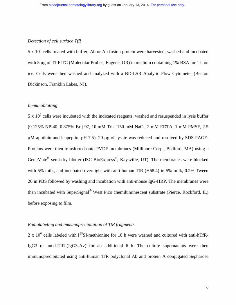
Detection of cell surface TfR
5 x 10⁵ cells treated with buffer, Ab or Ab fusion protein were harvested, washed and incubated
with 5 µg of Tf-FITC (Molecular Probes, Eugene, OR) in medium containing 1% BSA for 1 h on
ice. Cells were then washed and analyzed with a BD-LSR Analytic Flow Cytometer (Becton
Dickinson, Franklin Lakes, NJ).
Immunoblotting
5 x 10⁵ cells were incubated with the indicated reagents, washed and resuspended in lysis buffer
(0.125% NP-40, 0.875% Brij 97, 10 mM Tris, 150 mM NaCl, 2 mM EDTA, 1 mM PMSF, 2.5
µM apotinin and leupeptin, pH 7.5). 20 µg of lysate was reduced and resolved by SDS-PAGE.
Proteins were then transferred onto PVDF membranes (Millipore Corp., Bedford, MA) using a
GeneMate semi-dry blotter (ISC BioExpress, Kaysville, UT). The membranes were blocked
with 5% milk, and incubated overnight with anti-human TfR (H68.4) in 5% milk, 0.2% Tween
20 in PBS followed by washing and incubation with anti-mouse IgG-HRP. The membranes were
then incubated with SuperSignal West Pico chemiluminescent substrate (Pierce, Rockford, IL)
before exposing to film.
Radiolabeling and immunoprecipitation of TfR fragments
2 x 10⁶ cells labeled with [35S]-methionine for 18 h were washed and cultured with anti-hTfR-
IgG3 or anti-hTfR-(IgG3-Av) for an additional 6 h. The culture supernatants were then
immunoprecipitated using anti-human TfR polyclonal Ab and protein A conjugated Sepharose
For personal use only. by guest on January 13, 2014. bloodjournal.hematologylibrary.orgFrom
8
beads (Sigma). Samples were reduced and resolved by SDS-PAGE. The gel was dried and
exposed to film.
Apoptosis and mitochondrial membrane potential assays
75,000 cells were treated with the indicated reagents, washed and stained with propidium iodide
(PI) and annexin V Alexa Fluor 350 or 488 conjugates following procedures suggested by the
manufacturer of Vybrant Apoptosis kit (Molecular Probes). Up to 10,000 events were recorded
for each flow cytometry measurement. To measure mitochondrial depolarization, 40 nM
DiOC6(3) (Molecular Probes) was added to the culture for 30 min. Samples were analyzed by
flow cytometry. DFO and Z-VAD-FMK were purchased from Calbiochem (La Jolla, CA).
Caspase activity assay
25,000 cells per well of a 96-well plate were treated with buffer or anti-hTfR-(IgG3-Av), and
caspase activity was measured at 12, 24, 36, 44, 48 and 52 h using fluorogenic substrates specific
for caspases 9 (Ac-LEHD-AMC), 8 (Ac-IETD-AMC) and 3 (Ac-DMQD-AMC) (Alexis
Biochemicals, San Diego, CA). Ac-DMQD-AMC, instead of Ac-DEVD-AMC, was used
because it has been shown to be more specific for caspase 3 26,27. At each time point, reaction
buffer (1.5% NP-40, 0.3% CHAPS, 30% sucrose, 30 mM MgCl2, 150 mM KCl, 450 mM NaCl,
150 mM HEPES, 1.2 mM EDTA, 30 mM DTT, 3 mM PMSF, pH 7.4) containing 150 µM
fluorogenic substrate was added to each well and incubated for 4 h at 37ºC. Fluorescence was
measured by a Synergy HT Microplate Reader (Bio-Tek Instruments, Inc., Winooski, VT) at
360/460 nm. Substrate incubation for 4 h yields greater differences between the control and
experimental sample values when compared to other incubation times.
For personal use only. by guest on January 13, 2014. bloodjournal.hematologylibrary.orgFrom
9
Confocal microscopy
2 x 10⁶ cells treated with anti-hTfR-(IgG3-Av) were fixed with 4% paraformaldehyde and
permeabilized with 0.2% Tween 20 in PBS. After blocking for 2 h on ice in confocal buffer (2%
calf serum, 0.1% sodium azide in PBS) containing 10% human serum (Omega Scientific, Inc.,
Tarzana, CA), cells were incubated on ice for 2 h with 1 µg of anti-TfR (H68.4) or isotype
control mAb, and for 1 h with anti-mouse IgG-FITC (1:100). Cells were then fixed with 4%
paraformaldehyde and incubated on ice for 2 h with 0.1 µg of biotinylated anti-LAMP-1 or
isotype control, and for 1 h with streptavidin-Alexa Fluor 568 (1:500) (Molecular Probes). Cells
were washed twice with confocal buffer after each incubation. Stained cells were resuspended in
Prolong solution (Molecular Probes) and mounted onto slides. Analyses were performed at
room temperature on the MRC1024ES confocal system (Bio-Rad Laboratories, Hercules CA)
equipped with a Nikon E800 microscope. Images were taken at 63X magnification and analyzed
using ImageJ (NIH, Bethesda, MD). The mean fluorescence intensity (MFI) of the red and green
channels in each image field was determined and a threshold for positive staining of: MFI + 2SD
set. A pixel was considered colocalized when the overlapping area of the two channels was at
least 50% of the area of the pixel. The percent colocalization for each field was the number of
colocalized pixels, divided by the total number of positive pixels. The significance of values
compared to 0 min was determined using the Student’s t test.
Results
Anti-hTfR-(IgG3-Av) inhibits the growth of a panel of malignant B and plasma cell lines, and
primary cells from patients
For personal use only. by guest on January 13, 2014. bloodjournal.hematologylibrary.orgFrom
10
We previously reported that anti-hTfR-(IgG3-Av) is an anti-proliferative/pro-apoptotic drug that
strongly inhibits the human erythroleukemia cell line K562 13. To explore the potential of anti-
hTfR-(IgG3-Av) as a therapeutic for plasma cell malignancies, we measured the proliferation of
a panel of human malignant B and plasma cell lines incubated with the fusion protein. K562 cells
were included in the assay for comparison. Anti-hTfR-(IgG3-Av) inhibits the growth of all the
cell lines tested, but to different extents (Fig. 1A). Anti-hTfR-(IgG3-Av) also significantly
inhibits the in vitro growth of primary cells isolated from two patients diagnosed with multiple
myeloma and plasma cell leukemia, respectively (Fig. 1B). Further studies with a larger sample
size are necessary to confirm the spectrum of sensitivities to treatment with anti-hTR-(IgG3-Av)
in primary cells. The two most sensitive cell lines, ARH-77 and IM-9 were used in subsequent
experiments to determine the mechanism of growth inhibition by anti-hTfR-(IgG3-Av).
Anti-hTfR-(IgG3-Av) or anti-hTfR-IgG3 cross-linked with a secondary Ab inhibits the growth
and induces apoptosis in ARH-77 cells
Previous studies suggest that anti-hTfR-(IgG3-Av) inhibits cell growth by cross-linking the
TfR13. To test this hypothesis, we measured proliferation and apoptosis in ARH-77 cells treated
with different anti-hTfR molecules. Anti-hTfR-(IgG3-Av) completely inhibited [3H]-thymidine
incorporation (Figure 1C) and resulted in a significant increase in the number of dead (annexin
V bright, PI bright) and apoptotic (annexin V bright, PI dim) cells from 48 to 96 h (Figure 2A).
The total percentage of dead cells including apoptotic cells (annexin V bright, PI bright and
annexin V bright, PI dim) resulting from anti-hTfR-(IgG3-Av) treatment increased from 63% at
48 h, to 79% at 72 h, and 86% at 96 h. In another experiment, treatment with anti-hTfR-(IgG3-
Av) induced cell death in 85% of the cells compared to 47% in cells treated with a 3 fold higher
For personal use only. by guest on January 13, 2014. bloodjournal.hematologylibrary.orgFrom

11
concentration of anti-hTfR-IgG3 (Figure 2B). However, the growth inhibitory and pro-apoptotic
effects of anti-hTfR-IgG3 could be enhanced to levels comparable to those of anti-hTfR-(IgG3-
Av) by cross-linking with a secondary mAb (Figure 1C, 2B). Similar results were obtained using
IM-9 cells (data not shown). These results suggest that the tetravalent (4 antigen binding sites)
anti-hTfR-(IgG3-Av) has a stronger cytotoxic effect than anti-hTfR-IgG3 due, at least in part, to
its increased cross-linking of cell surface TfR.
Anti-hTfR-(IgG3-Av) down-regulates cell surface expression of TfR
The level of surface TfR on ARH-77 cells incubated with anti-hTfR molecules was measured by
flow cytometry using a Tf-FITC conjugate. Based on our findings (data not shown) and
published data on 128.1 21, anti-hTfR-IgG3 and anti-hTfR-(IgG3-Av) do not inhibit the binding
of Tf to the TfR. Therefore, their presence does not affect the binding of Tf-FITC to the receptor.
Both anti-TfR Abs caused an extensive and rapid decrease in surface TfR expression, which
remained low throughout the experiment (Figure 3, Figure 1S). Although the differences were
small, anti-hTfR-(IgG3-Av) consistently induced greater cell surface TfR down-regulation than
anti-hTfR-IgG3. Treatment of IM-9 cells yielded similar results (data not shown). Comparable
TfR down-regulation was also observed in cells treated with anti-hTfR-IgG3 cross-linked with a
secondary mAb (data not shown).
Anti-hTfR-(IgG3-Av) or anti-hTfR-IgG3 cross-linked with a secondary Ab induces intracellular
degradation of the TfR
To determine the fate of down-regulated surface TfR, we analyzed the lysates of treated cells by
immunoblotting. Intact TfR vanished between 4 to 12 h after treatment with anti-hTfR-(IgG3-
For personal use only. by guest on January 13, 2014. bloodjournal.hematologylibrary.orgFrom

12
Av). In contrast, anti-hTfR-IgG3 induced less extensive receptor loss (Figure 4A). However, TfR
loss following binding of anti-hTfR-IgG3 could be increased by cross-linking with a secondary
mAb (Figure 4B). When cells were treated with anti-hTfR-(IgG3-Av) for shorter times, over half
of the intact receptors had disappeared by 20 min (Figure 4C).
The rapid disappearance of TfR could be a result of shedding of surface TfR. However, we were
unable to detect any TfR fragments in the culture media of treated cells (data not shown).
Alternatively, TfR could be degraded intracellularly. In fact, immunoblotting of lysates from
cells treated with anti-hTfR-(IgG3-Av) showed small fragments of TfR (~16 and 20 kDa)
concomitant with the disappearance of full length TfR (95 kDa) (Figure 4D). Based on their sizes
and the fact that they were recognized by a mAb against the cytoplasmic N-terminus of TfR (a
type II transmembrane protein), these fragments appeared to be a result of proteolytic cleavages
in the extracellular region of the TfR. The degradation may take place in the lumen of an
intracellular compartment since anti-hTfR-(IgG3-Av) is internalized through TfR-mediated
endocytosis 13.
Iron supplement blocks the anti-proliferative and pro-apoptotic effects of anti-hTfR-(IgG3-Av)
To determine if anti-hTfR-(IgG3-Av) inhibits growth by disrupting TfR-dependent iron uptake,
we incubated cells with the fusion protein in the presence of metal supplements. Iron, but not
another divalent transition metal, zinc, completely blocked the growth inhibitory and pro-
apoptotic effects of anti-hTfR-(IgG3-Av) (Figure 5A, B). Similar results were obtained in cells
treated with anti-hTfR-IgG3 alone or cross-linked with a secondary Ab (data not shown). Iron
supplement in untreated cells did not promote cell growth (data not shown). In conclusion, these
data suggest that the anti-hTfR Abs induce apoptosis, at least in part, by causing iron deprivation.
For personal use only. by guest on January 13, 2014. bloodjournal.hematologylibrary.orgFrom

13
Anti-hTfR-(IgG3-Av) directs TfR to an intracellular compartment expressing LAMP-1, but
degradation of the receptor is not sensitive to increase of pH in the lysosome
To determine if anti-hTfR-(IgG3-Av) directs TfR to the lysosome for degradation, we analyzed
treated cells by confocal microscopy. Increased colocalization of TfR and LAMP-1 was
observed beginning 15 min after treatment with anti-hTfR-(IgG3-Av) (Figure 6, Table 1).
Despite the data indicating that TfR travels to the lysosome, the degradation of the receptor could
not be blocked by co-incubation with 20 mM ammonium chloride or 200 µM chloroquine (data
not shown), chemicals that increase the pH of the lysosome. This suggests that the proteases
responsible for the degradation of TfR are still functional at higher pHs.
A papain-like cysteine protease is involved in the degradation of TfR induced by anti-hTfR-
(IgG3-Av)
To identify the proteases that degrade the TfR, we used immunoblotting to detect TfR and its
cleavage products in the lysates of ARH-77 cells treated with anti-hTfR-(IgG3-Av) and a variety
of proteases inhibitors (Figure 7). None of the agents completely inhibited the destruction of the
95 kDa TfR, indicating that the process involves more than one protease family. PMSF, pepstatin
A, and bestatin, specific inhibitors of serine, aspartic acid proteases and aminopeptidases,
respectively 28,29, did not inhibit the formation of the 16 and 20 kDa fragments containing the N-
terminus. In contrast, antipain and leupeptin, inhibitors of both cysteine and serine proteases, and
E64d which inhibits most cysteine proteases and trypsin 28-30, blocked the cleavage that
generated the 16 and 20 kDa fragments. However, they were not able to block cleavage at a more
C-terminal site that resulted in a ~ 23 kDa fragment (Figure 7). The fact that only papain-like
For personal use only. by guest on January 13, 2014. bloodjournal.hematologylibrary.orgFrom

14
cysteine proteases are inhibited by antipain, leupeptin and E64d, but not by PMSF suggests that
one or more of these enzymes are involved in the generation of the 16 and 20 kDa fragments.
Inhibition of metalloproteinases using metal chelators such as o-phenanthroline and GM6001
also did not interfere with the production of the 16 and 20 kDa fragments (data not shown). In
conclusion, these results indicate that upon treatment with anti-hTfR-(IgG3-Av), the extracellular
portion of the internalized TfR is cleaved at multiple sites by different proteases including a
papain-like cysteine protease.
Anti-hTfR-(IgG3-Av) induces the activation of caspases 9,8 and 3
To determine if treatment with anti-hTfR-(IgG3-Av) induces caspase activation, we measured
the activity of caspases 9, 8, and 3 in ARH-77 cells treated with the fusion protein. Caspases 9
and 8 are key components of the mitochondrial and death receptor-mediated apoptotic pathway,
respectively, while caspase 3 is the executioner caspase of both pathways 31. Caspase activity
determined by using a specific fluorogenic substrate has been shown to correlate with
immunoblots detecting the active form of the protease 32. Activation of all three caspases was
seen only between 44 h and 52 h with the highest increase observed 48 h after treatment with
anti-hTfR-(IgG3-Av) (Figure 8A). We did not find a significant difference in the timing of
activation among the caspases.
Cell death induced by DFO or anti-hTfR-(IgG3-Av) is partially blocked by a broad-spectrum
caspase inhibitor
Although we had evidence of caspase activation in cells treated with anti-hTfR-(IgG3-Av), it
was unclear whether cell death was caspase-depended. To answer this question, we co-incubated
For personal use only. by guest on January 13, 2014. bloodjournal.hematologylibrary.orgFrom
15
cells with anti-hTfR-(IgG3-Av) and the broad-spectrum caspase inhibitor Z-VAD-FMK. We also
treated cells with DFO, a membrane permeable iron chelator, as a positive control for iron
deprivation. The levels of apoptosis and viability of cells were determined.
In anti-hTfR-(IgG3-Av) treated cells, inhibition of caspase activity by treatment with Z-VAD-
fmk only partially blocked cell death (Figure 8B). The number of dead cells (annexin V bright,
PI bright and annexin V bright, PI dim) decreased from 48% to 29% in cells treated with anti-
hTfR-(IgG3-Av) alone or pretreated with Z-VAD-fmk, respectively, at 48 h. Similar results were
observed after 96 h of treatment. These results suggest that anti-hTfR-(IgG3-Av) induced cell
death, in part, through a caspase-independent mechanism. However, it is important to note that in
several independent experiments we have observed that Z-VAD-FMK was less efficient in
blocking apoptosis induced by anti-hTfR-(IgG3-Av) than by DFO, suggesting that the two
reagents may induce apoptosis through slightly different pathways.
DFO and anti-hTfR-(IgG3-Av) induce mitochondrial depolarization
To determine if the PI negative, annexin V negative cells that survived the treatment of DFO or
anti-hTfR-(IgG3-Av) in the presence of Z-VAD-FMK were truly healthy, we stained cells
treated for 48 h (Figure 7B) with a third fluorescent reagent, DiOC6(3), that measures
mitochondrial potential. A cell with normal mitochondrial potential accumulates DiOC6(3) in the
organelle 33. All apoptotic cells lost mitochondrial potential regardless of the treatment or the
presence of Z-VAD-FMK (data not shown). In contrast, non-apoptotic (annexin V, PI double
negative) cells present at 48 h varied in their DiOC6(3) staining profiles (Figure 8C). When cells
were treated with DFO or anti-hTfR-(IgG3-Av) without Z-VAD-FMK, a majority of the non-
apoptotic cells had mitochondrial potentials comparable to those of control cells (Figure 8C, left
For personal use only. by guest on January 13, 2014. bloodjournal.hematologylibrary.orgFrom
16
column). In contrast, when Z-VAD-FMK was added, there was an increase in the number of
non-apoptotic cells (Figure 8B, 48 h) but a large percentage of these cells had sustained
significant mitochondrial depolarization (Figure 7C, right column). These results suggest that in
the absence of Z-VAD-FMK, cells that were sensitive to DFO or anti-hTfR-(IgG3-Av)
underwent apoptosis, leaving mostly bona fide healthy cells in the non-apoptotic population. In
contrast, in the presence of Z-VAD-FMK, many cells suffering from mitochondrial damage did
not undergo apoptosis by 48 h. However, with impaired mitochondrial potentials, many of these
cells eventually died 34(Figure 8B, 96 h).
Discussion
We have studied the growth inhibitory effect of anti-hTfR-(IgG3-Av) using a panel of
hematopoietic malignant cell lines and primary cells isolated from MM and PCL patients. We
observed a wide range of sensitivities in the cell lines studied, while primary cells from the two
patients displayed intermediate sensitivity. However, it is important to note that additional
studies using a larger cohort of MM patients are required.
Using the two most sensitive cell lines, ARH-77 and IM-9, we found that the tetravalent anti-
hTfR-(IgG3-Av) induces significantly more apoptosis than does the bivalent anti-hTfR-IgG3.
Cytotoxicity is associated with cell surface TfR down-regulation, rapid TfR degradation, and
iron deprivation. The fact that these events can be reproduced in cells treated with anti-hTfR-
IgG3 cross-linked with a secondary Ab suggests that: first, the potent cytotoxicity of anti-hTfR-
(IgG3-Av) stems from its ability to increase the cross-linking of cell surface TfR; second,
increased cross-linking of TfR results in more efficient degradation of the receptor and more
For personal use only. by guest on January 13, 2014. bloodjournal.hematologylibrary.orgFrom
17
severe cytotoxicity. These observations provide new insights into the mechanism of growth
inhibition by anti-TfR Abs.
The growth inhibitory property of anti-TfR Abs has been appreciated since the 1980s 35-39. A rat
anti-murine TfR IgM (RI7 208) that extensively cross-linked the TfR was reported to block the
internalization of the TfR-Tf complex, resulting in iron deprivation and growth inhibition 40,41. In
addition, a murine anti-human TfR IgA (42/6) showed potent growth inhibitory activity that
could not be attributed completely to its ability to block Tf binding to the receptor 21,42-47. A rat
anti-murine TfR IgG (RI7 217) and two murine anti-human TfR IgGs (OKT9 and B3/25) did not
cause significant growth inhibition unless used in combination or cross-linked with secondary
Abs 21,40,41,47,48. Based on these data, it was suggested that polymeric molecules such as anti-TfR
IgA and IgM, and anti-TfR IgGs cross-linked with secondary Abs inhibit growth by blocking
internalization of the TfR-Tf complex 18,21,41,47. Whether TfR on cells treated with anti-TfR IgA
or cross-linked anti-TfR IgGs is degraded intracellularly was not determined.
In contrast to the above model, our data indicate for the first time that some polymeric molecules
[anti-hTfR-(IgG3-Av), cross-linked anti-hTfR-IgG3] induce significant levels of apoptosis not
by blocking TfR internalization but instead by down-regulating cell surface TfR and causing its
intracellular degradation. Consistent with this hypothesis, previous studies of the anti-human TfR
IgA 42/6, a tetravalent Ab, had shown that treated cells readily internalized 42/6 and down-
regulated surface TfR 44,47. However, neither the fate of the internalized TfR, nor the contribution
of surface TfR down-regulation to the overall inhibitory effect of 42/6, which blocks Tf binding
to TfR, was known. In addition, the growth of the human leukemic T cell line CCRF-CEM was
strongly inhibited by treatment using pairs of two anti-TfR IgG mAbs targeting different
epitopes on the TfR, which likely increases the level of TfR cross-linking. It was found that these
For personal use only. by guest on January 13, 2014. bloodjournal.hematologylibrary.orgFrom

18
pairs of anti-TfR IgGs down-regulated cell surface TfR 21. Overall, these studies suggest that
polymeric molecules such as anti-hTfR-(IgG3-Av), anti-TfR IgA, and anti-TfR IgG cross-linked
with a secondary Ab inhibit growth by a mechanism different from what had been reported for
the anti-TfR IgM.
Although anti-hTfR-IgG3, anti-hTfR-IgG3 cross-linked with a secondary Ab and anti-hTfR-
(IgG3-Av) all down-regulate cell surface TfR, the latter two are far more efficient in directing
the receptor for degradation. This may explain the difference in the pro-apoptotic activities of the
different treatments. Therefore, iron uptake may not be completely blocked in cells treated with
anti-hTfR-IgG3. This hypothesis is supported by the finding that TfR bound by the anti-TfR IgG
OKT9 continued to cycle and mediate iron uptake 48. In addition, cells that were not sensitive to
the growth inhibitory activity of anti-TfR IgG B3/25 continued to take up a small amount of Tf
despite down-regulation of the TfR. In contrast, in the same experiment cells inhibited by the
IgA 42/6 did not internalize any Tf 47. Therefore, these studies suggest that polymeric anti-TfR
Abs have higher growth inhibitory activities than bivalent anti-TfR Abs due to their ability to
induce more efficient degradation of the TfR, resulting in more severe iron deprivation.
Collectively, the results discussed so far indicate that all anti-TfR Abs inhibit cell growth
through iron deprivation but the underlying mechanism may differ and depend on the extent of
TfR cross-linking. Increase in valence from an anti-TfR IgG (v = 2) to anti-hTfR-(IgG3-Av) and
anti-hTfR IgA 42/6 (v = 4) may increase cell surface TfR down-regulation with an
accompanying enhancement of TfR degradation, and more severe iron deprivation. On the other
hand, rat anti-murine TfR IgMs (v = 10, 12) extensively cross-linked cell surface TfR, thereby
blocking TfR-Tf complex internalization resulting in iron deprivation but through a different
mechanism. It is important to mention that the studies done to date have used different anti-TfR
For personal use only. by guest on January 13, 2014. bloodjournal.hematologylibrary.orgFrom
19
Abs binding to different epitopes. To confirm the contribution of valence to TfR down-regulation
and degradation, it would be important to compare IgG, IgA and IgM containing the same
variable regions. For anti-hTfR-(IgG3-Av), it is also possible that avidin, a positively charged
molecule with heparin binding ability 49, the extended hinge region of human IgG3 50, and/or the
binding of the Fc fragment of IgG3 to Fc receptors may contribute to TfR degradation.
Studies conducted in our laboratory and elsewhere indicate that the ability of anti-TfR IgGs to
inhibit growth depends on the cell type. Anti-human TfR IgGs B3/25 and 43/31 inhibited the
growth of normal granulocyte/macrophage progenitor cells 45 but not mitogen stimulated
mononuclear 47 and CCRF-CEM cells 21. The murine IgG1 128.1, from which we cloned the
variable regions to construct anti-hTfR-IgG3 and anti-hTfR-(IgG3-Av), did not inhibit the
growth of CCRF-CEM 21 or K562 cells 13. However, 128.1 inhibited the growth of ARH-77 cells
(data not shown). Consistent with these observations, we show that there are also significant
differences among malignant plasma cell lines and primary cells in sensitivity to anti-hTfR-
(IgG3-Av). Further studies are needed to determine if the proposed mechanism of growth
inhibition operates in cell lines other than ARH-77 and IM-9, and to identify additional factors
that contribute to the overall cellular sensitivity to anti-hTfR-(IgG3-Av). A possibility is that TfR
cross-linking initiates a pro-apoptotic signal independent of iron deprivation. It has been reported
that treatment with an anti-TfR Ab results in tyrosine phosphorylation of the ζ-chain of T cell
receptor (TCR), which participate in intracellular signaling 51. Furthermore, stimulation of either
receptor induces tyrosine phosphorylation on the other. Receptors in B cells may also interact
with the TfR. However, the fact that the inhibitory effect of anti-hTfR-(IgG3-Av) can be
reversed by iron supplement suggests that this signaling event, if it exists, does not play a major
role in determining the sensitivity of ARH-77 and IM-9 to anti-hTfR-(IgG3-Av).
For personal use only. by guest on January 13, 2014. bloodjournal.hematologylibrary.orgFrom

20
Earlier studies showed that the degradation of an internalized anti-TfR IgG could be inhibited by
leupeptin and chloroquine, and suggested that the TfR-Ab complex was degraded in the
lysosome 48. Electron microscopy by Hopkins et al. indicated that the internalized TfR-Ab
complex trafficked to a compartment that is morphologically similar to the lysosome 52. In this
report, we provide direct evidence that TfR travels to the lysosome after treatment with anti-
hTfR-(IgG3-Av) based on its co-localization with LAMP-1. However, an unexpected result is
that we could not inhibit TfR degradation by adding chloroquine or ammonium chloride to the
cell culture. This result suggests that the proteases responsible for the degradation of TfR are
functional at higher pHs. Thus, even though the TfR traffics to the lysosome it may be degraded
elsewhere such as the proteasome through the ubiquitin pathway similar to the degradation
reported for HER/neu targeted by the recombinant humanized Ab Herceptin 53. However,
preliminary studies using anti-ubiquitin in Western blots and proteosomal inhibitors such as MG-
132 (10 µM), lactacystin (20 µM), and PS-341 (500 nM) found no evidence of TfR degradation
through this pathway (unpublished results).
Several studies have shown that iron deprivation leads to mitochondrial damage and subsequent
activation of caspases of the mitochondrial apoptosis pathway 32,54-59. Leukemic T-cells have
been shown to undergo mitochondrial depolarization after incubation with a murine Ab that
competes with Tf for binding to TfR 60 and the human promyelocytic leukemic cell line HL-60
showed increased release of cytochrome c from the mitochondria, activation of caspases and
apoptosis after treatment with DFO 58,61. ARH-77 cells treated with either anti-hTfR-(IgG3-Av)
or DFO exhibit similar mitochondrial depolarization and apoptosis, with iron supplement
completely blocking the mitochondrial depolarization and cell death induced by anti-hTfR-
(IgG3-Av) (data not shown). Anti-hTfR-(IgG3-Av) induced activation of caspase 9, 8 and 3 in
For personal use only. by guest on January 13, 2014. bloodjournal.hematologylibrary.orgFrom

21
ARH-77 cells but we could not find a significant difference in the timing of activation among the
different caspases. Interestingly, we found that inhibition of caspase activity could not prevent
mitochondrial depolarization and only partially blocked cell death induced by DFO and anti-
hTfR-(IgG3-Av). It has been reported that damaged mitochondria release a variety of molecules,
such as apoptosis inducing factor (AIF), that induce cell death independent of caspases 62.
Further studies are needed to determine if anti-hTfR-(IgG3-Av) induces cell death through this
mechanism.
The anti-hTfR-(IgG3-Av) fusion protein, alone or in combination with biotinylated or non-
biotinylated therapeutics, can potentially be used both in vivo and ex vivo in the efficient purging
of hematopoietic malignant cells during the expansion of progenitors for use in autologous
transplantion. A concern regarding this antibody fusion protein is its potential cytotoxicity to
normal hematopoietic stem cells (HSC). However, malignant cells including myeloma cells
express much higher levels of TfR than normal tissues such as HSC 16,63,64. It is important to note
that the TfR is expressed at very low levels in early, non-committed HSC 65-69 and there is the
general agreement that the non-committed human adult bone marrow HSC has a CD34+/CD71lo
phenotype 66-68. In addition, there are subsets of bone marrow and peripheral blood HSC that are
negative for TfR expression (CD34+/CD71- phenotype) 65-68. Therefore, since the level of TfR is
minimal in these HSC, the anti-hTfR-(IgG3-Av) fusion protein is not expected to have a
significant effect on these cells. The HSC should be able to self renew and further differentiate to
give rise to the various subpopulations. In fact, it has even been shown that an anti-murine TfR
antibody conjugated to the potent plant toxin ricin is not cytotoxic to early mouse progenitors
and HSC in vitro 17. This issue needs to be explored in future studies to be certain that the anti-
hTfR-(IgG3-Av) fusion protein does not affect the viability of pluripotent HSC.
For personal use only. by guest on January 13, 2014. bloodjournal.hematologylibrary.orgFrom

22
In this report, we have described several key aspects of the mechanism of cell death induced by
anti-hTfR-(IgG3-Av) in ARH-77 cells, a widely used model for studying MM 23,24,70,71. This
information sheds light on the mechanism of inhibition by anti-TfR Abs in general, and will be
useful for designing therapeutics for MM and other hematopoietic malignancies based on anti-
hTfR-(IgG3-Av). These mechanistic studies were restricted to the two most sensitive cell lines
and further studies are needed to confirm the mechanism of cell death in the other cell lines and
primary cells from patients. Further studies are also needed to explain the differences in
sensitivity among the panel of hematopoietic malignant cell lines, since we have seen that the
sensitivity is not correlated to the levels of TfR expression (unpublished data). In addition to its
pro-apoptotic activity, anti-hTfR-(IgG3-Av) is the first anti-TfR to contain the human IgG3
constant regions, which are known to bind to Fc receptors and fix complement. These properties
have been shown to contribute to the anti-tumor activity of Abs in patients 72, and may enhance
the in vivo effectiveness of anti-hTfR-(IgG3-Av). The tumoricidal activity of anti-hTfR-(IgG3-
Av) can be further increased by combining it with a biotinylated toxin as suggested by
preliminary data using malignant hematopoietic cell lines (unpublished results). Further
development of this multifunctional fusion protein may lead to effective therapeutics for
hematopoietic malignancies such as multiple myeloma.
Acknowledgements
We thank Dr. Jay Dela Cruz (UCLA) for his assistance with confocal microscopy. The MM.1S,
S6B45, and OCI-My5 cell lines were kindly provided by Drs. Kenneth Anderson and Darminder
Chauhan (Harvard University), and Alan Lichtenstein (UCLA) generously provided the
8226/DOX40 cell line. The human erythroleukemia cell line K562 was kindly provided by J.
For personal use only. by guest on January 13, 2014. bloodjournal.hematologylibrary.orgFrom

23
Larrick (Palo Alto Institute of Molecular Medicine, Mountain View, CA). We also thank Drs.
Thomas Ganz, Elizabeth Neufeld, Koteswara Chintalacharuvu, Juran Kato-Stankiewicz, Milena
Pervan, and Lea Guo (UCLA) for their helpful suggestions.
For personal use only. by guest on January 13, 2014. bloodjournal.hematologylibrary.orgFrom

24
Figure legends
Figure 1. Anti-hTfR-(IgG3-Av) inhibits the growth of malignant hematopoietic cells.
A. Proliferation assay of a panel of hematopoietic malignant cell lines incubated with 34 nM
anti-hTfR-(IgG3-Av) for 72 h. Cells were incubated with 4µCi/mL [3H]-thymidine for an
additional 24 h prior to harvesting. Data represent the mean of quadruplicate samples of two
independent determinations of [3H]-thymidine incorporation. B. Proliferation assay of primary
cells isolated from the bone marrow of a patient with MM and a patient with PCL. Myeloma
cells from the patients were isolated in two independent experiments by positive selection. As a
control, two cell lines, IM-9 (highly sensitive) and U266 (less sensitive), were tested in parallel.
Control data from the experiment with the PCL patient are shown. All cells were treated in
triplicate with 100 nM anti-hTfR-(IgG3-Av) for 48 h and then incubated with 4µCi/mL [3H]-
thymidine for an additional 48 h prior to harvesting and determination of radioactivity. C. ARH-
77 cells were incubated with 11 nM anti-hTfR-(IgG3-Av), anti-hTfR-IgG3, anti-hTfR-IgG3
cross-linked with a 5-fold excess of secondary Ab, secondary Ab alone, the anti-dansyl IgG3
isotype control or the anti-dansyl IgG3-Av for 72 h. Cells were incubated with 4µCi/mL [3H]-
thymidine for an additional 24 h prior to harvesting and determination of radioactivity. Data
shown are representative of three independent experiments. All data are presented as the
percentage [3H]-thymidine incorporation compared to control cells treated with buffer. Error bars
indicate the standard deviation.
Figure 2. Anti-hTfR-(IgG3-Av) induces apoptosis in ARH-77 cells.
For personal use only. by guest on January 13, 2014. bloodjournal.hematologylibrary.orgFrom
25
A. Cells were incubated with 11 nM anti-hTfR-IgG3 or anti-hTfR-(IgG3-Av) for the indicated
times. Cells were then washed, stained with annexin V-Alexa Fluor 488 and PI, and analyzed
by flow cytometry. Data shown are representative of three independent experiments B. ARH-77
cells were treated with 1.2 nM anti-hTfR-(IgG3-Av), 3.7 nM anti-hTfR-IgG3, or 3.7 nM anti-
hTfR-IgG3 cross-linked with a 5-fold excess of secondary Ab for 96 h and analyzed by flow
cytometry. The percentage of cells located in each quadrant is shown in the corner. Results are
representative of two independent experiments.
Figure 3. Anti-hTfR-(IgG3-Av) down-regulates TfR expression on the surface of ARH-77
cells.
Cells treated with buffer (●), 11 nM anti-hTfR-IgG3 (�) or 11 nM anti-hTfR-(IgG3-Av) (▲)
were harvested at 4, 12, 24, 48, and 72 h. Cells were then incubated with Tf-FITC, washed and
analyzed by flow cytometry. This experiment has been repeated twice with similar results.
Figure 4. Anti-hTfR-(IgG3-Av) induces the highest levels of intracellular degradation of the
TfR.
A. ARH-77 and IM-9 cells were treated with buffer, 11 nM anti-hTfR-IgG3 or 11 nM anti-hTfR-
(IgG3-Av). ARH-77 was treated for 4, 12 and 24 h while IM-9 was treated for 4 h. B. ARH-77
cells were treated with buffer, 11 nM anti-hTfR-IgG3, 11 nM anti-hTfR-IgG3 cross-linked with
secondary mAb in 1:1 or 1:5 ratios, or 11 nM anti-hTfR-(IgG3-Av) for 8 h. C. ARH-77 cells
were treated with buffer or 11 nM anti-hTfR-(IgG3-Av) for 5, 10, and 20 min. D. ARH-77 cells
were treated with buffer or 11 nM anti-hTfR-(IgG3-Av) for 4 h. In all cases, cells were
harvested, whole cell lysates prepared and analyzed by immunoblotting with a murine mAb
For personal use only. by guest on January 13, 2014. bloodjournal.hematologylibrary.orgFrom

26
against the intracellular N-terminus of the human TfR followed by incubation with goat anti-
mouse IgG-HRP conjugate. The blot was then stripped and reblotted with a murine antiβ-actin
mAb. In panels A, B and C only the regions of the 95 kDa intact TfR and β-actin are shown. In
panel D the blot from 14 to 95 kDa is shown. These experiments have been repeated at least
twice with similar results.
Figure 5. Iron supplement blocks the anti-proliferative and pro-apoptotic effects of anti-
hTfR-(IgG3-Av).
A. ARH-77 and IM-9 cells were treated for 72 h with 11 nM anti-hTfR-(IgG3-Av) alone or, in
the presence of 30 µM zinc sulfate (ZnSO4) or 25 µM ferric ammonium citrate (FAC). The cells
were then cultured in the presence of [3H]-thymidine for an additional 24 h, harvested, and the
incorporation of [3H]-thymidine determined. Each value is the mean of quadruplicate samples
expressed as the percentage of the control mean (controls are cells treated with buffer alone, with
or without metal salt supplement, data not shown). B. ARH-77 cells were treated for 96 h with
buffer or 11 nM anti-hTfR-(IgG3-Av) with or without 50 µM FAC. The cells were then washed,
stained with annexin V-Alexa Fluor 488 and PI, and analyzed by flow cytometry. The
percentage of cells located in each quadrant is shown at the corner.
Figure 6. Anti-hTfR-(IgG3-Av) directs the TfR to an intracellular compartment containing
the lysosomal marker LAMP-1.
ARH-77 cells treated with 11 nM anti-hTfR-(IgG3-Av) for 0 (buffer control), 15, 30 or 45 min
were fixed, permeabilized and stained as described in Methods. Cells were then analyzed by
confocal microscopy. The image of a representative cell from each time point is shown
For personal use only. by guest on January 13, 2014. bloodjournal.hematologylibrary.orgFrom
27
immunostained for TfR (green) and LAMP-1 (red) with overlay. The area where colocalization
occurs is shown in white. The percent colocalization was calculated as described in Methods (see
Table 1).
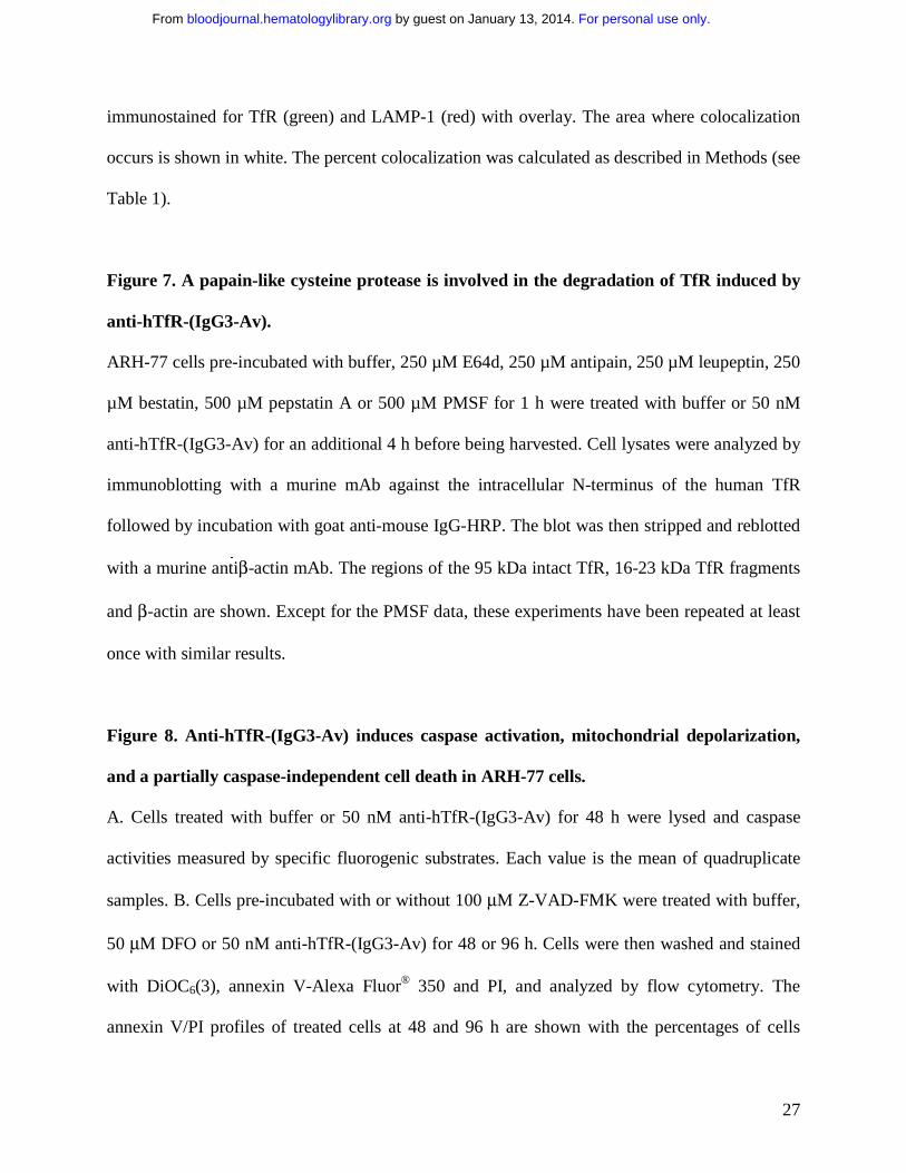
Figure 7. A papain-like cysteine protease is involved in the degradation of TfR induced by
anti-hTfR-(IgG3-Av).
ARH-77 cells pre-incubated with buffer, 250 µM E64d, 250 µM antipain, 250 µM leupeptin, 250
µM bestatin, 500 µM pepstatin A or 500 µM PMSF for 1 h were treated with buffer or 50 nM
anti-hTfR-(IgG3-Av) for an additional 4 h before being harvested. Cell lysates were analyzed by
immunoblotting with a murine mAb against the intracellular N-terminus of the human TfR
followed by incubation with goat anti-mouse IgG-HRP. The blot was then stripped and reblotted
with a murine antiβ-actin mAb. The regions of the 95 kDa intact TfR, 16-23 kDa TfR fragments
and β-actin are shown. Except for the PMSF data, these experiments have been repeated at least
once with similar results.
Figure 8. Anti-hTfR-(IgG3-Av) induces caspase activation, mitochondrial depolarization,
and a partially caspase-independent cell death in ARH-77 cells.
A. Cells treated with buffer or 50 nM anti-hTfR-(IgG3-Av) for 48 h were lysed and caspase
activities measured by specific fluorogenic substrates. Each value is the mean of quadruplicate
samples. B. Cells pre-incubated with or without 100 µM Z-VAD-FMK were treated with buffer,
50 µM DFO or 50 nM anti-hTfR-(IgG3-Av) for 48 or 96 h. Cells were then washed and stained
with DiOC6(3), annexin V-Alexa Fluor® 350 and PI, and analyzed by flow cytometry. The
annexin V/PI profiles of treated cells at 48 and 96 h are shown with the percentages of cells
For personal use only. by guest on January 13, 2014. bloodjournal.hematologylibrary.orgFrom

28
located in each quadrant. C. Non-apoptotic cells (lower left quadrants of Figure 7B) at 48 h were
gated and their mitochondrial membrane potentials shown in log fluorescence intensity. These
experiments have been repeated in both ARH-77 and IM-9 cells with similar results.
For personal use only. by guest on January 13, 2014. bloodjournal.hematologylibrary.orgFrom

29
References
1. Ponka P, Lok CN. The transferrin receptor: role in health and disease. Int J Biochem Cell
Biol. 1999;31:1111-1137.
2. Omary MB, Trowbridge IS, Minowada J. Human cell-surface glycoprotein with unusual
properties. Nature. 1980;286:888-891.
3. Faulk WP, Hsi BL, Stevens PJ. Transferrin and transferrin receptors in carcinoma of the
breast. Lancet. 1980;2:390-392.
4. Shindelman JE, Ortmeyer AE, Sussman HH. Demonstration of the transferrin receptor in
human breast cancer tissue. Potential marker for identifying dividing cells. Int J Cancer.
1981;27:329-334.
5. Trowbridge IS, Lesley J, Schulte R. Murine cell surface transferrin receptor: studies with
an anti-receptor monoclonal antibody. J Cell Physiol. 1982;112:403-410.
6. Prost AC, Menegaux F, Langlois P, et al. Differential transferrin receptor density in
human colorectal cancer: A potential probe for diagnosis and therapy. Int J Oncol.
1998;13:871-875.
7. Shinohara H, Fan D, Ozawa S, et al. Site-specific expression of transferrin receptor by
human colon cancer cells directly correlates with eradication by antitransferrin
recombinant immunotoxin. Int J Oncol. 2000;17:643-651.
8. Brooks D, Taylor C, Dos Santos B, et al. Phase Ia trial of murine immunoglobulin A
antitransferrin receptor antibody 42/6. Clin Cancer Res. 1995;1:1259-1265.
9. Mayers GL, Raghavan D, Hitt S, Glaves D. Transferrin-Gemcitabine conjugates:
application to chemotherapy. In Proceedings of the 89th Annual Meeting of the
For personal use only. by guest on January 13, 2014. bloodjournal.hematologylibrary.orgFrom

30
American Association for Cancer Research, New Orleans, Louisiana, USA, March 28-
April 1, 1998. 63. 1998.
10. Mayers GL, Razeq J, Abu-Hadid MM. Cytotoxic drug conjugates for treatment of
neoplastic disease. US Patent Application No. 5,393,737. 1995.
11. Laske DW, Muraszko KM, Oldfield EH, et al. Intraventricular immunotoxin therapy for
leptomeningeal neoplasia. Neurosurgery. 1997;41:1039-1049; discussion 1049-1051.
12. Weaver M, Laske DW. Transferrin receptor ligand-targeted toxin conjugate (Tf-
CRM107) for therapy of malignant gliomas. J Neurooncol. 2003;65:3-13.
13. Ng PP, Dela Cruz JS, Sorour DN, et al. An anti-transferrin receptor-avidin fusion protein
exhibits both strong proapoptotic activity and the ability to deliver various molecules into
cancer cells. Proc Natl Acad Sci U S A. 2002;99:10706-10711.
14. Habelshaw HA, Lister TA, Stansfeld AG. Correlation of transferrin receptor expression
with histological class and outcome in non-Hodgkin lymphoma. Lancet. 1983;1:498-500.
15. Beguin Y, Lampertz S, Degroote D, Igot D, Malaise M, Fillet G. Soluble Cd23 and Other
Receptors (Cd4, Cd8, Cd25, Cd71) In Serum Of Patients With Chronic Lymphocytic
Leukemia. Leukemia. 1993;7:2019-2025.
16. Jefferies WA, Brandon MR, Williams AF, Hunt SV. Analysis of lymphopoietic stem
cells with a monoclonal antibody to the rattransferrin receptor. Immunology.
1985;54:333-341.
17. Lesley J, Domingo DL, Schulte R, Trowbridge IS. Effect of an anti-murine transferrin
receptor-ricin A conjugate on bone marrow stem and progenitor cells treated in vitro. Exp
Cell Res. 1984;150:400-407.
For personal use only. by guest on January 13, 2014. bloodjournal.hematologylibrary.orgFrom

31
18. Trowbridge IS. Transferrin receptor as a potential therapeutic target. Prog Allergy.
1988;45:121-146.
19. Coloma MJ. Production and Characterization of Novel Recombinant Antibodies.
Microbiology and Molecular Genetics. Los Angeles: University of California at Los
Angeles; 1997:263.
20. Morrison SL, Wims L, Wallick S, Tan L, Oi VT. Genetically engineered antibody
molecules and their application. Ann N Y Acad Sci. 1987;507:187-198.
21. White S, Taetle R, Seligman PA, Rutherford M, Trowbridge IS. Combinations of anti-
transferrin receptor monoclonal antibodies inhibit human tumor cell growth in vitro and
in vivo: evidence for synergistic antiproliferative effects. Cancer Res. 1990;50:6295-
6301.
22. Shin SU, Wu D, Ramanathan R, Pardridge WM, Morrison SL. Functional and
pharmacokinetic properties of antibody-avidin fusion proteins. J Immunol.
1997;158:4797-4804.
23. Gado K, Silva S, Paloczi K, Domjan G, Falus A. Mouse plasmacytoma: an experimental
model of human multiple myeloma. Haematologica. 2001;86:227-236.
24. Cruz JC, Alsina M, Craig F, et al. Ibandronate decreases bone disease development and
osteoclast stimulatory activity in an in vivo model of human myeloma. Exp Hematol.
2001;29:441-447.
25. Hayman SR, Fonseca R. Plasma cell leukemia. Curr Treat Options Oncol. 2001;2:205-
216.
26. Garcia-Calvo M, Peterson EP, Rasper DM, et al. Purification and catalytic properties of
human caspase family members. Cell Death Differ. 1999;6:362-369.
For personal use only. by guest on January 13, 2014. bloodjournal.hematologylibrary.orgFrom

32
27. Tomita Y, Bilim V, Hara N, Kasahara T, Takahashi K. Role of IRF-1 and caspase-7 in
IFN-gamma enhancement of Fas-mediated apoptosis in ACHN renal cell carcinoma cells.
Int J Cancer. 2003;104:400-408.
28. Beynon RJ, Bond JS. Proteolytic Enzymes: A Practical Approach: IRL Press; 1989.
29. Zollner H. Handbook of Enzyme Inhibitors (ed 2nd); 1993.
30. Sreedharan SK, Verma C, Caves LS, et al. Demonstration that 1-trans-epoxysuccinyl-L-
leucylamido-(4-guanidino) butane (E-64) is one of the most effective low Mr inhibitors
of trypsin-catalysed hydrolysis. Characterization by kinetic analysis and by energy
minimization and molecular dynamics simulation of the E-64-beta-trypsin complex.
Biochem J. 1996;316 ( Pt 3):777-786.
31. Creagh EM, Conroy H, Martin SJ. Caspase-activation pathways in apoptosis and
immunity. Immunol Rev. 2003;193:10-21.
32. Greene BT, Thorburn J, Willingham MC, et al. Activation of caspase pathways during
iron chelator-mediated apoptosis. J Biol Chem. 2002;277:25568-25575.
33. Korchak HM, Rich AM, Wilkenfeld C, Rutherford LE, Weissmann G. A carbocyanine
dye, DiOC6(3), acts as a mitochondrial probe in human neutrophils. Biochem Biophys
Res Commun. 1982;108:1495-1501.
34. Green DR, Kroemer G. The pathophysiology of mitochondrial cell death. Science.
2004;305:626-629.
35. Trowbridge IS, Domingo DL. Anti-transferrin receptor monoclonal antibody and toxin-
antibody conjugates affect growth of human tumour cells. Nature. 1981;294:171-173.
For personal use only. by guest on January 13, 2014. bloodjournal.hematologylibrary.orgFrom

33
36. Lesley JF, Schulte RJ. Selection of cell lines resistant to anti-transferrin receptor
antibody: evidence for a mutation in transferrin receptor. Mol Cell Biol. 1984;4:1675-
1681.
37. Vaickus L, Levy R. Antiproliferative monoclonal antibodies: detection and initial
characterization. J Immunol. 1985;135:1987-1997.
38. Lesley J, Schulte R. Selection and characterization of transferrin receptor mutants using
receptor-specific antibodies. Immunogenetics. 1986;24:163-170.
39. Kemp JD, Thorson JA, McAlmont TH, Horowitz M, Cowdery JS, Ballas ZK. Role of the
transferrin receptor in lymphocyte growth: a rat IgG monoclonal antibody against the
murine transferrin receptor produces highly selective inhibition of T and B cell activation
protocols. J Immunol. 1987;138:2422-2426.
40. Lesley JF, Schulte RJ. Inhibition of cell growth by monoclonal anti-transferrin receptor
antibodies. Mol Cell Biol. 1985;5:1814-1821.
41. Lesley J, Schulte R, Woods J. Modulation of transferrin receptor expression and function
by anti- transferrin receptor antibodies and antibody fragments. Exp Cell Res.
1989;182:215-233.
42. Trowbridge IS, Lopez F. Monoclonal antibody to transferrin receptor blocks transferrin
binding and inhibits human tumor cell growth in vitro. Proc Natl Acad Sci U S A.
1982;79:1175-1179.
43. Mendelsohn J, Trowbridge I, Castagnola J. Inhibition of human lymphocyte proliferation
by monoclonal antibody to transferrin receptor. Blood. 1983;62:821-826.
For personal use only. by guest on January 13, 2014. bloodjournal.hematologylibrary.orgFrom

34
44. Neckers LM, Cossman J. Transferrin receptor induction in mitogen-stimulated human T
lymphocytes is required for DNA synthesis and cell division and is regulated by
interleukin 2. Proc Natl Acad Sci U S A. 1983;80:3494-3498.
45. Taetle R, Honeysett JM, Trowbridge I. Effects of anti-transferrin receptor antibodies on
growth of normal and malignant myeloid cells. Int J Cancer. 1983;32:343-349.
46. Taetle R, Rhyner K, Castagnola J, To D, Mendelsohn J. Role of transferrin, Fe, and
transferrin receptors in myeloid leukemia cell growth. Studies with an antitransferrin
receptor monoclonal antibody. J Clin Invest. 1985;75:1061-1067.
47. Taetle R, Castagnola J, Mendelsohn J. Mechanisms of growth inhibition by anti-
transferrin receptor monoclonal antibodies. Cancer Res. 1986;46:1759-1763.
48. Weissman AM, Klausner RD, Rao K, Harford JB. Exposure of K562 cells to anti-
receptor monoclonal antibody OKT9 results in rapid redistribution and enhanced
degradation of the transferrin receptor. J Cell Biol. 1986;102:951-958.
49. Kett WC, Osmond RI, Moe L, Skett SE, Kinnear BF, Coombe DR. Avidin is a heparin-
binding protein. Affinity, specificity and structural analysis. Biochim Biophys Acta.
2003;1620:225-234.
50. Phillips ML, Tao MH, Morrison SL, Schumaker VN. Human/mouse chimeric
monoclonal antibodies with human IgG1, IgG2, IgG3 and IgG4 constant domains:
electron microscopic and hydrodynamic characterization. Mol Immunol. 1994;31:1201-
1210.
51. Salmeron A, Borroto A, Fresno M, Crumpton MJ, Ley SC, Alarcon B. Transferrin
receptor induces tyrosine phosphorylation in T cells and is physically associated with the
TCR zeta-chain. J Immunol. 1995;154:1675-1683.
For personal use only. by guest on January 13, 2014. bloodjournal.hematologylibrary.orgFrom

35
52. Hopkins CR, Trowbridge IS. Internalization and processing of transferrin and the
transferrin receptor in human carcinoma A431 cells. J Cell Biol. 1983;97:508-521.
53. Klapper LN, Waterman H, Sela M, Yarden Y. Tumor-inhibitory antibodies to HER-
2/ErbB-2 may act by recruiting c-Cbl and enhancing ubiquitination of HER-2. Cancer
Res. 2000;60:3384-3388.
54. Ackrell BA, Maguire JJ, Dallman PR, Kearney EB. Effect of iron deficiency on
succinate- and NADH-ubiquinone oxidoreductases in skeletal muscle mitochondria. J
Biol Chem. 1984;259:10053-10059.
55. Ranasinghe AW, Johnson NW, Scragg MA, Williams RA. Iron deficiency reduces
cytochrome concentrations of mitochondria isolated from hamster cheek pouch
epithelium. J Oral Pathol Med. 1989;18:582-585.
56. Maclean K, Yang H, Cleveland JL. Serum suppresses myeloid progenitor apoptosis by
regulating iron homeostasis. J Cell Biochem. 2001;82:171-186.
57. Walter PB, Knutson MD, Paler-Martinez A, et al. Iron deficiency and iron excess damage
mitochondria and mitochondrial DNA in rats. Proc Natl Acad Sci U S A. 2002;99:2264-
2269.
58. Kim BS, Yoon KH, Oh HM, et al. Involvement of p38 MAP kinase during iron chelator-
mediated apoptotic cell death. Cell Immunol. 2002;220:96-106.
59. Buss JL, Neuzil J, Gellert N, Weber C, Ponka P. Pyridoxal isonicotinoyl hydrazone
analogs induce apoptosis in hematopoietic cells due to their iron-chelating properties.
Biochem Pharmacol. 2003;65:161-172.
For personal use only. by guest on January 13, 2014. bloodjournal.hematologylibrary.orgFrom

36
60. Moura IC, Lepelletier Y, Arnulf B, et al. A neutralizing monoclonal antibody (mAb A24)
directed against the transferrin receptor induces apoptosis of tumor T lymphocytes from
ATL patients. Blood. 2004;103:1838-1845.
61. Choi SC, Kim BS, Song MY, et al. Downregulation of p38 kinase pathway by cAMP
response element-binding protein protects HL-60 cells from iron chelator-induced
apoptosis. Free Radic Biol Med. 2003;35:1171-1184.
62. Lorenzo HK, Susin SA, Penninger J, Kroemer G. Apoptosis inducing factor (AIF): a
phylogenetically old, caspase-independent effector of cell death. Cell Death Differ.
1999;6:516-524.
63. Yeh CJ, Taylor CG, Faulk WP. Transferrin binding by peripheral blood mononuclear
cells in human lymphomas, myelomas and leukemias. Vox Sang. 1984;46:217-223.
64. Lesley J, Hyman R, Schulte R, Trotter J. Expression of transferrin receptor on murine
hematopoietic progenitors. Cell Immunol. 1984;83:14-25.
65. Peschle C, Botta R, Muller R, Valtieri M, Ziegler BL. Purification and functional assay
of pluripotent hematopoietic stem cells. Rev Clin Exp Hematol. 2001;5:3-14.
66. Gross S, Helm K, Gruntmeir JJ, Stillman WS, Pyatt DW, Irons RD. Characterization and
phenotypic analysis of differentiating CD34+ human bone marrow cells in liquid culture.
Eur J Haematol. 1997;59:318-326.
67. Bender JG, Unverzagt K, Walker DE, et al. Phenotypic analysis and characterization of
CD34+ cells from normal human bone marrow, cord blood, peripheral blood, and
mobilized peripheral blood from patients undergoing autologous stem cell
transplantation. Clin Immunol Immunopathol. 1994;70:10-18.
For personal use only. by guest on January 13, 2014. bloodjournal.hematologylibrary.orgFrom

37
68. Lansdorp PM, Dragowska W. Long-term erythropoiesis from constant numbers of
CD34+ cells in serum-free cultures initiated with highly purified progenitor cells from
human bone marrow. J Exp Med. 1992;175:1501-1509.
69. Sieff C, Bicknell D, Caine G, Robinson J, Lam G, Greaves MF. Changes in cell surface
antigen expression during hemopoietic differentiation. Blood. 1982;60:703-713.
70. Urashima M, Chen BP, Chen S, et al. The development of a model for the homing of
multiple myeloma cells to human bone marrow. Blood. 1997;90:754-765.
71. Mitsiades CS, Treon SP, Mitsiades N, et al. TRAIL/Apo2L ligand selectively induces
apoptosis and overcomes drug resistance in multiple myeloma: therapeutic applications.
Blood. 2001;98:795-804.
72. Carter P. Improving the efficacy of antibody-based cancer therapies. Nat Rev Cancer.
2001;1:118-129.
For personal use only. by guest on January 13, 2014. bloodjournal.hematologylibrary.orgFrom

38
Figure 1
K562
U266
MM.1S
S6B45
8226
/S
8226
Dox 4
0
OCI-My5
ARH-77 IM-9
0
20
40
60
80
100
120
[3 H]-
Th
ymid
ine
Inco
rpo
rati
on
(%
co
ntr
ol)
A
0
20
40
60
80
100
120
[3 H]-
Th
ymid
ine
Inco
rpo
rati
on
(%
co
ntr
ol)
MMPCL
IM-9
U266
B C
0
20
40
60
80
100
120
[3 H]-
Th
ymid
ine
Inco
rpo
rati
on
(%
co
ntr
ol)
Anti-hTfR
-(IgG3-A
v)
Anti-hTfR
IgG3
Cro
ss-lin
ked A
nti-hTfR
IgG3
Anti-dan
syl-(
IgG3-Av)
Cross
-linkin
g Ab
Anti-dan
syl IgG3
0
20
40
60
80
100
120
For personal use only. by guest on January 13, 2014. bloodjournal.hematologylibrary.orgFrom

39
15
28
26
53
21
21
51
27
22
61
17
16
70
14
73
12
15
37
21
42
46
21
33
41
14 45
48 h
96 h
72 h
Alexa Fluor 488 Labeled Annexin V (early apoptosis marker)
Pro
pid
ium
Iod
ide
(cel
l dea
th m
arke
r)
Buffer Anti-hTfR-(IgG3-Av)Anti-hTfR IgG3
A
57
B
15
46
39
44
31
24
73
15
12
52
26
21
Buffer Anti-hTfR IgG3-Av
Anti-hTfR IgG3 Anti-hTfR IgG3 + 2nd Ab
Alexa Fluor 488 Labeled Annexin V (early apoptosis marker)
Pro
pid
ium
Iod
ide
(cel
l dea
th m
arke
r) Buffer Anti-hTfR-(IgG3-Av)
Figure 2
For personal use only. by guest on January 13, 2014. bloodjournal.hematologylibrary.orgFrom

40
Time (hours)
Su
rfac
e T
fR E
xpre
ssio
n
(rat
io o
ver
bac
kgro
un
d s
ign
al)
Buffer
Anti- hTfR IgG3
Anti- hTfR-(IgG3-Av)
0
1
2
3
4
5
6
7
0 20 40 60 80
Figure 3
Figure 4
Buffer Control
4 h 24 h12 h
ARH-77
TfR
IM-9 4 h
TfR (95 kDa)
Buffer C
ontrol
Cross
-linke
d Anti-
hTfR Ig
G3
Anti-hTfR
(IgG3-
Av)
1:1 1:5
Anti-hTfR
IgG3
20' 5' 10' 20'
TfR
Cont. Anti-hTfR-(IgG3-Av)
β-actin
Anti-hTfR
-(IgG3-
Av)
Control
20 kDa 14 kDa
95 kDa
Anti-hTfR-(IgG3-Av)
Anti-hTfR IgG3
β-actin
β-actin
β-actin
A
B
C
D
For personal use only. by guest on January 13, 2014. bloodjournal.hematologylibrary.orgFrom

41
Figure 5
BBuffer Anti-hTfR-(IgG3-Av)
Anti- hTfR-(IgG3-Av) + FACBuffer + FAC
79
6
14
79
4
14
23
24
5214
11
74
Pro
pid
ium
Iod
ide
(cel
l dea
th m
arke
r)
Alexa Fluor 488 Labeled Annexin V (early apoptosis marker)
[3 H]-
Thym
idin
e In
corp
ora
tion
(%
co
ntr
ol)
0
10
20
30
40
50
60
70
80
90
100
110
ARH-77 IM-9
Buffe
r
ZnSO4FAC
ABuf
fer
ZnSO4FAC
Figure 6
0 Min.(Buffer�Control)
15 Min.
30 Min.
45 Min.
LAMP-1 TfR Overlay ColocalizedLAMP-1
15 Min.
0 Min. (Buffer
Control)
TfR Overlay Colocalized
30 Min.
45 Min.
For personal use only. by guest on January 13, 2014. bloodjournal.hematologylibrary.orgFrom

42
Experiment 1 Experiment 2 Experiment 3Incubation time 0' 15' 30' 90' 0' 30' 60' 0' 15' 30' 45'
Average % Colocalization 10.2 20.0 16.3 18.2 13.6 20.8 18.7 11.9 21.7 19.2 39.8
SD 1.5 4.0 2.6 3.4 3.8 3.0 3.9 3.5 8.1 1.4 9.8SE 0.5 1.4 0.9 1.4 1.4 1.1 1.5 1.0 2.5 0.5 3.1
Number of fields (n ) 11 8 8 6 7 8 7 12 10 9 10
Average no. of cells/field 17 14 16 18 19 21 24 19 11 17 7p value < 0.001 < 0.001 < 0.002 < 0.002 < 0.03 < 0.005 < 0.001 < 0.001
Figure 7
Table 1. Colocalization of TfR and LAMP-1 in ARH-77 cells incubated with 11 nM anti- hTfR-(IgG3-Av)
Anti-hTfR IgG3-AvBuffer
Buffer E64d
Antipain Leupeptin
Bestatin Pepstatin A
PMSF
20 kDa
β-actin
95 kDa
Anti-hTfR-(IgG3-Av)Buffer
For personal use only. by guest on January 13, 2014. bloodjournal.hematologylibrary.orgFrom

43
A
0
50
100
150
200
250
300
350
Caspase 9 Caspase 3 Caspase 8
Cas
pas
e A
ctiv
ity
(a
rbit
rary
flu
ore
scen
ce u
nit
)Buffer
Anti- hTfR-(IgG3-Av)
Figure 8
Alexa Fluor 350 Annexin V (early apoptosis marker)
R2
45
14
39 84
5
10
51 28
20
69
12
17
86
4
8 90 5
3
Buffer Z-VAD-FMK
72
6
21
6
9
15
26
59
29
42
29
22
28
50
43
29
27
84
Buffer Z-VAD-FMK
Bu
ffer
Co
ntr
ol
DF
O
An
ti-hT
fR-(
IgG
3-A
v)
48 h 96 h
Pro
pid
ium
Io
did
e (c
ell d
eath
mar
ker)
B
C
Log Fluorescence Intensity
Cel
l Nu
mb
er
Control
DFO
Anti- hTfR-(IgG3-Av)
Buffer Z-VAD-FMK
For personal use only. by guest on January 13, 2014. bloodjournal.hematologylibrary.orgFrom
Copyright © 2022 FDOKUMEN