Molecular dynamics simulation directed rational design of inhibitors targeting drug-resistant...
8
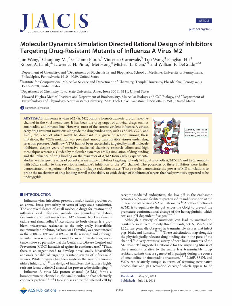
Published: July 11, 2011 r2011 American Chemical Society 12834 dx.doi.org/10.1021/ja204969m | J. Am. Chem. Soc. 2011, 133, 12834–12841 ARTICLE pubs.acs.org/JACS Molecular Dynamics Simulation Directed Rational Design of Inhibitors Targeting Drug-Resistant Mutants of Influenza A Virus M2 Jun Wang, † Chunlong Ma, # Giacomo Fiorin, § Vincenzo Carnevale, § Tuo Wang, || Fanghao Hu, || Robert A. Lamb, ^ Lawrence H. Pinto, # Mei Hong, || Michael L. Klein,* ,§ and William F. DeGrado* ,†,‡ † Department of Chemistry, and ‡ Department of Biochemistry and Biophysics, School of Medicine, University of Pennsylvania, Philadelphia, Pennsylvania 19104-6059, United States § Institute for Computational Molecular Science and Department of Chemistry, Temple University, Philadelphia, Pennsylvania 19122-6078, United States ) Department of Chemistry, Iowa State University, Ames, Iowa 50011-3111, United States ^ Howard Hughes Medical Institute and Department of Biochemistry, Molecular Biology and Cell Biology, and # Department of Neurobiology and Physiology, Northwestern University, 2205 Tech Drive, Evanston, Illinois 60208-3500, United States b S Supporting Information ’ INTRODUCTION Influenza virus infections present a major health problem on an annual basis, particularly in years of large-scale pandemics. The approved classes of small molecule drugs for treatment of influenza viral infections include neuraminidase inhibitors (zanamivir and oseltamivir) and M2 channel blockers (aman- tadine and rimantadine). 1 Resistance to both classes is a pro- blem: widespread resistance to the only orally bioavailable neuraminidase inhibitor, oseltamivir (Tamiflu), was encountered in the 20082009 2 and 20092010 flu seasons, 3 and although amantadine was successfully used for over three decades, resis- tance is now so pervasive that the Centers for Disease Control and Prevention (CDC) has advised against its continued use. 4,5 Thus, there is an urgent need to develop novel orally bioavailable antivirals capable of targeting resistant strains of influenza A viruses. While progress has been made in the area of neurami- nidase inhibitors, 68 the design of inhibitors that address highly resistant forms of the M2 channel has proven to be challenging. 1,9 Influenza A virus M2 proton channel (A/M2) forms a homotetrameric channel in the viral membrane that selectively conducts protons. 1012 Once viruses enter the infected cell by receptor-mediated endocytosis, the low pH in the endosome activates A/M2 and facilitates proton influx and disruption of the interaction of the viral RNA with its matrix. 13 Another function of A/M2 is to equilibrate the pH across the Golgi to prevent the premature conformational change of the hemagglutinin, which acts as a pH-dependent fusogen. 1416 Although a variety of mutations can lead to amantadine- resistance in vitro, 1719 only three mutants, S31N, V27A, and L26F, are generally observed in transmissible viruses that infect pigs, birds, and humans. 2022 These substitutions map alongside the physiologically relevant drug binding site in the pore of the channel. 23 A very extensive survey of pore-lining mutants of the M2 channel 24 suggested a rationale for the surprising fitness of these mutants relative to the many less transmissible drug- resistant variants that are generated in patients during the course of amantadine or rimantadine treatment. 19,25 L26F, S31N, and V27A are relatively unique in terms of retaining near-native proton flux and pH activation curves, 24 which appear to be Received: May 30, 2011 ABSTRACT: Influenza A virus M2 (A/M2) forms a homotetrameric proton selective channel in the viral membrane. It has been the drug target of antiviral drugs such as amantadine and rimantadine. However, most of the current virulent influenza A viruses carry drug-resistant mutations alongside the drug binding site, such as S31N, V27A, and L26F, etc., each of which might be dominant in a given flu season. Among these mutations, the V27A mutation was prevalent among transmissible viruses under drug selection pressure. Until now, V27A has not been successfully targeted by small molecule inhibitors, despite years of extensive medicinal chemistry research efforts and high throughput screening. Guided by molecular dynamics (MD) simulation of drug binding and the influence of drug binding on the dynamics of A/M2 from earlier experimental studies, we designed a series of potent spirane amine inhibitors targeting not only WT, but also both A/M2-27A and L26F mutants with IC 50 s similar to that seen for amantadine’s inhibition of the WT channel. The potencies of these inhibitors were further demonstrated in experimental binding and plaque reduction assays. These results demonstrate the power of MD simulations to probe the mechanism of drug binding as well as the ability to guide design of inhibitors of targets that had previously appeared to be undruggable.
-
Upload
independent -
Category
Documents
-
view
4 -
download
0
Transcript of Molecular dynamics simulation directed rational design of inhibitors targeting drug-resistant...
Published: July 11, 2011
r 2011 American Chemical Society 12834 dx.doi.org/10.1021/ja204969m | J. Am. Chem. Soc. 2011, 133, 12834–12841
ARTICLE
pubs.acs.org/JACS
Molecular Dynamics Simulation Directed Rational Design of InhibitorsTargeting Drug-Resistant Mutants of Influenza A Virus M2Jun Wang,† Chunlong Ma,# Giacomo Fiorin,§ Vincenzo Carnevale,§ Tuo Wang,|| Fanghao Hu,||
Robert A. Lamb,^ Lawrence H. Pinto,# Mei Hong,|| Michael L. Klein,*,§ and William F. DeGrado*,†,‡
†Department of Chemistry, and ‡Department of Biochemistry and Biophysics, School of Medicine, University of Pennsylvania,Philadelphia, Pennsylvania 19104-6059, United States§Institute for Computational Molecular Science and Department of Chemistry, Temple University, Philadelphia, Pennsylvania19122-6078, United States
)Department of Chemistry, Iowa State University, Ames, Iowa 50011-3111, United States^Howard Hughes Medical Institute and Department of Biochemistry, Molecular Biology and Cell Biology, and #Department ofNeurobiology and Physiology, Northwestern University, 2205 Tech Drive, Evanston, Illinois 60208-3500, United States
bS Supporting Information
’ INTRODUCTION
Influenza virus infections present a major health problem onan annual basis, particularly in years of large-scale pandemics.The approved classes of small molecule drugs for treatment ofinfluenza viral infections include neuraminidase inhibitors(zanamivir and oseltamivir) and M2 channel blockers (aman-tadine and rimantadine).1 Resistance to both classes is a pro-blem: widespread resistance to the only orally bioavailableneuraminidase inhibitor, oseltamivir (Tamiflu), was encounteredin the 2008�20092 and 2009�2010 flu seasons,3 and althoughamantadine was successfully used for over three decades, resis-tance is now so pervasive that the Centers for Disease Control andPrevention (CDC) has advised against its continued use.4,5 Thus,there is an urgent need to develop novel orally bioavailableantivirals capable of targeting resistant strains of influenza Aviruses. While progress has been made in the area of neurami-nidase inhibitors,6�8 the design of inhibitors that address highlyresistant forms of theM2 channel has proven to be challenging.1,9
Influenza A virus M2 proton channel (A/M2) forms ahomotetrameric channel in the viral membrane that selectivelyconducts protons.10�12 Once viruses enter the infected cell by
receptor-mediated endocytosis, the low pH in the endosomeactivates A/M2 and facilitates proton influx and disruption of theinteraction of the viral RNAwith itsmatrix.13 Another function ofA/M2 is to equilibrate the pH across the Golgi to prevent thepremature conformational change of the hemagglutinin, whichacts as a pH-dependent fusogen.14�16
Although a variety of mutations can lead to amantadine-resistance in vitro,17�19 only three mutants, S31N, V27A, andL26F, are generally observed in transmissible viruses that infectpigs, birds, and humans.20�22 These substitutions map alongsidethe physiologically relevant drug binding site in the pore of thechannel.23 A very extensive survey of pore-lining mutants of theM2 channel24 suggested a rationale for the surprising fitness ofthese mutants relative to the many less transmissible drug-resistant variants that are generated in patients during the courseof amantadine or rimantadine treatment.19,25 L26F, S31N, andV27A are relatively unique in terms of retaining near-nativeproton flux and pH activation curves,24 which appear to be
Received: May 30, 2011
ABSTRACT: Influenza A virus M2 (A/M2) forms a homotetrameric proton selectivechannel in the viral membrane. It has been the drug target of antiviral drugs such asamantadine and rimantadine. However, most of the current virulent influenza A virusescarry drug-resistant mutations alongside the drug binding site, such as S31N, V27A, andL26F, etc., each of which might be dominant in a given flu season. Among thesemutations, the V27A mutation was prevalent among transmissible viruses under drugselection pressure. Until now, V27A has not been successfully targeted by small moleculeinhibitors, despite years of extensive medicinal chemistry research efforts and highthroughput screening. Guided by molecular dynamics (MD) simulation of drug bindingand the influence of drug binding on the dynamics of A/M2 from earlier experimentalstudies, we designed a series of potent spirane amine inhibitors targeting not only WT, but also both A/M2-27A and L26F mutantswith IC50s similar to that seen for amantadine’s inhibition of the WT channel. The potencies of these inhibitors were furtherdemonstrated in experimental binding and plaque reduction assays. These results demonstrate the power of MD simulations toprobe the mechanism of drug binding as well as the ability to guide design of inhibitors of targets that had previously appeared to beundruggable.
12835 dx.doi.org/10.1021/ja204969m |J. Am. Chem. Soc. 2011, 133, 12834–12841
Journal of the American Chemical Society ARTICLE
parameters finely tuned to respond to the properties of a givenvirus’ hemagglutinin protein while minimizing toxicity for theparent cell until viral production is complete. Although S31N isthe substitution found in current resistant strains, in other yearsV27A has predominated.26 Recent studies showed that thecurrent predominance of S31N is not the result of drug selectionpressure, because S31N was prevalent before the introduction ofamantadine and has become widespread in regions whereamantadine was never used.20,22 Instead, V27A was identifiedto be the major mutation emerging from drug selection pressure.While the L26F and S31N mutation causes a 10�20-decrease inthe IC50s for amantadine inhibition, the corresponding V27Amutation renders the channel entirely resistant to both amanta-dine and rimantadine.27We therefore focused on this particularlychallenging mutant.
Over the last four decades, systematic studies of amantadineanalogues and library screening have elucidated structure�activ-ity relationships and helped to identify potent channel-blockers.1,9 However, there have been no confirmed reports ofsmall organic molecules that potently target highly amantadine-resistant variants of A/M2. Here, we use a combination ofmolecular dynamics (MD) and classical medicinal chemistryapproaches to design very potent inhibitors of V27A and L26F.MD was used to explore the mechanism of binding of amanta-dine to WT and of the designed inhibitor to V27A, therebyinforming the mechanism and potency of the designed com-pounds. Potent inhibitors in general require an ammoniumgroup, which associates with discrete, water-lined sites that mightbe hotspots for stabilizing a diffusing hydronium ion. These sitesappear to be retained in the L26F and V27Amutants and formedthe basis for the design of inhibitors that target these variantswhile maintaining affinity for WT.
’MATERIALS AND METHODS
Materials. All starting material chemicals were purchased fromcommercial vendors and used without purification. Reactions werecarried out using HPLC grade solvents under N2 atmosphere. Com-pounds were purified by silica gel flash column chromatography andcharacterized by ESI-MS, 1H NMR, and 13C NMR. Details about theinhibitor synthesis procedure and characterization can be found in theSupporting Information.Inhibitor Synthesis. Detailed synthesis procedure and compound
characterization can be found in the Supporting Information.Two-Electrode Voltage Clamp (TEVC) Assay and Plaque
Reduction Assay. The inhibitors were tested via a two-electrodepatch clamp (TEVC) assay using Xenopus laevis frog oocytes micro-injectected with RNA expressing the A/M2 protein as in a previousreport.27 The potency of the inhibitors was expressed as the percentageinhibition of A/M2 current observed after 2 min of incubation with 100μM compounds, and IC50 values were collected for selected potentcompounds.Peptide Synthesis. A/M2(22-46)-V27A peptide with A27, V28,
and G34 selectively 15N and 13C labeled was manually synthesizedusing Fmoc chemistry at elevated temperature (75 �C for both couplingand deprotection) in a semiautomated Quest synthesizer using RinkAmide Chemmatrix resin (Matrix Innovation Inc., Canada). Fiveequivalents of amino acid, 5 equiv of HCTU, and 10 equiv of DIEA inNMP were used for 5 min coupling. 5% piperazine and 0.1 M HOBt inDMF were used as the deprotection solution to minimize aspartamideformation. The peptide was cleaved from the resin using 95%TFA, 2.5%Tris, and 2.5% H2O and was precipitated from ether after removal ofTFA. Ether was decanted after centrifugation, and the peptide was
washed with cold ether again. Final peptide was dissolved in 50% B0
(59.9% isopropanol, 30% acetonitrile, 10%H2O, and 0.1%TFA)/50%A(99.9% H2O, 0.1% TFA) and was purified by preparative C4 reversephase HPLCwith a linear gradient of 70% B0 to 85% B0. The peptide waseluted at 78% B0. The purity and identity of the peptide were confirmedby analytical HPLC andMALDI-MS. CalculatedMS, 2755.24; observedMS, 2755.80.Molecular Dynamics Simulations. All simulations in this work
were performed on the transmembrane region of the A/M2 bundle,spanning residues 25�46 and identified in the following as M2TM. Thehigh-resolution structure of M2TM from Acharya et al.28 was used asinitial configuration. M2TM was embedded in a hydrated membrane of1-palmitoyl-2-oleoyl-sn-glycero-3-phosphocholine (POPC) molecules, andperiodic boundary conditions were applied with a 80 Å � 80 Å � 77 Åperiodic box, corresponding to a 38 Å thick layer of water between twoperiodic images of the bilayer. The hydrated bilayer was neutralized byadding 27 K+ and 28 Cl� ions, respectively (KCl concentration of about150 mM). The protein and the lipids were modeled using theCHARMM27 force field,29 and water molecules were modeled usingthe TIP3P force field.30 The electrostatic potential was solved by theparticle mesh Ewald (PME) method31 with an accuracy threshold of10�6, a real space spherical cutoff of 12 Å, and a fast Fourier transform(FFT) grid spacing of 0.8 Å. Lennard-Jones interactions were cut off at12 Å, with a switching function starting from 10 Å. The equations ofmotion were solved with the velocity Verlet integrator using a time stepof 1.5 fs. The lengths of all bonds involving hydrogen atoms were keptconstrained with the SHAKE method.32 The system was run at 310 Kand 1 atm using Langevin temperature33 and Langevin pistonpressure34,35 coupling schemes. Decay times for the thermostat andbarostat were chosen to be 1 and 0.1 ps, respectively. MD simulationswere performed with NAMD.36
Solid-State NMR (ssNMR). 1,2-Dimyristoyl-sn-glycero-3-phos-phoch-line (DMPC) was used to reconstitute A27, V28, and G34labeled A/M2(22-46)-V27A by detergent dialysis as described before.37
The dry lipid powder was suspended in 1mL of pH 7.5 phosphate buffer(10 mM NaH2PO4/Na2HPO4, 1 mM EDTA, and 0.01 mM NaN3),votexed, and freeze�thawed eight times to create uniform lipid vesicles.The peptide powder was codissolved with ∼20 mg of octyl-β-D-glucopyranoside (OG) in 1 mL of the same buffer. The solution wasthen mixed with 1 mL of lipid vesicle solution, votexed for 2 h, anddialyzed with a 3.5 kDa molecular weight cutoff against 1 L of buffer at4 �C for 3 days. The buffer was changed every 8�12 h to remove thedetergent. The protein�lipid precipitate appeared after one day. Theproteoliposome solution was centrifuged at 150 000g and 6 �C for 4 h toyield a membrane pellet with ∼40 wt % water. The final peptide:lipidmolar ratio was 1:8. Drugs were directly titrated into the membranepellet in the NMR rotor to a ratio of four per tetramer.
Solid-state NMR experiments were carried out on a 400MHz (9.4 T)and a 600 MHz (14.1 T) Bruker AVANCE spectrometer using 4 mmMAS probes. Typical radio frequency fields were 40�50 kHz for 13C and15N and 70 kHz for 1H. 13C and 15N chemical shifts were referenced tothe R-Gly CO signal at 176.49 ppm on the TMS scale and the 15N signalof N-acetyl-valine at 122 ppm on the liquid ammonia scale, respectively.2D 15N�13C correlation spectra were measured using a REDORsequence with a 0.8 ms mixing time for 13C�15N coherence transfer.38
2D 13C�13C correlation spectra were measured using a 1H-driven 13Cspin diffusion sequence39 with a 40 ms 13C mixing time. All experimentswere performed at 273 K and under 7 kHz spinning.
’RESULTS AND DISCUSSION
MD Simulations of Drug-Binding. A/M2 channel blockersbind to a site within the N-terminal half of the pore, displacingwater from a pore that leads to the pH-sensing H37 residues.
12836 dx.doi.org/10.1021/ja204969m |J. Am. Chem. Soc. 2011, 133, 12834–12841
Journal of the American Chemical Society ARTICLE
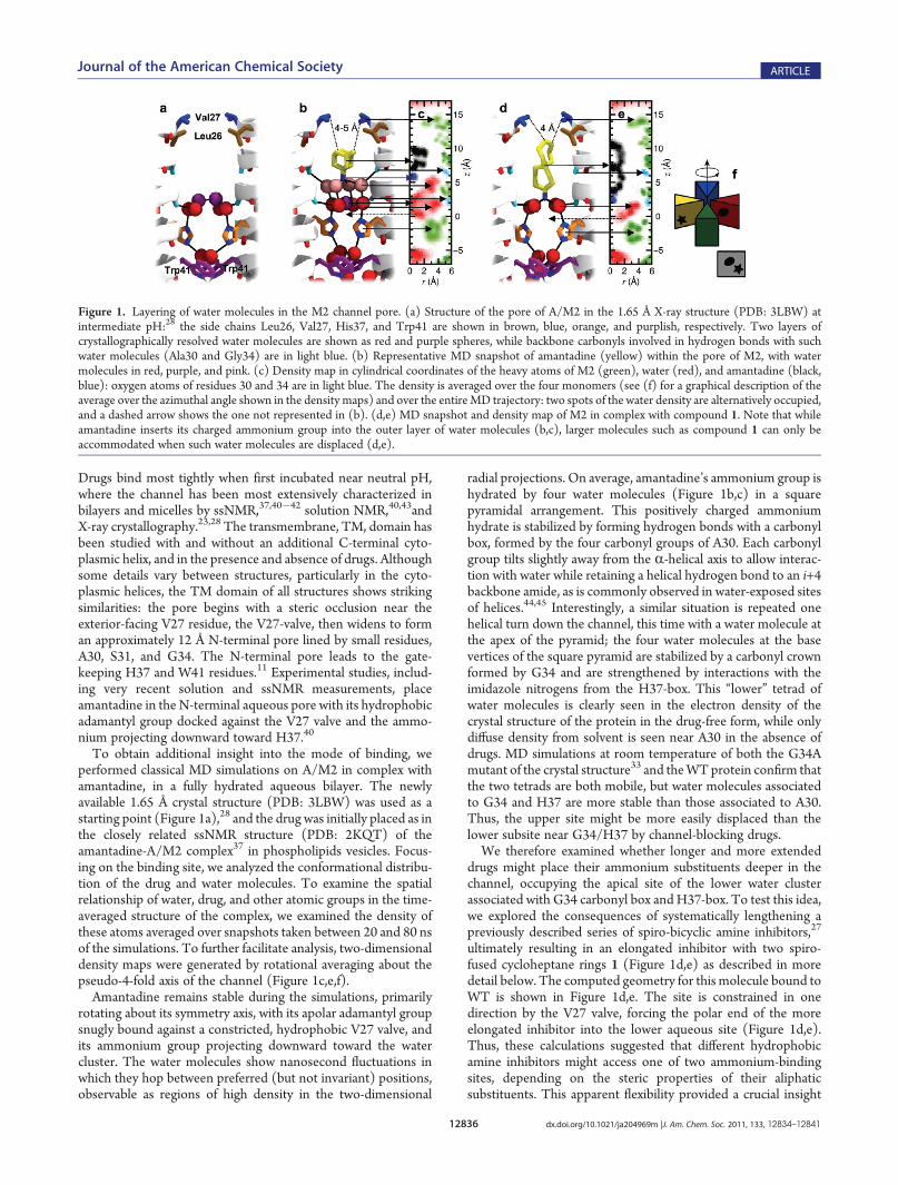
Drugs bind most tightly when first incubated near neutral pH,where the channel has been most extensively characterized inbilayers and micelles by ssNMR,37,40�42 solution NMR,40,43andX-ray crystallography.23,28 The transmembrane, TM, domain hasbeen studied with and without an additional C-terminal cyto-plasmic helix, and in the presence and absence of drugs. Althoughsome details vary between structures, particularly in the cyto-plasmic helices, the TM domain of all structures shows strikingsimilarities: the pore begins with a steric occlusion near theexterior-facing V27 residue, the V27-valve, then widens to forman approximately 12 Å N-terminal pore lined by small residues,A30, S31, and G34. The N-terminal pore leads to the gate-keeping H37 and W41 residues.11 Experimental studies, includ-ing very recent solution and ssNMR measurements, placeamantadine in the N-terminal aqueous pore with its hydrophobicadamantyl group docked against the V27 valve and the ammo-nium projecting downward toward H37.40
To obtain additional insight into the mode of binding, weperformed classical MD simulations on A/M2 in complex withamantadine, in a fully hydrated aqueous bilayer. The newlyavailable 1.65 Å crystal structure (PDB: 3LBW) was used as astarting point (Figure 1a),28 and the drug was initially placed as inthe closely related ssNMR structure (PDB: 2KQT) of theamantadine-A/M2 complex37 in phospholipids vesicles. Focus-ing on the binding site, we analyzed the conformational distribu-tion of the drug and water molecules. To examine the spatialrelationship of water, drug, and other atomic groups in the time-averaged structure of the complex, we examined the density ofthese atoms averaged over snapshots taken between 20 and 80 nsof the simulations. To further facilitate analysis, two-dimensionaldensity maps were generated by rotational averaging about thepseudo-4-fold axis of the channel (Figure 1c,e,f).Amantadine remains stable during the simulations, primarily
rotating about its symmetry axis, with its apolar adamantyl groupsnugly bound against a constricted, hydrophobic V27 valve, andits ammonium group projecting downward toward the watercluster. The water molecules show nanosecond fluctuations inwhich they hop between preferred (but not invariant) positions,observable as regions of high density in the two-dimensional
radial projections. On average, amantadine’s ammonium group ishydrated by four water molecules (Figure 1b,c) in a squarepyramidal arrangement. This positively charged ammoniumhydrate is stabilized by forming hydrogen bonds with a carbonylbox, formed by the four carbonyl groups of A30. Each carbonylgroup tilts slightly away from the R-helical axis to allow interac-tion with water while retaining a helical hydrogen bond to an i+4backbone amide, as is commonly observed in water-exposed sitesof helices.44,45 Interestingly, a similar situation is repeated onehelical turn down the channel, this time with a water molecule atthe apex of the pyramid; the four water molecules at the basevertices of the square pyramid are stabilized by a carbonyl crownformed by G34 and are strengthened by interactions with theimidazole nitrogens from the H37-box. This “lower” tetrad ofwater molecules is clearly seen in the electron density of thecrystal structure of the protein in the drug-free form, while onlydiffuse density from solvent is seen near A30 in the absence ofdrugs. MD simulations at room temperature of both the G34Amutant of the crystal structure33 and theWTprotein confirm thatthe two tetrads are both mobile, but water molecules associatedto G34 and H37 are more stable than those associated to A30.Thus, the upper site might be more easily displaced than thelower subsite near G34/H37 by channel-blocking drugs.We therefore examined whether longer and more extended
drugs might place their ammonium substituents deeper in thechannel, occupying the apical site of the lower water clusterassociated with G34 carbonyl box andH37-box. To test this idea,we explored the consequences of systematically lengthening apreviously described series of spiro-bicyclic amine inhibitors,27
ultimately resulting in an elongated inhibitor with two spiro-fused cycloheptane rings 1 (Figure 1d,e) as described in moredetail below. The computed geometry for this molecule bound toWT is shown in Figure 1d,e. The site is constrained in onedirection by the V27 valve, forcing the polar end of the moreelongated inhibitor into the lower aqueous site (Figure 1d,e).Thus, these calculations suggested that different hydrophobicamine inhibitors might access one of two ammonium-bindingsites, depending on the steric properties of their aliphaticsubstituents. This apparent flexibility provided a crucial insight
Figure 1. Layering of water molecules in the M2 channel pore. (a) Structure of the pore of A/M2 in the 1.65 Å X-ray structure (PDB: 3LBW) atintermediate pH:28 the side chains Leu26, Val27, His37, and Trp41 are shown in brown, blue, orange, and purplish, respectively. Two layers ofcrystallographically resolved water molecules are shown as red and purple spheres, while backbone carbonyls involved in hydrogen bonds with suchwater molecules (Ala30 and Gly34) are in light blue. (b) Representative MD snapshot of amantadine (yellow) within the pore of M2, with watermolecules in red, purple, and pink. (c) Density map in cylindrical coordinates of the heavy atoms of M2 (green), water (red), and amantadine (black,blue): oxygen atoms of residues 30 and 34 are in light blue. The density is averaged over the four monomers (see (f) for a graphical description of theaverage over the azimuthal angle shown in the density maps) and over the entireMD trajectory: two spots of the water density are alternatively occupied,and a dashed arrow shows the one not represented in (b). (d,e) MD snapshot and density map of M2 in complex with compound 1. Note that whileamantadine inserts its charged ammonium group into the outer layer of water molecules (b,c), larger molecules such as compound 1 can only beaccommodated when such water molecules are displaced (d,e).

12837 dx.doi.org/10.1021/ja204969m |J. Am. Chem. Soc. 2011, 133, 12834–12841
Journal of the American Chemical Society ARTICLE
to enable design of dual-specificity inhibitors for WT as wellas V27A.The V27A and L26F Mutants Have Larger, More Solvent-
Exposed N-Terminal Pores. To investigate how V27A andL26F mutations affect the size and shape of the binding site,we built theoretical models of these mutants in the drug-freeform using the recently solved high resolution structure of A/M228 (PDB: 3LBW) as an initial configuration. Initial modelswere obtained by replacing the side chains of V27 and L26 withAla and Phe, respectively, and refined by performing classicalMDsimulations in a bilayer of 1,2-dimyristoyl-sn-glycero-3-phospho-choline (DMPC) lipids hydrated by explicit solvent molecules.After a relaxation phase of several nanoseconds with the proteingradually released, the structures of themutants were observed tobe stable over a timespan of at least several tens of nanoseconds(backbone rmsd of the order of 1 Å). As expected, the mutationof the valines into alanines at position 27 does not perturbsignificantly the overall structure of the bundle. Therefore, theeffect of the mutation can be easily rationalized by observing thatreplacing the bulky Val side chain with a small Ala methyl moietyresults in an expansion of the pore radius near the top of theamantadine binding site (Figure 2c,d). A solutionNMR structureof this mutant,46 which was published subsequent to the com-pletion of these calculations, is in agreement with this conclusionand helps validate the modeling protocol. Particularly note-worthy and somewhat less anticipated is the fact that a similarnet effect is observed upon mutation of L26, which is not pore-lining, into phenylalanine (Figure 2e,f). In this case, the intro-duction of the phenyl groups results in less efficient packing nearthe N-terminus of the helix. As a result, the entry region of thepore is more disordered and features, on average, a larger radius.Most importantly,while thesemutations resulted in large changes in
the upper region of the pore, the structure of the channel and thewaterappeared largely unaffected near A30, G34, and H37. Thus, thechallenge was to design molecules that were larger than amantadineand able tofill the increased volumeof theupper pore createdbyV27Aand L26F mutant, while still being accommodated within the WTstructure. The ability of the pore to accommodate alkyl-ammoniumhydrates near either A30 or G34’s carbonyl groups provided flexibilityin the design of molecules that could penetrate different depthsdepending on the volume of the upper pore of the mutants.
Design and Synthesis of Inhibitors Targeting A/M2-V27A.Previously, we discovered a potent inhibitor of WT A/M2 (2;IC50 = 0.9 ( 0.1 μM) (Table 1) while investigating structure�activity relationships of a random screen hit (BL-174348).Although compound 2was ineffective against V27A,27,49 ssNMRcharacterization of this compound in complex withWT A/M2 inbilayers showed that this spiro-piperidine 2 had a greater impacton the dynamics and magnetic environment of the pore thanamantadine, and that it interacted over a more extended sitewithin the channel. Thus, it appeared to be an attractive scaffoldupon which to build functional groups to fill in the more spaciousvestibule created by the V27A mutation. Indeed, a one carbonextension of the spiro-piperidine 2 gave spirane amine 3, whichwas the first molecule to show weak but saturable inhibition ofV27A (IC50 = 84.9( 13.6 μM).27 Encouraged by this result, thelength of 3 was further extended by inserting a second carbonspacer, via a simple methylene in 4 and its methyl-substituted
Figure 2. Shape modulation of the drug-binding pocket in the pore of A/M2 channel. A cartoon representation of the A/M2-TM helix bundle is shownfor WT (a,b), V27A (c,d), and L26F (e,f). The molecular surface of the channel pore is highlighted, and pore-lining residues Leu/Phe26 (brown),Val/Ala27 (blue), His37 (orange), and Trp41 (violet) are shown as sticks. Pore water molecules and amantadine are shown for wt as red spheres andsticks, respectively. The positions of water oxygens and amantadine are obtained from the crystal structure28 (PDB: 3LBW) and the ssNMR structure ofthe A/M2-amantadine complex29 (PDB: 2KQT), respectively. For each structure, the radius of the pore (computed using Hole47) is plotted as afunction of the displacement along the channel axis in the region between His37 and Val-Ala27; the range of values corresponding to the entry region ofthe channel pore is highlighted in pink.
Table 1. Effect of Extending the Length of the 6,6-SpiraneCore on Potency against WT A/M2 and A/M2-V27Aa
a IC50 values were measured versus A/M2 from the Udorn strain of thevirus, or the V27A mutant expressed in Xenopus laevis oocytes. Allcompounds are tested in hydrochloride form. The proton currents andextent of inhibition in the presence of various concentrations of drugwere measured as described in the Materials and Methods.

12838 dx.doi.org/10.1021/ja204969m |J. Am. Chem. Soc. 2011, 133, 12834–12841
Journal of the American Chemical Society ARTICLE
analogue, 5. Their ability to inhibit proton currents wasmeasuredin Xenopus laevis oocytes expressing either WT A/M2 or A/M2-V27A. Encouragingly, 4 and 5 had potencies similar to that ofamantadine against WT; more importantly, they had similar lowIC50 values against V27A, which amantadine is completelyunable to inhibit.Similar potency increases could be achieved by homologating
the six-membered rings of 3. The potency increased as the fullyaliphatic ring was expanded from six to seven carbon atoms (7)(Table 2), and the same trend was observed when the amine-containing ring was expanded from six to seven carbon atoms(8). Combining these substitutions in the bis-cycloheptyl-spir-ane amine, 1, resulted in a compound with a 2-fold higherpotency than amantadine against WT A/M2, and great potencyagainst the amantadine-insensitive mutant V27A with IC50 of11.3( 0.7 μM. As a negative control, the six-membered aliphatic
ring of compound 3 was converted to the five-membered ring in6, which only showed minimal inhibition against V27A mutant.Design of Potent Spiro-adamantane Inhibitor of A/M2-
V27A and L26F.MD simulations were used to further probe themode of binding of amantadine and the potent bis-cycloheptyl-spirane amine 1 to both WT and V27A. Amantadine failed tobind a unique site when placed within V27A, and instead boundwith its ammonium occupying either the upper or the loweraqueous sites. Moreover, its apolar adamantane cage was sig-nificantly less well dehydrated in V27A versusWT, explaining theloss in potency for the mutant. This behavior contrasts with thatseen for the bis-cycloheptyl-spirane amine 1. As expected, thedrug shifted its position upward in V27A, to allow its alkyl groupto fill the larger cavity near the channel entrance (Figure 3e). Itsammonium group occupies the lower aqueous site in WT, whereit forms solvent-mediated hydrogen bonds with the carbonyl ofG34, versus A30 in V27A (Figure 3d,e). Although its apolarportion is more effectively dehydrated than amanatadine inV27A, the diffuse density of the upper ring of 1, when boundto V27A (Figure 3d), indicates that it averages between multipleorientations and does not fully fill the cavity. Thus, we suspectedthat its affinity might be further increased by modulating thesteric bulk of its apolar substituent. We first consider convertingthe “upper” cyclohexane ring of 3 to a bicyclo[3.3.1]nonane(intermediate I, Figure 4a), which can be further constrainedby a methylene linkage to give spiroadamantane 9 (Figure 4a).This inhibitor was simulated against WT, V27A, and L26F(Figure 3f,g). It was found that 9 had a pose similar to 1 inV27A with the hydrophobic adamantane filling in the extra spacenear A27. In the case of WT and L26F, compound 9 was pushedlower toward H37 and forms water-mediated hydrogen bondingwith H37. It is noted that high density of 9 was observed in allthree variants (including L26F, not shown), suggesting tightbinding with each mutant.Compound 9 was then synthesized using a Robinson annula-
tion reaction starting from 2-adamantanecarboaldehyde (Sup-porting Information Scheme 2), and its structure was confirmedby X-ray crystallography (Figure 4b, Cambridge CrystallographicData Centre deposition number: 824269). Spiroadamantane 9proved to be themost potent V27A inhibitor, showing more than280-fold lower IC50 for V27A than 3 (Figure 4c). Furthermore,this inhibitor was also highly active against another amantadine-resistant mutant L26F (Figure 4c), consistent with homology
Table 2. Effect of Ring Size on Potency of Spirane AmineInhibitors against WT A/M2 and A/M2-V27Aa
a IC50 values were measured versus A/M2 from the Udorn strain of thevirus, or the V27A mutant expressed in Xenopus laevis oocytes. Allcompounds were tested in hydrochloride salt form. The proton currentsand extent of inhibition in the presence of various concentrations of drugwere measured as described in the Materials and Methods.
Figure 3. Density profiles for amantadine, 1, and 9 for WT and V27A. (a) Graphical description of the average over the azimuthal angle shown thedensity maps. (b�g) Density of protein (green), water (red), and drug (blue, black) heavy atoms, computed from MD simulations, and averaged overthe four monomers (see illustration in (a)). Carbonyl groups from A30 and G34 are shown in light blue. Panels (b) and (c) show densities for thecomplex between amantadine andWT, A/M2-V27A, respectively, (d) and (e) between 1 andWT, A/M2-V27A, respectively, and (f) and (g) between 9and WT, A/M2-V27A, respectively.

12839 dx.doi.org/10.1021/ja204969m |J. Am. Chem. Soc. 2011, 133, 12834–12841
Journal of the American Chemical Society ARTICLE
modeling and MD results showing that L26F and V27A havesimilarly expanded central cavities.Activities in Plaque Reduction Assay. The inhibitory effect
of four of the most potent inhibitors on A/M2-V27Amutant wasconfirmed by plaque reduction assay of influenza A virus. Boththe size and the number of plaque formation were significantlyreduced by all four inhibitors at 10 μM concentration (Figure 5).Consistent with the electrophysiology result, 9 was most potentand inhibited plaque formation dramatically at concentrationsas low as 1 μM. As a negative control, amantadine showed no
inhibition of A/M2-V27A virus replication even at 50 μMconcentration. Thus, the inhibitory potency of the compoundsagainst amantadine-resistant A/M2-V27A mutant channels seenin electrophysiology assays results in potent inhibition of thereplication of influenza A virus.Direct Binding of Spirane Amine to the TM Domain of
V27A. Solid-state NMR (ssNMR) spectroscopy provides apowerful probe of the structure of proteins in phospholipidbilayer environments. We synthesized A/M2-V27A TM peptide(22�46, M2TM) containing uniformly 15N, 13C-labeled A27,V28, and G34 (Supporting Information). The labeled pep-tide was reconstituted into DMPC bilayers, and 13C and 15Nchemical shifts were measured using 2D magic-angle-spinningcorrelation experiments to examine drug-induced perturbation.Figure 6a and b shows that the V28 15N resonance was sharpenedand shifted downfield by 2 ppm upon incubation with excess 9.By contrast, incubation of A/M2TM-V27A with the weaker-binding 1 resulted in two partially overlapping peaks: the lessintense peak has a chemical shift close to that seen in the absenceof the inhibitor, indicative of either less complete binding or aminor conformation that is similar to the one found in theuncomplexed form. The more intense resonance in the pre-sence of 1 has a strongly perturbed chemical shift, although the
Figure 4. Spiroadamantane inhibitor 9 design and its dose responsecurve on WT, V27A, L26F, and S31N inhibition. (a) Structure-baseddesign of spiroadamantane 9. (b) X-ray crystal structure of spiroada-mantane 9. (c) Dose response curve of 9 against WT, V27A, and S31NA/M2 inhibition.
Figure 5. Plaque reduction assay of spirane amines on A/M2-V27Amutant virus. Effects of compounds on influenza A virus (A/Udorn/72)V27Amutant were evaluated by plaque formation onMDCK cells in thepresence or absence of the compounds (10 μM or dose dependent) asdescribed previously.27 9 is able to significantly reduce A/M2-V27Areplication in as low as 5 μM concentration. As control, amantadine hasno effect on virus replication at up to 50 μM.
Figure 6. 2D 15N�13C correlation spectra of A/M2-V27A in DMPCbilayers without and with spirane amine 9 and 1 bound. (a) Drug-freepeptide. (b) With 9 bound. (c) With 1 bound. Four drugs per tetramerwere used in (b,c). (d�f) 1D 15N cross sections without drug (top) andwith spirane amine 9 (bottom). (d) A27. (e) V28. (f) G34. (g) 13C and15N chemical shifts (ppm) of A/M2-V27A without drug and with drugs9 and 1. Where two peaks are observed per site, intensities are labeled asstrong (s), weak (w), or medium (m).
12840 dx.doi.org/10.1021/ja204969m |J. Am. Chem. Soc. 2011, 133, 12834–12841
Journal of the American Chemical Society ARTICLE
perturbation was less pronounced than that occasioned by thebinding of 9. Furthermore, the 15N and 13C chemical shifts ofG34 have previously been shown to be broad, heterogeneous,and sensitive to drug-binding inWTA/M2 (Figure 6a).50 As wasthe case for binding spirane inhibitors to WT M2TM,49 incuba-tion of V27A with 9 or 1 sharpened the peak, altered the intensityratios, and shifted the chemical shift positions for the resonancesassociated with G34 in V27A (Figure 6b,c). Finally, compound 9caused greater peak sharpening to the A27 peak than 1. Thesefindings indicate a direct interaction of the drug with V27A andsuggest that the 10-fold higher potency of 9 as compared to 1 is aresult of a tighter and more extended interaction between thedrug and the channel.
’CONCLUSION
Despite extensive efforts, there have been no well-documen-ted examples of inhibitors that target amantadine-resistant A/M2mutants. This difficulty reflects the rather small size of thebinding site in theWTprotein, and the fact that resistant mutantstend to increase the polarity of the pore-lining residues and/orthe hydration of themouth of the channel. Thus, it was challengingto devise inhibitors that simultaneously targeted both mutant andWT forms, which was further exacerbated by the important role ofrelatively mobile pore water molecules, for both proton transduc-tion as well as drug-binding. Thus, our early attempts to useautomated docking to discover new inhibitors failed, and weturned to MD simulations to gain insight into the mechanism ofproton transduction and its relation to drug-binding. PreviousMDsimulations from our group focused onmethyl-ammonium, whichwas chosen because unsubstituted ammonium enhances protonconduction, possibly by mimicking hydronium ion in the conduc-tion mechanism.51 Simulations of methyl�ammonium traversingthe channel showed a sawtoothed potential of mean force, whoselocal minima are now seen to coincide with preferred locations ofthe charged ammonium in the drugs simulated here. The minimacorrespond to square planar arrays of carbonyl groups that stabilizethe mobile hydrated ammonium groups in a manner analogous tothe stabilization of hydrated cations in cation-specific channels.11,52
We hypothesized that more hydrophobic amine drugs obtainadditional interactions by deepening the energy wells such that theyact as inhibitors rather than enhancers of proton conduction as is thecase for unsubstituted ammonia.53 Thus, we hypothesized thatamine drugs act as reaction intermediate analogues, tapping intothe ability of the protein to stabilize cations at specific locations ofthe channel. This insight paved the way to an understanding of thepotential mode of binding of our initial hits for V27A, and toenhance their affinity by maximizing the fit with this mutant whilesimultaneously retaining affinity for WT through a related butdistinct predicted binding mode.
In summary, these data show the central role ofMD simulationsprobing the mechanism of conduction and inhibition of a pre-viously “undruggable” target. Relatively long simulations haveallowed one to address mobile water as well as conformationalmobility and resulted in a deeper understanding of the mechanismof inhibition that translated into tight-binding inhibitors of V27A.
’ASSOCIATED CONTENT
bS Supporting Information. Chemical compound informa-tion. This material is available free of charge via the Internet athttp://pubs.acs.org.
’AUTHOR INFORMATION
Corresponding [email protected]; [email protected]
’ACKNOWLEDGMENT
This work was supported by NIH grants GM56423 andAI74571 to W.F.D. and GM088204 to M.H. J.W. thanksDr. Patrick J. Carroll (University of Pennsylvania) for assistancein obtaining X-ray crystallographic data.
’REFERENCES
(1) Lagoja, I. M.; De Clercq, E. Med. Res. Rev. 2008, 28, 1–38.(2) Moscona, A. N. Engl. J. Med. 2009, 360, 953–956.(3) Baz, M.; Abed, Y.; Papenburg, J.; Bouhy, X.; Hamelin, M.-~A. v.;
Boivin, G. N. Engl. J. Med. 2009, 361, 2296–2297.(4) Bright, R. A.; Shay, D.; Bresee, J.; Klimov, A.; Cox, N.; Ortiz, J.
MMWR Morb Mortal Wkly Rep. 2006, 55, 44–46.(5) Fiore, A. E.; Shay, D. K.; Broder, K.; Iskander, J. K.; Uyeki, T. M.;
Mootrey, G.; Bresee, J. S.; Cox, N. J. MMWR Recomm. Rep. 2008,57, 1–60.
(6) Mitrasinovic, P. M. Curr. Drug Targets 2010, 11, 319–326.(7) Liu, Y.; Zhang, J.; Xu,W.Curr. Med. Chem. 2007, 14, 2872–2891.(8) von Itzstein, M. Curr. Opin. Chem. Biol. 2008, 12, 102–108.(9) De Clercq, E. Nat. Rev. Drug Discovery 2006, 5, 1015–1025.(10) Pinto, L. H.; Holsinger, L. J.; Lamb, R. A. Cell 1992,
69, 517–528.(11) Wang, J.; Qiu, J. X.; Soto, C.; DeGrado, W. F.Curr. Opin. Struct.
Biol. 2011, 21, 68–80.(12) Chizhmakov, I. V.; Geraghty, F. M.; Ogden, D. C.; Hayhurst,
A.; Antoniou, M.; Hay, A. J. J. Physiol. 1996, 494, 329–336.(13) Martin, K.; Helenius, A. Cell 1991, 67, 117–130.(14) Sugrue, R. J.; Bahadur, G.; Zambon, M. C.; Hallsmith, M.;
Douglas, A. R.; Hay, A. J. EMBO J. 1990, 9, 3469–3476.(15) Sakaguchi, T.; Leser, G. P.; Lamb, R. A. J. Cell Biol. 1996,
133, 733–747.(16) Grambas, S.; Hay, A. J. Virology 1992, 190, 11–18.(17) Abed, Y.; Goyette, N.; Boivin, G. Antimicrob. Agents Chemother.
2005, 49, 556–559.(18) Brown, A. N.; McSharry, J. J.; Weng, Q.; Driebe, E. M.;
Engelthaler, D. M.; Sheff, K.; Keim, P. S.; Nguyen, J.; Drusano, G. L.Antimicrob. Agents Chemother. 2010, 54, 3442–3450.
(19) Li, D.; Saito, R.; Suzuki, Y.; Sato, I.; Zaraket, H.; Dapat, C.;Caperig-Dapat, I. M.; Suzuki, H. J. Clin. Microbiol. 2009, 47, 466–468.
(20) Furuse, Y.; Suzuki, A.; Oshitani, H. Antimicrob. Agents Che-mother. 2009, 53, 4457–4463.
(21) Bright, R. A.;Medina,M. J.; Xu, X. Y.; Perez-Oronoz, G.;Wallis,T. R.; Davis, X. H.M.; Povinelli, L.; Cox, N. J.; Klimov, A. I. Lancet 2005,366, 1175–1181.
(22) Furuse, Y.; Suzuki, A.; Kamigaki, T.; Oshitani, H. Virol. J. 2009,6, ???.
(23) Stouffer, A. L.; Acharya, R.; Salom, D.; Levine, A. S.;Di Costanzo, L.; Soto, C. S.; Tereshko, V.; Nanda, V.; Stayrook, S.;DeGrado, W. F. Nature 2008, 452, 380–380.
(24) Balannik, V.; Carnevale, V.; Fiorin, G.; Levine, B. G.; Lamb,R. A.; Klein, M. L.; DeGrado, W. F.; Pinto, L. H. Biochemistry 2009,49, 696–708.
(25) Shiraishi, K.;Mitamura, K.; Sakai-Tagawa, Y.; Goto, H.; Sugaya,N.; Kawaoka, Y. J. Infect. Dis. 2003, 188, 57–61.
(26) Saito, R.; Sakai, T.; Sato, I.; Sano, Y.; Oshitani, H.; Sato, M.;Suzuki, H. J. Clin. Microbiol. 2003, 41, 2164–2165.
(27) Balannik, V.; Wang, J.; Ohigashi, Y.; Jing, X. H.; Magavern, E.;Lamb, R. A.; DeGrado, W. F.; Pinto, L. H. Biochemistry 2009,48, 11872–11882.
12841 dx.doi.org/10.1021/ja204969m |J. Am. Chem. Soc. 2011, 133, 12834–12841
Journal of the American Chemical Society ARTICLE
(28) Acharya, R.; Carnevale, V.; Fiorin, G.; Levine, B. G.; Polishchuk,A. L.; Balannik, V.; Samish, I.; Lamb, R. A.; Pinto, L. H.; Degrado, W. F.;Klein, M. L. Proc. Natl. Acad. Sci. U.S.A. 2010, 107, 15075–80.(29) Foloppe, N.; MacKerell, J. A. D. J. Comput. Chem. 2000,
21, 86–104.(30) Jorgensen, W. L.; Chandrasekhar, J.; Madura, J. D.; Impey,
R. W.; Klein, M. L. J. Chem. Phys. 1983, 79, 926–935.(31) Darden, T.; York, D.; Pedersen, L. J. Chem. Phys. 1993,
98, 10089–10092.(32) Ryckaert, J.-P.; Ciccotti, G.; Berendsen, H. J. C. J. Comput. Phys.
1977, 23, 327–341.(33) Adelman, S. A.; Doll, J. D. J. Chem. Phys. 1976, 64, 2375–2388.(34) Feller, S. E.; Zhang, Y. H.; Pastor, R. W.; Brooks, B. R. J. Chem.
Phys. 1995, 103, 4613–4621.(35) Martyna, G. J.; Tobias, D. J.; Klein, M. L. J. Chem. Phys. 1994,
101, 4177–4189.(36) Phillips, J. C.; Braun, R.; Wang, W.; Gumbart, J.; Tajkhorshid,
E.; Villa, E.; Chipot, C.; Skeel, R. D.; Kale, L.; Schulten, K. J. Comput.Chem. 2005, 26, 1781–1802.(37) Cady, S. D.; Schmidt-Rohr, K.; Wang, J.; Soto, C. S.; DeGrado,
W. F.; Hong, M. Nature 2010, 463, 689–692.(38) Hong, M.; Griffin, R. G. J. Am. Chem. Soc. 1998, 120,
7113–7114.(39) Takegoshi, K.; Nakamura, S.; Terao, T. Chem. Phys. Lett. 2001,
344, 631–637.(40) Cady, S. D.; Wang, J.; Wu, Y.; DeGrado, W. F.; Hong, M. J. Am.
Chem. Soc. 2011, 133, 4274–4284.(41) Sharma, M.; Yi, M.; Dong, H.; Qin, H.; Peterson, E.; Busath,
D. D.; Zhou, H.-X.; Cross, T. A. Science 2010, 330, 509–512.(42) Hu, F.; Luo, W.; Hong, M. Science 2010, 330, 505–508.(43) Schnell, J. R.; Chou, J. J. Nature 2008, 451, 591–U12.(44) Manas, E. S.; Getahun, Z.; Wright, W. W.; DeGrado, W. F.;
Vanderkooi, J. M. J. Am. Chem. Soc. 2000, 122, 9883–9890.(45) Walsh, S. T. R.; Cheng, R. P.; Wright, W. W.; Alonso, D. O. V.;
Daggett, V.; Vanderkooi, J. M.; DeGrado, W. F. Protein Sci. 2003,12, 520–531.(46) Pielak, R. M.; Chou, J. J. Biochem. Biophys. Res. Commun. 2010,
401, 58–63.(47) Smart, O. S.; Neduvelil, J. G.; Wang, X.; Wallace, B. A.; Sansom,
M. S. P. J. Mol. Graphics 1996, 14, 354–360.(48) Kurtz, S.; Luo, G.; Hahnenberger, K.M.; Brooks, C.; Gecha, O.;
Ingalls, K.; Numata, K.; Krystal, M. Antimicrob. Agents Chemother. 1995,39, 2204–2209.(49) Wang, J.; Cady, S. D.; Balannik, V.; Pinto, L. H.; DeGrado,
W. F.; Hong, M. J. Am. Chem. Soc. 2009, 131, 8066–8076.(50) Hu, F.; Luo, W.; Cady, S. D.; Hong, M. Biochim. Biophys. Acta
2011, 1808, 415–423.(51) Carnevale, V.; Fiorin, G.; Levine, B. G.; DeGrado, W. F.; Klein,
M. L. J. Phys. Chem. C 2010, 114, 20856–20863.(52) Gouaux, E.; MacKinnon, R. Science 2005, 310, 1461–1465.(53) Mould, J. A.; Drury, J. E.; Frings, S. M.; Kaupp, U. B.; Pekosz,
A.; Lamb, R. A.; Pinto, L. H. J. Biol. Chem. 2000, 275, 31038–31050.