Molecular dynamic simulation to explore the molecular basis of Btk-PH domain interaction with...
10
Hindawi Publishing Corporation e Scientific World Journal Volume 2013, Article ID 580456, 10 pages http://dx.doi.org/10.1155/2013/580456 Research Article Molecular Dynamic Simulation to Explore the Molecular Basis of Btk-PH Domain Interaction with Ins(1,3,4,5)P4 Dan Lu, 1 Junfeng Jiang, 1 Zhongjie Liang, 1,2 Maomin Sun, 1 Cheng Luo, 1,2 Bairong Shen, 1,3 and Guang Hu 1 1 Center for Systems Biology, Soochow University, Suzhou 215006, China 2 Drug Discovery and Design Center, State Key Laboratory of Drug Research, Shanghai Institute of Materia Medica, Chinese Academy of Sciences, Shanghai 201203, China 3 Department of Bioinformatics, Medical College, Soochow University, Suzhou 215123, China Correspondence should be addressed to Guang Hu; [email protected] Received 28 July 2013; Accepted 7 September 2013 Academic Editors: P. Minkiewicz, Y. Muto, and W. A. ompson Copyright © 2013 Dan Lu et al. is is an open access article distributed under the Creative Commons Attribution License, which permits unrestricted use, distribution, and reproduction in any medium, provided the original work is properly cited. Bruton’s tyrosine kinase contains a pleckstrin homology domain, and it specifically binds inositol 1,3,4,5-tetrakisphosphate (Ins(1,3,4,5)P4), which is involved in the maturation of B cells. In this paper, we studied 12 systems including the wild type and 11 mutants, K12R, S14F, K19E, R28C/H, E41K, L11P, F25S, Y40N, and K12R-R28C/H, to investigate any change in the ligand binding site of each mutant. Molecular dynamics simulations combined with the method of molecular mechanics/Poisson-Boltzmann solvent- accessible surface area have been applied to the twelve systems, and reasonable mutant structures and their binding free energies have been obtained as criteria in the final classification. As a result, five structures, K12R, K19E, R28C/H, and E41K mutants, were classified as “functional mutations,” whereas L11P, S14F, F25S, and Y40N were grouped into “folding mutations.” is rigorous study of the binding affinity of each of the mutants and their classification provides some new insights into the biological function of the Btk-PH domain and related mutation-causing diseases. 1. Introduction Bruton’s tyrosine kinase (Btk) is a member of the Tec family of kinases and the only known one associated with human disease [1, 2]. Previous studies have indicated the significance of Btk in B-cell development, differentiation, and signaling [3, 4]. Once the Btk-dependent signal transduction pathway is inactivated, B cells remain at the pre-B-cells stage leading to X-linked agammaglobulinemia (XLA) in humans, which is one of the most frequently inherited immunodeficient dis- orders in human, and X-linked immunodeficiency (Xid) in mice [5–8]. e Btk protein contains Src-homology 2 and 3 domains (SH2 and SH3), a catalytic SH1 domain, a Tec-homology (TH) domain, and an N-terminal pleckstrin homology (PH) domain [9–11]. Studies have shown that XLA mutations in Btk can be mapped to all five domains of the kinase, which are critical for signal transmission. Several missense mutations in the PH domain have been widely studied and so far con- sidered to be the only known cause of the disease [12]. e PH domain is responsible for binding with phosphatidylinositols, showing the importance for the regulation of membrane targeting. us, a mutation in the PH domain can influence the binding affinity with a ligand, membrane targeting, and the activation of Btk [13, 14]. Of the various phos- phatidylinositols, the Btk-PH domain has higher specificity and binding affinity with inositol 1,3,4,5-tetrakisphosphate (Ins(1,3,4,5)P4) [15–17]. e crystal structure of a complex of the Btk-PH domain with Ins(1,3,4,5)P4 (PDB ID code: 2Z0P [18]) shows that the Btk-PH domain recognizes Ins(1,3,4,5)P4 in a canonical manner [19, 20]. e PH domain is a structural protein domain contain- ing approximately 120 amino acid residues that retains a highly conserved three-dimensional organization of different
Transcript of Molecular dynamic simulation to explore the molecular basis of Btk-PH domain interaction with...
Hindawi Publishing CorporationThe Scientific World JournalVolume 2013, Article ID 580456, 10 pageshttp://dx.doi.org/10.1155/2013/580456
Research ArticleMolecular Dynamic Simulation to Explore the MolecularBasis of Btk-PH Domain Interaction with Ins(1,3,4,5)P4
Dan Lu,1 Junfeng Jiang,1 Zhongjie Liang,1,2 Maomin Sun,1
Cheng Luo,1,2 Bairong Shen,1,3 and Guang Hu1
1 Center for Systems Biology, Soochow University, Suzhou 215006, China2Drug Discovery and Design Center, State Key Laboratory of Drug Research, Shanghai Institute of Materia Medica,Chinese Academy of Sciences, Shanghai 201203, China
3Department of Bioinformatics, Medical College, Soochow University, Suzhou 215123, China
Correspondence should be addressed to Guang Hu; [email protected]
Received 28 July 2013; Accepted 7 September 2013
Academic Editors: P. Minkiewicz, Y. Muto, and W. A. Thompson
Copyright © 2013 Dan Lu et al. This is an open access article distributed under the Creative Commons Attribution License, whichpermits unrestricted use, distribution, and reproduction in any medium, provided the original work is properly cited.
Bruton’s tyrosine kinase contains a pleckstrin homology domain, and it specifically binds inositol 1,3,4,5-tetrakisphosphate(Ins(1,3,4,5)P4), which is involved in the maturation of B cells. In this paper, we studied 12 systems including the wild type and 11mutants, K12R, S14F, K19E, R28C/H, E41K, L11P, F25S, Y40N, andK12R-R28C/H, to investigate any change in the ligand binding siteof each mutant. Molecular dynamics simulations combined with the method of molecular mechanics/Poisson-Boltzmann solvent-accessible surface area have been applied to the twelve systems, and reasonable mutant structures and their binding free energieshave been obtained as criteria in the final classification. As a result, five structures, K12R, K19E, R28C/H, and E41K mutants, wereclassified as “functional mutations,” whereas L11P, S14F, F25S, and Y40Nwere grouped into “foldingmutations.”This rigorous studyof the binding affinity of each of the mutants and their classification provides some new insights into the biological function of theBtk-PH domain and related mutation-causing diseases.
1. Introduction
Bruton’s tyrosine kinase (Btk) is amember of the Tec family ofkinases and the only known one associated with humandisease [1, 2]. Previous studies have indicated the significanceof Btk in B-cell development, differentiation, and signaling[3, 4]. Once the Btk-dependent signal transduction pathwayis inactivated, B cells remain at the pre-B-cells stage leadingto X-linked agammaglobulinemia (XLA) in humans, which isone of the most frequently inherited immunodeficient dis-orders in human, and X-linked immunodeficiency (Xid) inmice [5–8].
The Btk protein contains Src-homology 2 and 3 domains(SH2 and SH3), a catalytic SH1 domain, a Tec-homology(TH) domain, and an N-terminal pleckstrin homology (PH)domain [9–11]. Studies have shown thatXLAmutations inBtkcan be mapped to all five domains of the kinase, which are
critical for signal transmission. Several missense mutationsin the PH domain have been widely studied and so far con-sidered to be the only known cause of the disease [12].The PHdomain is responsible for bindingwith phosphatidylinositols,showing the importance for the regulation of membranetargeting. Thus, a mutation in the PH domain can influencethe binding affinity with a ligand, membrane targeting,and the activation of Btk [13, 14]. Of the various phos-phatidylinositols, the Btk-PH domain has higher specificityand binding affinity with inositol 1,3,4,5-tetrakisphosphate(Ins(1,3,4,5)P4) [15–17]. The crystal structure of a complex ofthe Btk-PH domain with Ins(1,3,4,5)P4 (PDB ID code: 2Z0P[18]) shows that the Btk-PHdomain recognizes Ins(1,3,4,5)P4in a canonical manner [19, 20].
The PH domain is a structural protein domain contain-ing approximately 120 amino acid residues that retains ahighly conserved three-dimensional organization of different

2 The Scientific World Journal
proteins, despite their poorly conserved primary sequences[21–23]. The core structure is a 𝛽-sandwich of two almostorthogonal 𝛽-sheets consisting of three and four strands,respectively. The opposite edge of the structure is capped byan amphipathic C-terminal 𝛼-helix. Six loops connect the𝛽-strands, while the amino acids in the 𝛽1-𝛽2 and 𝛽3-𝛽4loops form a hydrogen bond network that interacts withIns(1,3,4,5)P4 [19].
According to previous studies, XLA-causing mutationscan be mainly classified into two groups [24, 25]. The firstgroup of mutations appears to prevent the formation ofstable native-like structures, which are identified as “foldingmutations.” In the second group named as “functional muta-tions,” the mutations do not affect the overall fold, thoughthey may disrupt the ligand binding affinity leading tofunctional defects. Specially, K12 and R28 are significant inmost functional mutations involved in the interaction with aligand because a functionally based covariant pair is created,which contacts with a bound negatively charged ligand [26].Generally, experimental studies have indicated the biologicalinfluence of amino acid mutation [24, 25, 27], but theproposed explanations do not exactly reveal much detailedmolecular basis of the effects of amino acid mutations, suchas a change in hydrogen bonds, binding sites, and binding freeenergy.
In this paper, we have studied Btk-PH domain bindingwith the Ins(1,3,4,5)P4 structure in wild type and 11 well-known mutants [24, 25, 27], L11P, K12R, S14F, K19E, F25S,R28C/H, Y40N, E41K, and K12R-R28C/H. The method ofmolecular mechanics/Poisson-Boltzmann solvent-accessiblesurface area (MM/PBSA) was used to compare the bindingaffinity [28–32]. Three main aims were mainly raised anddiscussed in this study: (1) the classification of the elevenmutants, (2) the effects of amino acid mutations on the bio-logical function of the Btk-PH domain, and (3) the possiblecoordinate interactions of amino acid residues. Our simula-tions indicated that mutations K12R, K19E, R28C/H, E41K,andK12R-R28C/Hwere “functionalmutations,” because neg-ative binding free energies were identified in the MM/PBSAcalculation for these structures, whereas the others wereconsidered to be “folding mutations” whose binding freeenergies are positive. The ligand clearly moves away fromthe binding pocket in R28C/H mutation structures. In K12Rmutation structures, the ligand sites in the top of the bindingPocket, which results in the evident decrease of binding freeenergy. Thus, the biological function of Btk is weakened inall three mutant structures. The K19E mutation leads to amajor change in the 𝛽1-𝛽2 loop, leading to weak membranetargeting and the suppression of Btk activation. The E41Kmutation increases the positive charge of the 𝛽3-strand,which increases the positively charged surface. This mayprovide a cavity for binding with another ligand, whichcould enhance themembrane targeting and activation of Btk.Remarkably, the two double amino acids mutant structuresK12R-R28C/H retain a similar ligand binding affinity to thewild type structure, because new coordinate interactions bythe amino acid pairs R12 and K53 are found to play importantroles in the binding with Ins(1,3,4,5)P4. We hope that our
findings help to explain the relationship between residuemutations and biological function, as well as the molecularbasis of related diseases.
2. Materials and Methods
2.1. Btk PH-Ins(1,3,4,5)P4 and Mutation Structures. The X-ray structure of the PH domain of Btk (PDB ID code: 2Z0P(A chain); resolution: 2.58 A) [18], which is bound toIns(1,3,4,5)P4, was used as the initial structure for all simu-lations. The PH domain has the common PH domain fold,which has a seven-stranded antiparallel 𝛽-sheet grouped intoa 𝛽-sandwich and a 𝛼1 helix. For far distance away from thebinding pocket, the Zn2+ ion in the structure was deletedfor simplification and the forces between the Zn2+ ion andoriginal four amino acids, His143 and Cys154/155/165, weremanually added to reduce the flexibility of protein structure.Mutant structures (L11P, K12R, S14F, K19E, F25S, R28C/H,Y40N, E41K, and K12R-R28C/H) were obtained and visu-alized using the Sybyl software package (Tripos, St. Louis,MO). Ligand of Ins(1,3,4,5)P4 was extracted from the PHdomain complex as an independent small molecule for themolecular dynamics (MD) and MM/PBSA simulation. Allinitial mutation structures were minimized by Sybyl usinga distance-dependent dielectric function, with a nonbondedcut-off of 8 A. Amber charges were assigned to the proteinwhile Gasteiger-Huckel charges were given to Ins(1,3,4,5)P4.The whole system was minimized until no more atom colli-sions were found.
2.2. MD Simulation. Before the MD simulation, the chargeinformation of Ins(1,3,4,5)P4 was calculated using the RESPmethod [33] encoded in the AMBER suite program (version9) [34], followed by Gaussian 03 for calculations at theHartree-Fock (HF)/6-31G∗ level. After the application of theAMBER03 force field to proteins [35] and the generalAMBERforce field (gaff) to ligands [36], each complex was loadedinto the AMBER9 program, adding Na+ ions with a 1 A gridto neutralize the system. Finally, an 8 A water TIP3PBOXwas loaded into the system to form a complex environment.Subsequently, energyminimizationwas performed to removeany inappropriate contacts.
MD simulations were conducted with a nonbonded cut-off of 8 A, the dielectric constant of 1.0, integration step ofa 2 fs time interval, and Particle Mesh Ewald (PEM) forlong-range electrostatic interaction calculation [37], using thefollowing protocol: (1) 500 ps of heating equilibration withweak restraints on each complex in Cartesian space usinga harmonic potential, where the temperature was graduallyincreased to 300K, with constant volume, and Langevindynamics for temperature control; (2) 500 ps of density equi-libration, 300K thermal bath at 1.0 atm of pressure (atm =101.3 kPa) periodic boundary conditions with isotropic posi-tion scaling, and Langevin dynamics for temperature control;(3) 1 ns of constant pressure equilibration with weak restrainton the ligand and 20 ns of constant pressure equilibration at300K.

The Scientific World Journal 3
3
2
1
03
2
1
03
2
1
03
2
1
03
2
1
03
2
1
0
3
2
1
0
3
2
1
0
3
2
1
0
3
2
1
0
34
210
3
2
1
0K12R
R28H
K12R
R28C
E41K
K19E
K12R
WT
Y40N
F25S
S14F
L11P
R28H
R28C
RMSD
(ang
strom
)
RMSD
(ang
strom
)
0.0 4.0 8.0 12.0 16.0 20.0Time (ns)
0.0 4.0 8.0 12.0 16.0 20.0Time (ns)
Figure 1: Timedependence of the root-mean-square deviations (RMSDs) for theC𝛼 atoms from their initial structure of 20 nsMDsimulationsof all complex structures.
2.3.MM-PBSACalculations for Binding Free Energy. Bindingfree energy (Δ𝐺bind) of ligand on Btk-PH domain was calcu-lated by single trajectory method of MM/PBSA, according tothe following equation:
Δ𝐺bind = 𝐺complex − 𝐺PH − 𝐺li, (1)
where the free energies of complex, PH domain, and ligandare denoted as𝐺complex,𝐺PH, and𝐺li, respectively. Free energy(𝐺) was calculated according to following equations:
𝐺 = 𝐸gas + 𝐺sol − 𝑇𝑆config,
𝐸gas = 𝐸vdw + 𝐸ele + 𝐸int,
𝐺sol = 𝐺elec + 𝐺SA,
(2)
where 𝐸gas defines the molecular mechanical energy, 𝐺soldefines a solvation free energy, which is further decomposedto polar (𝐺elec) and nonpolar (𝐺SA) terms, and 𝑆config definedas configurational entropy, which is normally derived fromnormal model analysis. 𝐸gas was the sum of contributionsfrom internal energies including bond, angle, and torsionangle energies (𝐸int), electrostatic energy (𝐸ele), and van
der Waals energy (𝐸vdw), which were calculated using thesame force field as that of MD simulations with no cut-off. Notably, the 𝐸int was assigned to zero in (1), since thetorsion angle energies of the complex and the separated partwere calculated from the same trajectory. In the MM/PBSAcalculation, 2000 snapshots of each model were extractedfrom the last 10 ns of the trajectories at time intervals of 5 ps.
3. Results and Discussions
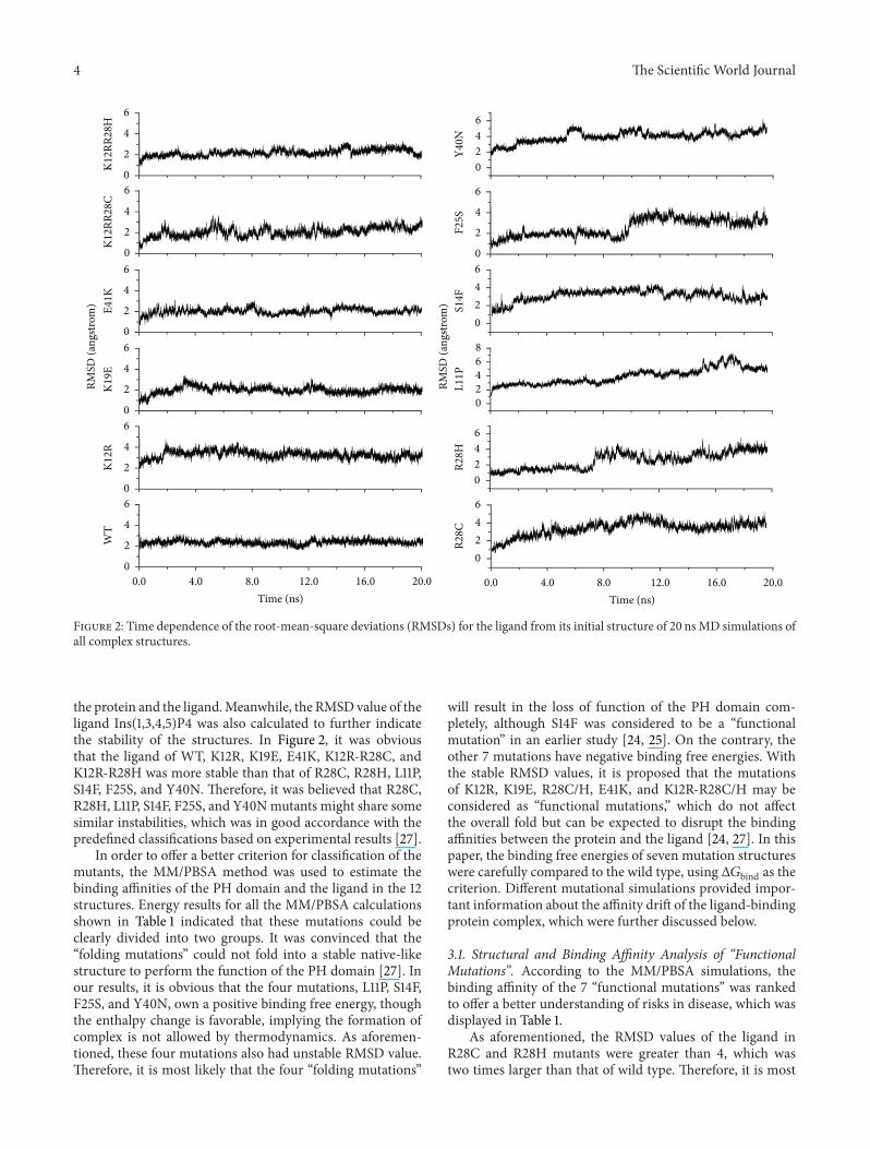
MD simulations were used to investigate the complexes of 11mutants andwild type PH domain and the interaction behav-ior between the PH domain and the ligand Ins(1,3,4,5)P4.Details of the simulation setup are provided in Materials andMethods.The root-mean-square deviation (RMSD) values ofthe C𝛼 atoms relative to the initial coordinates were calcu-lated using the 20 ns trajectory data. As shown in Figure 1, theRMSD tended to be flat after 5 ns’ simulation in WT, K12R,K19E, E41K, K12R-R28C, K12R-R28H, R28C, and R28H atabout 2 A, indicating stable conformations. As for the RMSDvalues for other four mutants, relative large fluctuations wereobserved during the 20 ns’ simulation, suggesting unsta-ble structures and maybe poor binding affinities between
4 The Scientific World Journal
K12R
R28H
K12R
R28C
E41K
K19E
K12R
WT
Y40N
F25S
S14F
L11P
R28H
R28C
RMSD
(ang
strom
)
RMSD
(ang
strom
)
0.0 4.0 8.0 12.0 16.0 20.0Time (ns)
0.0 4.0 8.0 12.0 16.0 20.0Time (ns)
6420
6420
68
420
6420
6
4
2
0
6420
6
4
2
06
4
2
0
6
4
2
0
6
4
2
0
6
4
2
0
6
4
2
0
Figure 2: Time dependence of the root-mean-square deviations (RMSDs) for the ligand from its initial structure of 20 ns MD simulations ofall complex structures.
the protein and the ligand.Meanwhile, the RMSDvalue of theligand Ins(1,3,4,5)P4 was also calculated to further indicatethe stability of the structures. In Figure 2, it was obviousthat the ligand of WT, K12R, K19E, E41K, K12R-R28C, andK12R-R28H was more stable than that of R28C, R28H, L11P,S14F, F25S, and Y40N. Therefore, it was believed that R28C,R28H, L11P, S14F, F25S, and Y40Nmutants might share somesimilar instabilities, which was in good accordance with thepredefined classifications based on experimental results [27].
In order to offer a better criterion for classification of themutants, the MM/PBSA method was used to estimate thebinding affinities of the PH domain and the ligand in the 12structures. Energy results for all the MM/PBSA calculationsshown in Table 1 indicated that these mutations could beclearly divided into two groups. It was convinced that the“folding mutations” could not fold into a stable native-likestructure to perform the function of the PH domain [27]. Inour results, it is obvious that the four mutations, L11P, S14F,F25S, and Y40N, own a positive binding free energy, thoughthe enthalpy change is favorable, implying the formation ofcomplex is not allowed by thermodynamics. As aforemen-tioned, these four mutations also had unstable RMSD value.Therefore, it is most likely that the four “folding mutations”
will result in the loss of function of the PH domain com-pletely, although S14F was considered to be a “functionalmutation” in an earlier study [24, 25]. On the contrary, theother 7 mutations have negative binding free energies. Withthe stable RMSD values, it is proposed that the mutationsof K12R, K19E, R28C/H, E41K, and K12R-R28C/H may beconsidered as “functional mutations,” which do not affectthe overall fold but can be expected to disrupt the bindingaffinities between the protein and the ligand [24, 27]. In thispaper, the binding free energies of seven mutation structureswere carefully compared to the wild type, using Δ𝐺bind as thecriterion. Different mutational simulations provided impor-tant information about the affinity drift of the ligand-bindingprotein complex, which were further discussed below.
3.1. Structural and Binding Affinity Analysis of “FunctionalMutations”. According to the MM/PBSA simulations, thebinding affinity of the 7 “functional mutations” was rankedto offer a better understanding of risks in disease, which wasdisplayed in Table 1.
As aforementioned, the RMSD values of the ligand inR28C and R28H mutants were greater than 4, which wastwo times larger than that of wild type. Therefore, it is most

The Scientific World Journal 5
Table 1: Ranked calculated binding free energies (kcal/mol) using MM/PBSA for the twelve protein-ligand systems.
ELE VDW PB SA PBTOT 𝑇Δ𝑆 Δ𝐺bind
WT −3031.77(9.01a)
−0.16(0.07)
2961.60(7.63)
−4.28(0.47)
−74.61(5.10)
−45.29(1.43)
−29.32(1.49)
K12R-R28C −3119.66(10.33)
−0.12(0.05)
3044.04(8.61)
−4.25(0.18)
−79.99(3.37)
−52.50(1.45)
−27.49(1.06)
K19E −3143.08(8.92)
−0.17(0.03)
3069.08(7.38)
−4.21(0.16)
−78.38(5.33)
−51.09(1.60)
−27.29(0.73)
K12R-R28H −3029.46(8.74)
−0.05(0.02)
2962.73(6.43)
−4.12(0.21)
−73.30(2.02)
−51.66(1.37)
−21.64(0.66)
E41K −2855.47(7.73)
−0.28(0.16)
2789.39(5.95)
−4.16(0.23)
−70.52(3.12)
−50.08(1.52)
−20.44(0.38)
K12R −2922.34(9.17)
−0.08(0.03)
2859.80(8.38)
−4.30(0.21)
−66.92(3.62)
−52.84(1.57)
−14.08(0.94)
R28C −2612.17(5.95)
−1.90(0.23)
2552.82(5.03)
−3.69(0.16)
−64.94(1.87)
−57.28(2.47)
−7.66(0.03)
R28H −2591.08(6.16)
−1.61(0.17)
2533.38(4.65)
−3.70(0.17)
−63.01(1.16)
−56.80(2.54)
−6.21(0.02)
S14F −2573.79(7.06)
−3.37(0.74)
2545.38(6.99)
−3.42(0.25)
−35.20(2.47)
−57.49(3.02)
22.29(0.98)
L11P −2599.24(7.61)
−1.75(0.41)
2578.10(6.70)
−3.94(0.33)
−26.83(1.42)
−58.75(4.47)
31.92(2.02)
Y40N −2550.35(7.0)
−2.57(0.59)
2531.48(6.23)
−3.92(0.27)
−25.36(1.84)
−58.34(3.89)
32.98(1.68)
F25S −2501.74(7.17)
−2.59(0.69)
2487.39(6.64)
−3.77(0.19)
−20.71(1.54)
−58.32(4.15)
37.61(1.82)
aThe statistical error was estimated on the basis of the deviation between block averages.ELE: electrostatic energy; VDW: van der Waals energy; PB: the PB solvation energy; SA: the surface area energy (nonpolar solvation energy); PBTOT: sum ofELE, VDW, PB, and SA.The absolute temperature (𝑇) was set to 300K in the MM-PBSA calculations. Δ𝐺bind: subtracting 𝑇Δ𝑆 from PBTOT.
likely that the ligand completelymoves away from the originalbinding pocket of the PHdomain and thiswas consistentwiththe result of the binding free energy calculation for R28C/Hmutants (Table 1). It is clear that the neutral charged cysteinehas a sulfur atom in its side chain, which imposes a largersteric restriction compared with arginine. Although it has thesame positive charge as arginine, histidine has a five-memberring that produces more steric hindrance than the side chainof arginine. Structural analysis in Figure 3 verified that theligand was already out of the binding pocket. The mutationsremoved the hydrogen bond interaction between R28 andthe ligand while forming new hydrogen bond interactionswith residues on the 𝛽4 strand (K53) and 𝛽1-𝛽2 loop (K17),respectively, which would not provide suitable side chains tobind with the ligand. Accordingly, it was considered that theR28C/H mutations had greatly reduced the binding affinitywith the ligand. And the biological function of the twomutants was severely weakened, which may be considered asa primary factor that leads to associated diseases such as XLAand Xid [5].
As shown in Table 1, according to the MM/PBSA simu-lations, the free binding free energies of the mutants K19E,E41K, and K12R-R28C/H were similar to the wild type,whereas the K12R mutant was relatively smaller (−14.08 kcal/mol). Furthermore, compared with the wild type, the ligandwas still located in the binding pocket of these mutants, asshown in Figures 3(d)–3(h), although there were different
ligand-conformational changes due to hydrogen bond rup-ture. The time evolution of the root-mean-square fluctuation(RMSF) from the initial structurewas also analyzed to furtherevaluate the ligand binding behavior of these mutations.
Structural analysis of the wild type showed that K12,which was located at the bottom of the binding pocket,formed hydrogen bonds with the 3- and 4-phosphates of theligand, which is critical for the twist of 𝛽1-𝛽2 loops [24]. Inthe K12R mutant, there was obvious decrease in binding freeenergy in the MM/PBSA simulation (Table 1), which couldbe explained by the difference in the conformation and thechange in the hydrogen bond interactions of the ligand.Sharing the same positive change as lysine, the three atomsin arginine tail formed a conjugated system, which decreasedthe flexibility and increased the steric hindrance of thisresidue, leading to the conformational change required forligand binding. The hydrogen bond with the 3-phosphatesof the ligand was also fully ruptured in K12R mutant,resulting in a loss of specificity in the Btk-PH domain for theIns(1,3,4,5)P4 ligand. As shown in Table 2, the K12R mutantabolished the hydrogen bond network formed by K12, R28,and the ligand; thereby the contribution of the conservedcovariation pair on the biological function of the Btk-PHdomain was removed.
In the wild type, as shown in Figure 3(a), K19 contactsD148 via a hydrogen bond, which moves the 𝛽1-𝛽2 loopcloser to the tail of the structure and moves it away from the

6 The Scientific World Journal
(a) (b)
(c) (d)
(e) (f)
(g) (h)
Figure 3: The snapshot structures taken at time 20 ns of wild type and all “functional mutations” complexes from MD simulation. Ligandsand surrounding important residues are labeled and shown in stick, while hydrogen bonds are displayed in yellow dashed lines. (a) Wildtype complex; (b) R28C mutation complex; (c) R28H mutation complex; (d) K12R mutation complex; (e) K19E mutation complex; (f) E41Kmutation complex; (g) K12R-R28C mutation complex; (h) K12R-R28H mutation complex.
The Scientific World Journal 7
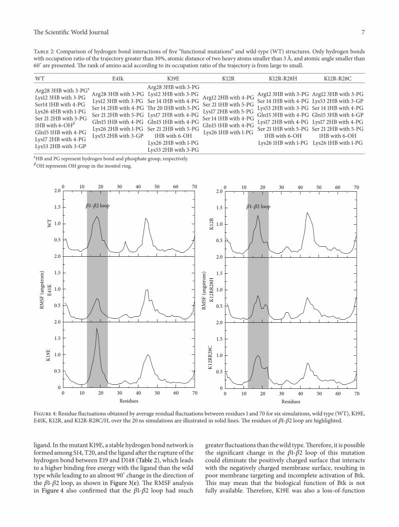
Table 2: Comparison of hydrogen bond interactions of five “functional mutations” and wild-type (WT) structures. Only hydrogen bondswith occupation ratio of the trajectory greater than 30%, atomic distance of two heavy atoms smaller than 3 A, and atomic angle smaller than60∘ are presented. The rank of amino acid according to its occupation ratio of the trajectory is from large to small.
WT E41k K19E K12R K12R-R28H K12R-R28C
Arg28 3HB with 3-PGa
Lys12 3HB with 3-PGSer14 1HB with 4-PGLys26 4HB with 1-PGSer 21 2HB with 5-PG1HB with 6-OH𝛽Gln15 1HB with 4-PGLys17 2HB with 4-PGLys53 2HB with 3-GP
Arg28 3HB with 3-PGLys12 3HB with 3-PGSer 14 2HB with 4-PGSer 21 2HB with 5-PGGln15 1HB with 4-PGLys26 2HB with 1-PGLys53 2HB with 3-GP
Arg28 3HB with 3-PGLys12 3HB with 3-PGSer 14 1HB with 4-PGThr 20 1HB with 5-PGLys17 2HB with 4-PGGln15 1HB with 4-PGSer 21 2HB with 5-PG
1HB with 6-OHLys26 2HB with 1-PGLys53 2HB with 3-PG
Arg12 2HB with 4-PGSer 21 1HB with 5-PGLys17 2HB with 5-PGSer 14 1HB with 4-PGGln15 1HB with 4-PGLys26 1HB with 1-PG
Arg12 3HB with 3-PGSer 14 1HB with 4-PGLys53 2HB with 3-PGGln15 3HB with 4-PGLys17 2HB with 4-PGSer 21 1HB with 5-PG
1HB with 6-OHLys26 1HB with 1-PG
Arg12 3HB with 3-PGLys53 2HB with 3-GPSer 14 1HB with 4-PGGln15 3HB with 4-GPLys17 2HB with 4-PGSer 21 2HB with 5-PG
1HB with 6-OHLys26 1HB with 1-PG
aHB and PG represent hydrogen bond and phosphate group, respectively.𝛽OH represents OH group in the inositol ring.
2.0
1.5
1.0
0.5
2.0
1.5
1.0
0.5
2.0
1.5
1.0
0.5
0
0 10 20 30 40 50 60 70
0 10 20 30 40 50 60 70Residues
RMSF
(ang
strom
)W
TE4
1KK1
9E
2.0
1.5
1.0
0.5
2.0
1.5
1.0
0.5
2.0
1.5
1.0
0.5
0
0 10 20 30 40 50 60 70
0 10 20 30 40 50 60 70Residues
RMSF
(ang
strom
)K1
2RK1
2RR2
8HK1
2RR2
8C𝛽1-𝛽2 loop 𝛽1-𝛽2 loop
Figure 4: Residue fluctuations obtained by average residual fluctuations between residues 1 and 70 for six simulations, wild type (WT), K19E,E41K, K12R, and K12R-R28C/H, over the 20 ns simulations are illustrated in solid lines. The residues of 𝛽1-𝛽2 loop are highlighted.
ligand. In themutantK19E, a stable hydrogen bondnetwork isformed among S14, T20, and the ligand after the rupture of thehydrogen bond between E19 and D148 (Table 2), which leadsto a higher binding free energy with the ligand than the wildtype while leading to an almost 90∘ change in the direction ofthe 𝛽1-𝛽2 loop, as shown in Figure 3(e). The RMSF analysisin Figure 4 also confirmed that the 𝛽1-𝛽2 loop had much
greater fluctuations than thewild type.Therefore, it is possiblethe significant change in the 𝛽1-𝛽2 loop of this mutationcould eliminate the positively charged surface that interactswith the negatively charged membrane surface, resulting inpoor membrane targeting and incomplete activation of Btk.This may mean that the biological function of Btk is notfully available. Therefore, K19E was also a loss-of-function

8 The Scientific World Journal
mutation, although it had a similar binding affinity with theligand than the wild type, which was in good agreement withthe experimental results [24, 27].
Compared with K19E, the mutant of E41K has theopposite effect on the biological function of Btk. As shownin Figure 3(a), E41 was located in the 𝛽3 strand with somedistance from the ligand, but it is known to have greatimportance in the contact with the membrane. Despite thehigher steric hindrance after the substitution of glutamatewith lysine, the ligand slightly shifts to the 𝛽1-𝛽2 loop inFigure 3(f), which results in a reduced flexibility of the 𝛽1-𝛽2loop (RMSF in Figure 4). The binding free energy of theE41K mutant was also smaller than the wild type, as shownin Table 1. Thus, E41K may retain most of the biologicalfunctionality of the wild type. However, the mutation inE41K increased the positively charged complex surfaceof the region located above the 𝛽3 and 𝛽4 strand, whichwas favorable for interaction with the negatively chargedmembrane surface, while it also offered a second ligandbinding site. Therefore, it is likely that the E41K mutant mayincrease the activation of Btk, which is known as a gain offunction [24, 27].
3.2. Covariant Pair Identification in K12R-R28C/HMutations.Of the disease-causing mutations, the mutations in K12 andR28 always coexist with othermutations, althoughwith lowerfrequencies, which may be due to the diversity of the PHdomain. Single mutations of K12 and R28 have been knownto weaken the biological function of the Btk-PH domain todifferent degrees, while it is also reported that K12 and R28can form a conserved covariant pair that maintains the bind-ing affinity of Ins(1,3,4,5)P4, which plays an important role inthe biological function of the Btk-PHdomain [26].Therefore,the double residue mutations of K12R-R28C/H were closelyanalyzed to underly the molecular basis. Compared with thewild type structure, the ligand was still located in the bottomof the binding pocket of the two mutations (Figures 3(g)and 3(h)). The MM/PBSA simulations indicated a similarfree binding energy forK12R-R28HandK12R-R28C (Table 1).This could be explained by the formation of a new covariantpair between R12 and K53 as K12-R28. Unlike the K12Rmutation, the hydrogen bond network of 3-phosphates waskept through R12 and K53 in the mutation of K12R-R28C/H,which ensured the preservation of ligand specificity, whereasall hydrogen bond interactions between H28/C28 and lig-and were ruptured (Table 2). Interestingly, a new conservedhydrogen bond was discovered between K53 and the ligandduring theMD simulations. Instead of the covariant pair K12-R28, we believe that R12-K53 formed a similar odd positivelycharged pair that made contact with a bound negativelycharged inositol phosphate and thereby can maintain thebiological function of the K12R-R28C/H mutants.
4. Conclusions
Twelve complex systems, including eleven mutants and onewild type PH domain, have been comprehensively studiedby integrating MD and MM/PBSA simulations, to revealthe detailed molecular basis. As a result, L11P, S14F, F25S, and
Y40N were considered to be “folding mutations,” whereasmutants K12R, K19E, R28C/H, E41K, and K12R-R28C/Hwere classified into “functional mutations.” The effects of“functional mutations” on the biological function of theBtk-PH domain have also been predicted. In the R28C/Hmutation, the ligands completely left the binding pocket andformed new interactions with the 𝛽1-𝛽2 loop and the 𝛽4strand, respectively. For the K12R mutation, the biologicalfunction of the Btk-PH domain may be reduced by the lackof hydrogen bond interaction with 3-phosphates when theligand is formed, which determines the specificity of the Btk-PH domain. As for the K19E mutation, the change in theresidue charge and the trend of the 𝛽1-𝛽2 loop eliminated thepositively charged surface that interacted with the negativelycharged membrane surface, resulting in poor membranetargeting and incomplete activation of Btk. In contrast, E41Kincreased the positive surface of the Btk-PH domain, therebyincreasing the possibility of the binding with a second ligand,which may result in a gain of function. Of note, the K12R-R28C/H mutant retained a high binding affinity without thedisplacement of the ligand from the binding pocket. A newodd positively charged pair was formed between R12 andK53 in the K12R-R28C/H mutation, which was similar to theK12-R28 in the wild type. This formation may interact witha bound negatively charged inositol phosphate, which maypreserve the biological function of the Btk-PH domain andthis should be further investigated experimentally.
In summary, our MD and MM/PBSA simulations of thetwelve structures may provide some new insights into PHdomain and their associated diseases. Based on the bindingfree energy, binding site of the ligand, and hydrogen bondinteractions, we have predicted the effects ofmutations on thebiological function of the Btk-PH domain.
Authors’ Contribution
Dan Lu and Junfeng Jiang contributed equally to this work.
Acknowledgments
The authors gratefully acknowledge financial support fromthe National Natural Science Foundation of China Grants(21203131, 31170795, 20872107, 20972174, and 91029704), the863 Program (SS2012AA021103), and the Chinese Academyof Sciences (XDA01040305).
References
[1] J. Gomez-Rodriguez, J. A. Readinger, I. C. Viorritto, K. L. Muel-ler, R. A. Houghtling, and P. L. Schwartzberg, “Tec kinases,actin, and cell adhesion,” Immunological Reviews, vol. 218, no.1, pp. 45–64, 2007.
[2] C. I. Smith, T.C. Islam, P. T.Mattsson,A. J.Mohamed, B. F.Nore,and M. Vihinen, “The Tec family of cytoplasmic tyrosine kina-ses: mammalian Btk, Bmx, Itk, Tec, Txk and homologs in otherspecies,” Bioessays, vol. 23, no. 5, pp. 436–446, 2001.
[3] A. Maas and R. W. Hendriks, “Role of Bruton’s tyrosine kinasein B cell development,” Developmental Immunology, vol. 8, no.3-4, pp. 171–181, 2001.

The Scientific World Journal 9
[4] G. Matthew and G. K. William, “Role of inositol phospholipidsignaling in natural killer cell biology,” Front Immunology, vol.4, article 47, 2013.
[5] A. J. Mohamed, L. Yu, C. M. Backesjo et al., “Bruton’s tyrosinekinase (Btk): function, regulation, and transformation withspecial emphasis on the PH domain,” Immunological Reviews,vol. 228, no. 1, pp. 58–73, 2009.
[6] T. S. Rans and R. England, “The evolution of gene therapy in X-linked severe combined immunodeficiency,” Annals of Allergy,Asthma and Immunology, vol. 102, no. 5, pp. 357–363, 2009.
[7] J. D. Thomas, P. Sideras, C. I. Smith, I. Vorechovsky, V. Chap-man, andW. E. Paul, “Colocalization of X-linked agammaglob-ulinemia and X-linked immunodeficiency genes,” Science, vol.261, no. 5119, pp. 355–358, 1993.
[8] M. Vihinen, B. H. Belohradsky, R. N. Haire et al., “BTKbase,mutation database for X-linked agammaglobulinemia (XLA),”Nucleic Acids Research, vol. 25, no. 9, pp. 166–171, 1997.
[9] T. Kaneko, R. Joshi, S. M. Feller, and S. S. Li, “Phosphotyrosinerecognition domains: the typical, the atypical and the versatile,”Cell Communication and Signaling, vol. 10, no. 1, p. 32, 2012.
[10] G. Koytiger, A. Kaushansky, A. Gordus, J. Rush, P. K. Sorger, andG. Macbeath, “Phosphotyrosine signaling proteins that driveoncogenesis tend to be highly interconnected,” Molecular &Cellular Proteomics, vol. 12, no. 5, pp. 1204–1213, 2013.
[11] K. Chudasama, J. Winnay, S. Johansson et al., “Short syndromewith partial lipodystrophy due to impaired phosphatidylinositol3 kinase signaling,” The American Journal of Human Genetics,vol. 93, no. 1, pp. 150–157, 2013.
[12] M. Vihinen, M. D. Cooper, G. de Saint Basile et al., “BTKbase:a database of XLA-causing mutations,” Immunology Today, vol.16, no. 10, pp. 460–465, 1995.
[13] T. G. Kutateladze, “Molecular analysis of protein—phosphoi-nositide interactions,” Current Topics in Microbiology andImmunology, vol. 362, pp. 111–126, 2012.
[14] P. Mayinger, “Phosphoinositides and vesicular membrane traf-fic,” Biochimica et Biophysica Acta, vol. 1821, no. 8, pp. 1104–1113,2012.
[15] L. E. Rameh, A. Arvidsson, K. L. Carraway et al., “A comparativeanalysis of the phosphoinositide binding specificity of pleck-strin homology domains,” Journal of Biological Chemistry, vol.272, no. 35, pp. 22059–22066, 1997.
[16] A.M. Scharenberg, O. El-Hillal, D. A. Fruman et al., “Phosphat-idylinositol-3,4,5-trisphosphate (Ptdlns-3,4,5-P3)/Tec kinase-dependent calcium signaling pathway: a target for SHIP-medi-ated inhibitory signals,” The EMBO Journal, vol. 17, no. 7, pp.1961–1972, 1998.
[17] K. Saito, A. M. Scharenberg, and J.-P. Kinet, “Interaction bet-ween the Btk PH Domain and Phosphatidylinositol-3,4,5-trisphosphate Directly Regulates Btk,” Journal of Biological Che-mistry, vol. 276, no. 19, pp. 16201–16206, 2001.
[18] K. Murayama, M. Kato-Murayama, C. Mishima et al., “Crystalstructure of the Bruton’s tyrosine kinase PH domain withphosphatidylinositol,” Biochemical and Biophysical ResearchCommunications, vol. 377, no. 1, pp. 23–28, 2008.
[19] T. J. Gibson, M. Hyvonen, A.Musacchio, M. Saraste, and E. Bir-ney, “PH domain: the first anniversary,” Trends in BiochemicalSciences, vol. 19, no. 9, pp. 349–353, 1994.
[20] C.C.Milburn,M.Deak, S.M.Kelly,N.C. Price,D. R.Alessi, andD. M. F. van Aalten, “Binding of phosphatidylinositol 3,4,5-trisphosphate to the pleckstrin homology domain of proteinkinase B induces a conformational change,” Biochemical Jour-nal, vol. 375, no. 3, pp. 531–538, 2003.
[21] T. F. Jenny and S. A. Benner, “A prediction of the secondarystructure of the pleckstrin homology domain,” Proteins, vol. 20,no. 1, pp. 1–3, 1994.
[22] B. J. Mayer, R. Ren, K. L. Clark, and D. Baltimore, “A putativemodular domain present in diverse signaling proteins,”Cell, vol.73, no. 4, pp. 629–630, 1993.
[23] H. S. Yoon, P. J. Hajduk, A. M. Petros, E. T. Olejniczak, R. P.Meadows, and S. W. Fesik, “Solution structure of a pleckstrin-homology domain,” Nature, vol. 369, no. 6482, pp. 672–675,1994.
[24] E. Baraldi, K.D.Carugo,M.Hyvonen et al., “Structure of the PHdomain from Bruton’s tyrosine kinase in complex with inositol1,3,4,5-tetrakisphosphate,” Structure, vol. 7, no. 4, pp. 449–460,1999.
[25] T. Kojima,M. Fukuda, Y.Watanabe, F.Hamazato, andK.Mikos-hiba, “Characterization of the pleckstrin homology domain ofBtk as an inositol polyphosphate and phosphoinositide bindingdomain,” Biochemical and Biophysical Research Communica-tions, vol. 236, no. 2, pp. 333–339, 1997.
[26] B. Shen and M. Vihinen, “Conservation and covariance in PHdomain sequences: physicochemical profile and informationtheoretical analysis of XLA-causing mutations in the Btk PHdomain,” Protein Engineering, Design and Selection, vol. 17, no.3, pp. 267–276, 2004.
[27] M. Hyvonen and M. Saraste, “Stucture of the PH domain andBtkmotif fromBruton’s tyrosine kinase:molecular explanationsfor X-linked agammaglobulinaemia,” The EMBO Journal, vol.16, no. 12, pp. 3396–3404, 1997.
[28] S. Fujiwara and T. Amisaki, “Identification of high affinity fattyacid binding sites on human serum albumin by MM-PBSAmethod,” Biophysical Journal, vol. 94, no. 1, pp. 95–103, 2008.
[29] T. Hou and R. Yu, “Molecular dynamics and free energy studieson the wild-type and double mutant HIV-1 protease complexedwith amprenavir and two amprenavir-related inhibitors: mech-anism for binding and drug resistance,” Journal of MedicinalChemistry, vol. 50, no. 6, pp. 1177–1188, 2007.
[30] P. A. Kollman, I. Massova, C. Reyes et al., “Calculatingstructures and free energies of complex molecules: combiningmolecular mechanics and continuum models,” Accounts ofChemical Research, vol. 33, no. 12, pp. 889–897, 2000.
[31] J. Wang, P. Morin, W. Wang, and P. A. Kollman, “Use of MM-PBSA in reproducing the binding free energies to HIV-1 RTof TIBO derivatives and predicting the binding mode to HIV-1 RT of efavirenz by docking and MM-PBSA,” Journal of theAmerican Chemical Society, vol. 123, no. 22, pp. 5221–5230, 2001.
[32] T.Hou, J.Wang, Y. Li, andW.Wang, “Assessing the performanceof the MM/PBSA and MM/GBSA methods. 1. The accuracy ofbinding free energy calculations based on molecular dynamicssimulations,” Journal of Chemical Information and Modeling,vol. 51, no. 1, pp. 69–82, 2011.
[33] C. I. Bayly, P. Cieplak, W. Cornell, and P. A. Kollman, “Awell-behaved electrostatic potential based method using chargerestraints for deriving atomic charges: the RESPmodel,” Journalof Physical Chemistry, vol. 97, no. 40, pp. 10269–10280, 1993.
[34] J. Wang, W. Wang, P. A. Kollman, and D. A. Case, “Automaticatom type and bond type perception in molecular mechanicalcalculations,” Journal of Molecular Graphics and Modelling, vol.25, no. 2, pp. 247–260, 2006.
[35] Y. Duan, C. Wu, S. Chowdhury et al., “A point-charge forcefield for molecular mechanics simulations of proteins based oncondensed-phase quantummechanical calculations,” Journal ofComputational Chemistry, vol. 24, no. 16, pp. 1999–2012, 2003.

10 The Scientific World Journal
[36] J. Wang, R. M. Wolf, J. W. Caldwell, P. A. Kollman, and D. A.Case, “Development and testing of a general Amber force field,”Journal of Computational Chemistry, vol. 25, no. 9, pp. 1157–1174,2004.
[37] T. Darden, L. Perera, L. Li, and L. Pedersen, “New tricks formodelers from the crystallography toolkit: the particle meshEwald algorithm and its use in nucleic acid simulations,”Structure, vol. 7, no. 3, pp. R55–R60, 1999.


![¨2¤!/P4(. !,]« - Kurtzman Carson Consultants LLC](https://static.fdokumen.com/doc/165x107/631e562b25add517740afee9/2p4-kurtzman-carson-consultants-llc.jpg)