Molecular characterization of strains isolated from Portuguese freshwaters
9
Molecular characterization of Cylindrospermopsis raciborskii strains isolated from Portuguese freshwaters E. Vale ´rio a , P. Pereira b , M.L. Saker c , S. Franca b , R. Tenreiro a, * a Universidade de Lisboa, Faculdade de Cie ˆncias, Centro de Gene ´tica e Biologia Molecular, and Instituto de Cie ˆncia Aplicada e Tecnologia, Edifı ´cio ICAT, Campus da FCUL, Campo Grande, 1749-016 Lisboa, Portugal b Instituto Nacional de Sau ´de Dr. Ricardo Jorge, Av. Padre Cruz, 1649-016 Lisboa, Portugal c Centro de Investigac ¸a ˜o Marinha e Ambiental, Rua dos Bragas 289, 4050-123 Porto, Portugal Received 5 August 2004; received in revised form 28 February 2005; accepted 10 March 2005 Abstract Cylindrospermopsis raciborskii is a toxic bloom forming cyanobacteria that is a common component of the phytoplankton assemblage in temperate freshwaters, as well as in temperate climates. This species is of major concern in public health, due to its known ability to produce toxins, including cylindrospermopsin and paralytic shellfish poisoning toxin (PSP). In this study, M13 PCR fingerprinting, ERIC PCR fingerprinting and amplification of the internal transcribed spacer (ITS) region were used to characterize nine cultured strains of C. raciborskii, sourced from several freshwater lakes and rivers in Portugal, and two other Australian. Strains belonging to other taxa including Microcystis aeruginosa, Aphanizomenon spp., Planktothrix agardhii and Oscillatoria neglecta were also analysed to evaluate the taxonomical potential of the fingerprinting methods. Data obtained from genomic fingerprinting were used to perform hierarchical cluster analysis and demonstrated ability to differentiate strains at intra-specific level. However, the high level of variability prevents their use as an identification tool. ITS amplification displayed intra-specific polymorphism both in number and length of the obtained amplicons, but revealed itself as a good method for strain clustering. The unsuccessful amplification of peptide synthetase (PS) and polyketide synthase (PKS) genes pointed to the inability of Portuguese C. raciborskii strains to produce cylindrospermopsin. HPLC analysis further confirmed this lack of toxicity, since negative results were obtained for cylindrospermopsin and PSP toxins. # 2005 Elsevier B.V. All rights reserved. Keywords: Cylindrospermopsis raciborskii; ERIC PCR fingerprinting; ITS; M13 PCR fingerprinting; PS and PKS genes; PSP toxins 1. Introduction Cylindrospermopsis raciborskii is a well-known toxic bloom-forming cyanobacteria, originally described as a species of tropical origin (Woloszynska, 1912), but increasingly found across a wide range of www.elsevier.com/locate/hal Harmful Algae 4 (2005) 1044–1052 * Corresponding author. Tel.: +351 21 750 00 06; fax: +351 21 750 01 72. E-mail address: [email protected] (R. Tenreiro). 1568-9883/$ – see front matter # 2005 Elsevier B.V. All rights reserved. doi:10.1016/j.hal.2005.03.002
-
Upload
independent -
Category
Documents
-
view
0 -
download
0
Transcript of Molecular characterization of strains isolated from Portuguese freshwaters
Molecular characterization of Cylindrospermopsis raciborskii
strains isolated from Portuguese freshwaters
E. Valerio a, P. Pereira b, M.L. Saker c, S. Franca b, R. Tenreiro a,*
a Universidade de Lisboa, Faculdade de Ciencias, Centro de Genetica e Biologia Molecular, and
Instituto de Ciencia Aplicada e Tecnologia, Edifıcio ICAT, Campus da FCUL, Campo Grande, 1749-016 Lisboa, Portugalb Instituto Nacional de Saude Dr. Ricardo Jorge, Av. Padre Cruz, 1649-016 Lisboa, Portugal
c Centro de Investigacao Marinha e Ambiental, Rua dos Bragas 289, 4050-123 Porto, Portugal
Received 5 August 2004; received in revised form 28 February 2005; accepted 10 March 2005
Abstract
Cylindrospermopsis raciborskii is a toxic bloom forming cyanobacteria that is a common component of the phytoplankton
assemblage in temperate freshwaters, as well as in temperate climates. This species is of major concern in public health, due to
its known ability to produce toxins, including cylindrospermopsin and paralytic shellfish poisoning toxin (PSP).
In this study, M13 PCR fingerprinting, ERIC PCR fingerprinting and amplification of the internal transcribed spacer (ITS)
region were used to characterize nine cultured strains of C. raciborskii, sourced from several freshwater lakes and rivers in
Portugal, and two other Australian. Strains belonging to other taxa including Microcystis aeruginosa, Aphanizomenon spp.,
Planktothrix agardhii and Oscillatoria neglecta were also analysed to evaluate the taxonomical potential of the fingerprinting
methods.
Data obtained from genomic fingerprinting were used to perform hierarchical cluster analysis and demonstrated ability to
differentiate strains at intra-specific level. However, the high level of variability prevents their use as an identification tool. ITS
amplification displayed intra-specific polymorphism both in number and length of the obtained amplicons, but revealed itself as
a good method for strain clustering. The unsuccessful amplification of peptide synthetase (PS) and polyketide synthase (PKS)
genes pointed to the inability of Portuguese C. raciborskii strains to produce cylindrospermopsin. HPLC analysis further
confirmed this lack of toxicity, since negative results were obtained for cylindrospermopsin and PSP toxins.
# 2005 Elsevier B.V. All rights reserved.
Keywords: Cylindrospermopsis raciborskii; ERIC PCR fingerprinting; ITS; M13 PCR fingerprinting; PS and PKS genes; PSP toxins
www.elsevier.com/locate/hal
Harmful Algae 4 (2005) 1044–1052
* Corresponding author. Tel.: +351 21 750 00 06;
fax: +351 21 750 01 72.
E-mail address: [email protected] (R. Tenreiro).
1568-9883/$ – see front matter # 2005 Elsevier B.V. All rights reserved
doi:10.1016/j.hal.2005.03.002
1. Introduction
Cylindrospermopsis raciborskii is a well-known
toxic bloom-forming cyanobacteria, originally
described as a species of tropical origin (Woloszynska,
1912), but increasingly found across a wide range of
.
E. Valerio et al. / Harmful Algae 4 (2005) 1044–1052 1045
latitudes. This species is of major concern from a water
quality and public health perspective, due to its known
ability to produce toxins, including the potent hepato-
toxic alkaloid cylindrospermopsin (Hawkins et al.,
1985; Li et al., 2001; Saker and Neilan, 2001; Fastner
et al., 2003), and the highly toxic paralytic shellfish
poisons (PSP) (Lagos et al., 1999). Suspected causation
of human sickness by cylindrospermopsin (Hayman,
1992) and cattle mortality associated with C. raciborskii
(Saker et al., 1999) has, so far, been restricted to
Australia. However, the recent reports of C. raciborskii
from many temperate countries have highlighted the
invasive nature of this species, spreading worldwide.
Saker et al. (2003) recently reported the occurrence
of C. raciborskii in several Portuguese water bodies
used for potable and recreational purposes. Although
tests for the presence of cylindrospermopsin and PSP
were negative, a number of strains grown in pure
culture showed atypical toxicity by mouse bioassay,
suggesting that a new type of toxin should be present.
While the chemical structure of the causative
compound(s) was not determined, this report drew
attention to the need for a comprehensive research on
the genetic variability of Portuguese C. raciborskii and
pointed to the need for an adequate monitoring of this
cyanobacterium in freshwater reservoirs.
So far, several molecular methods have been used
to characterize C. raciborskii, including the genetic
analysis of the 16S rRNA (Chonudomkul et al., 2004;
Saker and Neilan, 2001) and rpoC1 (Wilson et al.,
2000) genes, which display a considerable consensus
within the species. On the contrary, other genomic
targets such as Hip1 (Saker and Neilan, 2001; Smith
et al., 1998), STRR sequences (Fergusson et al., 2000;
Wilson et al., 2000), nifH (Dyble et al., 2002) and PC-
IGS (Baker et al., 2001; Dyble et al., 2002) showed
significant variations among different strains of C.
raciborskii.
The M13 PCR fingerprinting has never been used in
cyanobacteria, although its use in other bacteria (Grif
et al., 1998) and yeasts (Valerio et al., 2002) pointed to
its potential as an identification tool. On the other
hand, techniques based on highly repetitive sequences
such as enterobacterial repetitive intergenic consensus
(ERIC) have already been used for differentiation of
some cyanobacterial genera showing different profiles
for all the strains tested (Lyra et al., 2001). The
amplification of the internal transcribed spacers (ITS)
of rDNA operons from planktonic heterocystous
cyanobacteria (Iteman et al., 2002) also showed that
these regions displayed size variability.
In the search for molecular tools able to differentiate
the toxic from non-toxic strains of C. raciborskii, nine
cultured strains of this species, sourced from several
Portuguese freshwater lakes and rivers, were char-
acterized by using M13 and ERIC fingerprinting and
amplification of ITS region, techniques that have not
been applied yet to this particular species. The
Portuguese isolates were compared with two Australian
ones and strains belonging to other taxa, including
Microcystis aeruginosa, Aphanizomenon spp., Plank-
tothrix agardhii and Oscillatoria neglecta, were also
analysed in order to evaluate the taxonomical potential
of these molecular methods.
The ability of C. raciborskii to produce cylindros-
permopsin was evaluated by the amplification of the
polyketide synthase (PKS) and peptide synthetase
(PS) genes. Detection of cylindrospermopsin and PSP
toxins was performed by HPLC analysis.
2. Materials and methods
2.1. Strains
The strains used in this study are listed in Table 1,
including their code, origin and references to toxicity
assessment. Lyophilised samples of the isolates were
obtained as described by Saker et al. (2003). All
strains were identified morphologically at species
level. All C. raciborskii strains shared common
morphological traits and presented straight trichome
morphology, as described for this species.
2.2. DNA extraction
Genomic DNA of cyanobacterial strains was
extracted following the method described by Pitcher
et al. (1989), with some modifications. Briefly, an
aliquot of 100 ml of lyophilised culture was suspended
in a lysis buffer containing 50 mM Tris, 250 mM NaCl,
50 mM EDTA, 0.3% SDS, pH 8 and mechanically
broken with glass beads (400–600 mm) by vortex
shaking for 2 min. The suspension was incubated for
1 h at 65 8C. After new vortex shaking for 2 min,
1000 ml of GES (5 M guanidium thiocyanate, 100 mM
E. Valerio et al. / Harmful Algae 4 (2005) 1044–10521046
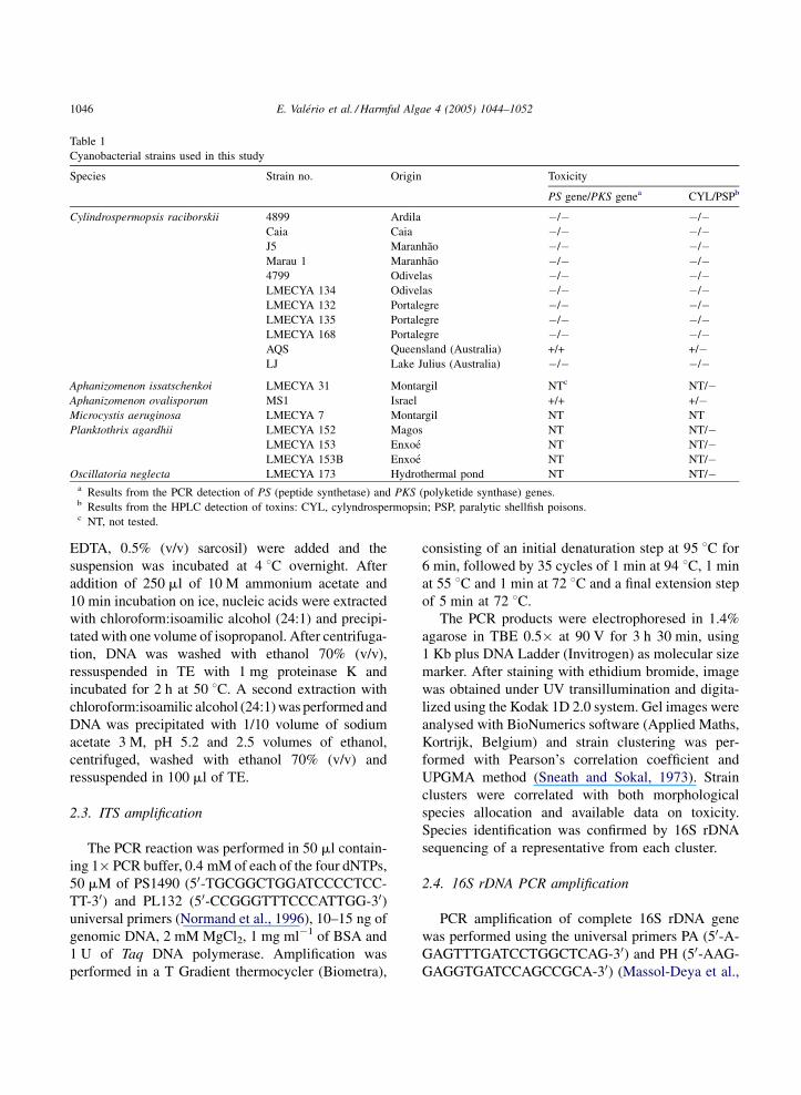
Table 1
Cyanobacterial strains used in this study
Species Strain no. Origin Toxicity
PS gene/PKS genea CYL/PSPb
Cylindrospermopsis raciborskii 4899 Ardila �/� �/�Caia Caia �/� �/�J5 Maranhao �/� �/�Marau 1 Maranhao �/� �/�4799 Odivelas �/� �/�LMECYA 134 Odivelas �/� �/�LMECYA 132 Portalegre �/� �/�LMECYA 135 Portalegre �/� �/�LMECYA 168 Portalegre �/� �/�AQS Queensland (Australia) +/+ +/�LJ Lake Julius (Australia) �/� �/�
Aphanizomenon issatschenkoi LMECYA 31 Montargil NTc NT/�Aphanizomenon ovalisporum MS1 Israel +/+ +/�Microcystis aeruginosa LMECYA 7 Montargil NT NT
Planktothrix agardhii LMECYA 152 Magos NT NT/�LMECYA 153 Enxoe NT NT/�LMECYA 153B Enxoe NT NT/�
Oscillatoria neglecta LMECYA 173 Hydrothermal pond NT NT/�a Results from the PCR detection of PS (peptide synthetase) and PKS (polyketide synthase) genes.b Results from the HPLC detection of toxins: CYL, cylyndrospermopsin; PSP, paralytic shellfish poisons.c NT, not tested.
EDTA, 0.5% (v/v) sarcosil) were added and the
suspension was incubated at 4 8C overnight. After
addition of 250 ml of 10 M ammonium acetate and
10 min incubation on ice, nucleic acids were extracted
with chloroform:isoamilic alcohol (24:1) and precipi-
tated with one volume of isopropanol. After centrifuga-
tion, DNA was washed with ethanol 70% (v/v),
ressuspended in TE with 1 mg proteinase K and
incubated for 2 h at 50 8C. A second extraction with
chloroform:isoamilic alcohol (24:1) was performed and
DNA was precipitated with 1/10 volume of sodium
acetate 3 M, pH 5.2 and 2.5 volumes of ethanol,
centrifuged, washed with ethanol 70% (v/v) and
ressuspended in 100 ml of TE.
2.3. ITS amplification
The PCR reaction was performed in 50 ml contain-
ing 1� PCR buffer, 0.4 mM of each of the four dNTPs,
50 mM of PS1490 (50-TGCGGCTGGATCCCCTCC-
TT-30) and PL132 (50-CCGGGTTTCCCATTGG-30)universal primers (Normand et al., 1996), 10–15 ng of
genomic DNA, 2 mM MgCl2, 1 mg ml�1 of BSA and
1 U of Taq DNA polymerase. Amplification was
performed in a T Gradient thermocycler (Biometra),
consisting of an initial denaturation step at 95 8C for
6 min, followed by 35 cycles of 1 min at 94 8C, 1 min
at 55 8C and 1 min at 72 8C and a final extension step
of 5 min at 72 8C.
The PCR products were electrophoresed in 1.4%
agarose in TBE 0.5� at 90 V for 3 h 30 min, using
1 Kb plus DNA Ladder (Invitrogen) as molecular size
marker. After staining with ethidium bromide, image
was obtained under UV transillumination and digita-
lized using the Kodak 1D 2.0 system. Gel images were
analysed with BioNumerics software (Applied Maths,
Kortrijk, Belgium) and strain clustering was per-
formed with Pearson’s correlation coefficient and
UPGMA method (Sneath and Sokal, 1973). Strain
clusters were correlated with both morphological
species allocation and available data on toxicity.
Species identification was confirmed by 16S rDNA
sequencing of a representative from each cluster.
2.4. 16S rDNA PCR amplification
PCR amplification of complete 16S rDNA gene
was performed using the universal primers PA (50-A-
GAGTTTGATCCTGGCTCAG-30) and PH (50-AAG-
GAGGTGATCCAGCCGCA-30) (Massol-Deya et al.,

E. Valerio et al. / Harmful Algae 4 (2005) 1044–1052 1047
1995). The PCR reaction was performed in 50 ml
containing 1� PCR buffer (Invitrogen), 0.2 mM of
each of the four dNTPs (Invitrogen), 25 mM of each
primer, 10–15 ng of genomic DNA, 2 mM MgCl2,
0.5 mg ml�1 of BSA and 1 U of Taq DNA polymerase
(Invitrogen). Amplification was performed in a T
Gradient thermocycler (Biometra), consisting of an
initial denaturation step at 94 8C for 5 min, followed
by 30 cycles of 1 min at 94 8C, 1 min at 55 8C and
1 min at 72 8C and a final extension step of 3 min at
72 8C. The amplification reaction products were
purified with a JETquick spin column (Genomed,
Germany) and sequenced on a Beckman-Coulter
automated DNA sequencer (model CEQ-2000) with
dye terminators, using standard protocols and the
cyanobacterial specific primers CYAN16S (50-ATA
CCC CWG TAG TCC TAG C-30) and CYAN16SR (50-GCA ATT ACT AGC GAT TCC TCC-30) designed in
this study. The first primer recognition site is located
in positions 738–765 and the other primer recognition
site is located in positions 1281–1302 of the 16S rRNA
operon of Synechocystis PCC 6803 (GenBank
accession no. NC000911). The confirmation of the
identification was performed by using the BLAST tool
of the Nacional Center for Biotechnology Information
(http://www.ncbi.nlm.nih.gov).
2.5. PCR fingerprinting
M13 PCR fingerprinting was performed using the
single primer M13 (50-GAGGGTGGCGGTTCT-30)(Meyer et al., 1993). For ERIC PCR fingerprinting both
ERICIR (50-ATGTAAGCTCCTGGGGATTCAC-30)and ERIC2 (50-AAGTAAGTGACTGGGGTGAGCG-
30) primers (Versalovic et al., 1991) were used.
The PCR reactions were performed in 50 ml
containing 1� PCR buffer (Invitrogen), 0.4 mM of
each of the four dNTPs (Invitrogen), 100 mM of each
primer, 10–15 ng of genomic DNA, 3 mM (for M13
fingerprinting) or 2 mM (for ERIC fingerprinting)
MgCl2, 1 mg ml�1 of BSA and 1 U of Taq DNA
polymerase (Invitrogen). Amplification was per-
formed in a T Gradient thermocycler (Biometra),
consisting of an initial denaturation step at 95 8C for
10 min, followed by 35 cycles of 90 s at 95 8C, 2 min
at 56 8C (for M13 fingerprinting) or 40 8C (for ERIC
fingerprinting) and 2 min at 72 8C and a final
extension step of 5 min at 72 8C.
The PCR products were electrophoresed in 1.4%
agarose gel in TBE 0.5� at 90 V for 3 h 30 min, with
1 Kb plus DNA Ladder as size marker. Gel images
were obtained as described above.
DNA banding patterns obtained with both finger-
printing methods were analysed using the BioNu-
merics software (Applied Maths, Kortrijk, Belgium).
Similarities among strains were estimated using
Pearson’s correlation coefficient and clustering was
based on the UPGMA method.
2.6. PS gene and PKS gene amplification
For the PS gene amplification, the PCR reaction
was performed in 50 ml containing 1� PCR buffer,
0.4 mM of each of the four dNTPs, 50 mM of PS M13
(50-GGCAAATTGTGATAGCCACGAGC-30) and PS
M14 (50-GATGGAACATCGCTCACTGGTG-30)primers (Schembri et al., 2001), 10–15 ng of genomic
DNA, 2.5 mM MgCl2, 1 mg ml�1 of BSA and 1 U of
Taq DNA polymerase. Amplification was performed
in a T Gradient thermocycler (Biometra), consisting
of an initial denaturation step at 95 8C for 6 min,
followed by 35 cycles of 1 min at 94 8C, 1 min at
55 8C and 1 min at 72 8C and a final extension step
of 5 min at 72 8C. For the PKS gene amplification,
the PCR reaction was performed as above, using
PKS M4 (50-GAAGCTCTGGAATCCGGTAA-30)and PKS M5 (50-AATCCTTACGGGATCCGGTGC-
30) primers (Schembri et al., 2001). The PCR products
were electrophoresed in 0.8% agarose in TBE 0.5� at
90 V for 2 h, with 1 Kb plus DNA Ladder as size
marker.
To confirm that the PCR amplicons corresponded
to the PS and PKS genes, the amplification reaction
products were purified with a JETquick spin column
(Genomed, Germany) and sequenced on a Beckman-
Coulter automated DNA sequencer (model CEQ-
2000) with dye terminators, using standard protocols
with the same primers used for the amplification.
Sequences were analysed with the BLAST tool of the
Nacional Center for Biotechnology Information
(http://www.ncbi.nlm.nih.gov).
2.7. Toxin analysis by HPLC
Analysis of C. raciborskii for the presence of
cylindrospermopsin was performed by reverse phase
E. Valerio et al. / Harmful Algae 4 (2005) 1044–10521048
HPLC and mass spectroscopy according to Eaglesham
et al. (1999).
Presence of PSP toxins was assessed by HPLC-
FLD according to the method of Oshima (1995).
3. Results
3.1. Molecular identification
The set of primers used to amplify the ITS regions
of the 11 Portuguese isolates of C. raciborskii strains
revealed polymorphisms both in number and length of
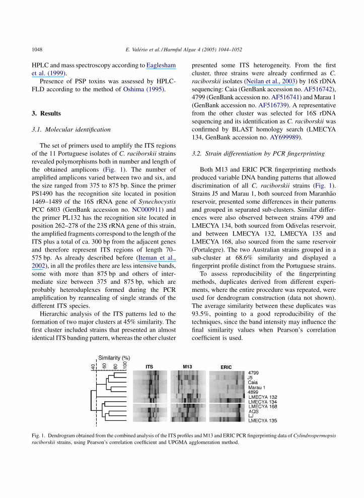
the obtained amplicons (Fig. 1). The number of
amplified amplicons varied between two and six, and
the size ranged from 375 to 875 bp. Since the primer
PS1490 has the recognition site located in position
1469–1489 of the 16S rRNA gene of Synechocystis
PCC 6803 (GenBank accession no. NC000911) and
the primer PL132 has the recognition site located in
position 262–278 of the 23S rRNA gene of this strain,
the amplified fragments correspond to the length of the
ITS plus a total of ca. 300 bp from the adjacent genes
and therefore represent ITS regions of length 70–
575 bp. As already described before (Iteman et al.,
2002), in all the profiles there are less intensive bands,
some with more than 875 bp and others of inter-
mediate size between 375 and 875 bp, which are
probably heteroduplexes formed during the PCR
amplification by reannealing of single strands of the
different ITS species.
Hierarchic analysis of the ITS patterns led to the
formation of two major clusters at 45% similarity. The
first cluster included strains that presented an almost
identical ITS banding pattern, whereas the other cluster
Fig. 1. Dendrogram obtained from the combined analysis of the ITS profile
raciborskii strains, using Pearson’s correlation coefficient and UPGMA a
presented some ITS heterogeneity. From the first
cluster, three strains were already confirmed as C.
raciborskii isolates (Neilan et al., 2003) by 16S rDNA
sequencing: Caia (GenBank accession no. AF516742),
4799 (GenBank accession no. AF516741) and Marau 1
(GenBank accession no. AF516739). A representative
from the other cluster was selected for 16S rDNA
sequencing and its identification as C. raciborskii was
confirmed by BLAST homology search (LMECYA
134, GenBank accession no. AY699989).
3.2. Strain differentiation by PCR fingerprinting
Both M13 and ERIC PCR fingerprinting methods
produced variable DNA banding patterns that allowed
discrimination of all C. raciborskii strains (Fig. 1).
Strains J5 and Marau 1, both sourced from Maranhao
reservoir, presented some differences in their patterns
and grouped in separated sub-clusters. Similar differ-
ences were also observed between strains 4799 and
LMECYA 134, both sourced from Odivelas reservoir,
and between LMECYA 132, LMECYA 135 and
LMECYA 168, also sourced from the same reservoir
(Portalegre). The two Australian strains grouped in a
sub-cluster at 68.6% similarity and displayed a
fingerprint profile distinct from the Portuguese strains.
To assess reproducibility of the fingerprinting
methods, duplicates derived from different experi-
ments, where the entire procedure was repeated, were
used for dendrogram construction (data not shown).
The average similarity between these duplicates was
93.5%, pointing to a good reproducibility of the
techniques, since the band intensity may influence the
final similarity values when Pearson’s correlation
coefficient is used.
s and M13 and ERIC PCR fingerprinting data of Cylindrospermopsis
gglomeration method.
E. Valerio et al. / Harmful Algae 4 (2005) 1044–1052 1049
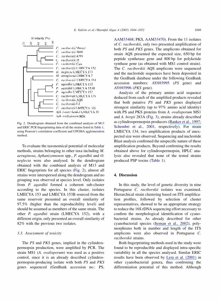
Fig. 2. Dendrogram obtained from the combined analysis of M13
and ERIC PCR fingerprinting data of all the strains listed in Table 1,
using Pearson’s correlation coefficient and UPGMA agglomeration
method.
To evaluate the taxonomical potential of molecular
methods, strains belonging to other taxa including M.
aeruginosa, Aphanizomenon spp., P. agardhii and O.
neglecta were also analysed. In the dendrogram
obtained with the combined analysis of M13 and
ERIC fingerprints for all species (Fig. 2), almost all
strains were interspersed along the dendrogram and no
grouping was observed at species level. Only isolates
from P. agardhii formed a coherent sub-cluster
according to the species. In this cluster, isolates
LMECYA 153 and LMECYA 153B sourced from the
same reservoir presented an overall similarity of
97.5% (higher than the reproducibility level) and
should be assumed as members of the same strain. The
other P. agardhii strain (LMECYA 152), with a
different origin, only presented an overall similarity of
82% with the previous two isolates.
3.3. Assessment of toxicity
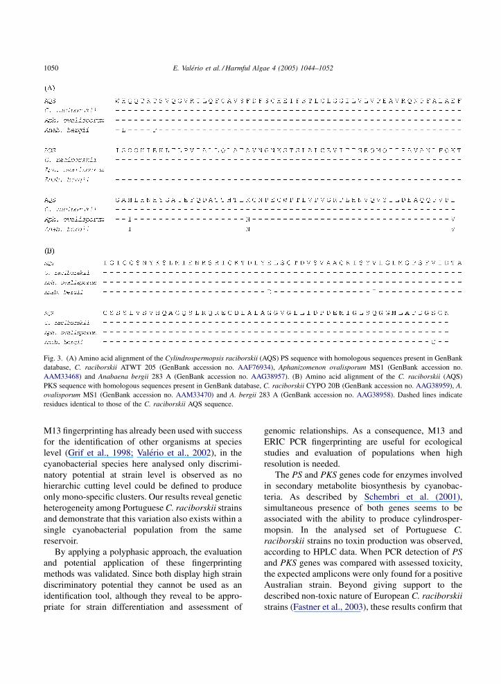
The PS and PKS genes, implied in the cylindros-
permopsin production, were amplified by PCR. The
strain MS1 (A. ovalisporum) was used as a positive
control, since it is an already described cylindros-
permopsin-producing isolate with both PS and PKS
genes sequenced (GenBank accession no.: PS,
AAM33468; PKS, AAM33470). From the 11 isolates
of C. raciborskii, only two presented amplification of
both PS and PKS genes. The amplicons obtained for
strain AQS presented the expected size, 650 bp for
peptide synthetase gene and 800 bp for polyketide
synthase gene (as obtained with MS1 control strain).
The C. raciborskii AQS amplicons were sequenced
and the nucleotide sequences have been deposited in
the GenBank database under the following GenBank
accession numbers: AY693995 (PS gene) and
AY693996 (PKS gene).
Analysis of the primary amino acid sequence
deduced from each of the amplified products revealed
that both putative PS and PKS genes displayed
strongest similarity (up to 97% amino acid identity)
with PS and PKS proteins from A. ovalisporum MS1
and A. bergii 283A (Fig. 3), strains already described
as cylindrospermopsin producers (Banker et al., 1997;
Schembri et al., 2001, respectively). For strain
LMECYA 134, two amplification products of unex-
pected size were observed. Sequencing and nucleotide
Blast analysis confirmed the unspecific nature of these
amplification products. Beyond confirming the results
obtained above for cylindrospermopsin, HPLC ana-
lysis also revealed that none of the tested strains
produced PSP toxins (Table 1).
4. Discussion
In this study, the level of genetic diversity in nine
Portuguese C. raciborskii isolates was examined.
Hierarchical strain clustering based on ITS amplifica-
tion profiles, followed by selection of cluster
representatives, showed to be an appropriate strategy
to reduce the 16S rDNA sequencing effort necessary to
confirm the morphological identification of cyano-
bacterial strains. As already described for other
cyanobacterial species (Iteman et al., 2002), poly-
morphisms both in number and length of the ITS
amplicons were also observed in Portuguese C.
raciborskii strains.
Both fingerprinting methods used in the study were
found to be reproducible and displayed intra-specific
variability in all the species analysed. Similar ERIC
results have been observed by Lyra et al. (2001) in
other cyanobacterial genera, thus confirming the
differentiation potential of this method. Although
E. Valerio et al. / Harmful Algae 4 (2005) 1044–10521050
Fig. 3. (A) Amino acid alignment of the Cylindrospermopsis raciborskii (AQS) PS sequence with homologous sequences present in GenBank
database, C. raciborskii ATWT 205 (GenBank accession no. AAF76934), Aphanizomenon ovalisporum MS1 (GenBank accession no.
AAM33468) and Anabaena bergii 283 A (GenBank accession no. AAG38957). (B) Amino acid alignment of the C. raciborskii (AQS)
PKS sequence with homologous sequences present in GenBank database, C. raciborskii CYPO 20B (GenBank accession no. AAG38959), A.
ovalisporum MS1 (GenBank accession no. AAM33470) and A. bergii 283 A (GenBank accession no. AAG38958). Dashed lines indicate
residues identical to those of the C. raciborskii AQS sequence.
M13 fingerprinting has already been used with success
for the identification of other organisms at species
level (Grif et al., 1998; Valerio et al., 2002), in the
cyanobacterial species here analysed only discrimi-
natory potential at strain level is observed as no
hierarchic cutting level could be defined to produce
only mono-specific clusters. Our results reveal genetic
heterogeneity among Portuguese C. raciborskii strains
and demonstrate that this variation also exists within a
single cyanobacterial population from the same
reservoir.
By applying a polyphasic approach, the evaluation
and potential application of these fingerprinting
methods was validated. Since both display high strain
discriminatory potential they cannot be used as an
identification tool, although they reveal to be appro-
priate for strain differentiation and assessment of
genomic relationships. As a consequence, M13 and
ERIC PCR fingerprinting are useful for ecological
studies and evaluation of populations when high
resolution is needed.
The PS and PKS genes code for enzymes involved
in secondary metabolite biosynthesis by cyanobac-
teria. As described by Schembri et al. (2001),
simultaneous presence of both genes seems to be
associated with the ability to produce cylindrosper-
mopsin. In the analysed set of Portuguese C.
raciborskii strains no toxin production was observed,
according to HPLC data. When PCR detection of PS
and PKS genes was compared with assessed toxicity,
the expected amplicons were only found for a positive
Australian strain. Beyond giving support to the
described non-toxic nature of European C. raciborskii
strains (Fastner et al., 2003), these results confirm that
E. Valerio et al. / Harmful Algae 4 (2005) 1044–1052 1051
this two-gene directed PCR method is a powerful, easy
to perform and rapid approach to evaluate the
cylindrospermopsin-related toxicity of cyanobacterial
strains.
5. Conclusion
C. raciborskii strains isolated from the Portuguese
freshwaters were shown to be genomically hetero-
geneous and revealed a non-toxic behaviour. PS and
PKS genes of the Australian C. raciborskii AQS strain
code for putative proteins displaying 97–100% of
amino acid identity with those from other strains of C.
raciborskii, A. ovalisporum and A. bergii.
16S rDNA sequencing of cluster representatives
selected from ITS analysis is an appropriate strategy to
confirm the morphological identification of cyano-
bacterial strains. M13 and ERIC PCR fingerprinting
are appropriate methods for strain differentiation and
assessment of genomic relationships.
Acknowledgements
The authors thank G.K. Eaglesham for the HPLC
determination of cylindrospermopsin. Elisabete Valerio
thanks the financial support (Grant SFRH/BD/8272/
2002) by Fundacao para a Ciencia e a Tecnologia.
References
Baker, J.A., Neilan, B.A., Entsch, B., McKay, D.B., 2001. Identi-
fication of cyanobacteria and their toxigenicity in environmental
samples by rapid molecular analysis. Environ. Toxicol. 16, 472–
482.
Banker, R., Carmeli, S., Hadas, O., Teltsch, B., Porat, R., Sukenik,
A., 1997. Identification of cylindrospermopsin in the cyanobac-
terium Aphanizomenon ovalisporum (Cyanophyceae) isolated
from Lake Kinneret. Isr. J. Phycol. 33, 613–616.
Chonudomkul, D., Yongmanitchai, W., Theeragool, G., Kawachi,
M., Kasai, F., Kaya, K., Watanabe, M.M., 2004. Morphology,
genetic diversity, temperature tolerance and toxicity of Cylin-
drospermopsis raciborskii (Nostocales Cyanobacteria) strains
from Thailand and Japan. FEMS Microbiol. Ecol. 48, 639–649.
Dyble, J., Paerl, H.W., Neilan, B.A., 2002. Genetic characterization
of Cylindrospermopsis raciborskii (cyanobacteria) isolates from
diverse geographic origins based on nifH and cpcBA-IGS
nucleotide analysis. Appl. Environ. Microbiol. 68, 2567–
2571.
Eaglesham, G.K., Norris, R.L., Shaw, G.R., Smith, M.J., Chiswell,
R.K., Davis, B.C., Neville, G.R., Seawright, A.A., Moore, M.R.,
1999. Use of HPLC-MS/MS to monitor cylindrospermopsin, a
blue-green algal toxin, for public health purposes. Environ.
Toxicol. 14, 151–155.
Fastner, J., Heinze, R., Humpage, A.R., Mischke, U., Eaglesham,
G.K., Chorus, I., 2003. Cylindrospermopsin occurence in two
German lakes and preliminary assessment of toxicity and toxin
production of Cylindrospermopsis raciborskii (Cyanobacteria)
isolates. Toxicon 42, 313–321.
Fergusson, K.M., Schembri, M.A., Saint, C.P., 2000. The use of
molecular techniques to characterise toxic cyanobacteria. In:
Proceedings of the Ninth Conference on Harmful Algal Blooms,
Sandy Bay, Tasmania, Australia, 6–11 February, pp. 234–237.
Grif, K., Karch, H., Schneider, C., Daschner, F.D., Beutin, L.,
Cheasty, T., Smit, H., Rowe, B., Dierich, M.P., Allerberger,
F., 1998. Comparative study of five different techniques for
epidemiological typing of Escherichia coli O157. Diagn. Micro-
biol. Infect. Dis. 32, 165–176.
Hawkins, P.R., Runnegar, M.T.C., Jackson, A.R.B., Falconer, I.R.,
1985. Severe hepatotoxicity caused by the tropical cyanobacter-
ium (blue-green alga) Cylindrospermopsis raciborskii (Wolos-
zynska) Seenaya and Subba Raju isolated from a domestic water
supply reservoir. Appl. Environ. Microbiol. 50, 1292–1295.
Hayman, J., 1992. Beyond the Barcoo-probable human tropical
cyanobacterium poisoning in outback Autralia. Med. J. Aust.
157, 794–796.
Iteman, I., Rippka, R., Tandeau de Marsac, N., Herdman, M., 2002.
rDNA analyses of planktonic heterocystous cyanobacteria,
including members of the genera Anabaenopsis and Cyanospira.
Microbiology 148, 481–496.
Lagos, N., Onodera, H., Zagatto, P.A., Andrinolo, D., Azevedo,
S.M.F.O., Oshima, Y., 1999. The first evidence of paralytic
shellfish toxins in the freshwater cyanobacterium Cylindrosper-
mopsis raciborskii, isolated from Brazil. Toxicon 37, 1359–
1373.
Li, R., Carmichael, W.W., Brittain, S., Eaglesham, G.K., Shaw,
G.R., Mahakhant, A., Noparatnaraporn, N., Yongmanitchai, W.,
Kaya, K., Watanabe, M.M., 2001. Isolation and identification of
the cyanotoxin cylindrospermopsin and deoxy-cylindrosper-
mopsin from a Thailand strain of Cylindrospermopsis racibors-
kii (Cyanobacteria). Toxicon 39, 973–980.
Lyra, C., Suomalainen, S., Gugger, M., Vezie, C., Sundman, P.,
Paulin, L., Sivonen, K., 2001. Molecular characterization of
planktic cyanobacteria of Anabaena, Aphanizomenon, Micro-
cystis and Planktothrix genera. Int. J. Syst. Evol. Microbiol. 51,
513–526.
Massol-Deya, A.A., Odelson, D., Hickey, R.F., Tiedje, J.M., 1995.
Bacterial community fingerprinting of amplified 16S and 16S-23S
ribosomal DNA gene sequences and restriction endonuclease
analysis (ARDRA). Mol. Microb. Ecol. Methods 3.3.2, 1–8.
Meyer, W., Lieckfeldt, E., Kuhls, K., Freedman, E.Z., Borner, T.,
Mitchell, T.G., 1993. DNA and PCR-fingerprinting in fungi.
EXS 67, 311–320.
Neilan, B.A., Saker, M.L., Fastner, J., Torokne, A., Burns, B.P.,
2003. Phylogeography of the invasive cyanobacterium Cylin-
drospermopsis raciborskii. Molec. Ecol. 12, 133–140.
E. Valerio et al. / Harmful Algae 4 (2005) 1044–10521052
Normand, P., Ponsonnet, C., Nesme, X., Neyra, M., Simonet, P.,
1996. ITS analysis in prokaryotes. Mol. Microb. Ecol. Manual
2.4.5, 1–12.
Oshima, Y., 1995. Postcolumn derivatization liquid chromato-
graphic methods for paralytic shellfish toxins. J. AOAC Int.
78, 528–532.
Pitcher, D., Saunders, N., Owen, R., 1989. Rapid extraction of
bacterial DNA with guanidium thyocinate. Lett. Appl. Micro-
biol. 8, 151–156.
Saker, M.L., Thomas, A.D., Norton, J.H., 1999. Cattle mortality
attributed to the toxic cyanobacterium Cylindrospermopsis raci-
borskii in an outback region of north Queensland. Environ.
Toxicol. 14, 179–183.
Saker, M.L., Neilan, B.A., 2001. Varied diazotrophies, morphol-
ogies, and toxicities of genetically similar isolates of Cylin-
drospermopsis raciborskii (Nostocales, Cyanophyceae) from
Northern Australia. Appl. Environ. Microbiol. 67, 1839–
1845.
Saker, M.L., Nogueira, I.C.G., Vasconcelos, V.M., Neilan, B.A.,
Eaglesham, G.H., Pereira, P., 2003. First report and toxicological
assessment of the cyanobacterium Cylindrospermopsis racibors-
kii from Portuguese freshwaters. Ecotoxicol. Environ. Saf. 55,
243–250.
Schembri, M.A., Neilan, B.A., Saint, C.P., 2001. Identification of
genes implicated in toxin production in the cyanobacterium
Cylindrospermopsis raciborskii. Environ. Toxicol. 16, 413–421.
Smith, J.K., Parry, J.D., Day, J.G., Smith, R.J., 1998. A PCR
technique based on the Hip1 interspersed repetitive sequence
distinguishes cyanobacterial species and strains. Microbiology
144, 2791–2801.
Sneath, P.H.A., Sokal, R.R., 1973. Numerical Taxonomy: The
Principles and Practice of Numerical Classification. Freeman,
San Francisco.
Valerio, E., Gadanho, M., Sampaio, J.P., 2002. Sporobolomyces
odoratus sp. nov., a new species in the Sporidiobolus ruineniae
clade. FEMS Yeast Res. 2, 9–16.
Versalovic, J., Koeuth, T., Lupsk, J.R., 1991. Distribution of repetitive
DNA sequences in eubacteria and application to fingerprinting of
bacterial genomes. Nucleic Acids Res. 19, 6823–6831.
Wilson, K.M., Schembri, M.A., Baker, P.D., Saint, C.P., 2000.
Molecular characterization of the toxic cyanobacterium Cylin-
drospermopsis raciborskii and design of a species-specific PCR.
Appl. Environ. Microbiol. 66, 332–338.
Woloszynska, J., 1912. Das Phytoplankton einiger Javanian Seen
mit Berucksichtigung des sawa-planktons.. Bull. Int. Acad. Sci.
Cracoviae, Ser. B 649–709.