Modulatory profiling identifies mechanisms of small molecule-induced cell death
10
Modulatory profiling identifies mechanisms of small molecule-induced cell death Adam J. Wolpaw a , Kenichi Shimada a , Rachid Skouta a,b , Matthew E. Welsch b , Uri David Akavia a,c , Dana Pe’er a,c , Fatima Shaik a , J. Chloe Bulinski a , and Brent R. Stockwell a,b,d,1 a Department of Biological Sciences, b Department of Chemistry, c Center for Computational Biology and Bioinformatics, and d The Howard Hughes Medical Institute, Columbia University, New York, NY 10027 Edited by James A. Wells, University of California, San Francisco, CA, and approved August 15, 2011 (received for review April 23, 2011) Cell death is a complex process that plays a vital role in de- velopment, homeostasis, and disease. Our understanding of and ability to control cell death is impeded by an incomplete charac- terization of the full range of cell death processes that occur in mammalian systems, especially in response to exogenous pertur- bations. We present here a general approach to address this problem, which we call modulatory profiling. Modulatory profiles are composed of the changes in potency and efficacy of lethal compounds produced by a second cell death-modulating agent in human cell lines. We show that compounds with the same characterized mechanism of action have similar modulatory pro- files. Furthermore, clustering of modulatory profiles revealed relationships not evident when clustering lethal compounds based on gene expression profiles alone. Finally, modulatory profiling of compounds correctly predicted three previously uncharacter- ized compounds to be microtubule-destabilizing agents, classi- fied numerous compounds that act nonspecifically, and identified compounds that cause cell death through a mechanism that is morphologically and biochemically distinct from previously estab- lished ones. apoptosis | chemical biology | small molecules C ell death has historically been viewed as a binary phenome- non. Cells were described to die in one of two ways—through a controlled and ordered process (apoptosis) or an unregulated and chaotic process (necrosis) (1, 2). Not only were these often considered the only two possible mechanisms, but they were also frequently viewed as morphologically and biochemically uniform (3, 4). A great deal of research in recent decades has not only shown the complexity and heterogeneity of apoptotic and ne- crotic signaling, but also that cells can die in physiological and nonphysiological contexts through processes morphologically and biochemically distinct from both apoptosis and necrosis. Activation of caspases, a family of cysteine proteases, is es- sential for producing the full morphological characteristics of apoptosis. There are at least three distinct pathways that can lead to the activation of effector caspases—the extrinsic death re- ceptor pathway, the intrinsic mitochondrial pathway, and the Granzyme B pathway (5)—but numerous mechanisms feed into these three pathways. There is substantial evidence that necrosis, long considered to be a disorganized and unregulated process, can proceed through an evolutionarily conserved pathway in a highly orchestrated fashion. Necrosis-like morphology has been observed after death receptor stimulation (6–12), after treatment with DNA-damaging agents (13–15), and in Caenorhabditis ele- gans in response to a variety of stresses (16, 17). These examples illustrate the heterogeneity of cell death processes resembling necrosis. A widely debated nonapoptotic, nonnecrotic cell death mechanism is autophagic cell death, which has been implicated in vivo in the involution of the salivary gland in Drosophila (18) and in death because of the hypersensitivity response in Arabi- dopsis (19) as well as a number of cell culture systems (20, 21). Additional forms of cell death have been described and reviewed elsewhere (22–24). Many of these alternative death programs have been observed only in specialized cells or under unusual conditions, and they are often limited to morphological rather than molecular-level descriptions. Rigorous functional under- standing of these processes and their mechanistic relationship to each other is lacking. New lethal reagents are routinely generated in anticancer drug discovery, chemical biology screens, and cell death research. However, despite our improved understanding of cell death, the investigation and characterization of such lethal reagents typi- cally proceeds in an ad hoc way. There is no standardized process to compare lethal compounds or identify those compounds act- ing through specific mechanisms. Hence, rigorous characteriza- tion is only performed on lethal compounds that are active in more selective and inherently interesting secondary assays. The remaining uncharacterized orphan lethal compounds represent an untapped resource. Investigation into the mechanism of ac- tion of such compounds could reveal information about the scope and detail of death pathways that can be activated in cells. A major use of specific secondary assays is for the elimination of compounds that kill cells through nonspecific mechanisms. Such compounds are not useful in investigating the signaling pathways that govern cell death, and they are not attractive leads for drug development. For instance, compounds can act non- specifically on cells through chemical reactivity (25), by forming small molecule aggregates (26), or disrupting membranes (27). These properties of small molecules are not easy to predict a priori. Although reactivity can often be predicted from chem- ical structure, some reactive compounds kill cells specifically, whereas others are not lethal at all (28). Small molecule aggre- gates are well-established as nonspecific inhibitors in in vitro biochemical assays (29, 30), but only limited studies have been performed on their persistence in the presence of high protein concentrations (31) and cell culture (26). Most drug-like small molecules are lipophilic (32), and determining those molecules that act primarily through membrane disruption is not obvious. Here, we present a methodology for systematically charac- terizing lethal compounds based on functional profiles. This method, called modulatory profiling, systematically analyzes the changes in the lethality of a compound when used in combina- tion with each member of a panel of cell death modulators. These modulators were selected to modulate established cell death processes. Applying this method to both characterized and uncharacterized compounds has allowed us to identify previously unidentified microtubule-destabilizing agents, segre- gate compounds that act nonspecifically, and identify compounds Author contributions: A.J.W. and B.R.S. designed research; A.J.W., K.S., R.S., M.E.W., F.S., and J.C.B. performed research; A.J.W., K.S., R.S., M.E.W., U.D.A., D.P., J.C.B., and B.R.S. analyzed data; and A.J.W. and B.R.S. wrote the paper. The authors declare no conflict of interest. This article is a PNAS Direct Submission. Freely available online through the PNAS open access option. 1 To whom correspondence should be addressed. E-mail: [email protected]. See Author Summary on page 16151. This article contains supporting information online at www.pnas.org/lookup/suppl/doi:10. 1073/pnas.1106149108/-/DCSupplemental. www.pnas.org/cgi/doi/10.1073/pnas.1106149108 PNAS | September 27, 2011 | vol. 108 | no. 39 | E771–E780 PHARMACOLOGY PNAS PLUS
Transcript of Modulatory profiling identifies mechanisms of small molecule-induced cell death

Modulatory profiling identifies mechanisms of smallmolecule-induced cell deathAdam J. Wolpawa, Kenichi Shimadaa, Rachid Skoutaa,b, Matthew E. Welschb, Uri David Akaviaa,c, Dana Pe’era,c,Fatima Shaika, J. Chloe Bulinskia, and Brent R. Stockwella,b,d,1
aDepartment of Biological Sciences, bDepartment of Chemistry, cCenter for Computational Biology and Bioinformatics, and dThe Howard Hughes MedicalInstitute, Columbia University, New York, NY 10027
Edited by James A. Wells, University of California, San Francisco, CA, and approved August 15, 2011 (received for review April 23, 2011)
Cell death is a complex process that plays a vital role in de-velopment, homeostasis, and disease. Our understanding of andability to control cell death is impeded by an incomplete charac-terization of the full range of cell death processes that occur inmammalian systems, especially in response to exogenous pertur-bations. We present here a general approach to address thisproblem, which we call modulatory profiling. Modulatory profilesare composed of the changes in potency and efficacy of lethalcompounds produced by a second cell death-modulating agentin human cell lines. We show that compounds with the samecharacterized mechanism of action have similar modulatory pro-files. Furthermore, clustering of modulatory profiles revealedrelationships not evident when clustering lethal compounds basedon gene expression profiles alone. Finally, modulatory profilingof compounds correctly predicted three previously uncharacter-ized compounds to be microtubule-destabilizing agents, classi-fied numerous compounds that act nonspecifically, and identifiedcompounds that cause cell death through a mechanism that ismorphologically and biochemically distinct from previously estab-lished ones.
apoptosis | chemical biology | small molecules
Cell death has historically been viewed as a binary phenome-non. Cells were described to die in one of two ways—through
a controlled and ordered process (apoptosis) or an unregulatedand chaotic process (necrosis) (1, 2). Not only were these oftenconsidered the only two possible mechanisms, but they were alsofrequently viewed as morphologically and biochemically uniform(3, 4). A great deal of research in recent decades has not onlyshown the complexity and heterogeneity of apoptotic and ne-crotic signaling, but also that cells can die in physiological andnonphysiological contexts through processes morphologically andbiochemically distinct from both apoptosis and necrosis.Activation of caspases, a family of cysteine proteases, is es-
sential for producing the full morphological characteristics ofapoptosis. There are at least three distinct pathways that can leadto the activation of effector caspases—the extrinsic death re-ceptor pathway, the intrinsic mitochondrial pathway, and theGranzyme B pathway (5)—but numerous mechanisms feed intothese three pathways. There is substantial evidence that necrosis,long considered to be a disorganized and unregulated process,can proceed through an evolutionarily conserved pathway in ahighly orchestrated fashion. Necrosis-like morphology has beenobserved after death receptor stimulation (6–12), after treatmentwith DNA-damaging agents (13–15), and in Caenorhabditis ele-gans in response to a variety of stresses (16, 17). These examplesillustrate the heterogeneity of cell death processes resemblingnecrosis. A widely debated nonapoptotic, nonnecrotic cell deathmechanism is autophagic cell death, which has been implicatedin vivo in the involution of the salivary gland in Drosophila (18)and in death because of the hypersensitivity response in Arabi-dopsis (19) as well as a number of cell culture systems (20, 21).Additional forms of cell death have been described and reviewedelsewhere (22–24). Many of these alternative death programshave been observed only in specialized cells or under unusual
conditions, and they are often limited to morphological ratherthan molecular-level descriptions. Rigorous functional under-standing of these processes and their mechanistic relationship toeach other is lacking.New lethal reagents are routinely generated in anticancer drug
discovery, chemical biology screens, and cell death research.However, despite our improved understanding of cell death, theinvestigation and characterization of such lethal reagents typi-cally proceeds in an ad hoc way. There is no standardized processto compare lethal compounds or identify those compounds act-ing through specific mechanisms. Hence, rigorous characteriza-tion is only performed on lethal compounds that are active inmore selective and inherently interesting secondary assays. Theremaining uncharacterized orphan lethal compounds representan untapped resource. Investigation into the mechanism of ac-tion of such compounds could reveal information about thescope and detail of death pathways that can be activated in cells.A major use of specific secondary assays is for the elimination
of compounds that kill cells through nonspecific mechanisms.Such compounds are not useful in investigating the signalingpathways that govern cell death, and they are not attractive leadsfor drug development. For instance, compounds can act non-specifically on cells through chemical reactivity (25), by formingsmall molecule aggregates (26), or disrupting membranes (27).These properties of small molecules are not easy to predicta priori. Although reactivity can often be predicted from chem-ical structure, some reactive compounds kill cells specifically,whereas others are not lethal at all (28). Small molecule aggre-gates are well-established as nonspecific inhibitors in in vitrobiochemical assays (29, 30), but only limited studies have beenperformed on their persistence in the presence of high proteinconcentrations (31) and cell culture (26). Most drug-like smallmolecules are lipophilic (32), and determining those moleculesthat act primarily through membrane disruption is not obvious.Here, we present a methodology for systematically charac-
terizing lethal compounds based on functional profiles. Thismethod, called modulatory profiling, systematically analyzes thechanges in the lethality of a compound when used in combina-tion with each member of a panel of cell death modulators.These modulators were selected to modulate established celldeath processes. Applying this method to both characterizedand uncharacterized compounds has allowed us to identifypreviously unidentified microtubule-destabilizing agents, segre-gate compounds that act nonspecifically, and identify compounds
Author contributions: A.J.W. and B.R.S. designed research; A.J.W., K.S., R.S., M.E.W., F.S.,and J.C.B. performed research; A.J.W., K.S., R.S., M.E.W., U.D.A., D.P., J.C.B., and B.R.S.analyzed data; and A.J.W. and B.R.S. wrote the paper.
The authors declare no conflict of interest.
This article is a PNAS Direct Submission.
Freely available online through the PNAS open access option.1To whom correspondence should be addressed. E-mail: [email protected].
See Author Summary on page 16151.
This article contains supporting information online at www.pnas.org/lookup/suppl/doi:10.1073/pnas.1106149108/-/DCSupplemental.
www.pnas.org/cgi/doi/10.1073/pnas.1106149108 PNAS | September 27, 2011 | vol. 108 | no. 39 | E771–E780
PHARM
ACO
LOGY
PNASPL
US

that induce death through morphologically and biochemicallydistinct mechanisms.
ResultsCreating and Clustering Modulatory Profiles. Our initial tasks wereto (i) assemble a collection of reagents that can modulate celldeath-related processes (cell death modulators), (ii) identifya test pool of characterized lethal compounds, and (iii) choosecell lines for testing. First, we chose chemical modulators (SIAppendix, Table S1) based on literature precedent. These com-pounds include reactive oxygen species scavengers and inhibitorsof calcium signaling, protein synthesis, and proteases, amongothers. We also included four genetic modulators of cell death(SI Appendix, Fig. S1).For the lethal compounds, we chose a set of 28 well-character-
ized compounds (SI Appendix, Table S2), including inhibitorsof topoisomerases, microtubule assembly, proteasomes, histonedeacetylases (HDACs), kinases, and various stages of mitochon-drial respiration. We chose two cell lines, a fibrosarcoma cell line(HT-1080) and an engineered tumorigenic line derived from hu-man foreskin fibroblasts (BJ-TERT/LT/ST/RASV12). Both ofthese cell lines grow rapidly and uniformly in 384-well assay plates.We reasoned that it would be advantageous to explore both anengineered tumorigenic line lacking mutations in uncharacterizeddeath pathways thatmay be found in cancers aswell as a cancer cellline with WT p53 that could be easily infected with lentivirus.We then tested each lethal compound in combination with
each cell death modulator (Fig. 1A). We chose a single con-centration of each modulator (based on literature precedent and
our optimization experiments) and used a 14-point, twofold di-lution series of each lethal compound. Comparing survival in thepresence of each modulator to survival without a modulatorallowed us to construct comparative concentration responsecurves (Fig. 1B and SI Appendix, Fig. S2). We extracted twoparameters from each graph—the change in potency (Fig. 1BUpper) and the change in efficacy (Fig. 1B Lower) caused by eachmodulator. The modulatory profile of a lethal compound wasdefined as the dimensional vector of its activity changes (i.e.,changes in potency and efficacy) induced by each modulatoracross distinct cell lines. The modulatory profiles of known lethalcompounds are graphically depicted in Fig. 1C and numericallydepicted in SI Appendix, Table S3. We tested the data require-ments of the profiles by calculating the parameters based ona subset of the data (SI Appendix, SI Text and SI Appendix, Fig.S3A). Experiments to determine the reproducibility of themeasurements between batches, the similarities and differencesin the parameters between cell lines, the dependence of themeasured parameters on the detection reagent, and the effects ofthe modulators on the detection reagent and the cells are shownin SI Appendix, Fig. S3 B–F and discussed in SI Appendix, SI Text.Classifying the lethal compounds based on the effects of a single
modulator was ineffective (SI Appendix, SI Text and SI Appendix,Fig. S4), necessitating the use of the full modulatory profiles. Wecompared modulatory profiles of lethal compounds using Spear-man correlations between pairs of compounds (the similaritymatrix is shown in Fig. 2A). Hierarchically clustering the axes ofthe matrix caused compounds with well-correlated modulatoryprofiles to appear clearly along the axis. The clustering was also
Fig. 1. Creating modulatory profiles. (A) Cells with orwithout modulator were seeded in 384-well plates. Lethalcompounds in dilution series were then added, and theplates were incubated for 48 h before the addition of thecell viability dye Alamar blue. Readout of fluorescence aftera 14-h incubation with Alamar blue allowed the construc-tion of comparative concentration response curves. (B) Twoexamples of comparative concentration response curvesand an illustration of the two parameters—the changein efficacy and change in potency—extracted from eachpairwise combination of modulators and lethal com-pounds. Both examples use HT-1080 cells. (C) Heat mapdepicting 32 modulatory profiles of characterized lethalcompounds (28 distinct compounds and 4 repeat com-pounds). Lethal compounds are listed on the y axis, andmodulators, cell lines, and parameter types are on the xaxis. Missing values are depicted in gray.
E772 | www.pnas.org/cgi/doi/10.1073/pnas.1106149108 Wolpaw et al.
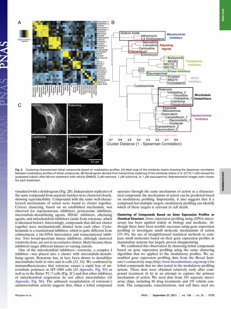
visualized with a dendrogram (Fig. 2B). Independent replicates ofthe same compound from separate batches were clustered closely,showing reproducibility. Compounds with the same well-charac-terized mechanisms of action were found to cluster together.Correct clustering, based on an established mechanism, wasobserved for topoisomerase inhibitors, proteasome inhibitors,microtubule-destabilizing agents, HDAC inhibitors, alkylatingagents, and mitochondrial inhibitors (aside from rotenone, whichis discussed below). Interestingly, compounds that did not clustertogether were mechanistically distinct from each other. Cyclo-heximide is a translational inhibitor, which is quite different fromechinomycin, a bis-DNA intercalator and transcriptional inhib-itor. Two broad-spectrum kinase inhibitors, although clusteredrelatively close, are not in an exclusive cluster, likely because theseinhibitors target different kinases to varying extents.One of the mitochondrial inhibitors—rotenone, a complex I
inhibitor—was placed into a cluster with microtubule-destabi-lizing agents. Rotenone has, in fact, been shown to destabilizemicrotubules both in vitro and in cells (33, 34). We confirmed byimmunofluorescence that rotenone causes a rapid loss of mi-crotubule polymers in HT-1080 cells (SI Appendix, Fig. S5) aswell as in the flatter TC-7 cells (Fig. 2C) and that other inhibitorsof mitochondrial respiration do not affect microtubules (SIAppendix, Fig. S6). The unbiased recapitulation of rotenone’santimicrotubule activity suggests that, when a lethal compound
operates through the same mechanism of action as a character-ized compound, the mechanism of action can be predicted basedon modulatory profiling. Importantly, it also suggests that if acompound has multiple targets, modulatory profiling can identifywhich of these targets is relevant to cell death.
Clustering of Compounds Based on Gene Expression Profiles orChemical Structure.Gene expression profiling using cDNA micro-arrays has been applied widely in biology and medicine. Al-though there have been notable successes using gene expressionprofiling to investigate small molecule mechanisms of action(35–39), the use of straightforward statistical methods to com-pare small molecules based on their gene expression profiles inmammalian systems has largely proven disappointing.We confirmed this observation by clustering lethal compounds
based on gene expression profiling using the same clusteringalgorithm that we applied to the modulatory profiles. We as-sembled gene expression profiling data from the Broad Insti-tute’s connectivity map (http://www.broadinstitute.org/cmap/) forlethal compounds that we also tested in the modulatory profilingsystem. These data were obtained relatively early after com-pound treatment (6 h) in an attempt to capture the primarymechanism of action. We used data from 281 separate micro-array chips, including 86 drug treatments and 195 vehicle con-trols. The compounds, concentrations, and cell lines used are
Fig. 2. Clustering characterized lethal compounds based on modulatory profiles. (A) Heat map of the similarity matrix showing the Spearman correlationbetween modulatory profiles of lethal compounds. (B) Dendrogram derived from hierarchical clustering of the similarity matrix in A. (C) TC-7 cells stained foracetylated tubulin after 60-min treatment with vehicle (DMSO), 5 μM rotenone, 1 μM colchicine, or 1 μM staurosporine. Representative images were chosenfor each treatment.
Wolpaw et al. PNAS | September 27, 2011 | vol. 108 | no. 39 | E773
PHARM
ACO
LOGY
PNASPL
US
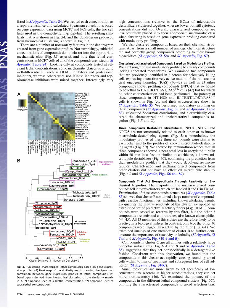
listed in SI Appendix, Table S4. We treated each concentration asa separate instance and calculated Spearman correlations basedon gene expression data using MCF7 and PC3 cells, the two celllines used in the connectivity map pipeline. The resulting simi-larity matrix is shown in Fig. 3A, and the dendrogram producedfrom hierarchical clustering is shown in Fig. 3B.There are a number of noteworthy features in the dendrogram
created from gene expression profiles. Not surprisingly, sublethalconcentrations of compounds do not cluster into the appropriatemechanistic class (Fig. 3B, asterisk and note that lethal con-centrations in MCF7 cells of all of the compounds are listed in SIAppendix, Table S4). Looking only at compounds tested at rel-evant lethal concentrations, some mechanistic classes were quitewell-differentiated, such as HDAC inhibitors and proteasomeinhibitors, whereas others were not. Kinase inhibitors and top-oisomerase inhibitors were mixed together. Interestingly, very
high concentrations (relative to the EC50) of microtubuledestabilizers clustered together, whereas lower but still cytotoxicconcentrations did not. Overall, characterized compounds areless accurately placed into their appropriate mechanistic classwhen clustering is based on gene expression profiling comparedwith modulatory profiling.We also clustered compounds based on their chemical struc-
ture. Apart from a small number of analogs, chemical structuredid not correctly group compounds according to their knownbioactivities (SI Appendix, SI Text and SI Appendix, Fig. S7).
Clustering Uncharacterized Compounds Based on Modulatory Profiles.We next sought to use modulatory profiling to classify compoundslacking annotated mechanisms. We examined two compoundsthat we previously identified in a screen for selectively killingcells expressing a constitutively active mutant of the rat sarcomaviral oncogene homolog (RAS) (40–42) as well as 23 othercompounds [novel profiling compounds (NPC)] that we foundto be lethal to BJ-TERT/LT/ST/RASV12 cells (42) but for whichno other characterization had been performed. The potency ofthese compounds in HT-1080 and BJ-TERT/LT/ST/RASV12
cells is shown in Fig. 4A, and their structures are shown inSI Appendix, Table S5. We performed modulatory profiling onthese compounds (SI Appendix, Fig. S8 and SI Appendix, TableS3), calculated Spearman correlations, and hierarchically clus-tered the characterized and uncharacterized compounds to-gether (Fig. 4 B and C).
Three Compounds Destabilize Microtubules. NPC4, NPC7, andNPC25 are not structurally related to each other or to knownmicrotubule-destabilizing agents (Fig. 5A); nonetheless, themodulatory profiles of these three compounds were similar toeach other and to the profiles of known microtubule-destabiliz-ing agents (Fig. 5B). We showed by immunofluorescence that allthree compounds showed a near total loss in acetylated tubulinwithin 60 min in a fashion similar to colchicine, a known mi-crotubule destabilizer (Fig. 5C), confirming the prediction fromtheir modulatory profiles that they would depolymerize micro-tubules. Characterized and uncharacterized compounds fromother clusters did not have an effect on microtubule stability(Fig. 4C and SI Appendix, Figs. S6 and S9).
Compounds That Act Nonspecifically Through Reactivity or Bio-physical Properties. The majority of the uncharacterized com-pounds fell into two clusters, which are labeled B and C in Fig. 4C.Examination of these compounds’ structures (SI Appendix, TableS5) showed that cluster B contained a large number of compoundswith reactive functionalities, including known alkylating agents.To quantify the relative reactivity of this cluster, we applied anestablished set of predictive reactivity filters (43); 10 of 13 com-pounds were scored as reactive by this filter, but the other 3compounds are activated chloroarenes, also known electrophiles(44, 45). All 13 members of this cluster are therefore likely to bereactive in a biological milieu. In contrast, only 6 of the other 40compounds were flagged as reactive by the filter (Fig. 6A). Weexamined analogs of one member of cluster B to further dem-onstrate the importance of reactivity on lethality (SI Appendix, SIText and SI Appendix, Fig. S10 A and B).Compounds in cluster C are all amines with a relatively large
nonpolar surface area (Fig. 6 A and B and SI Appendix, TableS5), suggesting that they act nonspecifically in a detergent-likefashion. Consistent with this observation, we found that thecompounds in this cluster act rapidly, causing rounding up ofcells within 40 min of treatment and subsequent loss of cell ad-hesion (SI Appendix, Fig. S10C).Small molecules are more likely to act specifically at low
concentrations, whereas at higher concentrations, they can actmore promiscuously (46). We examined the potency of thecompounds in the different lethal compound clusters (Fig. 6C),omitting the characterized compounds to avoid selection bias.
Fig. 3. Clustering characterized lethal compounds based on gene expres-sion profiles. (A) Heat map of the similarity matrix showing the Spearmancorrelation between gene expression profiles of lethal compounds. (B)Dendrogram derived from hierarchical clustering of the similarity matrixin A. *Compound used at sublethal concentration. **Compound used atsupralethal concentration.
E774 | www.pnas.org/cgi/doi/10.1073/pnas.1106149108 Wolpaw et al.

Consistent with our expectation, the compounds in clusters Band C that appear to act nonspecifically were the least potent.Although a compound targeting a specific cellular pathway is
susceptible to modulation, compounds causing nonspecific cel-lular damage are unlikely to be susceptible to such modulation.Thus, nonspecifically acting compounds should have modulatoryprofiles consisting of minimal changes. To test this theory, wecomputed the average of the absolute value of each profile andcalled this value the modulatability score. We compared theaverage modulatability of the different lethal compound clusters(Fig. 6D). As expected, cluster D (containing compounds knownto target specific cellular processes) had high modulatability,whereas compounds in clusters B and C, which appear to actthrough reactivity or biophysical mechanisms, had consistentlylow modulatability (this finding can also be seen in the heat mapshown in SI Appendix, Fig. S8).There were two clusters of uncharacterized compounds re-
maining: clusters A and E. Cluster E had potent compounds withhigh modulatability. Cluster A had one potent and one less potentcompound, both with low modulatability. We investigated furtherthe compounds in cluster E, because based on these metrics, theyaremore likely to act throughmodulation of specific cellular targets.
Cluster of Compounds Induces BAX-/BAK-Independent MitochondrialCell Death. Cluster E in Fig. 4C contains three compounds. Wepreviously found that two of these compounds, erastin and Ras-selective-lethal compound 3 (RSL3), induce a nonapoptotic, iron-dependent form of death that involves the generation of reactive
oxygen species. Further confirming this finding in the presentcontext, we observed that erastin and RSL3 caused no detectablecaspase activation, which was measured by cleavage of a fluoro-genic caspase substrate (Fig. 7B).Death independent of caspase activity can be dependent on mi-
tochondrial outer membrane permeabilization mediated by BCL2-associated X protein (BAX) or BCL2-antagonist/killer (BAK)(47). We therefore tested the lethality of RSL3 and erastin inWT and Bax−/−Bak−/− double KO mouse embryonic fibroblasts(MEFs). The absence of BAX and BAK did not suppress deathinduced by erastin, and it sensitized cells to death induced by RSL3(Fig. 7C). Thus, modulatory profiling correctly predicted thaterastin and RSL3 act through a form of cell death that is non-apoptotic and, in fact, is independent of the core apoptotic ma-chinery—caspases, BAX, and BAK.We then asked whether the other compound in cluster E,
NPC26, also acts through a nonapoptotic cell death process. Werepeated a subset of the changes in the modulatory profile in twoadditional cell lines and found good consistency between the celllines (SI Appendix, Fig. S11A), suggesting that the modulatoryprofile is a good marker of the compound’s action independent ofcell line. Within the profile, however, there were some immediatemechanistic differences apparent between NPC26 and erastin/RSL3. Erastin and RSL3 are virtually inactive in the presence ofall of the reactive oxygen species scavengers tested [α-tocopherol,Trolox, butylated hydroxyanisole (BHA), and butylated hydroxy-toluene (BHT)], the iron chelator deferoxamine, and the MAPK/ERK Kinase (MEK) inhibitor U0126. NPC26 was only slightly
Fig. 4. Clustering uncharacterized lethal compounds based on modulatory profiles. (A) Potency of uncharacterized lethal compounds in BJ-TERT/LT/ST/RASV12 and HT-1080 cells after 48 h. Values represent the average of three replicates ± SEM. (B) Heat map of the similarity matrix showing the Spearmancorrelation between modulatory profiles of characterized and uncharacterized lethal compounds. (C) Dendrogram derived from hierarchical clustering of thesimilarity matrix in B.
Wolpaw et al. PNAS | September 27, 2011 | vol. 108 | no. 39 | E775
PHARM
ACO
LOGY
PNASPL
US

inhibited by α-tocopherol, Trolox, and deferoxamine, and it wasinsensitive to the presence of BHA, BHT, or U0126 (SI Appendix,Table S3). Thus, reactive oxygen species, iron, andMEK signalingplay a less central (if any) role in NPC26-induced death. NPC26caused a small increase in cleavage of a fluorogenic caspasesubstrate (Fig. 7B), but death induced by NPC26 was not blockedby caspase inhibitors (SI Appendix, Fig. S11B) or affected by theabsence of BAX and BAK (Fig. 7C). These observations led us tothe hypothesis that NPC26 induced a different but perhaps dis-tantly related death process from the process induced by erastinand RSL3.We had previously found by EM that erastin does not affect
nuclear morphology but does cause a loss of mitochondrialstructural integrity (41). To test the hypothesis that NPC26induces a distantly related form of death, we performed EM todetermine if NPC26 induced similar features. The images, shownin Fig. 7D, reveal no change in nuclear morphology and a drasticmitochondrial phenotype. By 3 h in BJ-TERT/LT/ST/RASV12
and 6 h in HT-1080 cells, mitochondria have become circular andlost their cristae completely. We investigated these mitochon-drial morphological changes further by expressing a mitochond-rially targeted red fluorescent protein (DsRed2-mito). NPC26induced a conversion from elongated to punctate mitochondria,a phenotype that has been previously observed during apoptosis(48) and by death induced by inhibitors of oxidative phosphor-ylation and uncoupling agents (49). However, typical apoptotic-inducing stimuli failed to induce similar mitochondrial frag-
mentation in HT-1080 cells (SI Appendix, Fig. S11C). Unlikewith inhibitors of oxidative phosphorylation, NPC26-inducedmitochondrial fragmentation is independent of the activity ofthe dynamin-related protein (DRP1) (Fig. 7E and SI Appendix,Fig. S11D). NPC26 is also not a direct uncoupling agent (SIAppendix, Fig. S11E). Thus, similar to erastin and RSL3, NPC26induces a distinct form of BAX-/BAK-independent mitochon-drial cell death.
NPC26-Induced Death Depends on a Unique Kinase Signaling Pathway.Wenext askedwhether the signaling pathways controllingNPC26-induced death are unique from those pathways controlling deathinduced by characterized compounds or erastin. We first testeda collection of kinase inhibitors for their ability to inhibit NPC26-induced death (compounds and activity are shown in SI Appendix,Table S6). We then tested active inhibitors and related inactiveinhibitors for their ability to inhibit erastin and a representativesubset of the characterized lethal compounds. The results (Fig. 7F)show that NPC26-induced death depends on kinase signalingpathways distinct from the other compounds.Although this pattern serves as a measure of uniqueness, it did
not allow us to identify a specific kinase involved, because notwo redundant inhibitors were effective in preventing death.For example, one JNK inhibitor (SP600125) prevented death,whereas another inhibitor did not (JNK Inh VIII). Similar resultswere obtained with MEK inhibitors (PD98059 protects and U0126does not protect) and PI3K inhibitors (LY294002 protects and
Fig. 5. Previously uncharacterized compounds destabilizemicrotubules. (A) Structures of the well-characterized mi-crotubule destabilizers vinblastine, podophyllotoxin, andcolchicine, the previously reported destabilizer rotenone,and the three compounds predicted to destabilize micro-tubules based on their modulatory profiles. (B) Heat map ofthe Spearman correlations between the modulatory profilesof the uncharacterized compounds NPC4, NPC7, and NPC25and the characterized compounds colchicine, vinblastine,carmustine, trichostatin A, MG132, and doxorubicin. (C)Time course of TC-7 cells stained for acetylated tubulin aftertreatment with 1 μM colchicine, 28 μM NPC4, 28 μM NPC7,or 14 μM NPC25. Staining for total tubulin and acetylatedtubulin after a 60-min treatment with vehicle (DMSO) isshown at the top right. Representative images were chosenfrom each time point.
E776 | www.pnas.org/cgi/doi/10.1073/pnas.1106149108 Wolpaw et al.

wortmannin does not protect). Such inhibitors have significantpromiscuity within the kinase family, raising two possibilities forthese results: (i) the compounds that suppress NPC26-induceddeath inhibit the same off-target kinase or (ii) the compounds thatsuppress NPC26-induced death inhibit multiple kinases, and thepathway is robust to the inhibition of any single kinase.
Structure Activity Relationships of NPC26. Because of the high po-tency and modulatability of its cluster, modulatory profilingpredicted that NPC26 would exert its effect through a specificprocess. To further confirm this hypothesis, we tested structuralanalogs of NPC26 (17 purchased and 7 synthesized analogs) andscored their activity based on their ability to induce cell deaththat could be inhibited by the kinase inhibitor SP600125 (SIAppendix, Table S7). In addition, we resynthesized NPC26, pro-viding the most rigorous confirmation that the assigned structureis correct.Analog testing provided evidence supporting a specific mech-
anism for NPC26-induced death. Subtle structural changes thatwould not be predicted to alter the compound’s reactivity orlipophilicity eliminated activity. For example, although analog26A14 is active, analog 26A3, in which a seven-membered ring in26A14 is replaced by a six-membered ring, is inactive (SI Ap-pendix, Fig. S11F).In addition, we tested an analog containing a linker with a
protected amine attached at the end. This analog (SI Appendix,Table S7, SRS1-78) retained activity, suggesting the potential forthe use of NPC26 as a probe for future affinity purification of itstarget. In summary, NPC26 acts through a nonapoptotic, mito-chondrial-driven mechanism, and it has a modulatory profile andstructure activity relationship to suggest a specific molecule tar-get and mechanism. Thus, modulatory profiling is useful not onlyfor classifying compounds that act through known cell deathmechanisms and targets but also for flagging for additional studycompounds that are likely to act through previously unidentifiedcell death mechanisms.
DiscussionMisregulation of cell death plays a central role in tumorigenesis,neurodegeneration, damage from infarction, and a host of otherhuman diseases. Greater understanding of the scope of cell deathpathways that can be activated in cells and gaining pharmacolog-ical control over such pathways hold the promise of improvinghuman health. In this study, we present modulatory profiling asa methodology for categorizing diverse inducers of cell death andidentifying inducers of previously uncharacterized cell deathprocesses, with the goal of ultimately creating a global view of celldeath mechanisms.Over the past decade, technologies have enabled the collection
of large-scale molecular profiles and led to their use in charac-terizing cellular states and perturbations to those states. Thesetechnologies include gene expression and copy number variationusing DNA microarrays, protein expression and metabolite pro-filing usingMS, enzyme activity usingfluorescence probes, and cellmorphology using high-content imaging (50). Although thesetechnologies have proven invaluable for investigating a wide rangeof basic biological mechanisms and disease processes, it can bedifficult to separate causal changes from correlative changes usingsuch methods. For example, deletion, knockdown, or inhibition ofmany of the proteins that are altered in abundance in a specific cellstate has no consequence, because these changes are products ofthe cell state rather than regulators of it. A great deal of effort isbeing put to identifying the smaller number of driver mutationsresponsible for producing cell states of interest (51–53). Suchtechniques are computationally and data intensive, and they re-quire expert adaptation and fine tuning to each new biologicaldomain. Thus, despite these technological and computationaladvances, there remains a need for the development of high-di-mensionality profiles of functionally relevant measurements thatcan be used to characterize cell states.We developed modulatory profiling to address this deficiency,
which is particularly acute with regards to small molecule-inducedcell states, particularly cell death. Small molecules, even approveddrugs, can have pleiotropic effects on cells (54, 55). Identifyingwhich of the many effects is relevant in a given context is impor-
Fig. 6. Two clusters of previously un-characterized lethal compounds act non-specifically. (A) Distribution of reactivecompounds and amines throughout theclusters from Fig. 4C. Reactivity was cal-culated by adding activated chloroarenesto filters described previously (43). (B)Fraction nonpolar van der Waals surfacearea vs. pKa of the most basic residue.Fraction nonpolar surface area was cal-culated with the built-in parameter in thesoftware MOE. The most basic pKa wascalculated with the web-based softwareSPARC (SI Appendix, SI Methods). Usinga one-way ANOVA and Tukey’s multiplecomparison test, compounds in cluster Care significantly different in their fractionnonpolar surface area from those com-pounds in cluster D (P < 0.01) and signif-icantly more basic than those compoundsin cluster B or D (P < 0.01). (C) Averagepotency of uncharacterized compounds.The average of the potency in HT-1080and BJ-TERT/LT/ST/RASV12 cells is shown.Using a one-way ANOVA and Tukey’smultiple comparison test, the compoundsin clusters B and C are significantly lesspotent than the compounds in cluster D,and the compounds in cluster C are significantly less potent than the compounds in cluster E (P < 0.05). (D) Average modulatability of compounds. Modu-latability was calculated by taking the mean of the absolute value of all of the normalized changes in a compound’s modulatory profile. Lines represent clusteraverages ± SEM. Using a one-way ANOVA and Tukey’s multiple comparison test, compounds in clusters A, B, and C each are significantly less modulatable thanthose compounds in clusters D and E (P < 0.05).
Wolpaw et al. PNAS | September 27, 2011 | vol. 108 | no. 39 | E777
PHARM
ACO
LOGY
PNASPL
US

Fig. 7. Previously uncharacterized compounds induce BAX-/BAK-independent mitochondrial cell death. (A) Structures of erastin, RSL3, and NPC26, which arethe three compounds in cluster E. (B) Caspase activity in HT-1080 cells treated for 12–15 h with erastin, RSL3, NPC26, and staurosporine. Caspase activity wasmeasured by the cleavage of a fluorogenic caspase substrate and is shown on the left y axis (points represent the mean of three replicates ± SEM). Cellviability is shown by the shaded region and on the right y axis (points represent the mean of three replicates ± SEM). (C) Lethality of erastin, RSL3, NPC26, andcamptothecin in WT or Bax−/−Bak−/− MEFs after 48 h of treatment. Data represent the mean of three replicates ± SEM. (D) EM images taken of BJ-TERT/LT/ST/RASV12 cells after a 3-h treatment with either DMSO or 9 μM NPC26 and EM images taken of HT-1080 cells after a 6-h treatment with either DMSO or 9 μMNPC26. Arrows show the nuclei, and arrowheads show mitochondria. Boxes show the portion of the image shown at higher magnification in the next imageto the right. (E) Fluorescence images of HT-1080 cells expressing a mitochondrially targeted dsRed construct infected with either shRNA targeting DRP1or a control nontargeting shRNA treated with either DMSO or 9 μM NPC26 for 6 h. (F) Heat map depicting the ability of various kinase inhibitors to alterdeath induced by NPC26 and other lethal compounds. Protection is depicted in yellow, and sensitization is depicted in blue. Experiments were performed inHT-1080 cells.
E778 | www.pnas.org/cgi/doi/10.1073/pnas.1106149108 Wolpaw et al.

tant and nontrivial. Rotenone provided an illustrative example ofthis problem within the data presented here. Rotenone has beenused extensively as an inhibitor of mitochondrial complex I (56),but it has also been shown to destabilize microtubules (33, 34).Using a measurement such as gene expression profiling, it is notobvious which of these two effects will be most strongly repre-sented. If one is interested in the way in which rotenone inducesa downstream phenotype such as cell death, it is also not neces-sarily the case that the lethal effect will be the one with thestrongest gene expression signature. However, modulatory pro-filing, by using functional measurements, highlighted the lethalmechanism of rotenone in the system tested.Other systems have been developed using collections of func-
tional assays to investigate small molecule activities, includingcomparing pairwise interactions of antibiotics in bacteria (57, 58),profiling the hypersensitivity of yeast haploid deletionmutants (59,60), profiling cytotoxicity across 60 cancer cell lines (61, 62), andmost recently, measuring changes in cancer cell survival in thepresence of various shRNAs (63). Although these other systemshave provided valuable insights, they also have certain drawbacks.Nonapoptotic cell death differs markedly even between mamma-lian cells and other metazoans, such as D. melanogaster and C.elegans (47), making the use of models in bacteria and yeast lessrelevant. The NCI60 approach has produced useful signatures forfinding compounds with similar modes of action, but the use of 60genetically diverse cell lines does not give any specific informationabout cell death mechanisms and is less amenable to geneticperturbations and the use of neurons and other specialized celltypes. The advantages of modulatory profiling are that (i) it usesquantitative, functional assays that measure and perturb celldeath, the process that is specifically under study; (ii) it uses a fulldilution series of each compound, eliminating the possibility ofmaking measurements at irrelevant concentrations; and (iii) theassays are performed in human cells, the species in which we ul-timately want to apply our findings.However, we made some choices that proved somewhat limiting.
We used exclusively small molecules as death-inducing agents.Although small molecules are tractable experimentally, we werelimited by the number of existing classes of characterized lethalcompounds. It will be desirable to extend modulatory profiling toinclude larger numbers of other stimuli, particularly gene knock-down and overexpression. In addition, we chose two fibroblast celllines that are well-suited for the type ofminiaturized assays requiredfor performing high-throughput assays; however, the use of thesetwo cell lines is likely to prevent the analysis of more specializedpathways. We chose to focus, therefore, on capturing the morecommon mechanisms. Last, we showed here the utility of modula-tory profiling as applied only to cell death. It would be desirable toextend modulatory profiling to other cell fates and processes.We measured viability with Alamar blue, a fluorogenic dye
that measures cellular reductive potential. Although this reagenthas shown excellent correlation with other viability metrics, bothin our hands and in the hands of others (42, 64), in certain cases,it can give false positives and negatives (65). It should be em-phasized, however, that to disrupt the output of the profiling,a modulator would have to cause a viability-independent effecton Alamar blue reduction to varying extents for different lethalcompounds. Such a finding is expected to be a rare event.As a proof of principle, we showed that modulatory profiling
correctly classified characterized compounds according to theirknown mechanisms of action. It did this without ambiguity, de-spite using a relatively naïve clustering algorithm. Clusteringcompounds based on gene expression data using the same algo-rithmwas less accurate. Althoughmore sophisticated methods foranalyzing gene expression data have shown an improved ability toidentify compounds with the same mechanism of action (37, 39),it should be emphasized that modulatory profiling accomplishedaccurate clustering without sophisticated computational methods.These studies identified three microtubule destabilizers from
structural classes that had not previously been shown to de-
stabilize microtubules. It also allowed us to identify two groups ofcompounds that likely act nonspecifically, one through chemicalreactivity and the other through detergent-like properties. Suchclassification is highly desirable, because these compounds areuninteresting as both probes and pharmaceuticals. Identifyingsuch compounds without modulatory profiling would not havebeen possible without also removing a number of specificallyacting characterized compounds and some highly interesting pre-viously uncharacterized compounds. Finally, modulatory profilingidentified compounds that kill cells through pathways distinctfrom characterized pathways. Expanding the number of charac-terized cell death pathways could contribute greatly to our basicunderstanding of cellular fates and provide avenues for cancercell-specific therapies. Erastin and RSL3 were originally identi-fied in our laboratory (40–42), and the current work serves toshow the uniqueness of their mode of death relative to a largenumber of other compounds.One uncharacterized compound, NPC26, had a modulatory
profile similar to the profiles of erastin and RSL3. Although themechanism of action of this compound has important differencesfrom the mechanisms of erastin and RSL3, it also inducesa BAX-/BAK-independent death that involves severe distur-bances to mitochondria. The death process can be prevented bya unique set of kinase inhibitors, and structural activity rela-tionships testing shows that this compound acts specifically andcan be modified for use as an affinity probe.In summary, we have developed a system of modulatory pro-
filing, and we have shown its ability to map cell death pathwaysand identify small molecules operating through known and un-known mechanisms. Use of this system should improve our un-derstanding of cell death and provide small molecule tools todissect and control diverse biological and disease processes.
Experimental ProceduresCell Lines. BJ-TERT/LT/ST/RASV12 and HT-1080 cells were cultured as describedpreviously (42). The WT and Bax−/−Bak−/− MEFs were provided by CraigThompson (Memorial Sloan-Kettering Cancer Center) (SI Appendix, SI Methods).
Cell Survival Assays. Cells were trypsinized, counted, and combined withmodulators or vehicle and seeded into 384-well plates. Cell death-inducingagents were then added. After 48 h, Alamar blue was added to a finalconcentration of 10%. After 16 h of incubation, the fluorescence intensitywas determined using a Victor 3 plate reader. All assays were performed in atleast triplicate.
Determination of Changes in Potency and Efficacy. Background was subtractedfrom raw fluorescence measurements, and values were normalized to vehicleor modulator-only controls. Four-parameter logistic best-fit dose curves wereconstructed for each of the replicates using GraphPad Prism. The change inpotency was defined as the log ratio of the concentration of compound in thepresence of modulator to the concentration in the absence of modulatorrequired to produce a level of cell survival equal to the one-half maximalreduction in viability produced in the absence of the modulator. Efficacychanges were defined as the difference between the curves in the presencevs. the absence of modulator at the highest concentration of lethal com-pound tested. The efficacy measurement was deemed unreliable when nomeasurement was taken at a concentration close to the bottom of the curve,and a missing value was assigned in its place (additional details on calculatingthe parameters and using them to cluster the compounds are in SI Appendix,SI Methods).
Preprocessing and Clustering Based on Gene Expression Profiles. Microarraydata were downloaded from the Broad’s Connectivity Map website (http://www.broadinstitute.org/cmap/) as cell intensity files (CEL files). For the ex-periments included in our analysis, cells were treated with compound for 6 hbefore lysis and mRNA collection. More detailed descriptions of the experi-mental protocols are available on the website and in the publication about theproject (37).Weperformedprobe set summarizationusing theMAS5algorithm(66). Values were thresholded, and consistently low or invariant probe setswere removed. Values were then normalized to a batch-matched vehicle-onlycontrol, and redundant probe sets for the same gene were averaged together(SI Appendix, SI Methods has additional details of processing and clustering).
Wolpaw et al. PNAS | September 27, 2011 | vol. 108 | no. 39 | E779
PHARM
ACO
LOGY
PNASPL
US

ACKNOWLEDGMENTS. We thank Todd Golub and Justin Lamb at the BroadInstitute for generating and providing the microarray data, Craig Thompsonfor the Bax−/−Bak−/− double KO MEFs, and Lloyd Greene, Todd Golub, andAndrea Califano for comments on the manuscript. A.J.W. was supported inpart by a National Institutes of Health Medical Scientist Training Program
grant to Columbia University. B.R.S. is an Early Career Scientist of theHoward Hughes Medical Institute and is supported by additional fundingfrom the Arnold and Mabel Beckman Foundation, New York Science, Tech-nology, and Academic Research (NYSTAR), and National Institutes of HealthGrants R01CA097061, R01GM085081, and RC2CA148308.
1. Wyllie AH (1981) Cell death: A new classification separating apoptosis from necrosis.Cell Death in Biology and Pathology, eds Bowen ID, Lockshin RA (Chapman & Hall,London), pp 9–34.
2. Kerr JF, Wyllie AH, Currie AR (1972) Apoptosis: A basic biological phenomenon withwide-ranging implications in tissue kinetics. Br J Cancer 26:239–257.
3. Dive C, et al. (1992) Analysis and discrimination of necrosis and apoptosis (programmedcell death) by multiparameter flow cytometry. Biochim Biophys Acta 1133:275–285.
4. Duvall E, Wyllie AH (1986) Death and the cell. Immunol Today 7:115–119.5. Taylor RC, Cullen SP, Martin SJ (2008) Apoptosis: Controlled demolition at the cellular
level. Nat Rev Mol Cell Biol 9:231–241.6. Vercammen D, et al. (1998) Dual signaling of the Fas receptor: Initiation of both
apoptotic and necrotic cell death pathways. J Exp Med 188:919–930.7. Degterev A, et al. (2005) Chemical inhibitor of nonapoptotic cell death with
therapeutic potential for ischemic brain injury. Nat Chem Biol 1:112–119.8. Degterev A, et al. (2008) Identification of RIP1 kinase as a specific cellular target of
necrostatins. Nat Chem Biol 4:313–321.9. Hitomi J, et al. (2008) Identification of a molecular signaling network that regulates
a cellular necrotic cell death pathway. Cell 135:1311–1323.10. Cho YS, et al. (2009) Phosphorylation-driven assembly of the RIP1-RIP3 complex
regulates programmed necrosis and virus-induced inflammation. Cell 137:1112–1123.11. He S, et al. (2009) Receptor interacting protein kinase-3 determines cellular necrotic
response to TNF-alpha. Cell 137:1100–1111.12. Zhang DW, et al. (2009) RIP3, an energy metabolism regulator that switches TNF-
induced cell death from apoptosis to necrosis. Science 325:332–336.13. Ha HC, Snyder SH (1999) Poly(ADP-ribose) polymerase is a mediator of necrotic cell
death by ATP depletion. Proc Natl Acad Sci USA 96:13978–13982.14. Zong WX, Ditsworth D, Bauer DE, Wang ZQ, Thompson CB (2004) Alkylating DNA
damage stimulates a regulated form of necrotic cell death. Genes Dev 18:1272–1282.15. Yu SW, et al. (2002) Mediation of poly(ADP-ribose) polymerase-1-dependent cell
death by apoptosis-inducing factor. Science 297:259–263.16. Luke CJ, et al. (2007) An intracellular serpin regulates necrosis by inhibiting the
induction and sequelae of lysosomal injury. Cell 130:1108–1119.17. Syntichaki P, Xu K, Driscoll M, Tavernarakis N (2002) Specific aspartyl and calpain
proteases are required for neurodegeneration in C. elegans. Nature 419:939–944.18. Berry DL, Baehrecke EH (2007) Growth arrest and autophagy are required for salivary
gland cell degradation in Drosophila. Cell 131:1137–1148.19. Hofius D, et al. (2009) Autophagic components contribute to hypersensitive cell death
in Arabidopsis. Cell 137:773–783.20. Shimizu S, et al. (2004) Role of Bcl-2 family proteins in a non-apoptotic programmed
cell death dependent on autophagy genes. Nat Cell Biol 6:1221–1228.21. Yu L, et al. (2004) Regulation of an ATG7-beclin 1 program of autophagic cell death
by caspase-8. Science 304:1500–1502.22. Kroemer G, et al. (2008) Classification of cell death: Recommendations of the
Nomenclature Committee on Cell Death 2009. Cell Death Differ 16:3–11.23. Fink SL, Cookson BT (2005) Apoptosis, pyroptosis, and necrosis: Mechanistic
description of dead and dying eukaryotic cells. Infect Immun 73:1907–1916.24. Bredesen DE (2007) Key note lecture: Toward a mechanistic taxonomy for cell death
programs. Stroke 38(Suppl):652–660.25. Fang J, Koen YM, Hanzlik RP (2009) Bioinformatic analysis of xenobiotic reactive
metabolite target proteins and their interacting partners. BMC Chem Biol 9:5.26. Feng BY, et al. (2008) Small-molecule aggregates inhibit amyloid polymerization. Nat
Chem Biol 4:197–199.27. Jackson JK, et al. (1994) A comparison of the lytic properties of two ether-linked
lipids, one with and one without antineoplastic activity. Biochem Cell Biol 72:297–303.28. Wong HL, Liebler DC (2008) Mitochondrial protein targets of thiol-reactive
electrophiles. Chem Res Toxicol 21:796–804.29. McGovern SL, Caselli E, Grigorieff N, Shoichet BK (2002) A common mechanism
underlying promiscuous inhibitors from virtual and high-throughput screening. J MedChem 45:1712–1722.
30. McGovern SL, Helfand BT, Feng B, Shoichet BK (2003) A specific mechanism ofnonspecific inhibition. J Med Chem 46:4265–4272.
31. Coan KE, Shoichet BK (2007) Stability and equilibria of promiscuous aggregates inhigh protein milieus. Mol Biosyst 3:208–213.
32. Lipinski CA, Lombardo F, Dominy BW, Feeney PJ (2001) Experimental andcomputational approaches to estimate solubility and permeability in drug discoveryand development settings. Adv Drug Deliv Rev 46:3–26.
33. Brinkley BR, Barham SS, Barranco SC, Fuller GM (1974) Rotenone inhibition of spindlemicrotubule assembly in mammalian cells. Exp Cell Res 85:41–46.
34. Marshall LE, Himes RH (1978) Rotenone inhibition of tubulin self-assembly. BiochimBiophys Acta 543:590–594.
35. Hughes TR, et al. (2000) Functional discovery via a compendium of expression profiles.Cell 102:109–126.
36. Waring JF, et al. (2001) Clustering of hepatotoxins based on mechanism of toxicityusing gene expression profiles. Toxicol Appl Pharmacol 175:28–42.
37. Lamb J, et al. (2006) The Connectivity Map: Using gene-expression signatures toconnect small molecules, genes, and disease. Science 313:1929–1935.
38. Boshoff HI, et al. (2004) The transcriptional responses of Mycobacterium tuberculosisto inhibitors of metabolism: Novel insights into drug mechanisms of action. J BiolChem 279:40174–40184.
39. Iorio F, et al. (2010) Discovery of drug mode of action and drug repositioning fromtranscriptional responses. Proc Natl Acad Sci USA 107:14621–14626.
40. Dolma S, Lessnick SL, Hahn WC, Stockwell BR (2003) Identification of genotype-selective antitumor agents using synthetic lethal chemical screening in engineeredhuman tumor cells. Cancer Cell 3:285–296.
41. Yagoda N, et al. (2007) RAS-RAF-MEK-dependent oxidative cell death involvingvoltage-dependent anion channels. Nature 447:864–868.
42. Yang WS, Stockwell BR (2008) Synthetic lethal screening identifies compoundsactivating iron-dependent, nonapoptotic cell death in oncogenic-RAS-harboringcancer cells. Chem Biol 15:234–245.
43. Oprea TI (2000) Property distribution of drug-related chemical databases. J ComputAided Mol Des 14:251–264.
44. Ghosh PB, Whitehouse MW (1968) 7-chloro-4-nitrobenzo-2-oxa-1,3-diazole: A newfluorigenic reagent for amino acids and other amines. Biochem J 108:155–156.
45. Anwair MA, et al. (2003) Lipophilicity of aminopyridazinone regioisomers. J AgricFood Chem 51:5262–5270.
46. Stockwell BR (2004) Exploring biology with small organic molecules. Nature 432:846–854.
47. Tait SW, Green DR (2008) Caspase-independent cell death: Leaving the set withoutthe final cut. Oncogene 27:6452–6461.
48. Youle RJ, Karbowski M (2005) Mitochondrial fission in apoptosis. Nat Rev Mol Cell Biol6:657–663.
49. De Vos KJ, Allan VJ, Grierson AJ, Sheetz MP (2005) Mitochondrial function and actinregulate dynamin-related protein 1-dependent mitochondrial fission. Curr Biol 15:678–683.
50. Feng Y, Mitchison TJ, Bender A, Young DW, Tallarico JA (2009) Multi-parameterphenotypic profiling: Using cellular effects to characterize small-moleculecompounds. Nat Rev Drug Discov 8:567–578.
51. Chen Y, et al. (2008) Variations in DNA elucidate molecular networks that causedisease. Nature 452:429–435.
52. Basso K, et al. (2005) Reverse engineering of regulatory networks in human B cells.Nat Genet 37:382–390.
53. Lim WK, Lyashenko E, Califano A (2009) Master regulators used as breast cancermetastasis classifier. Pac Symp Biocomput 2009:504–515.
54. Keiser MJ, et al. (2009) Predicting new molecular targets for known drugs. Nature462:175–181.
55. Campillos M, Kuhn M, Gavin AC, Jensen LJ, Bork P (2008) Drug target identificationusing side-effect similarity. Science 321:263–266.
56. Lindahl PE, Oberg KE (1960) Mechanism of the physiological action of rotenone.Nature 187:784.
57. Farha MA, Brown ED (2010) Chemical probes of Escherichia coli uncovered throughchemical-chemical interaction profiling with compounds of known biological activity.Chem Biol 17:852–862.
58. Yeh P, Tschumi AI, Kishony R (2006) Functional classification of drugs by properties oftheir pairwise interactions. Nat Genet 38:489–494.
59. Parsons AB, et al. (2004) Integration of chemical-genetic and genetic interaction datalinks bioactive compounds to cellular target pathways. Nat Biotechnol 22:62–69.
60. Parsons AB, et al. (2006) Exploring the mode-of-action of bioactive compounds bychemical-genetic profiling in yeast. Cell 126:611–625.
61. Paull KD, et al. (1989) Display and analysis of patterns of differential activity of drugsagainst human tumor cell lines: Development of mean graph and COMPAREalgorithm. J Natl Cancer Inst 81:1088–1092.
62. Weinstein JN, et al. (1997) An information-intensive approach to the molecularpharmacology of cancer. Science 275:343–349.
63. Jiang H, Pritchard JR, Williams RT, Lauffenburger DA, Hemann MT (2011) Amammalian functional-genetic approach to characterizing cancer therapeutics. NatChem Biol 7:92–100.
64. Nociari MM, Shalev A, Benias P, Russo C (1998) A novel one-step, highly sensitive fluo-rometric assay to evaluate cell-mediated cytotoxicity. J Immunol Methods 213:157–167.
65. Hamid R, Rotshteyn Y, Rabadi L, Parikh R, Bullock P (2004) Comparison of alamar blueand MTT assays for high through-put screening. Toxicol In Vitro 18:703–710.
66. Hubbell E, Liu WM, Mei R (2002) Robust estimators for expression analysis.Bioinformatics 18:1585–1592.
E780 | www.pnas.org/cgi/doi/10.1073/pnas.1106149108 Wolpaw et al.