Modulation of Interleukin1 Transcriptional Response by the Interaction between VRK2 and the JIP1...
15
Modulation of Interleukin-1 Transcriptional Response by the Interaction between VRK2 and the JIP1 Scaffold Protein Sandra Blanco, Marta Sanz-Garcı´a, Claudio R. Santos, Pedro A. Lazo* Programa de Oncologı ´a Traslacional, Instituto de Biologı ´a Molecular y Celular del Ca ´ncer, Consejo Superior de Investigaciones Cientı ´ficas (CSIC), Universidad de Salamanca, Salamanca, Spain Abstract Background: Cellular biological responses to specific stimulation are determined by a balance among signaling pathways. Protein interactions are likely to modulate these pathways. Vaccinia-related kinase-2 (VRK2) is a novel human kinase that can modulate different signaling pathways. Principal Findings: We report that in vivo, the activity of JIP1-JNK complexes is downregulated by VRK2 in response to interleukin-1b. Also the reduction of endogenous VRK2 with shRNA increases the transcriptional response to IL-1b. The JIP1 scaffold protein assembles three consecutive members of a given MAPK pathway forming signaling complexes and their signal can be modulated by interactions with regulatory proteins that remain to be identified. Knocking-down JIP1 with siRNA resulted in elimination of the AP1 transcriptional response to IL-1b. VRK2, a member of novel Ser-Thr kinase family, is able to stably interact with JIP1, TAK1 and MKK7, but not JNK, and can be isolated forming oligomeric complexes with different proportions of TAK1, MKK7b1 and JNK. JIP1 assembles all these proteins in an oligomeric signalosome. VRK2 binding to the JIP1 signalosome prevents the association of JNK and results in a reduction in its phosphorylation and downregulation of AP1-dependent transcription. Conclusions/Significance: This work suggests that the intracellular level of VRK2 protein can modulate the flow through a signaling pathway and alter the response from a receptor that can be distributed by more than one pathway, and thus contribute to the cellular specificity of the response by forming alternative signaling complexes. Furthermore, the effect might be more general and affect other signaling routes assembled on the JIP1 scaffold protein for which a model is proposed. Citation: Blanco S, Sanz-Garcı ´a M, Santos CR, Lazo PA (2008) Modulation of Interleukin-1 Transcriptional Response by the Interaction between VRK2 and the JIP1 Scaffold Protein. PLoS ONE 3(2): e1660. doi:10.1371/journal.pone.0001660 Editor: Karl-Wilhelm Koch, University of Oldenburg, Germany Received December 12, 2007; Accepted January 24, 2008; Published February 20, 2008 Copyright: ß 2008 Blanco et al. This is an open-access article distributed under the terms of the Creative Commons Attribution License, which permits unrestricted use, distribution, and reproduction in any medium, provided the original author and source are credited. Funding: S.B., M.S-G, and C.R.S. have predoctoral fellowships from Ministerio de Educacio ´ n y Ciencia, CSIC (Spain) and Fundac ¸a ˜o para a Cie ˆncia e a Tecnologia (Portugal) respectively. This work was funded by grants from Ministerio de Educacio ´ n y Ciencia (SAF2004-02900, SAF2007-60242 and Consolider CSD-2007-0017), Fundacio ´ n de Investigacio ´n Me ´dica MM and Federacio ´ n de Cajas de Ahorro de Castilla y Leo ´ n to P.A.L. Competing Interests: The authors have declared that no competing interests exist. *E-mail: [email protected] Introduction The cell response to specific stimulation, such as interleukins, may be transmitted by more than one signaling pathway, as is the case with interleukin-1 (IL-1) that regulates multiple biological effects [1–4]. In this context the modular assembly of different components of signaling pathways permits the possibility to distribute the signal among them, and depending on the interactions of the module with other proteins not only the flux, but also the subcellular localization, might be controlled [5,6]. Among the most characterized signaling pathways are those of mitogen-associated protein kinases (MAPK), by which three different and consecutive kinases channel the response initiated at a large number of receptors in the membrane to transcription factors in the nucleus [7,8], whose biological effects range from mitogenic to growth inhibitory responses [9–12], and cell survival or apoptosis [10,13]. For each individual step there are several possible kinases thus increasing the diversity and specificity of the signal [14–16]. These kinases can be further modulated by their assembly with scaffold proteins [6,17] of which the best known are the JIP protein family [18], thus contributing to achieve the specificity of particular biological effects, depending on cell type [6]. JIP1 interacts with upstream components of the c-Jun pathway in the cytosol, specifically JNK, MKK7 and some members of the MLK family [19,20], and has been implicated in the cell response to oxidative stress [21,22]; the regulation of apoptosis in neural cells[23,24]; the response to some cytokines, such as IL-1b or TNF-a [1]; and with pathological conditions such as Alzheimer disease [2,3] and type 2 diabetes [4]. The balances between positive and negative responses determine the biological effects induced by this cytokine [25,26]. Recently modulation of signaling cascades with other interacting proteins is acquiring more relevance, as is the case in the STAT pathway [27] and in NFAT responses [28]. In the kinome there is a novel family of serine-threonine kinases composed by three members, the VRK (vaccinia-related kinase) PLoS ONE | www.plosone.org 1 February 2008 | Volume 3 | Issue 2 | e1660
Transcript of Modulation of Interleukin1 Transcriptional Response by the Interaction between VRK2 and the JIP1...
Modulation of Interleukin-1 Transcriptional Response bythe Interaction between VRK2 and the JIP1 ScaffoldProteinSandra Blanco, Marta Sanz-Garcıa, Claudio R. Santos, Pedro A. Lazo*
Programa de Oncologıa Traslacional, Instituto de Biologıa Molecular y Celular del Cancer, Consejo Superior de Investigaciones Cientıficas (CSIC), Universidad de
Salamanca, Salamanca, Spain
Abstract
Background: Cellular biological responses to specific stimulation are determined by a balance among signaling pathways.Protein interactions are likely to modulate these pathways. Vaccinia-related kinase-2 (VRK2) is a novel human kinase that canmodulate different signaling pathways.
Principal Findings: We report that in vivo, the activity of JIP1-JNK complexes is downregulated by VRK2 in response tointerleukin-1b. Also the reduction of endogenous VRK2 with shRNA increases the transcriptional response to IL-1b. The JIP1scaffold protein assembles three consecutive members of a given MAPK pathway forming signaling complexes and theirsignal can be modulated by interactions with regulatory proteins that remain to be identified. Knocking-down JIP1 withsiRNA resulted in elimination of the AP1 transcriptional response to IL-1b. VRK2, a member of novel Ser-Thr kinase family, isable to stably interact with JIP1, TAK1 and MKK7, but not JNK, and can be isolated forming oligomeric complexes withdifferent proportions of TAK1, MKK7b1 and JNK. JIP1 assembles all these proteins in an oligomeric signalosome. VRK2binding to the JIP1 signalosome prevents the association of JNK and results in a reduction in its phosphorylation anddownregulation of AP1-dependent transcription.
Conclusions/Significance: This work suggests that the intracellular level of VRK2 protein can modulate the flow through asignaling pathway and alter the response from a receptor that can be distributed by more than one pathway, and thuscontribute to the cellular specificity of the response by forming alternative signaling complexes. Furthermore, the effectmight be more general and affect other signaling routes assembled on the JIP1 scaffold protein for which a model isproposed.
Citation: Blanco S, Sanz-Garcıa M, Santos CR, Lazo PA (2008) Modulation of Interleukin-1 Transcriptional Response by the Interaction between VRK2 and the JIP1Scaffold Protein. PLoS ONE 3(2): e1660. doi:10.1371/journal.pone.0001660
Editor: Karl-Wilhelm Koch, University of Oldenburg, Germany
Received December 12, 2007; Accepted January 24, 2008; Published February 20, 2008
Copyright: � 2008 Blanco et al. This is an open-access article distributed under the terms of the Creative Commons Attribution License, which permitsunrestricted use, distribution, and reproduction in any medium, provided the original author and source are credited.
Funding: S.B., M.S-G, and C.R.S. have predoctoral fellowships from Ministerio de Educacion y Ciencia, CSIC (Spain) and Fundacao para a Ciencia e a Tecnologia(Portugal) respectively. This work was funded by grants from Ministerio de Educacion y Ciencia (SAF2004-02900, SAF2007-60242 and Consolider CSD-2007-0017),Fundacion de Investigacion Medica MM and Federacion de Cajas de Ahorro de Castilla y Leon to P.A.L.
Competing Interests: The authors have declared that no competing interests exist.
*E-mail: [email protected]
Introduction
The cell response to specific stimulation, such as interleukins,
may be transmitted by more than one signaling pathway, as is the
case with interleukin-1 (IL-1) that regulates multiple biological
effects [1–4]. In this context the modular assembly of different
components of signaling pathways permits the possibility to
distribute the signal among them, and depending on the
interactions of the module with other proteins not only the flux,
but also the subcellular localization, might be controlled [5,6].
Among the most characterized signaling pathways are those of
mitogen-associated protein kinases (MAPK), by which three
different and consecutive kinases channel the response initiated
at a large number of receptors in the membrane to transcription
factors in the nucleus [7,8], whose biological effects range from
mitogenic to growth inhibitory responses [9–12], and cell survival
or apoptosis [10,13]. For each individual step there are several
possible kinases thus increasing the diversity and specificity of the
signal [14–16]. These kinases can be further modulated by their
assembly with scaffold proteins [6,17] of which the best known are
the JIP protein family [18], thus contributing to achieve the
specificity of particular biological effects, depending on cell type
[6]. JIP1 interacts with upstream components of the c-Jun pathway
in the cytosol, specifically JNK, MKK7 and some members of the
MLK family [19,20], and has been implicated in the cell response
to oxidative stress [21,22]; the regulation of apoptosis in neural
cells[23,24]; the response to some cytokines, such as IL-1b or
TNF-a [1]; and with pathological conditions such as Alzheimer
disease [2,3] and type 2 diabetes [4]. The balances between
positive and negative responses determine the biological effects
induced by this cytokine [25,26]. Recently modulation of signaling
cascades with other interacting proteins is acquiring more
relevance, as is the case in the STAT pathway [27] and in NFAT
responses [28].
In the kinome there is a novel family of serine-threonine kinases
composed by three members, the VRK (vaccinia-related kinase)
PLoS ONE | www.plosone.org 1 February 2008 | Volume 3 | Issue 2 | e1660
[29], which are likely to have important biological roles in cellular
signaling. These kinases are expressed in many cell types [30,31],
but their integration in new signaling pathways, or their effect in
the context of known pathways are not yet known. Thus VRK1,
the better known member [32], appears to be implicated in the
cellular response to cellular stress based on the nature of its
substrates, p53 [31,33] forming an autoregulatory circuit [34],
transcription factors ATF-2 [35] and c-Jun [36], and Baf [37,38]
required for nuclear envelope assembly. The VRK2 gene can
generate by alternative splicing two isoforms, A and B, of 508 and
397 aminoacids respectively that differ in their C-terminal region.
The VRK2A isoform, expressed in all cell types, has a C-terminal
hydrophobic tail that anchors it to the endoplasmic reticulum and
mitochondria [39,40]. The rare VRK2B isoform, which can also
stabilize p53 [40], lacks the membrane-anchor region and is
detected in both cytosol and nucleus. VRK2B expression is very
restricted to cell types in which VRK1 is cytosolic and thus
functionally replaces it in the nucleus [40]. VRK3, another
member of this kinase family, inactivates ERK signaling by a
mechanism independent of its kinase activity but dependent on
protein-protein interactions, promoting the interaction between
ERK and its specific phosphatase VHR [41]
In this report we have studied the role of VRK2 in JNK
pathway in response to IL-1b, which implicates the activation of c-
Jun transcription factor [42,43] via TAK1 [43–46]. We show that
an interaction between VRK2 proteins and the JIP1 scaffold can
modulate JNK signaling in response to IL-1b by modifying MAP
kinase complexes. VRK2 protein stably interacts with the JIP1
scaffold protein and TAK1 downregulating the signals transmitted
by JNK, without affecting the interaction with the other kinases of
the complex, which can have wider implications in the context of
cell responses mediated by MAP kinases.
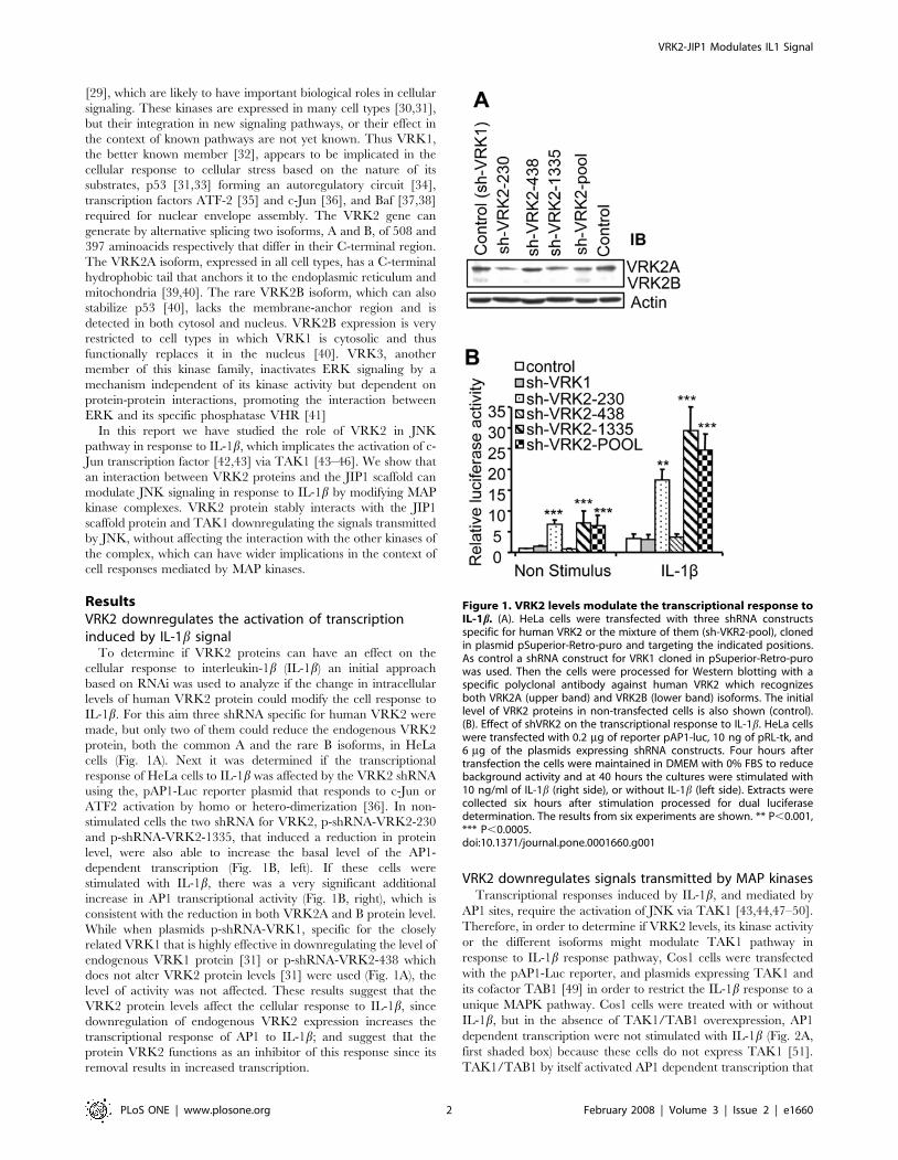
ResultsVRK2 downregulates the activation of transcriptioninduced by IL-1b signal
To determine if VRK2 proteins can have an effect on the
cellular response to interleukin-1b (IL-1b) an initial approach
based on RNAi was used to analyze if the change in intracellular
levels of human VRK2 protein could modify the cell response to
IL-1b. For this aim three shRNA specific for human VRK2 were
made, but only two of them could reduce the endogenous VRK2
protein, both the common A and the rare B isoforms, in HeLa
cells (Fig. 1A). Next it was determined if the transcriptional
response of HeLa cells to IL-1b was affected by the VRK2 shRNA
using the, pAP1-Luc reporter plasmid that responds to c-Jun or
ATF2 activation by homo or hetero-dimerization [36]. In non-
stimulated cells the two shRNA for VRK2, p-shRNA-VRK2-230
and p-shRNA-VRK2-1335, that induced a reduction in protein
level, were also able to increase the basal level of the AP1-
dependent transcription (Fig. 1B, left). If these cells were
stimulated with IL-1b, there was a very significant additional
increase in AP1 transcriptional activity (Fig. 1B, right), which is
consistent with the reduction in both VRK2A and B protein level.
While when plasmids p-shRNA-VRK1, specific for the closely
related VRK1 that is highly effective in downregulating the level of
endogenous VRK1 protein [31] or p-shRNA-VRK2-438 which
does not alter VRK2 protein levels [31] were used (Fig. 1A), the
level of activity was not affected. These results suggest that the
VRK2 protein levels affect the cellular response to IL-1b, since
downregulation of endogenous VRK2 expression increases the
transcriptional response of AP1 to IL-1b; and suggest that the
protein VRK2 functions as an inhibitor of this response since its
removal results in increased transcription.
VRK2 downregulates signals transmitted by MAP kinasesTranscriptional responses induced by IL-1b, and mediated by
AP1 sites, require the activation of JNK via TAK1 [43,44,47–50].
Therefore, in order to determine if VRK2 levels, its kinase activity
or the different isoforms might modulate TAK1 pathway in
response to IL-1b response pathway, Cos1 cells were transfected
with the pAP1-Luc reporter, and plasmids expressing TAK1 and
its cofactor TAB1 [49] in order to restrict the IL-1b response to a
unique MAPK pathway. Cos1 cells were treated with or without
IL-1b, but in the absence of TAK1/TAB1 overexpression, AP1
dependent transcription were not stimulated with IL-1b (Fig. 2A,
first shaded box) because these cells do not express TAK1 [51].
TAK1/TAB1 by itself activated AP1 dependent transcription that
Figure 1. VRK2 levels modulate the transcriptional response toIL-1b. (A). HeLa cells were transfected with three shRNA constructsspecific for human VRK2 or the mixture of them (sh-VKR2-pool), clonedin plasmid pSuperior-Retro-puro and targeting the indicated positions.As control a shRNA construct for VRK1 cloned in pSuperior-Retro-purowas used. Then the cells were processed for Western blotting with aspecific polyclonal antibody against human VRK2 which recognizesboth VRK2A (upper band) and VRK2B (lower band) isoforms. The initiallevel of VRK2 proteins in non-transfected cells is also shown (control).(B). Effect of shVRK2 on the transcriptional response to IL-1b. HeLa cellswere transfected with 0.2 mg of reporter pAP1-luc, 10 ng of pRL-tk, and6 mg of the plasmids expressing shRNA constructs. Four hours aftertransfection the cells were maintained in DMEM with 0% FBS to reducebackground activity and at 40 hours the cultures were stimulated with10 ng/ml of IL-1b (right side), or without IL-1b (left side). Extracts werecollected six hours after stimulation processed for dual luciferasedetermination. The results from six experiments are shown. ** P,0.001,*** P,0.0005.doi:10.1371/journal.pone.0001660.g001
VRK2-JIP1 Modulates IL1 Signal
PLoS ONE | www.plosone.org 2 February 2008 | Volume 3 | Issue 2 | e1660
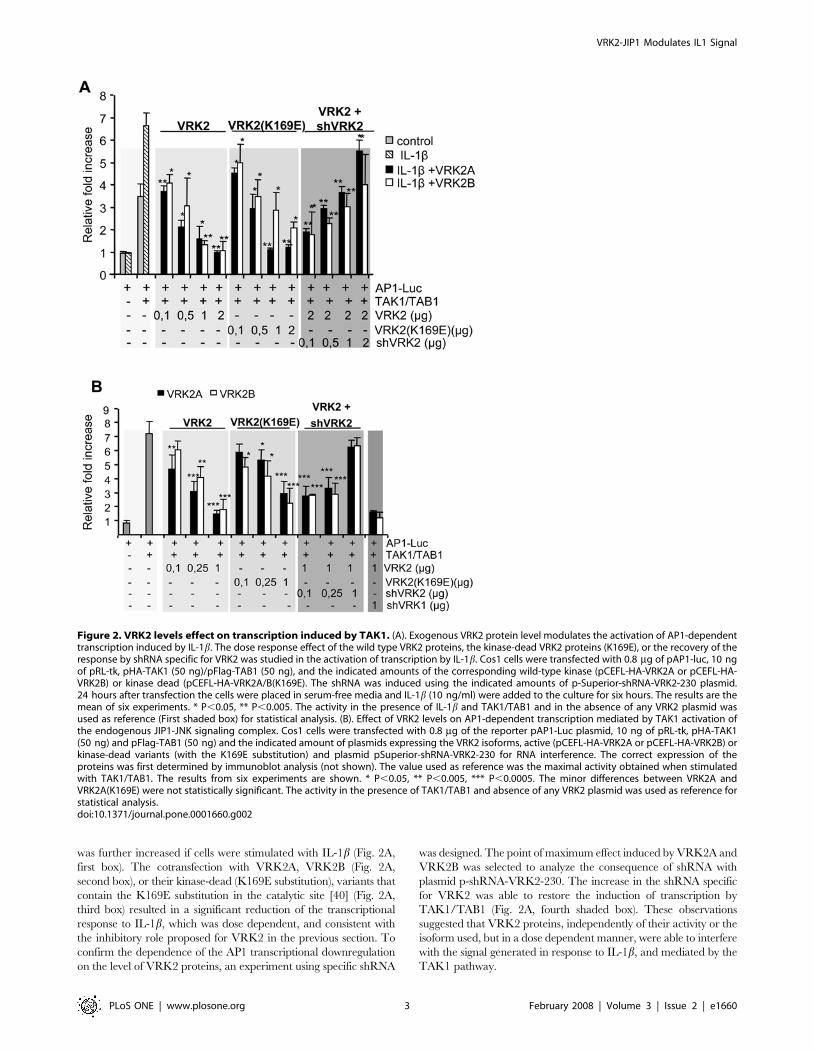
was further increased if cells were stimulated with IL-1b (Fig. 2A,
first box). The cotransfection with VRK2A, VRK2B (Fig. 2A,
second box), or their kinase-dead (K169E substitution), variants that
contain the K169E substitution in the catalytic site [40] (Fig. 2A,
third box) resulted in a significant reduction of the transcriptional
response to IL-1b, which was dose dependent, and consistent with
the inhibitory role proposed for VRK2 in the previous section. To
confirm the dependence of the AP1 transcriptional downregulation
on the level of VRK2 proteins, an experiment using specific shRNA
was designed. The point of maximum effect induced by VRK2A and
VRK2B was selected to analyze the consequence of shRNA with
plasmid p-shRNA-VRK2-230. The increase in the shRNA specific
for VRK2 was able to restore the induction of transcription by
TAK1/TAB1 (Fig. 2A, fourth shaded box). These observations
suggested that VRK2 proteins, independently of their activity or the
isoform used, but in a dose dependent manner, were able to interfere
with the signal generated in response to IL-1b, and mediated by the
TAK1 pathway.
Figure 2. VRK2 levels effect on transcription induced by TAK1. (A). Exogenous VRK2 protein level modulates the activation of AP1-dependenttranscription induced by IL-1b. The dose response effect of the wild type VRK2 proteins, the kinase-dead VRK2 proteins (K169E), or the recovery of theresponse by shRNA specific for VRK2 was studied in the activation of transcription by IL-1b. Cos1 cells were transfected with 0.8 mg of pAP1-luc, 10 ngof pRL-tk, pHA-TAK1 (50 ng)/pFlag-TAB1 (50 ng), and the indicated amounts of the corresponding wild-type kinase (pCEFL-HA-VRK2A or pCEFL-HA-VRK2B) or kinase dead (pCEFL-HA-VRK2A/B(K169E). The shRNA was induced using the indicated amounts of p-Superior-shRNA-VRK2-230 plasmid.24 hours after transfection the cells were placed in serum-free media and IL-1b (10 ng/ml) were added to the culture for six hours. The results are themean of six experiments. * P,0.05, ** P,0.005. The activity in the presence of IL-1b and TAK1/TAB1 and in the absence of any VRK2 plasmid wasused as reference (First shaded box) for statistical analysis. (B). Effect of VRK2 levels on AP1-dependent transcription mediated by TAK1 activation ofthe endogenous JIP1-JNK signaling complex. Cos1 cells were transfected with 0.8 mg of the reporter pAP1-Luc plasmid, 10 ng of pRL-tk, pHA-TAK1(50 ng) and pFlag-TAB1 (50 ng) and the indicated amount of plasmids expressing the VRK2 isoforms, active (pCEFL-HA-VRK2A or pCEFL-HA-VRK2B) orkinase-dead variants (with the K169E substitution) and plasmid pSuperior-shRNA-VRK2-230 for RNA interference. The correct expression of theproteins was first determined by immunoblot analysis (not shown). The value used as reference was the maximal activity obtained when stimulatedwith TAK1/TAB1. The results from six experiments are shown. * P,0.05, ** P,0.005, *** P,0.0005. The minor differences between VRK2A andVRK2A(K169E) were not statistically significant. The activity in the presence of TAK1/TAB1 and absence of any VRK2 plasmid was used as reference forstatistical analysis.doi:10.1371/journal.pone.0001660.g002
VRK2-JIP1 Modulates IL1 Signal
PLoS ONE | www.plosone.org 3 February 2008 | Volume 3 | Issue 2 | e1660
Next we attempted to establish at what stage, between the
receptor and the transcription factor, was VRK2 acting in the IL-
1b response pathway. For this aim, a similar assay was used, but
the endogenous MAPK pathway was stimulated only by
overexpression of the active form of TAK1 with TAB1. TAK1/
TAB1 strongly activated the transcription mediated by the AP1
response element (Fig. 2B, first shaded box). Increasing amounts of
either VRK2A (black bars) or VRK2B (white bars) resulted in a
significant downregulation of the TAK1/TAB1 activation of
transcription (Fig. 2B, second shaded box). The kinase-dead,
VRK2 (K169E) proteins similarly induced a downregulation of the
activation of transcription (Fig. 2B, third shaded box). The
negative effect on transcription induced by the maximum amount
of VRK2A or VRK2B was reversible in a dose dependent manner
using p-sh-RNA-VRK2-230 plasmid, specific for human VRK2
(Fig. 2B, fourth box). The shRNA specific for the closely related
VRK1 was used as before as a negative control and had no effect
(Fig. 2B, fifth shaded box). These results indicate that VRK2
interferes with the IL-1b signal at the MAP kinase level. And the
most likely explanation for these results is by a physical interaction
of VRK2 with some component of the signaling pathway, located
between TAK1 and the activation of the transcription factor.
VRK2 stably interacts with JIP1The effect of VRK2 is independent of its kinase activity; therefore
it is likely to be mediated by protein-protein interactions with MAPK
kinase complexes formed in response to IL-1b signaling. One likely
candidate is the scaffold protein JIP1, which assembles and regulates
the MAP kinases of the JNK signal transduction pathway MLK3,
MKK7 and JNK [20,52–54], and is required for JNK activation in
response to cytokine stimulation such as IL-1b but unnecessary for
JNK activation induced by UV radiation or anisomycin [1,20,55]. In
order to confirm that JIP1 is necessary for JNK activation in
response to IL-1b, an experiment based on RNAi silencing was
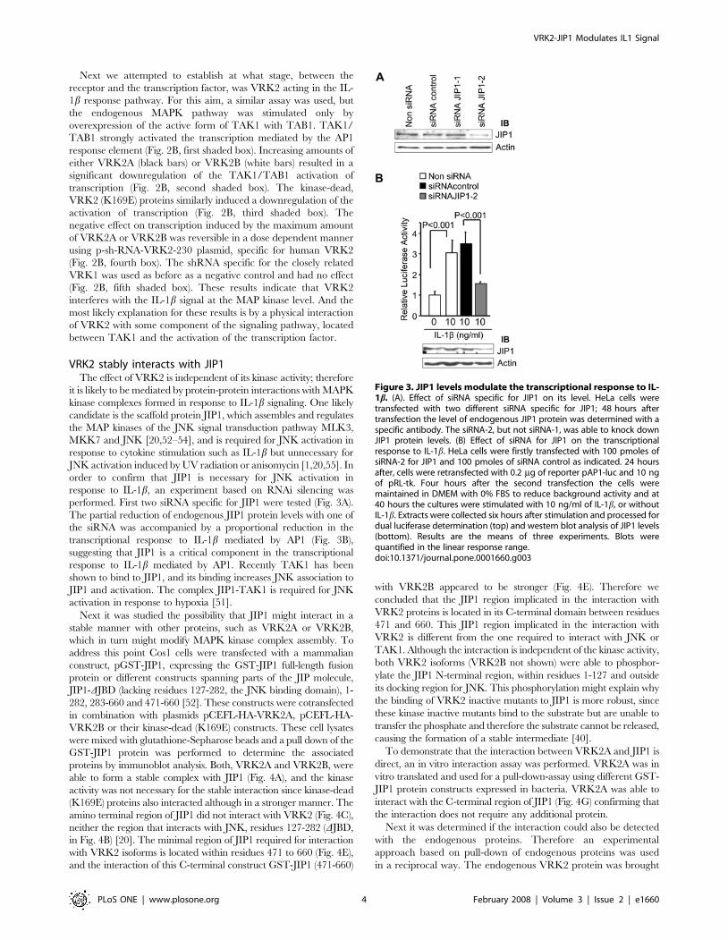
performed. First two siRNA specific for JIP1 were tested (Fig. 3A).
The partial reduction of endogenous JIP1 protein levels with one of
the siRNA was accompanied by a proportional reduction in the
transcriptional response to IL-1b mediated by AP1 (Fig. 3B),
suggesting that JIP1 is a critical component in the transcriptional
response to IL-1b mediated by AP1. Recently TAK1 has been
shown to bind to JIP1, and its binding increases JNK association to
JIP1 and activation. The complex JIP1-TAK1 is required for JNK
activation in response to hypoxia [51].
Next it was studied the possibility that JIP1 might interact in a
stable manner with other proteins, such as VRK2A or VRK2B,
which in turn might modify MAPK kinase complex assembly. To
address this point Cos1 cells were transfected with a mammalian
construct, pGST-JIP1, expressing the GST-JIP1 full-length fusion
protein or different constructs spanning parts of the JIP molecule,
JIP1-DJBD (lacking residues 127-282, the JNK binding domain), 1-
282, 283-660 and 471-660 [52]. These constructs were cotransfected
in combination with plasmids pCEFL-HA-VRK2A, pCEFL-HA-
VRK2B or their kinase-dead (K169E) constructs. These cell lysates
were mixed with glutathione-Sepharose beads and a pull down of the
GST-JIP1 protein was performed to determine the associated
proteins by immunoblot analysis. Both, VRK2A and VRK2B, were
able to form a stable complex with JIP1 (Fig. 4A), and the kinase
activity was not necessary for the stable interaction since kinase-dead
(K169E) proteins also interacted although in a stronger manner. The
amino terminal region of JIP1 did not interact with VRK2 (Fig. 4C),
neither the region that interacts with JNK, residues 127-282 (DJBD,
in Fig. 4B) [20]. The minimal region of JIP1 required for interaction
with VRK2 isoforms is located within residues 471 to 660 (Fig. 4E),
and the interaction of this C-terminal construct GST-JIP1 (471-660)
with VRK2B appeared to be stronger (Fig. 4E). Therefore we
concluded that the JIP1 region implicated in the interaction with
VRK2 proteins is located in its C-terminal domain between residues
471 and 660. This JIP1 region implicated in the interaction with
VRK2 is different from the one required to interact with JNK or
TAK1. Although the interaction is independent of the kinase activity,
both VRK2 isoforms (VRK2B not shown) were able to phosphor-
ylate the JIP1 N-terminal region, within residues 1-127 and outside
its docking region for JNK. This phosphorylation might explain why
the binding of VRK2 inactive mutants to JIP1 is more robust, since
these kinase inactive mutants bind to the substrate but are unable to
transfer the phosphate and therefore the substrate cannot be released,
causing the formation of a stable intermediate [40].
To demonstrate that the interaction between VRK2A and JIP1 is
direct, an in vitro interaction assay was performed. VRK2A was in
vitro translated and used for a pull-down-assay using different GST-
JIP1 protein constructs expressed in bacteria. VRK2A was able to
interact with the C-terminal region of JIP1 (Fig. 4G) confirming that
the interaction does not require any additional protein.
Next it was determined if the interaction could also be detected
with the endogenous proteins. Therefore an experimental
approach based on pull-down of endogenous proteins was used
in a reciprocal way. The endogenous VRK2 protein was brought
Figure 3. JIP1 levels modulate the transcriptional response to IL-1b. (A). Effect of siRNA specific for JIP1 on its level. HeLa cells weretransfected with two different siRNA specific for JIP1; 48 hours aftertransfection the level of endogenous JIP1 protein was determined with aspecific antibody. The siRNA-2, but not siRNA-1, was able to knock downJIP1 protein levels. (B) Effect of siRNA for JIP1 on the transcriptionalresponse to IL-1b. HeLa cells were firstly transfected with 100 pmoles ofsiRNA-2 for JIP1 and 100 pmoles of siRNA control as indicated. 24 hoursafter, cells were retransfected with 0.2 mg of reporter pAP1-luc and 10 ngof pRL-tk. Four hours after the second transfection the cells weremaintained in DMEM with 0% FBS to reduce background activity and at40 hours the cultures were stimulated with 10 ng/ml of IL-1b, or withoutIL-1b. Extracts were collected six hours after stimulation and processed fordual luciferase determination (top) and western blot analysis of JIP1 levels(bottom). Results are the means of three experiments. Blots werequantified in the linear response range.doi:10.1371/journal.pone.0001660.g003
VRK2-JIP1 Modulates IL1 Signal
PLoS ONE | www.plosone.org 4 February 2008 | Volume 3 | Issue 2 | e1660
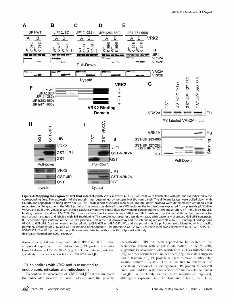
down in a pull-down assay with GST-JIP1 (Fig. 4H). In the
reciprocal experiment, the endogenous JIP1 protein was also
brought down by GST-VRK2A (Fig. 4I). These data supports the
specificity of the interaction between VRK2A and JIP1.
JIP1 colocalizes with VRK2 and is associated toendoplasmic reticulum and mitochondria
To confirm the association of VRK2 and JIP1 it was analyzed
the subcellular location of each molecule and the possible
colocalization. JIP1 has been reported to be located in the
perinuclear region with a particulate pattern in neural cells,
suggesting its association with membranes such as mitochondria
[56], or other organelles still unidentified [57]. These data suggests
that a fraction of JIP1 protein is likely to have a subcellular
location similar to VRK2. This led us first to determine the
subcellular location of the endogenous JIP1 protein in two cell
lines; Cos1 and HeLa (human cervical carcinoma cell line), given
that JIP1 is the family member more ubiquitously expressed,
although is expression is more abundant in brain, testis, lung,
Figure 4. Mapping the region of JIP1 that interacts with VRK2 isoforms. (A–F). Cos1 cells were transfected with plasmids as indicated in thecorresponding lane. The expression of the proteins was determined by western blot (bottom panel). The different lysates were pulled down withGlutathione-Sepharose to bring down the GST-JIP1 protein and associated molecules. The pull-down proteins were detected with antibodies thatrecognize the HA epitope in the VRK2 proteins. The constructs derived from VRK2 includes the two isoforms expressed from plasmids pCEFL-HA-VRK2A and pCEFL-HA-VRK2B as well as their catalytically inactive kinase-dead (KD) variants containing the K169E substitution. JIP1DJBD lacks the JNKbinding domain (residues 127-282). (G). In vitro interaction between human VRK2 and JIP1 proteins. The human VRK2 protein was in vitrotranscribed-translated and labeled with 35S methionine. This protein was used for a pulldown assay with bacterially expressed GST-JIP1 constructs.(F). Schematic representation of the GST-JIP1 proteins used in the pull-down assay and the interacting region with VRK2. (H). Binding of endogenousVRK2A to GST-JIP1. Cos1 cell were transfected with pCEFL-GST or pEBG-GST-JIP1, and the proteins in the pull-down were identified with a specificpolyclonal antibody for VRK2 and GST. (I). Binding of endogenous JIP1 protein to GST-VRK2A. Cos1 cells were transfected with pCEFL-GST or PCEFL-GST-VRK2A. The JIP1 protein in the pull-down was detected with a specific polyclonal antibody.doi:10.1371/journal.pone.0001660.g004
VRK2-JIP1 Modulates IL1 Signal
PLoS ONE | www.plosone.org 5 February 2008 | Volume 3 | Issue 2 | e1660
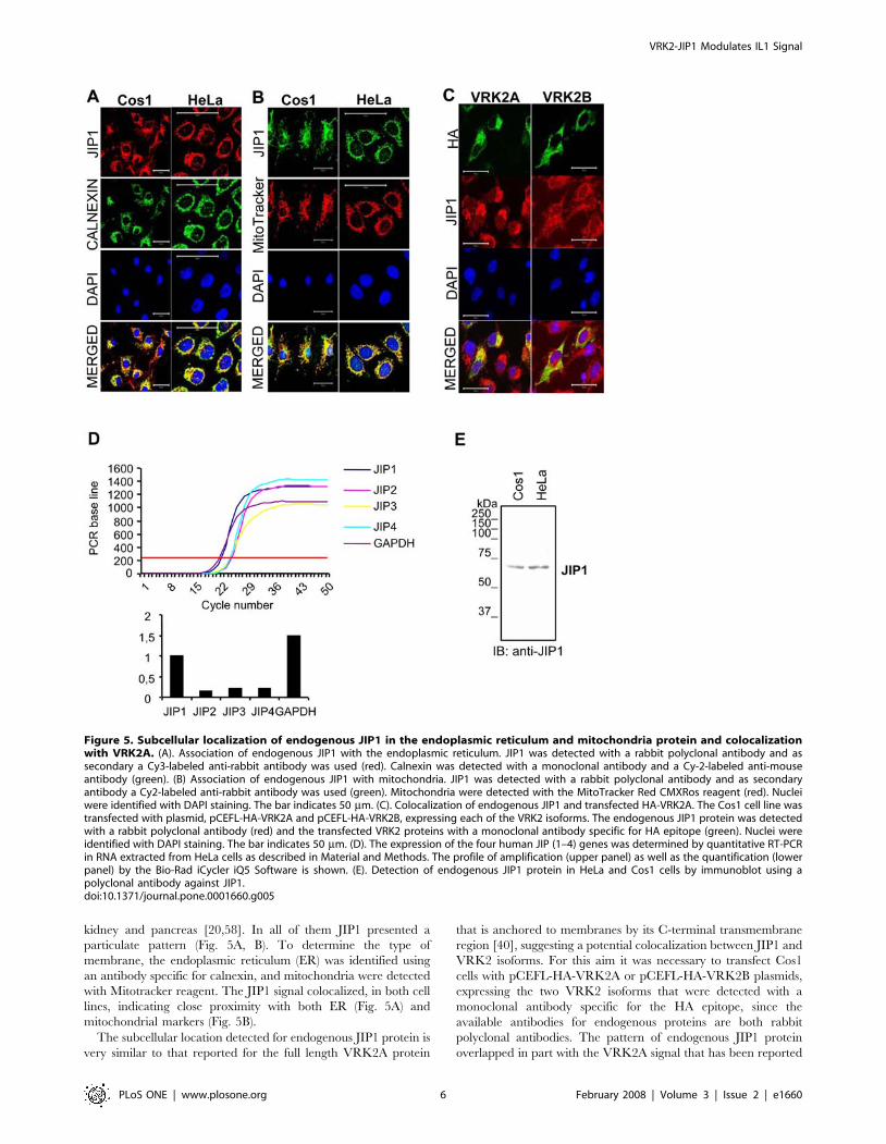
kidney and pancreas [20,58]. In all of them JIP1 presented a
particulate pattern (Fig. 5A, B). To determine the type of
membrane, the endoplasmic reticulum (ER) was identified using
an antibody specific for calnexin, and mitochondria were detected
with Mitotracker reagent. The JIP1 signal colocalized, in both cell
lines, indicating close proximity with both ER (Fig. 5A) and
mitochondrial markers (Fig. 5B).
The subcellular location detected for endogenous JIP1 protein is
very similar to that reported for the full length VRK2A protein
that is anchored to membranes by its C-terminal transmembrane
region [40], suggesting a potential colocalization between JIP1 and
VRK2 isoforms. For this aim it was necessary to transfect Cos1
cells with pCEFL-HA-VRK2A or pCEFL-HA-VRK2B plasmids,
expressing the two VRK2 isoforms that were detected with a
monoclonal antibody specific for the HA epitope, since the
available antibodies for endogenous proteins are both rabbit
polyclonal antibodies. The pattern of endogenous JIP1 protein
overlapped in part with the VRK2A signal that has been reported
Figure 5. Subcellular localization of endogenous JIP1 in the endoplasmic reticulum and mitochondria protein and colocalizationwith VRK2A. (A). Association of endogenous JIP1 with the endoplasmic reticulum. JIP1 was detected with a rabbit polyclonal antibody and assecondary a Cy3-labeled anti-rabbit antibody was used (red). Calnexin was detected with a monoclonal antibody and a Cy-2-labeled anti-mouseantibody (green). (B) Association of endogenous JIP1 with mitochondria. JIP1 was detected with a rabbit polyclonal antibody and as secondaryantibody a Cy2-labeled anti-rabbit antibody was used (green). Mitochondria were detected with the MitoTracker Red CMXRos reagent (red). Nucleiwere identified with DAPI staining. The bar indicates 50 mm. (C). Colocalization of endogenous JIP1 and transfected HA-VRK2A. The Cos1 cell line wastransfected with plasmid, pCEFL-HA-VRK2A and pCEFL-HA-VRK2B, expressing each of the VRK2 isoforms. The endogenous JIP1 protein was detectedwith a rabbit polyclonal antibody (red) and the transfected VRK2 proteins with a monoclonal antibody specific for HA epitope (green). Nuclei wereidentified with DAPI staining. The bar indicates 50 mm. (D). The expression of the four human JIP (1–4) genes was determined by quantitative RT-PCRin RNA extracted from HeLa cells as described in Material and Methods. The profile of amplification (upper panel) as well as the quantification (lowerpanel) by the Bio-Rad iCycler iQ5 Software is shown. (E). Detection of endogenous JIP1 protein in HeLa and Cos1 cells by immunoblot using apolyclonal antibody against JIP1.doi:10.1371/journal.pone.0001660.g005
VRK2-JIP1 Modulates IL1 Signal
PLoS ONE | www.plosone.org 6 February 2008 | Volume 3 | Issue 2 | e1660
to be bound to the endoplasmic reticulum by its C-terminal region
(Fig. 5C, left column). In the case of VRK2B the pattern detected
is much more diffuse in the cytosol (Fig. 5C, right column) [40].
These data support the physical interaction between JIP1 and the
membrane bound VRK2A protein.
To establish that JIP1 is the main human JIP gene expressed in
HeLa cells, the expression of the four JIP genes, 1 to 4, was
determined by RT-PCR. JIP1 expression is at least ten fold higher
than the rest of JIP messages expressed in this cell type (Fig. 5D).
This main form is recognized by the corresponding JIP antibody in
both cell lines (Fig. 5E).
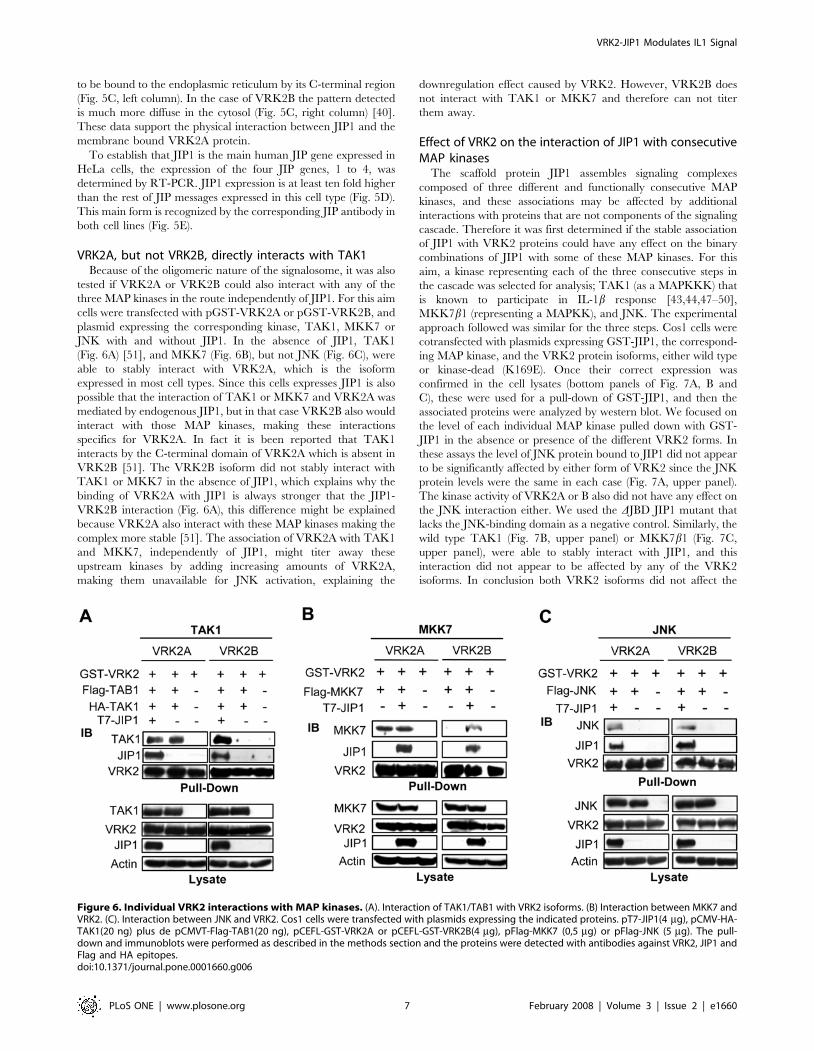
VRK2A, but not VRK2B, directly interacts with TAK1Because of the oligomeric nature of the signalosome, it was also
tested if VRK2A or VRK2B could also interact with any of the
three MAP kinases in the route independently of JIP1. For this aim
cells were transfected with pGST-VRK2A or pGST-VRK2B, and
plasmid expressing the corresponding kinase, TAK1, MKK7 or
JNK with and without JIP1. In the absence of JIP1, TAK1
(Fig. 6A) [51], and MKK7 (Fig. 6B), but not JNK (Fig. 6C), were
able to stably interact with VRK2A, which is the isoform
expressed in most cell types. Since this cells expresses JIP1 is also
possible that the interaction of TAK1 or MKK7 and VRK2A was
mediated by endogenous JIP1, but in that case VRK2B also would
interact with those MAP kinases, making these interactions
specifics for VRK2A. In fact it is been reported that TAK1
interacts by the C-terminal domain of VRK2A which is absent in
VRK2B [51]. The VRK2B isoform did not stably interact with
TAK1 or MKK7 in the absence of JIP1, which explains why the
binding of VRK2A with JIP1 is always stronger that the JIP1-
VRK2B interaction (Fig. 6A), this difference might be explained
because VRK2A also interact with these MAP kinases making the
complex more stable [51]. The association of VRK2A with TAK1
and MKK7, independently of JIP1, might titer away these
upstream kinases by adding increasing amounts of VRK2A,
making them unavailable for JNK activation, explaining the
downregulation effect caused by VRK2. However, VRK2B does
not interact with TAK1 or MKK7 and therefore can not titer
them away.
Effect of VRK2 on the interaction of JIP1 with consecutiveMAP kinases
The scaffold protein JIP1 assembles signaling complexes
composed of three different and functionally consecutive MAP
kinases, and these associations may be affected by additional
interactions with proteins that are not components of the signaling
cascade. Therefore it was first determined if the stable association
of JIP1 with VRK2 proteins could have any effect on the binary
combinations of JIP1 with some of these MAP kinases. For this
aim, a kinase representing each of the three consecutive steps in
the cascade was selected for analysis; TAK1 (as a MAPKKK) that
is known to participate in IL-1b response [43,44,47–50],
MKK7b1 (representing a MAPKK), and JNK. The experimental
approach followed was similar for the three steps. Cos1 cells were
cotransfected with plasmids expressing GST-JIP1, the correspond-
ing MAP kinase, and the VRK2 protein isoforms, either wild type
or kinase-dead (K169E). Once their correct expression was
confirmed in the cell lysates (bottom panels of Fig. 7A, B and
C), these were used for a pull-down of GST-JIP1, and then the
associated proteins were analyzed by western blot. We focused on
the level of each individual MAP kinase pulled down with GST-
JIP1 in the absence or presence of the different VRK2 forms. In
these assays the level of JNK protein bound to JIP1 did not appear
to be significantly affected by either form of VRK2 since the JNK
protein levels were the same in each case (Fig. 7A, upper panel).
The kinase activity of VRK2A or B also did not have any effect on
the JNK interaction either. We used the DJBD JIP1 mutant that
lacks the JNK-binding domain as a negative control. Similarly, the
wild type TAK1 (Fig. 7B, upper panel) or MKK7b1 (Fig. 7C,
upper panel), were able to stably interact with JIP1, and this
interaction did not appear to be affected by any of the VRK2
isoforms. In conclusion both VRK2 isoforms did not affect the
Figure 6. Individual VRK2 interactions with MAP kinases. (A). Interaction of TAK1/TAB1 with VRK2 isoforms. (B) Interaction between MKK7 andVRK2. (C). Interaction between JNK and VRK2. Cos1 cells were transfected with plasmids expressing the indicated proteins. pT7-JIP1(4 mg), pCMV-HA-TAK1(20 ng) plus de pCMVT-Flag-TAB1(20 ng), pCEFL-GST-VRK2A or pCEFL-GST-VRK2B(4 mg), pFlag-MKK7 (0,5 mg) or pFlag-JNK (5 mg). The pull-down and immunoblots were performed as described in the methods section and the proteins were detected with antibodies against VRK2, JIP1 andFlag and HA epitopes.doi:10.1371/journal.pone.0001660.g006
VRK2-JIP1 Modulates IL1 Signal
PLoS ONE | www.plosone.org 7 February 2008 | Volume 3 | Issue 2 | e1660
binary combinations of the MAP kinases mentioned above with
JIP1 or the effect can not be noticed when the complex is not
complete or active as in this case, since only one MAP kinase has
been overexpressed in each assay.
JIP1 signalosome: assembly of an oligomeric complexThe interaction between JIP1 and VRK2 may be exclusive of
JIP1-MAP kinase individual interactions since pull down experi-
ments performed before can not discriminate between complexes
formed by more than three proteins, and immunoprecipitation with
antibodies might interfere or compete with binding of additional
proteins, thus precipitating only the non complexed combinations
available to the antibody. Therefore the different possible combina-
tion of interacting proteins, or even the formation of large
complexes, was assayed when all of them are expressed at the same
level. The protein complexes were separated by performing a gel
filtration chromatography in a Superose 12 10/300 GL column that
specifically separates native molecules ranging from 50 to 1500 kDa
and permits to detect all different protein combinations present in
complexes. The different fractions were analyzed in western blots to
identify its components. First it was determined the complex
formation of oligomeric endogenous JIP1 and VRK2 proteins in
Cos1 cells. These two proteins are forming a large complexes of
different sizes (Fig. 8A), but the endogenous JNK is mostly free,
probably because the cells were not stimulated and therefore the
complex remains in a latent state, hence JNK is not gathered to the
whole complex. Some JNK is also detected in small complexes
formed by two or three proteins, as is the case for most of the
endogenous VRK2A protein (Fig. 8A). Surprisingly JIP1, endoge-
nous or transfected were forming large complexes in the range 300 to
1200 kDa (Fig. 8A, B). In the case of endogenous JIP1 also smaller
complexes were detected, but they contain bound endogenous
VRK2A, which is not detected free (Fig. 8A). A possible explanation
is that the polymerization of the complex might be a consequence of
JIP1 oligomerization that is known to be mediated by its SH3 to
form at least dimers of the signalosome [59].
Next it was determined the incorporation in these JIP1
complexes of different MAP kinases in the absence (Fig. 8C) or
presence of VRK2A (Fig. 8D) or VRK2B (Fig. 8E). For this aim
the whole cells extracts from Cos1 cells transfected with a mixture
of plasmids expressing the different MAP kinases without (Fig. 8C)
or with VRK2A (Fig. 8D) or VRK2B (Fig. 8E). The cell extracts
were fractionated and complexes at high molecular weigh
containing the proteins JIP1, TAK1, TAB1, MKK7 and JNK
were detected, suggesting that when the phosphorylation cascade
is activated, in this case by TAK1/TAB1 overexpression or by
hypoxia stimulation as we recently reported [51], all the MAP
kinases tend to be gathered by JIP1 forming an active signalosome.
But unexpectedly, the complex had a size of approximately of
1200 kDa (Fig. 8C), which is larger than the expected 340 kDa,
probably because the signalosome is an oligomeric complexes
Figure 7. Effect of VRK2 on the interaction of JIP1 withdifferent MAP kinases and detection of JIP1 signalosome. (A).Effect of VRK2A and B on the JIP1-JNK interaction. The plasmids usedwere pEBG-GST-JIP1(3 mg), pFlag-JNK(3 mg) and pCEFL-HA-VRK2A orpCEFL-HA-VRK2B wild-type or kinase-dead (5 mg). The proteins weredetected with antibodies for actin and the corresponding epitopes, HA,
r
GST, and Flag. (B). Effect of VRK2A orVRK2B on the TAK1-JIP1interaction. The plasmid used in Cos1 cell transfections were pEBG-GST-JIP1(3 mg), pCMV-HA-TAK1(50 ng) plus de pCMVT-Flag-TAB1(50 ng) and de pCEFL-HA-VRK2A/B wild-type or kinase-dead(5 mg). The proteins were detected with antibodies for actin and thecorresponding epitopes, HA, GST, and Flag. (C). Effect of VRK2AorVRK2B on the MKK7b1-JIP1 interaction. Cos1 cells were transfectedwith pEBG-GST-JIP1(3 mg), pFlag-MKK7b1(1 mg) and pCEFL-HA-VRK2A/Bwild-type or kinase-dead(5 mg). The proteins were detected withantibodies for actin and the corresponding epitopes, HA, GST, and Flag.doi:10.1371/journal.pone.0001660.g007
VRK2-JIP1 Modulates IL1 Signal
PLoS ONE | www.plosone.org 8 February 2008 | Volume 3 | Issue 2 | e1660
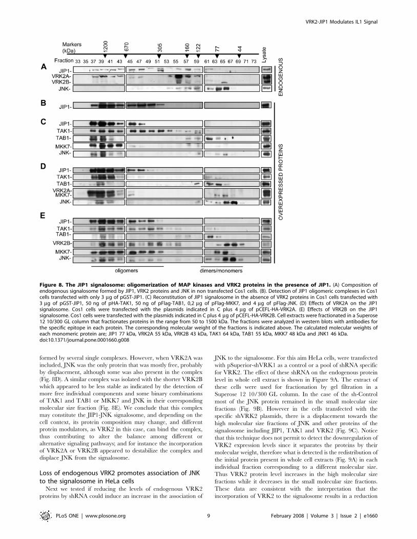
formed by several single complexes. However, when VRK2A was
included, JNK was the only protein that was mostly free, probably
by displacement, although some was also present in the complex
(Fig. 8D). A similar complex was isolated with the shorter VRK2B
which appeared to be less stable as indicated by the detection of
more free individual components and some binary combinations
of TAK1 and TAB1 or MKK7 and JNK in their corresponding
molecular size fraction (Fig. 8E). We conclude that this complex
may constitute the JIP1-JNK signalosome, and depending on the
cell context, its protein composition may change, and different
protein modulators, as VRK2 in this case, can bind the complex,
thus contributing to alter the balance among different or
alternative signaling pathways; and for instance the incorporation
of VRK2A or VRK2B appeared to destabilize the complex and
displace JNK from the signalosome.
Loss of endogenous VRK2 promotes association of JNKto the signalosome in HeLa cells
Next we tested if reducing the levels of endogenous VRK2
proteins by shRNA could induce an increase in the association of
JNK to the signalosome. For this aim HeLa cells, were transfected
with pSuperior-shVRK1 as a control or a pool of shRNA specific
for VRK2. The effect of these shRNA on the endogenous protein
level in whole cell extract is shown in Figure 9A. The extract of
these cells were used for fractionation by gel filtration in a
Superose 12 10/300 GL column. In the case of the sh-Control
most of the JNK protein remained in the small molecular size
fractions (Fig. 9B). However in the cells transfected with the
specific shVRK2 plasmids, there is a displacement towards the
high molecular size fractions of JNK and other proteins of the
signalosome including JIP1, TAK1 and VRK2 (Fig. 9C). Notice
that this technique does not permit to detect the downregulation of
VRK2 expression levels since it separates the proteins by their
molecular weight, therefore what is detected is the redistribution of
the initial protein present in whole cell extracts (Fig. 9A) in each
individual fraction corresponding to a different molecular size.
Thus VRK2 protein level increases in the high molecular size
fractions while it decreases in the small molecular size fractions.
These data are consistent with the interpretation that the
incorporation of VRK2 to the signalosome results in a reduction
Figure 8. The JIP1 signalosome: oligomerization of MAP kinases and VRK2 proteins in the presence of JIP1. (A) Composition ofendogenous signalosome formed by JIP1, VRK2 proteins and JNK in non transfected Cos1 cells. (B). Detection of JIP1 oligomeric complexes in Cos1cells transfected with only 3 mg of pGST-JIP1. (C) Reconstitution of JIP1 signalosome in the absence of VRK2 proteins in Cos1 cells transfected with3 mg of pGST-JIP1, 50 ng of pHA-TAK1, 50 ng of pFlag-TAB1, 0,2 mg of pFlag-MKK7, and 4 mg of pFlag-JNK. (D) Effects of VRK2A on the JIP1signalosome. Cos1 cells were transfected with the plasmids indicated in C plus 4 mg of pCEFL-HA-VRK2A. (E) Effects of VRK2B on the JIP1signalosome. Cos1 cells were transfected with the plasmids indicated in C plus 4 mg of pCEFL-HA-VRK2B. Cell extracts were fractionated in a Superose12 10/300 GL column that fractionates proteins in the range from 50 to 1500 kDa. The fractions were analyzed in western blots with antibodies forthe specific epitope in each protein. The corresponding molecular weight of the fractions is indicated above. The calculated molecular weights ofeach monomeric protein are; JIP1 77 kDa, VRK2A 55 kDa, VRK2B 43 kDa, TAK1 64 kDa, TAB1 55 kDa, MKK7 48 kDa and JNK1 46 kDa.doi:10.1371/journal.pone.0001660.g008
VRK2-JIP1 Modulates IL1 Signal
PLoS ONE | www.plosone.org 9 February 2008 | Volume 3 | Issue 2 | e1660
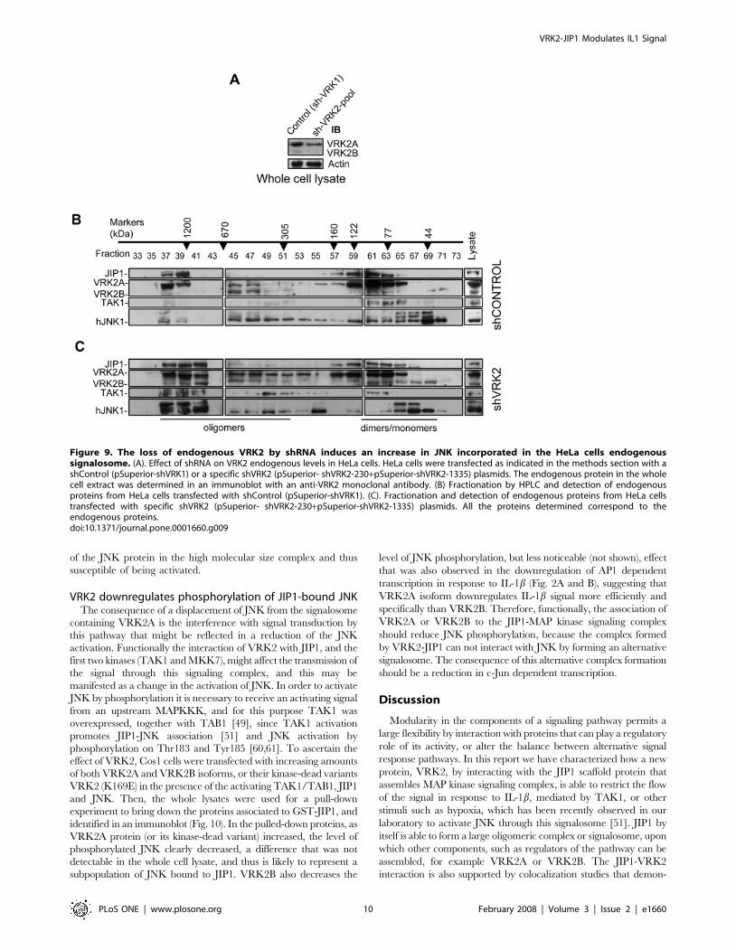
of the JNK protein in the high molecular size complex and thus
susceptible of being activated.
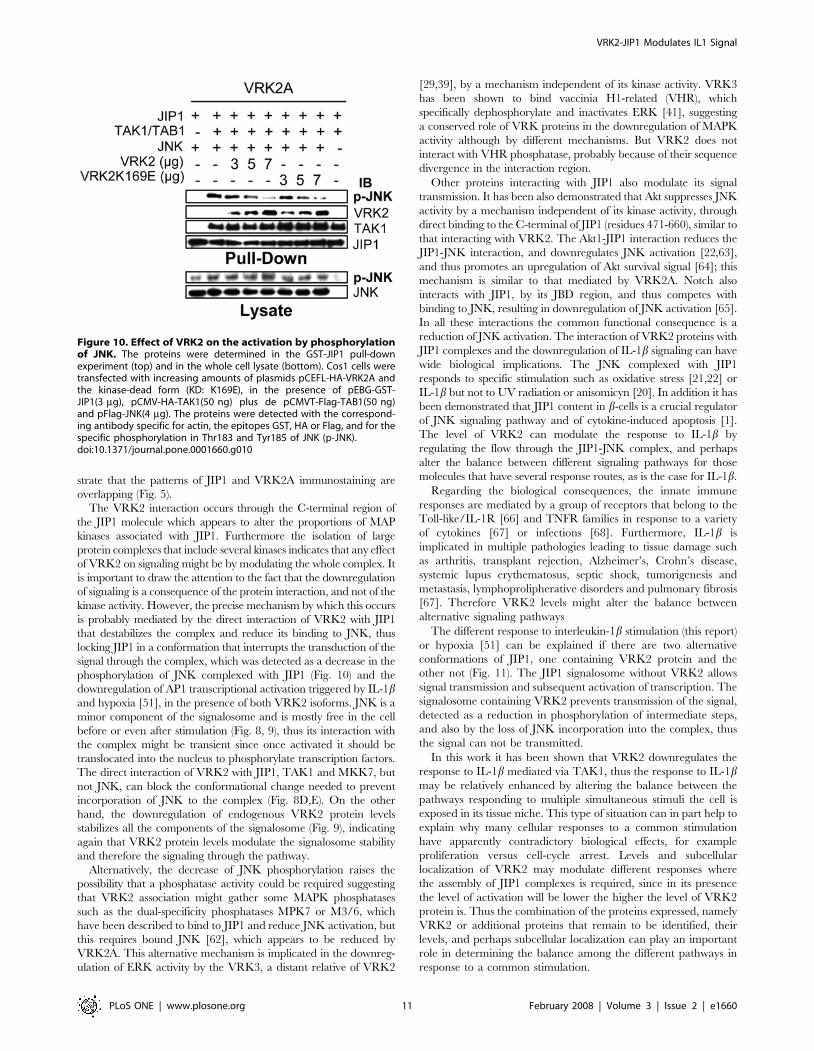
VRK2 downregulates phosphorylation of JIP1-bound JNKThe consequence of a displacement of JNK from the signalosome
containing VRK2A is the interference with signal transduction by
this pathway that might be reflected in a reduction of the JNK
activation. Functionally the interaction of VRK2 with JIP1, and the
first two kinases (TAK1 and MKK7), might affect the transmission of
the signal through this signaling complex, and this may be
manifested as a change in the activation of JNK. In order to activate
JNK by phosphorylation it is necessary to receive an activating signal
from an upstream MAPKKK, and for this purpose TAK1 was
overexpressed, together with TAB1 [49], since TAK1 activation
promotes JIP1-JNK association [51] and JNK activation by
phosphorylation on Thr183 and Tyr185 [60,61]. To ascertain the
effect of VRK2, Cos1 cells were transfected with increasing amounts
of both VRK2A and VRK2B isoforms, or their kinase-dead variants
VRK2 (K169E) in the presence of the activating TAK1/TAB1, JIP1
and JNK. Then, the whole lysates were used for a pull-down
experiment to bring down the proteins associated to GST-JIP1, and
identified in an immunoblot (Fig. 10). In the pulled-down proteins, as
VRK2A protein (or its kinase-dead variant) increased, the level of
phosphorylated JNK clearly decreased, a difference that was not
detectable in the whole cell lysate, and thus is likely to represent a
subpopulation of JNK bound to JIP1. VRK2B also decreases the
level of JNK phosphorylation, but less noticeable (not shown), effect
that was also observed in the downregulation of AP1 dependent
transcription in response to IL-1b (Fig. 2A and B), suggesting that
VRK2A isoform downregulates IL-1b signal more efficiently and
specifically than VRK2B. Therefore, functionally, the association of
VRK2A or VRK2B to the JIP1-MAP kinase signaling complex
should reduce JNK phosphorylation, because the complex formed
by VRK2-JIP1 can not interact with JNK by forming an alternative
signalosome. The consequence of this alternative complex formation
should be a reduction in c-Jun dependent transcription.
Discussion
Modularity in the components of a signaling pathway permits a
large flexibility by interaction with proteins that can play a regulatory
role of its activity, or alter the balance between alternative signal
response pathways. In this report we have characterized how a new
protein, VRK2, by interacting with the JIP1 scaffold protein that
assembles MAP kinase signaling complex, is able to restrict the flow
of the signal in response to IL-1b, mediated by TAK1, or other
stimuli such as hypoxia, which has been recently observed in our
laboratory to activate JNK through this signalosome [51]. JIP1 by
itself is able to form a large oligomeric complex or signalosome, upon
which other components, such as regulators of the pathway can be
assembled, for example VRK2A or VRK2B. The JIP1-VRK2
interaction is also supported by colocalization studies that demon-
Figure 9. The loss of endogenous VRK2 by shRNA induces an increase in JNK incorporated in the HeLa cells endogenoussignalosome. (A). Effect of shRNA on VRK2 endogenous levels in HeLa cells. HeLa cells were transfected as indicated in the methods section with ashControl (pSuperior-shVRK1) or a specific shVRK2 (pSuperior- shVRK2-230+pSuperior-shVRK2-1335) plasmids. The endogenous protein in the wholecell extract was determined in an immunoblot with an anti-VRK2 monoclonal antibody. (B) Fractionation by HPLC and detection of endogenousproteins from HeLa cells transfected with shControl (pSuperior-shVRK1). (C). Fractionation and detection of endogenous proteins from HeLa cellstransfected with specific shVRK2 (pSuperior- shVRK2-230+pSuperior-shVRK2-1335) plasmids. All the proteins determined correspond to theendogenous proteins.doi:10.1371/journal.pone.0001660.g009
VRK2-JIP1 Modulates IL1 Signal
PLoS ONE | www.plosone.org 10 February 2008 | Volume 3 | Issue 2 | e1660
strate that the patterns of JIP1 and VRK2A immunostaining are
overlapping (Fig. 5).
The VRK2 interaction occurs through the C-terminal region of
the JIP1 molecule which appears to alter the proportions of MAP
kinases associated with JIP1. Furthermore the isolation of large
protein complexes that include several kinases indicates that any effect
of VRK2 on signaling might be by modulating the whole complex. It
is important to draw the attention to the fact that the downregulation
of signaling is a consequence of the protein interaction, and not of the
kinase activity. However, the precise mechanism by which this occurs
is probably mediated by the direct interaction of VRK2 with JIP1
that destabilizes the complex and reduce its binding to JNK, thus
locking JIP1 in a conformation that interrupts the transduction of the
signal through the complex, which was detected as a decrease in the
phosphorylation of JNK complexed with JIP1 (Fig. 10) and the
downregulation of AP1 transcriptional activation triggered by IL-1band hypoxia [51], in the presence of both VRK2 isoforms. JNK is a
minor component of the signalosome and is mostly free in the cell
before or even after stimulation (Fig. 8, 9), thus its interaction with
the complex might be transient since once activated it should be
translocated into the nucleus to phosphorylate transcription factors.
The direct interaction of VRK2 with JIP1, TAK1 and MKK7, but
not JNK, can block the conformational change needed to prevent
incorporation of JNK to the complex (Fig. 8D,E). On the other
hand, the downregulation of endogenous VRK2 protein levels
stabilizes all the components of the signalosome (Fig. 9), indicating
again that VRK2 protein levels modulate the signalosome stability
and therefore the signaling through the pathway.
Alternatively, the decrease of JNK phosphorylation raises the
possibility that a phosphatase activity could be required suggesting
that VRK2 association might gather some MAPK phosphatases
such as the dual-specificity phosphatases MPK7 or M3/6, which
have been described to bind to JIP1 and reduce JNK activation, but
this requires bound JNK [62], which appears to be reduced by
VRK2A. This alternative mechanism is implicated in the downreg-
ulation of ERK activity by the VRK3, a distant relative of VRK2
[29,39], by a mechanism independent of its kinase activity. VRK3
has been shown to bind vaccinia H1-related (VHR), which
specifically dephosphorylate and inactivates ERK [41], suggesting
a conserved role of VRK proteins in the downregulation of MAPK
activity although by different mechanisms. But VRK2 does not
interact with VHR phosphatase, probably because of their sequence
divergence in the interaction region.
Other proteins interacting with JIP1 also modulate its signal
transmission. It has been also demonstrated that Akt suppresses JNK
activity by a mechanism independent of its kinase activity, through
direct binding to the C-terminal of JIP1 (residues 471-660), similar to
that interacting with VRK2. The Akt1-JIP1 interaction reduces the
JIP1-JNK interaction, and downregulates JNK activation [22,63],
and thus promotes an upregulation of Akt survival signal [64]; this
mechanism is similar to that mediated by VRK2A. Notch also
interacts with JIP1, by its JBD region, and thus competes with
binding to JNK, resulting in downregulation of JNK activation [65].
In all these interactions the common functional consequence is a
reduction of JNK activation. The interaction of VRK2 proteins with
JIP1 complexes and the downregulation of IL-1b signaling can have
wide biological implications. The JNK complexed with JIP1
responds to specific stimulation such as oxidative stress [21,22] or
IL-1b but not to UV radiation or anisomicyn [20]. In addition it has
been demonstrated that JIP1 content in b-cells is a crucial regulator
of JNK signaling pathway and of cytokine-induced apoptosis [1].
The level of VRK2 can modulate the response to IL-1b by
regulating the flow through the JIP1-JNK complex, and perhaps
alter the balance between different signaling pathways for those
molecules that have several response routes, as is the case for IL-1b.
Regarding the biological consequences, the innate immune
responses are mediated by a group of receptors that belong to the
Toll-like/IL-1R [66] and TNFR families in response to a variety
of cytokines [67] or infections [68]. Furthermore, IL-1b is
implicated in multiple pathologies leading to tissue damage such
as arthritis, transplant rejection, Alzheimer’s, Crohn’s disease,
systemic lupus erythematosus, septic shock, tumorigenesis and
metastasis, lymphoprolipherative disorders and pulmonary fibrosis
[67]. Therefore VRK2 levels might alter the balance between
alternative signaling pathways
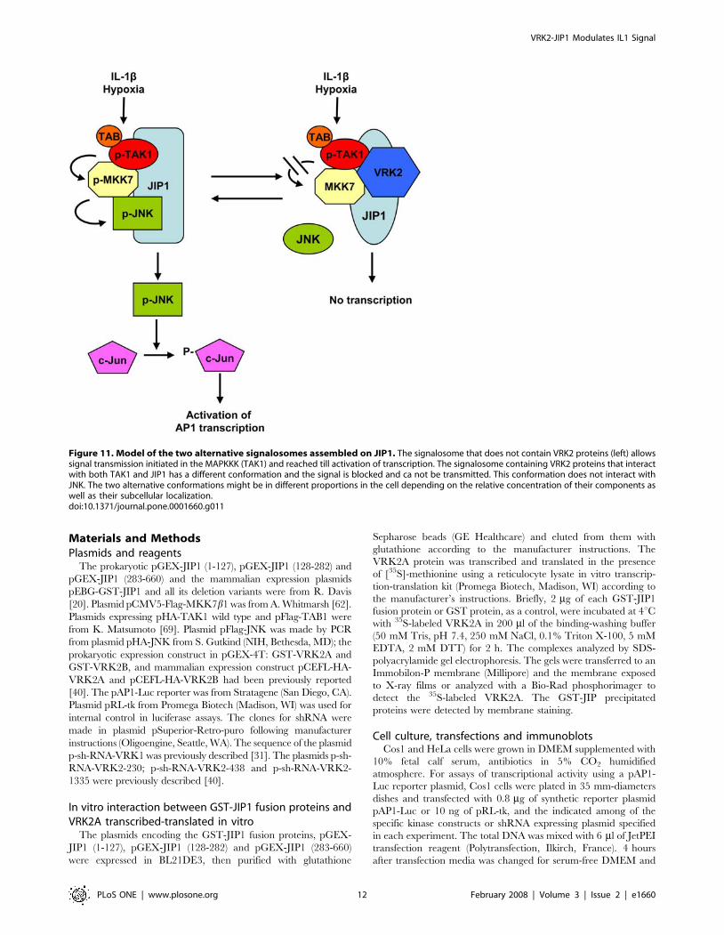
The different response to interleukin-1b stimulation (this report)
or hypoxia [51] can be explained if there are two alternative
conformations of JIP1, one containing VRK2 protein and the
other not (Fig. 11). The JIP1 signalosome without VRK2 allows
signal transmission and subsequent activation of transcription. The
signalosome containing VRK2 prevents transmission of the signal,
detected as a reduction in phosphorylation of intermediate steps,
and also by the loss of JNK incorporation into the complex, thus
the signal can not be transmitted.
In this work it has been shown that VRK2 downregulates the
response to IL-1b mediated via TAK1, thus the response to IL-1bmay be relatively enhanced by altering the balance between the
pathways responding to multiple simultaneous stimuli the cell is
exposed in its tissue niche. This type of situation can in part help to
explain why many cellular responses to a common stimulation
have apparently contradictory biological effects, for example
proliferation versus cell-cycle arrest. Levels and subcellular
localization of VRK2 may modulate different responses where
the assembly of JIP1 complexes is required, since in its presence
the level of activation will be lower the higher the level of VRK2
protein is. Thus the combination of the proteins expressed, namely
VRK2 or additional proteins that remain to be identified, their
levels, and perhaps subcellular localization can play an important
role in determining the balance among the different pathways in
response to a common stimulation.
Figure 10. Effect of VRK2 on the activation by phosphorylationof JNK. The proteins were determined in the GST-JIP1 pull-downexperiment (top) and in the whole cell lysate (bottom). Cos1 cells weretransfected with increasing amounts of plasmids pCEFL-HA-VRK2A andthe kinase-dead form (KD: K169E), in the presence of pEBG-GST-JIP1(3 mg), pCMV-HA-TAK1(50 ng) plus de pCMVT-Flag-TAB1(50 ng)and pFlag-JNK(4 mg). The proteins were detected with the correspond-ing antibody specific for actin, the epitopes GST, HA or Flag, and for thespecific phosphorylation in Thr183 and Tyr185 of JNK (p-JNK).doi:10.1371/journal.pone.0001660.g010
VRK2-JIP1 Modulates IL1 Signal
PLoS ONE | www.plosone.org 11 February 2008 | Volume 3 | Issue 2 | e1660
Materials and Methods
Plasmids and reagentsThe prokaryotic pGEX-JIP1 (1-127), pGEX-JIP1 (128-282) and
pGEX-JIP1 (283-660) and the mammalian expression plasmids
pEBG-GST-JIP1 and all its deletion variants were from R. Davis
[20]. Plasmid pCMV5-Flag-MKK7b1 was from A. Whitmarsh [62].
Plasmids expressing pHA-TAK1 wild type and pFlag-TAB1 were
from K. Matsumoto [69]. Plasmid pFlag-JNK was made by PCR
from plasmid pHA-JNK from S. Gutkind (NIH, Bethesda, MD); the
prokaryotic expression construct in pGEX-4T: GST-VRK2A and
GST-VRK2B, and mammalian expression construct pCEFL-HA-
VRK2A and pCEFL-HA-VRK2B had been previously reported
[40]. The pAP1-Luc reporter was from Stratagene (San Diego, CA).
Plasmid pRL-tk from Promega Biotech (Madison, WI) was used for
internal control in luciferase assays. The clones for shRNA were
made in plasmid pSuperior-Retro-puro following manufacturer
instructions (Oligoengine, Seattle, WA). The sequence of the plasmid
p-sh-RNA-VRK1 was previously described [31]. The plasmids p-sh-
RNA-VRK2-230; p-sh-RNA-VRK2-438 and p-sh-RNA-VRK2-
1335 were previously described [40].
In vitro interaction between GST-JIP1 fusion proteins andVRK2A transcribed-translated in vitro
The plasmids encoding the GST-JIP1 fusion proteins, pGEX-
JIP1 (1-127), pGEX-JIP1 (128-282) and pGEX-JIP1 (283-660)
were expressed in BL21DE3, then purified with glutathione
Sepharose beads (GE Healthcare) and eluted from them with
glutathione according to the manufacturer instructions. The
VRK2A protein was transcribed and translated in the presence
of [35S]-methionine using a reticulocyte lysate in vitro transcrip-
tion-translation kit (Promega Biotech, Madison, WI) according to
the manufacturer’s instructions. Briefly, 2 mg of each GST-JIP1
fusion protein or GST protein, as a control, were incubated at 4uCwith 35S-labeled VRK2A in 200 ml of the binding-washing buffer
(50 mM Tris, pH 7.4, 250 mM NaCl, 0.1% Triton X-100, 5 mM
EDTA, 2 mM DTT) for 2 h. The complexes analyzed by SDS-
polyacrylamide gel electrophoresis. The gels were transferred to an
Immobilon-P membrane (Millipore) and the membrane exposed
to X-ray films or analyzed with a Bio-Rad phosphorimager to
detect the 35S-labeled VRK2A. The GST-JIP precipitated
proteins were detected by membrane staining.
Cell culture, transfections and immunoblotsCos1 and HeLa cells were grown in DMEM supplemented with
10% fetal calf serum, antibiotics in 5% CO2 humidified
atmosphere. For assays of transcriptional activity using a pAP1-
Luc reporter plasmid, Cos1 cells were plated in 35 mm-diameters
dishes and transfected with 0.8 mg of synthetic reporter plasmid
pAP1-Luc or 10 ng of pRL-tk, and the indicated among of the
specific kinase constructs or shRNA expressing plasmid specified
in each experiment. The total DNA was mixed with 6 ml of JetPEI
transfection reagent (Polytransfection, Ilkirch, France). 4 hours
after transfection media was changed for serum-free DMEM and
Figure 11. Model of the two alternative signalosomes assembled on JIP1. The signalosome that does not contain VRK2 proteins (left) allowssignal transmission initiated in the MAPKKK (TAK1) and reached till activation of transcription. The signalosome containing VRK2 proteins that interactwith both TAK1 and JIP1 has a different conformation and the signal is blocked and ca not be transmitted. This conformation does not interact withJNK. The two alternative conformations might be in different proportions in the cell depending on the relative concentration of their components aswell as their subcellular localization.doi:10.1371/journal.pone.0001660.g011
VRK2-JIP1 Modulates IL1 Signal
PLoS ONE | www.plosone.org 12 February 2008 | Volume 3 | Issue 2 | e1660
cells were treated during 6 hours with 10 ng/ml of IL-1b(Preprotech, London, UK). Cells were lysed 48 hours after
transfection and luciferase activity determined with a Dual-
Luciferase reporter reagent from Promega.
For immunoblot analysis, cells were harvested 48 hours post-
transfection and lysed with buffer containing in 20 mM Tris-HCl
pH 7.4, 137 mM NaCl, 2 mM EDTA, 25 mM b-glycerophos-
phate, 10% (v/v) glycerol and 1% Triton-X100 with inhibitors of
proteases and phosphatases (1 mM PMSF, 10 mg/ml aprotinin,
10 mg/ml leupeptin, 1 mM Na orthovanadate). 50 mg of total
protein lysate were fractionated in a 10% SDS-polyacrylamide gel
and analyzed by western blot to identify the proteins present
depending on the experiment.
VRK2 knock-down by shRNAFor assays of transcriptional activity using shRNA specific for
VRK2, HeLa cells were plated in 35 mm-diameters dishes and
transfected with 0.2 mg of pAP1-Luc, 10 ng of pRL-tk and 6 mg of
the specific shRNA expressing plasmids indicated previously. The
total DNA was mixed with 12 ml of JetPEI transfection reagent.
Cells were treated as indicated before and luciferase activity was
determined 48 hours post-transfection with a Dual-Luciferase
reporter reagent from Promega.
JIP1 knock-down by siRNA interferenceHP Validated siRNA duplexes for JIP1 were purchased from
Quiagen (Valencia, CA). The targeted sequences for JIP1 (gene
accession number NM_005456) were TGGCATCAGCTTA-
CAGTGCAA (siRNA JIP1#1) and CTGGAGGAGTTTGAG-
GATGAA (siRNA JIP1#2). A functional siCONTROL nontarget-
ing siRNA pool from Dharmacon was used as a negative control,
and fluorescently labeled siGLO Lamin A/C siRNA was used for
lamin silencing and transfection efficiency. HeLa cells were plated in
35 mm-diameters dishes and transfected with 100 pmols of siRNAs
using 10 ml of LipofectamineTM 2000 transfection reagent (Invitro-
gen); 24 hours later, cells were retransfected with 0.2 mg of pAP1-
Luc and 10 ng of pRL-tk using Lipofectamine TM 2000 transfection
reagent. Cells were treated as indicated before and luciferase activity
was determined 48 hours post-transfection with a Dual-Luciferase
reporter reagent from Promega.
Detection of protein complexes by pull-downexperiments and gel filtration chromatography
For pull-down experiments Cos1 cells grown in 100 mm dishes
were transfected with different fragments of fusion proteins in
mammalian expression vectors. The amount and type of the
specific plasmid is indicated in each individual experiment. Whole
cell extracts prepared 48 hours after transfection were lysed in the
buffer mentioned before. To bring down the fusion protein with its
associated proteins the extract was mixed with glutathione-
Sepharose beads (GE Healthcare) for 12 hours at 4uC with gentle
shaking. The washed beads were loaded in a SDS-PAGE gel and
transferred to an Immobilon-P membrane (Millipore) and the
western blot was analyzed for the indicated proteins with the
corresponding antibody in individual experiments. For isolation of
protein complexes by gel filtration chromatography Cos1 cells
were transfected with 3 mg of pGST-JIP1, 50 ng of pHA-TAK1,
50 ng of pFlag-TAB1, 0.2 mg of pFlag-MKK7, 4 mg of pFlag-JNK
and 4 mg of pCEFL-HA-VRK2A or pCEFL-HA-VRK2B.
48 hours later protein extracts were prepared using buffer
containing in 20 mM Tris-HCl pH 7.4, 137 mM NaCl, 2 mM
EDTA, 25 mM b-glycerophosphate, 10% (v/v) glycerol and 1%
Triton-X100 with inhibitors of proteases and phosphatases (1 mM
PMSF, 10 mg/ml aprotinin, 10 mg/ml leupeptin, 1 mM Na
orthovanadate). Insoluble material was removed by centrifugation
at 16,0006g for 20 min. The supernatant, containing 1.5 mg of
dissolved protein, was fractionated by HPLC gel filtration through
a Superose 12 10/300 GL column (GE Healthcare). HPLC was
performed with an HP 1100 model from Agilent Technologies
(Germany) equipped with a ChemStation software, and developed
with a buffer containing 50 mM Tris-HCl, pH 7.5, 1 mM EDTA,
100 mM KCl at a flow rate of 0.1ml/min. 0,2 ml-fractions were
collected, precipitated and resolved on a 7,5 or 10% polyacryl-
amide gel and immunoblotted. Molecular weight markers used to
calibrate the column were: bovine thyroglobulin (670000),
apoferritin from horse spleen (440000), alcohol dehydrogenase
from yeast (150 000), bovine serum albumin (66000) and bovine
carbonic anhydrase (29000), all from Sigma. The effluent was
monitored at 280 nm.
Detection of endogenous JIP RNAThe expression of four endogenous human JIP genes was
determined by real time quantitative RT-PCR was performed as
previously described [40].Total RNA was extracted using the
‘‘RNeasy extraction kit’’ from Quiagen (Hilden, Germany). RNA
was analyzed and quantified using a Bioanalyzer 2100 nano-lab chip
from Agilent Technologies (Germany). 100 ng of total RNA were
used in a one-step reverse transcription real-time PCR amplification
reaction using the ‘‘Quantitec SYBR Green RT-PCR kit’’ from
Quiagen in an iCycler equipped with an iCycler iQ5 Software
(BioRad, Hercules, CA). The RT step was performed at 50uC for
30 minutes, and inactivated at 95uC for 30 seconds, the PCR phase
consisted of one cycle at 95uC for 15 minutes followed by 50 cycles
with three steps, of 94uC for 15 seconds, 58uC for 30 seconds and
72uC for 1 minute. PCR products were resolved in a 2% agarose
ethidium-bromide gel. The primers used for JIP1 amplification
were for JIP1(forward: 59-TCAGTCCAGGTTCCCTATCAC-39;
reverse: 59-TTGACGCCTATCTTCACACC-39), JIP2 (forward:
59-GCTTTTCCTCAGATCCGTTC-39, reverse: 59-CACTTGG-
AAGCCGACATTAC-39), JIP3 (forward: 59-AAGCCTCTATC-
CTGTCTGTC-39; reverse: 59-CCTCCAAGGTGAGTCTTC-
TG-39), JIP4 (forward: 59-AGCCCACAAAGTAGCAGTAG-39;
reverse: 59-GACAGAAGGTTCAAGTGGAAG-39), and for
GAPDH (forward: 59-GGTCTTACTCCTTGGAGGCCATGT-
39; reverse: 59-ACCTAACTACATGGTTTACATGTT-39).
Antibodies and reagentsHuman VRK2 was detected with a rabbit polyclonal antibody
[40]. Human JNK1 was detected with monoclonal (G151-333)
from BD Pharmingen. Human JIP1 protein was detected with
rabbit polyclonal (M-300) antibody; calnexin was detected with a
monoclonal (AF18); JNK phosphorylated in Thr183 and Tyr185
was detected with a monoclonal antibody (G7); endogenous TAK1
was detected with monoclonal (C9); and GST protein was detected
with a monoclonal (B-14), all from Santa Cruz. The HA epitope
was detected with a monoclonal (HA.11) from Covance (Berkeley,
CA). The FLAG epitope was detected with a rabbit polyclonal
antibody from Sigma. Actin was determined with a monoclonal
antibody (clone AC-15) from Sigma. A goat HRP-anti mouse
antibody was from GE Healthcare. A sheep HRP anti-rabbit
antibody was from Sigma. FluorolinkCy2 anti rabbit IgG,
FluorolinkCy3 anti rabbit IgG and FluorolinkCy2 anti mouse
IgG were from GE Healthcare. Mitochondria were detected using
the MitoTracker Red CMXRos reagent (Molecular Probes,
Invitrogen). Recombinant human IL-1b was from Peprotech
(London, UK).
VRK2-JIP1 Modulates IL1 Signal
PLoS ONE | www.plosone.org 13 February 2008 | Volume 3 | Issue 2 | e1660

The JIP1 region recognized by the anti-JIP1 rabbit polyclonal
antibody (M-300) was tested using 100 ng of the GST-fusion
proteins GST-JIP1 1-127, GST-JIP1 127-282, GST-JIP1 283-660
and GST that were subjected to immunoblot analysis with a GST
specific monoclonal antibody and the aJIP1 antibody. To test the
specificity of the anti JIP1 antibody, an aliquot of the diluted
antibody was incubated overnight at 4uC with 2 mg of GST-JIP1
(1-127) fusion protein, and as a control, another aliquot of the
diluted antibody was incubated with 2 mg of GST fusion protein.
The two aliquots were used to perform an immunoblot with HeLa
and Cos1 cell extracts to detect the endogenous JIP1 protein.
Confocal microscopyThe subcellular localization of JIP1, VRK2 endogenous or
transfected proteins were determined in the indicated cells lines
grown on coverslips and stained with the corresponding
antibodies. Cells were seeded in 60 mm dishes and transfected
24 hours later with 5 mg of pCEFL-HA-VRK2A and B mixed
with 10 ml of JetPEI transfection reagent (Polytransfection, Ilkirch,
France). 48 hours post-transfection the slides were collected and
fixed with 3% paraformaldehyde for 30 minutes at room
temperature, then treated with 100 mM glycine for 10 min at
room temperature and then permeabilized with 0.2% Triton X-
100 for 30 min at room temperature. The cells were blocked with
1% BSA in PBS for 30 min at room temperature followed by a
double immunostaining with the corresponding antibodies. Finally
cells were stained with DAPI (49, 69-diamidino-2-phenylindole)
(Sigma) 1:1000 in PBS for 10 min at room temperature, then cells
were washed with PBS, and slides were mounted with Gelvatol
(Monsanto). The images were acquired with a Zeiss LSM510
confocal microscope and the analysis was performed with the
LSM Image Examiner program (Zeiss).
Acknowledgments
The technical assistance by Virginia Gascon is greatly appreciated.
Author Contributions
Conceived and designed the experiments: PL SB. Performed the
experiments: SB MS CS. Analyzed the data: PL SB MS CS. Wrote the
paper: PL.
References
1. Haefliger JA, Tawadros T, Meylan L, Gurun SL, Roehrich ME, et al. (2003)
The scaffold protein IB1/JIP-1 is a critical mediator of cytokine-inducedapoptosis in pancreatic beta cells. J Cell Sci 116: 1463–1469.
2. Helbecque N, Abderrahamani A, Meylan L, Riederer B, Mooser V, et al. (2003)
Islet-brain1/C-Jun N-terminal kinase interacting protein-1 (IB1/JIP-1) promotervariant is associated with Alzheimer’s disease. Mol Psychiatry 8: 413–422, 363.
3. Scheinfeld MH, Matsuda S, D’Adamio L (2003) JNK-interacting protein-1
promotes transcription of A beta protein precursor but not A beta precursor-likeproteins, mechanistically different than Fe65. Proc Natl Acad Sci U S A 100:
1729–1734.
4. Waeber G, Delplanque J, Bonny C, Mooser V, Steinmann M, et al. (2000) Thegene MAPK8IP1, encoding islet-brain-1, is a candidate for type 2 diabetes. Nat
Genet 24: 291–295.
5. Kolch W, Calder M, Gilbert D (2005) When kinases meet mathematics: the
systems biology of MAPK signalling. FEBS Lett 579: 1891–1895.
6. Kolch W (2005) Coordinating ERK/MAPK signalling through scaffolds andinhibitors. Nat Rev Mol Cell Biol.
7. Hazzalin CA, Mahadevan LC (2002) MAPK-regulated transcription: a
continuously variable gene switch? Nat Rev Mol Cell Biol 3: 30–40.
8. Yang S-H, Sharrocks AD, Whitmarsh AJ (2003) Transcriptional regulation by
the MAP kinase signaling cascades. Gene 320: 3–21.
9. Whitmarsh AJ, Davis RJ (1999) Signal transduction by MAP kinases: regulationby phosphorylation-dependent switches. Sci STKE 1999: PE1.
10. Ichijo H (1999) From receptors to stress-activated MAP kinases. Oncogene 18:
6087–6093.
11. Dunn C, Wiltshire C, MacLaren A, Gillespie DA (2002) Molecular mechanism
and biological functions of c-Jun N-terminal kinase signalling via the c-Jun
transcription factor. Cell Signal 14: 585–593.
12. Cuevas BD, Abell AN, Johnson GL (2007) Role of mitogen-activated protein
kinase kinase kinases in signal integration. Oncogene 26: 3159–3171.
13. Liu J, Lin A (2005) Role of JNK activation in apoptosis: a double-edged sword.Cell Res 15: 36–42.
14. Tournier C, Whitmarsh AJ, Cavanagh J, Barret T, Davis RJ (1997) Mitogen-activated protein kinase 7 is an activator of the jun NH2-terminal kinase. Proc
Natl Acad Sci USA 94: 7337–7342.
15. Davis RJ (2000) Signal transduction by the JNK group of MAP kinases. Cell103: 239–252.
16. Karin M, Gallagher E (2005) From JNK to pay dirt: jun kinases, their
biochemistry, physiology and clinical importance. IUBMB Life 57: 283–295.
17. Dhanasekaran DN, Kashef K, Lee CM, Xu H, Reddy EP (2007) Scaffold
proteins of MAP-kinase modules. Oncogene 26: 3185–3202.
18. Yamano S, Tokino T, Yasuda M, Kaneuchi M, Takahashi M, et al. (1999)Induction of transformation and p53-dependent apoptosis by adenovirus type 5
E4orf6/7 cDNA. J Virol 73: 10095–10103.
19. Bonny C, Oberson A, Steinmann M, Schorderet DF, Nicod P, et al. (2000) IB1reduces cytokine-induced apoptosis of insulin-secreting cells. J Biol Chem 275:
16466–16472.
20. Whitmarsh AJ, Cavanagh J, Tournier C, Yasuda J, Davis RJ (1998) A
mammalian scaffold complex that selectively mediates MAP kinase activation.
Science 281: 1671–1674.
21. Whitmarsh AJ, Kuan CY, Kennedy NJ, Kelkar N, Haydar TF, et al. (2001)
Requirement of the JIP1 scaffold protein for stress-induced JNK activation.
Genes Dev 15: 2421–2432.
22. Song JJ, Lee YJ (2005) Dissociation of Akt1 from its negative regulator JIP1 is
mediated through the ASK1-MEK-JNK signal transduction pathway during
metabolic oxidative stress: a negative feedback loop. J Cell Biol 170: 61–72.
23. Scheinfeld MH, Roncarati R, Vito P, Lopez PA, Abdallah M, et al. (2002) Jun
NH2-terminal kinase (JNK) interacting protein 1 (JIP1) binds the cytoplasmic
domain of the Alzheimer’s beta-amyloid precursor protein (APP). J Biol Chem
277: 3767–3775.
24. Dong Z, Zhou L, Del Villar K, Ghanevati M, Tashjian V, et al. (2005) JIP1
regulates neuronal apoptosis in response to stress. Brain Res Mol Brain Res 134:
282–293.
25. Liew FY, Xu D, Brint EK, O’Neill LA (2005) Negative regulation of toll-like
receptor-mediated immune responses. Nat Rev Immunol 5: 446–458.
26. Symons A, Beinke S, Ley SC (2006) MAP kinase kinase kinases and innate
immunity. Trends Immunol 27: 40–48.
27. Nakahira M, Tanaka T, Robson BE, Mizgerd JP, Grusby MJ (2007) Regulation
of Signal Transducer and Activator of Transcription Signaling by the Tyrosine
Phosphatase PTP-BL. Immunity 26: 163–176.
28. Round JL, Humphries LA, Tomassian T, Mittelstadt P, Zhang M, et al. (2007)
Scaffold protein Dlgh1 coordinates alternative p38 kinase activation, directing T
cell receptor signals toward NFAT but not NF-kappaB transcription factors. Nat
Immunol 8: 154–161.
29. Manning G, Whyte DB, Martinez R, Hunter T, Sudarsanam S (2002) The
protein kinase complement of the human genome. Science 298: 1912–1934.
30. Nezu J, Oku A, Jones MH, Shimane M (1997) Identification of two novel human
putative serine/threonine kinases, VRK1 and VRK2, with structural similarity
to vaccinia virus B1R kinase. Genomics 45: 327–331.
31. Vega FM, Sevilla A, Lazo PA (2004) p53 Stabilization and Accumulation
Induced by Human Vaccinia-Related Kinase 1. Mol Cell Biol 24: 10366–10380.
32. Lazo PA, Vega FM, Sevilla A (2005) Vaccinia-related kinase-1. Afcs Nature
Molecule Page doi:10.1038/: mp.a003025.003001.
33. Lopez-Borges S, Lazo PA (2000) The human vaccinia-related kinase 1 (VRK1)
phosphorylates threonine-18 within the mdm-2 binding site of the p53 tumour
suppressor protein. Oncogene 19: 3656–3664.
34. Valbuena A, Vega FM, Blanco S, Lazo PA (2006) p53 Downregulates Its
Activating Vaccinia-Related Kinase 1, Forming a New Autoregulatory Loop.
Mol Cell Biol 26: 4782–4793.
35. Sevilla A, Santos CR, Vega FM, Lazo PA (2004) Human Vaccinia-related
Kinase 1 (VRK1) Activates the ATF2 Transcriptional Activity by Novel
Phosphorylation on Thr-73 and Ser-62 and Cooperates with JNK. J Biol Chem
279: 27458–27465.
36. Sevilla A, Santos CR, Barcia R, Vega FM, Lazo PA (2004) c-Jun
phosphorylation by the human vaccinia-related kinase 1 (VRK1) and its
cooperation with the N-terminal kinase of c-Jun (JNK). Oncogene 23:
8950–8958.
37. Nichols RJ, Wiebe MS, Traktman P (2006) The vaccinia-related kinases
phosphorylate the N’ terminus of BAF, regulating its interaction with DNA and
its retention in the nucleus. Mol Biol Cell 17: 2451–2464.
38. Gorjanacz M, Klerkx EP, Galy V, Santarella R, Lopez-Iglesias C, et al. (2007)
Caenorhabditis elegans BAF-1 and its kinase VRK-1 participate directly in post-
mitotic nuclear envelope assembly. Embo J 26: 132–143.
39. Nichols RJ, Traktman P (2004) Characterization of three paralogous members
of the Mammalian vaccinia related kinase family. J Biol Chem 279: 7934–7946.
VRK2-JIP1 Modulates IL1 Signal
PLoS ONE | www.plosone.org 14 February 2008 | Volume 3 | Issue 2 | e1660
40. Blanco S, Klimcakova L, Vega FM, Lazo PA (2006) The subcellular localization
of vaccinia-related kinase-2 (VRK2) isoforms determines their different effect onp53 stability in tumour cell lines. Febs J 273: 2487–2504.
41. Kang TH, Kim KT (2006) Negative regulation of ERK activity by VRK3-
mediated activation of VHR phosphatase. Nat Cell Biol 8: 863–869.42. Shim JH, Xiao C, Paschal AE, Bailey ST, Rao P, et al. (2005) TAK1, but not
TAB1 or TAB2, plays an essential role in multiple signaling pathways in vivo.Genes Dev 19: 2668–2681.
43. Hammaker DR, Boyle DL, Chabaud-Riou M, Firestein GS (2004) Regulation of
c-Jun N-terminal kinase by MEKK-2 and mitogen-activated protein kinasekinase kinases in rheumatoid arthritis. J Immunol 172: 1612–1618.
44. Jensen LE, Whitehead AS (2003) Pellino2 activates the mitogen activatedprotein kinase pathway. FEBS Lett 545: 199–202.
45. Cheung PC, Campbell DG, Nebreda AR, Cohen P (2003) Feedback control ofthe protein kinase TAK1 by SAPK2a/p38alpha. Embo J 22: 5793–5805.
46. Windheim M, Lang C, Peggie M, Plater LA, Cohen P (2007) Molecular
mechanisms involved in the regulation of cytokine production by muramyldipeptide. Biochem J 404: 179–190.
47. Holtmann H, Enninga J, Kalble S, Thiefes A, Dorrie A, et al. (2001) The MAPKkinase kinase TAK1 plays a central role in coupling the interleukin-1 receptor to
both transcriptional and RNA-targeted mechanisms of gene regulation. J Biol
Chem 276: 3508–3516.48. Takaesu G, Ninomiya-Tsuji J, Kishida S, Li X, Stark GR, et al. (2001)
Interleukin-1 (IL-1) Receptor-Associated Kinase Leads to Activation of TAK1by Inducing TAB2 Translocation in the IL-1 Signaling Pathway. Mol Cell Biol
21: 2475–2484.49. Jiang Z, Ninomiya-Tsuji J, Qian Y, Matsumoto K, Li X (2002) Interleukin-1 (IL-
1) Receptor-Associated Kinase-Dependent IL-1-Induced Signaling Complexes
Phosphorylate TAK1 and TAB2 at the Plasma Membrane and Activate TAK1in the Cytosol. Mol Cell Biol 22: 7158–7167.
50. Yao J, Kim TW, Qin J, Jiang Z, Qian Y, et al. (2007) Interleukin-1 (IL-1)-induced TAK1-dependent Versus MEKK3-dependent NFkappaB activation
pathways bifurcate at IL-1 receptor-associated kinase modification. J Biol Chem
282: 6075–6089.51. Blanco S, Santos C, Lazo PA (2007) Vaccinia-Related Kinase 2 Modulates the
Stress Response to Hypoxia Mediated by TAK1. Mol Cell Biol 27: 7273–7283.52. Yasuda J, Whitmarsh AJ, Cavanagh J, Sharma M, Davis RJ (1999) The JIP
Group of Mitogen-Activated Protein Kinase Scaffold Proteins. Mol Cell Biol 19:7245–7254.
53. Whitmarsh AJ, Davis RJ (2001) Analyzing JNK and p38 mitogen-activated
protein kinase activity. Methods Enzymol 332: 319–336.54. Dhanasekaran DN, Johnson GL (2007) MAPKs: function, regulation, role in
cancer and therapeutic targeting. Oncogene 26: 3097–3099.
55. Ling Z, Van de Casteele M, Dong J, Heimberg H, Haefliger JA, et al. (2003)
Variations in IB1/JIP1 expression regulate susceptibility of beta-cells tocytokine-induced apoptosis irrespective of C-Jun NH2-terminal kinase signaling.
Diabetes 52: 2497–2502.
56. Eminel S, Klettner A, Roemer L, Herdegen T, Waetzig V (2004) JNK2translocates to the mitochondria and mediates cytochrome c release in PC12
cells in response to 6-hydroxydopamine. J Biol Chem 279: 55385–55392.57. Li CH, Wang RM, Zhang QG, Zhang GY (2005) Activated mitogen-activated
protein kinase kinase 7 redistributes to the cytosol and binds to Jun N-terminal
kinase-interacting protein 1 involving oxidative stress during early reperfusion inrat hippocampal CA1 region. J Neurochem 93: 290–298.
58. Dickens M, Rogers JS, Cavanagh J, Raitano A, Xia Z, et al. (1997) Acytoplasmic inhibitor of the JNK signal transduction pathway. Science 277:
693–696.59. Kristensen O, Guenat S, Dar I, Allaman-Pillet N, Abderrahmani A, et al. (2006)
A unique set of SH3-SH3 interactions controls IB1 homodimerization. Embo J
25: 785–797.60. Tournier C, Whitmarsh AJ, Cavanagh J, Barrett T, Davis RJ (1999) The MKK7
gene encodes a group of c-Jun NH2-terminal kinase kinases. Mol Cell Biol 19:1569–1581.
61. Weston CR, Davis RJ (2002) The JNK signal transduction pathway. Curr Opin
Genet Dev 12: 14–21.62. Willoughby EA, Perkins GR, Collins MK, Whitmarsh AJ (2003) The JNK-
interacting protein-1 scaffold protein targets MAPK phosphatase-7 to dephos-phorylate JNK. J Biol Chem 278: 10731–10736.
63. Kim AH, Yano H, Cho H, Meyer D, Monks B, et al. (2002) Akt1 regulates aJNK scaffold during excitotoxic apoptosis. Neuron 35: 697–709.
64. Kim AH, Sasaki T, Chao MV (2003) JNK-interacting protein 1 promotes Akt1
activation. J Biol Chem 278: 29830–29836.65. Kim JW, Kim MJ, Kim KJ, Yun HJ, Chae JS, et al. (2005) Notch interferes with
the scaffold function of JNK-interacting protein 1 to inhibit the JNK signalingpathway. Proc Natl Acad Sci U S A 102: 14308–14313.
66. Muzio M, Polentarutti N, Bosisio D, Prahladan MK, Mantovani A (2000) Toll-
like receptors: a growing family of immune receptors that are differentiallyexpressed and regulated by different leukocytes. J Leukoc Biol 67: 450–456.
67. Aggarwal BB (2003) Signalling pathways of the TNF superfamily: a double-edged sword. Nat Rev Immunol 3: 745–756.
68. Beutler B, Jiang Z, Georgel P, Crozat K, Croker B, et al. (2006) Genetic analysisof host resistance: Toll-Like Receptor Signaling and Immunity at Large. Annu
Rev Immunol 24: 353–389.
69. Kishimoto K, Matsumoto K, Ninomiya-Tsuji J (2000) TAK1 mitogen-activatedprotein kinase kinase kinase is activated by autophosphorylation within its
activation loop. J Biol Chem 275: 7359–7364.
VRK2-JIP1 Modulates IL1 Signal
PLoS ONE | www.plosone.org 15 February 2008 | Volume 3 | Issue 2 | e1660