Modulated Binding of SATB1, a Matrix Attachment Region Protein, to the AT-Rich Sequence Flanking the...
10
MOLECULAR AND CELLULAR BIOLOGY, 0270-7306/00/$04.0010 Feb. 2000, p. 868–877 Vol. 20, No. 2 Copyright © 2000, American Society for Microbiology. All Rights Reserved. Modulated Binding of SATB1, a Matrix Attachment Region Protein, to the AT-Rich Sequence Flanking the Major Breakpoint Region of BCL2 MEERA RAMAKRISHNAN, 1 WEN-MAN LIU, 2 PATRICIA A. DICROCE, 1 ALEZA POSNER, 1 JIAN ZHENG, 1 TERUMI KOHWI-SHIGEMATSU, 2 AND THEODORE G. KRONTIRIS 1 * Division of Molecular Medicine, Beckman Research Institute of the City of Hope National Medical Center, Duarte, California 91010, 1 and Ernest Orlando Lawrence Berkeley National Laboratory, Life Sciences Division, University of California, Berkeley, California 94720 2 Received 17 May 1999/Returned for modification 6 July 1999/Accepted 22 October 1999 The t(14,18) chromosomal translocation that occurs in human follicular lymphoma constitutively activates the BCL2 gene and disrupts control of apoptosis. Interestingly, 70% of the t(14,18) translocations are confined to three 15-bp clusters positioned within a 150-bp region (major breakpoint region or [MBR]) in the untrans- lated portion of terminal exon 3. We analyzed DNA-protein interactions in the MBR, as these may play some role in targeting the translocation to this region. An 87-bp segment (87MBR) immediately 3* to breakpoint cluster 3 was essential for DNA-protein interaction monitored with mobility shift assays. We further delineated a core binding region within 87MBR: a 33-bp, very AT-rich sequence highly conserved between the human and mouse BCL2 gene (37MBR). We have purified and identified one of the core factors as the matrix attachment region (MAR) binding protein, SATB1, which is known to bind to AT-rich sequences with a high propensity to unwind. Additional factors in nuclear extracts, which we have not yet characterized further, increased SATB1 affinity for the 37MBR target four- to fivefold. Specific binding activity within 37MBR displayed cell cycle regulation in Jurkat T cells, while levels of SATB1 remained constant throughout the cell cycle. Finally, we demonstrated in vivo binding of SATB1 to the MBR, strongly suggesting the BCL2 major breakpoint region is a MAR. We discuss the potential consequences of our observations for both MBR fragility and regulatory function. The t(14,18) translocation of human follicular lymphoma activates the oncogene BCL2 (22, 25) and aborts programmed cell death (11, 12, 18). Although the BCL2 gene is more than 200 kb long, at least 70% of translocations occur within a 150-bp sequence located in the untranslated portion of the terminal exon, designated the major breakpoint region (MBR) (4, 25, 26). We have shown that the targeting of BCL2 trans- location within the MBR is considerably more precise than previously appreciated (27). Nearly all breakpoints that we examined (51 of 62) occurred within three 15-bp-wide clusters spaced evenly at approximately 50-bp intervals along the MBR. The breakpoints demonstrated fusion of BCL2 sequences to J H coding sequences in a canonical form suggesting rearrange- ment into immunoglobulin gene coding joints. Like immuno- globulin heavy-chain (IgH) rearrangements, untemplated nu- cleotides were usually present at the breakpoints. Tsujimoto et al. (26) suggested that these recombinations could be explained by the presence of a pseudo-D H element within the MBR. Under this model, aberrant events leading to translocation would involve the V(D)J recombinase recogniz- ing and using immunoglobulin signal sequences within the MBR to target the MBR-J H fusion. The inappropriate use of ectopic immunoglobulin signal sequences has been convinc- ingly demonstrated in the translocation of other genes, such as TTG1 (19). However, the clustering of translocation break- points as described in our study is completely inconsistent with the pseudo-D model. In this latter instance, translocations should occur in one cluster—the junction of pseudo-D coding and signal sequences—rather than at the three observed clus- ter sites. Furthermore, no signal joint has ever been found on the reciprocal product, der 18q-. Therefore, while the V(D)J recombinase probably participates in BCL2 translocation by site-directed cleavage and DNA end generation within the IGH locus, some other determinant(s) must be responsible for the involvement of the MBR. Two general alternatives to the pseudo-D model, which are not mutually exclusive, may be offered in explanation of BCL2 translocation. The first is that translocations take place through the stochastic intervention of any one of a host of recombina- tion/repair mechanisms (RRM). The hyperrecombination phe- notype of several chromosome instability disorders, for exam- ple, is likely to be explained by deficiencies of products important in these several pathways; the rare errors of an otherwise intact RRM could, therefore, generate transloca- tions. It should be noted that this hypothesis includes the possible existence of DNA signals that target RRM, creating translocation hot spots. Although such hot spots may be dis- tributed throughout the genome, in those (presumably fortu- itous) instances in which hot spots and growth-regulatory genes, such as BCL2, are located in the same region of DNA, rearrangements targeted by the hot spot may create a select- able phenotype through regulatory or coding sequence alter- ations in the nearby gene. We have previously shown that an octamer consensus se- quence, CC(A/T)CC(A/T)GC, present in 50% of highly re- combinogenic minisatellite repeat unit motifs, also occurs at or near the breakpoints of several oncogene translocations, in- cluding those involving MYC and BCL2 (16). Of particular * Corresponding author. Mailing address: Division of Molecular Medicine, Beckman Research Institute, City of Hope National Medi- cal Center, 1500 E. Duarte Rd., Duarte, CA 91010-3000. Phone: (626) 359-8111, ext. 4297. Fax: (626) 301-8862. E-mail: [email protected]. 868 on May 28, 2015 by guest http://mcb.asm.org/ Downloaded from
-
Upload
independent -
Category
Documents
-
view
0 -
download
0
Transcript of Modulated Binding of SATB1, a Matrix Attachment Region Protein, to the AT-Rich Sequence Flanking the...
MOLECULAR AND CELLULAR BIOLOGY,0270-7306/00/$04.0010
Feb. 2000, p. 868–877 Vol. 20, No. 2
Copyright © 2000, American Society for Microbiology. All Rights Reserved.
Modulated Binding of SATB1, a Matrix Attachment RegionProtein, to the AT-Rich Sequence Flanking the Major
Breakpoint Region of BCL2MEERA RAMAKRISHNAN,1 WEN-MAN LIU,2 PATRICIA A. DICROCE,1 ALEZA POSNER,1
JIAN ZHENG,1 TERUMI KOHWI-SHIGEMATSU,2 AND THEODORE G. KRONTIRIS1*
Division of Molecular Medicine, Beckman Research Institute of the City of Hope National Medical Center, Duarte,California 91010,1 and Ernest Orlando Lawrence Berkeley National Laboratory, Life Sciences Division, University of
California, Berkeley, California 947202
Received 17 May 1999/Returned for modification 6 July 1999/Accepted 22 October 1999
The t(14,18) chromosomal translocation that occurs in human follicular lymphoma constitutively activatesthe BCL2 gene and disrupts control of apoptosis. Interestingly, 70% of the t(14,18) translocations are confinedto three 15-bp clusters positioned within a 150-bp region (major breakpoint region or [MBR]) in the untrans-lated portion of terminal exon 3. We analyzed DNA-protein interactions in the MBR, as these may play somerole in targeting the translocation to this region. An 87-bp segment (87MBR) immediately 3* to breakpointcluster 3 was essential for DNA-protein interaction monitored with mobility shift assays. We further delineateda core binding region within 87MBR: a 33-bp, very AT-rich sequence highly conserved between the human andmouse BCL2 gene (37MBR). We have purified and identified one of the core factors as the matrix attachmentregion (MAR) binding protein, SATB1, which is known to bind to AT-rich sequences with a high propensity tounwind. Additional factors in nuclear extracts, which we have not yet characterized further, increased SATB1affinity for the 37MBR target four- to fivefold. Specific binding activity within 37MBR displayed cell cycleregulation in Jurkat T cells, while levels of SATB1 remained constant throughout the cell cycle. Finally, wedemonstrated in vivo binding of SATB1 to the MBR, strongly suggesting the BCL2 major breakpoint region isa MAR. We discuss the potential consequences of our observations for both MBR fragility and regulatoryfunction.
The t(14,18) translocation of human follicular lymphomaactivates the oncogene BCL2 (22, 25) and aborts programmedcell death (11, 12, 18). Although the BCL2 gene is more than200 kb long, at least 70% of translocations occur within a150-bp sequence located in the untranslated portion of theterminal exon, designated the major breakpoint region (MBR)(4, 25, 26). We have shown that the targeting of BCL2 trans-location within the MBR is considerably more precise thanpreviously appreciated (27). Nearly all breakpoints that weexamined (51 of 62) occurred within three 15-bp-wide clustersspaced evenly at approximately 50-bp intervals along the MBR.The breakpoints demonstrated fusion of BCL2 sequences toJH coding sequences in a canonical form suggesting rearrange-ment into immunoglobulin gene coding joints. Like immuno-globulin heavy-chain (IgH) rearrangements, untemplated nu-cleotides were usually present at the breakpoints.
Tsujimoto et al. (26) suggested that these recombinationscould be explained by the presence of a pseudo-DH elementwithin the MBR. Under this model, aberrant events leading totranslocation would involve the V(D)J recombinase recogniz-ing and using immunoglobulin signal sequences within theMBR to target the MBR-JH fusion. The inappropriate use ofectopic immunoglobulin signal sequences has been convinc-ingly demonstrated in the translocation of other genes, such asTTG1 (19). However, the clustering of translocation break-points as described in our study is completely inconsistent with
the pseudo-D model. In this latter instance, translocationsshould occur in one cluster—the junction of pseudo-D codingand signal sequences—rather than at the three observed clus-ter sites. Furthermore, no signal joint has ever been found onthe reciprocal product, der 18q-. Therefore, while the V(D)Jrecombinase probably participates in BCL2 translocation bysite-directed cleavage and DNA end generation within theIGH locus, some other determinant(s) must be responsible forthe involvement of the MBR.
Two general alternatives to the pseudo-D model, which arenot mutually exclusive, may be offered in explanation of BCL2translocation. The first is that translocations take place throughthe stochastic intervention of any one of a host of recombina-tion/repair mechanisms (RRM). The hyperrecombination phe-notype of several chromosome instability disorders, for exam-ple, is likely to be explained by deficiencies of productsimportant in these several pathways; the rare errors of anotherwise intact RRM could, therefore, generate transloca-tions. It should be noted that this hypothesis includes thepossible existence of DNA signals that target RRM, creatingtranslocation hot spots. Although such hot spots may be dis-tributed throughout the genome, in those (presumably fortu-itous) instances in which hot spots and growth-regulatorygenes, such as BCL2, are located in the same region of DNA,rearrangements targeted by the hot spot may create a select-able phenotype through regulatory or coding sequence alter-ations in the nearby gene.
We have previously shown that an octamer consensus se-quence, CC(A/T)CC(A/T)GC, present in 50% of highly re-combinogenic minisatellite repeat unit motifs, also occurs at ornear the breakpoints of several oncogene translocations, in-cluding those involving MYC and BCL2 (16). Of particular
* Corresponding author. Mailing address: Division of MolecularMedicine, Beckman Research Institute, City of Hope National Medi-cal Center, 1500 E. Duarte Rd., Duarte, CA 91010-3000. Phone: (626)359-8111, ext. 4297. Fax: (626) 301-8862. E-mail: [email protected].
868
on May 28, 2015 by guest
http://mcb.asm
.org/D
ownloaded from
interest is the observation that tandem 8-for-8 and 7-for-8matches of this octamer, designated x because of its homologyto the recognition signal of the RecBCD recombinase of Esch-erichia coli (23), form the immediate 59 boundary of the MBR:The first translocation cluster begins at the first MBR base 39to the 8/8 x. On the basis of this association, we have suggestedthe hypothesis that the consensus is a targeting signal for anRRM and, as such, defines a recombination hot spot for trans-location within the MBR.
The alternative model is that BCL2 translocations take placeat precise locations within the MBR because such rearrange-ments disrupt or alter functional configurations that occurthere. Other than polyadenylation and message stability sig-nals, however, little is known of the potential functional fea-tures of untranslated, terminal exons. In the case of BCL2,mRNA produced from translocated genes shows no half-lifealteration (22), although an advantage over wild-type mRNAin posttranscriptional processing has been noted (21). There-fore, a functional basis for targeting rearrangements wouldnecessarily involve a currently unknown (or unexpected) use ofDNA sequence.
To begin distinguishing between these possibilities, we haveidentified and characterized several complexes that form whennuclear extracts are incubated with DNA probes from thisregion of BCL2. Our initial studies indicated complexes form-ing with a 279-bp target that included the three breakpointclusters described by Wyatt et al. (27) and approximately 175bp of 59 and 39 flanking DNA. The most prominent of thesewas a very slowly migrating complex with a binding site withinthe AT-rich region immediately flanking breakpoint cluster 3(7). We now report purification and characterization of a ma-trix attachment region (MAR) binding protein present in thiscomplex.
MATERIALS AND METHODS
Cell lines. Jurkat is a human T-cell leukemia cell line. SUDHL-6 is a cell linederived from a human diffuse B-cell lymphoma and carries the t(14,18) translo-cation characteristic of BCL2-IGH fusions. HL60 is derived from a human acutepromyelocytic leukemia, and MCF-7 is a human breast cancer cell line.
Preparation of nuclear extract. Nuclear extracts were prepared as described byDignam et al. (8). The nuclear extracts from sorted cells and transient transfec-tions were prepared as described by Andrews and Faller (1). Protease inhibitorsphenylmethylsulfonyl fluoride (PMSF) and benzamidine to 1 mM and aprotinin,leupeptin, and pepstatin to 1 mg/ml were used in all nuclear extract preparations.
Preparation of target DNA. The 279-bp double-stranded DNA probe for gelshift analysis was PCR labeled as described by DiCroce and Krontiris (7), usingprimers RTW2 and RTW3. The 87MBR (87-bp segment of the MBR) probe wasPCR labeled by using oligomers PDC4 (59AATGATCAGACCTTTGAATGA39) and the 39 oligomer, RTW3. The 37MBR oligomer probe was prepared byannealing 37-bp sense and antisense oligonucleotides in the presence of 50 mMNaCl. The ends of the oligonucleotides containing SalI and XhoI sites were thenfilled in the presence of [a-32P]dATP (3,000 Ci/mmol), [a-32P]dCTP (3,000Ci/mmol), dTTP, and dGTP, using the Klenow fragment of DNA polymerase I(New England Biolabs). The radiolabeled 37MBR probe was then purified byusing a QIAquick nucleotide removal kit (Qiagen) or probeQuant G-50 micro-columns (Pharmacia). Gel-purified 37MBR oligomers for the DNA affinity col-umn were prepared similarly to the method for gel-purified 279MBR probepreparation (7).
MSA. DNA-protein binding reactions were carried out in 20 ml of a mixturecontaining 10 mM Tris HCl (pH 7.5), 40 mM NaCl, 1 mM EDTA, 1 mMb-mercaptoethanol, 4% glycerol, 0.5 mg of poly(dI-dC), 3 ng of probe, and 2 mgof nuclear extract. The samples were incubated for 30 min at room temperature.The reaction products were separated by electrophoresis through a 4% poly-acrylamide gel in 13 TBE (89 mM Tris HCl [pH 8.0], 89 mM boric acid, 2 mMEDTA). In competition experiments, nonradioactive oligonucleotide competi-tors were added along with labeled probe to the binding reaction prior to theaddition of nuclear extract. For Resource Q column fractions, assays were per-formed with 100 ng of poly(dI-dC) per reaction; for 37MBR and poly(rA)-oligo(dT) columns, 50 ng of poly(dI-dC) and 4 mg of bovine serum albumin(BSA) per reaction were used. Some gel mobility shift assays (MSAs) (Fig. 6 and7A) were done as described by Dickinson et al. (6), with slight modifications. Thereaction volume was 20 ml containing 10 mM HEPES (pH 7.9), 1 mM dithio-threitol (DTT), 50 mM KCl, 2.5 mM MgCl2, 10% glycerol, BSA (0.2 mg/ml),
poly(dI-dC) (0.05 to 0.25 mg), and purified SATB1 (special AT-rich bindingprotein 1) at room temperature for 20 min. The samples were separated over a6% acrylamide minigel (8 by 10 cm) containing 0.05% bisacrylamide, 5% glyc-erol, and 0.53 TBE. The gel was run at 120 V at 4°C for 2 h.
Chromatographic purification of SATB1 from Jurkat cells. The nuclear ex-tract (5 mg) was passed through a NAP-10 column equilibrated with buffer A (20mM HEPES [pH 7.9], 0.5 mM EDTA, 0.5 mM dithiothreitol, 5% glycerol, 0.5mM PMSF, 0.1 M NaCl). The extract in buffer A and 0.1 M NaCl was appliedonto a Resource Q column (1 ml; Pharmacia) attached to an AKTA purifiersystem (Pharmacia). The column was washed with 10 ml of buffer A, and proteinwas eluted with an increasing linear gradient of NaCl in buffer A. Fractionscontaining complex B1 or B3/B4 were diluted in buffer B (20 mM HEPES [pH7.9], 12.5 mM MgCl2, 1 mM DTT, 20% glycerol, 0.1% NP-40, 1 mM PMSF, 0.075M NaCl) and passed over the 37-mer DNA affinity column. The proteins wereeluted with an NaCl step gradient. The fraction containing SATB1 and poly-(ADP-ribose) polymerase (PARP) (0.3 M NaCl) was diluted in buffer B (withoutNP-40) and passed through a poly(rA)-oligo(dT)-cellulose column. The boundproteins were eluted with an NaCl step gradient.
Preparation of DNA affinity and poly(rA)-oligo(dT) columns. Gel-purifiedoligomers of 37MBR were annealed and ligated as described by Kadonaga andTjian (13). The oligomers (400 mg) were coupled to 0.8 g of cyanogen bromide-activated Sepharose 4B (Pharmacia). The coupling of the oligomers to the resinwas performed as described by Kadonaga and Tjian (13). The resin was stored incolumn storage buffer (10 mM Tris HCl [pH 7.6], 1 mM EDTA, 0.3 M NaCl,0.02% sodium azide) at 4°C. The poly(rA)-oligo(dT)-cellulose column was pre-pared by suspending 1 g of oligo(dT)-cellulose (Pharmacia) in buffer D (50 mMTris HCl [pH 8.0], 0.5 mM EDTA, 12 mM b-mercaptoethanol, 5 mM MgCl2, 1mM PMSF, 5% glycerol) for 10 min at room temperature (15). A solution ofpoly(rA) (10 mg/ml in 10 mM Tris HCl [pH 8.0]–1 mM EDTA) was incubatedwith the oligo(dT)-cellulose suspension [100 A260 units/g (dry powder) of oli-go(dT)] with shaking at room temperature for 30 min. The unbound poly(rA)was removed with equilibration buffer washes.
Western blot analysis. Protein samples were mixed with equal volumes of 23sample buffer (100 mM Tris HCl [pH 6.8], 200 mM DTT, 4% sodium dodecylsulfate (SDS), 0.2% bromophenol blue, 20% glycerol), boiled for 5 min, andresolved by SDS-polyacrylamide gel electrophoresis (SDS-PAGE) in 6% mini-gels. The proteins were electrophoretically transferred to Immobilon P mem-brane (Millipore) in 25 mM Tris HCl (pH 8.3)–192 mM glycine for 1 h at 60 V.The membrane was blocked in 5% BSA in TST buffer (20 mM Tris HCl [pH 7.4],0.5 M NaCl, 0.05% Tween 20), washed in TST buffer and incubated with a1:2,000 dilution of rabbit anti-SATB1 serum (20) at 4°C overnight. The mem-brane was washed and incubated with anti-rabbit antibody conjugated to horse-radish peroxidase as the secondary antibody (1:10,000) for 2 h at room temper-ature. The blot was extensively washed with TST buffer, incubated with enhancedchemiluminescence reagent solution (Amersham), and exposed to X-ray film forvisualization of the SATB1 band. For detection of PARP protein, the primaryantibody was goat polyclonal anti-PARP antibody (1:500; Santa Cruz Biotech-nology), and the secondary antibody was an anti-goat antibody conjugated toalkaline phosphatase (Bio-Rad). This blot was developed with an Immune-Litechemiluminescence assay kit (Biorad).
Staining of protein gels. Protein gels were silver stained by using a Bio-RadSilver Stain Plus kit.
Transient transfection. MCF-7 cells were transfected with a total of 30 mg ofDNA from pECE (vector alone) or pECH (SATB1 expression plasmid) (6),using a calcium phosphate transfection kit (Promega). Cells were incubated withDNA for 20 h following transfection. The medium was then changed, and thecells were harvested 48 h posttransfection in TEN (40 mM Tris HCl [pH 7.5], 1mM EDTA, 150 mM NaCl) and washed once with phosphate-buffered saline.The nuclear extracts were prepared by the protocol of Andrews and Faller (1).
FACS. The Jurkat cells (5 3 106 cells/ml) were incubated with Hoeschst 33342dye (10 mg/ml) at 37°C for 1 h. The cells were then concentrated to 3 3 107/mland incubated on ice for 30 min. Cells were sorted into six fractions with afluorescence-activated cell sorting (FACS) analyzer (MoFlo; Cytomation, Inc.).
Liquid chromatography-MS/MS sequencing analysis. The purified proteinswere separated on an SDS–6% polyacrylamide gel. The protein in the gel wasthen subjected to the in-gel reduction-alkylation-digestion procedure as de-scribed by Hellman et al. (10). The peptides obtained following tryptic digestionwere then subjected to liquid chromatography-tandem mass spectrometry (MS)analysis (24) on a MAT TSQ-700 triple-quadrupole mass spectrometer (FinniganCorp., San Jose, Calif.).
In vivo SATB1 binding assay. Jurkat cells were grown in RPMI 1640 mediumcontaining 10% fetal bovine serum in a humidified atmosphere of 5% CO2. Invivo cross-linking and immunoprecipitation with anti-SATB1 and control anti-bodies were performed by the published protocol (5, 15a) except that the cross-linked chromatin was digested with RsaI and HindIII (New England Biolabs).After the reversal of formaldehyde cross-links and DNA purification, DNA wasPCR amplified to identify sequences binding SATB1 in vivo. Six oligonucleotideswere used as primers to PCR amplify three loci: BCL2 locus (59CTTTAGAGTTGCTTTACGTTG39/59TCCATATTCATCACTTTGACAA39), SBS3 locus (59CAAGATTTTGATGGCCCCAAGCA39/59GGGCTTTACCAGATGAGAAT39), and HBB (59AGAAACTGCAGATTCTCTGCAT39/59CCTTCTACTTAGCCTACTTTTGA39). SBS3 is a locus isolated from Jurkat cell DNA that served as
VOL. 20, 2000 SATB1 BINDING IN THE BCL2 MAJOR BREAKPOINT REGION 869
on May 28, 2015 by guest
http://mcb.asm
.org/D
ownloaded from

a positive control for SATB1 binding in vivo (5; GenBank/EMBL/DDBJ acces-sion no. AF051676). The sequence chosen from human b-globin locus (HBB;accession no. U013171, positions 6789 to 7098), in which there is no SATB1binding site, served as a negative control. The accession number for the MBRregion is M13994.
PCR was performed in a 50-ml volume including 2.5 U of Taq polymerase(Display Systems Biotech), 13 manufacturer’s buffer, 1.5 mM MgCl2, four (200mM each), deoxynucleoside triphosphates and 2 mM each primer. Cycling con-ditions were as follows: 2 cycles of 95°C for 3 min, 53°C for 3 min, and 72°C for3 min, 38 cycles of 94°C for 1 min, 53°C for 1 min, and 72°C for 1 min, and anadditional 5 min at 72°C. PCR products were examined by 2% agarose gelelectrophoresis in the presence of ethidium bromide.
RESULTS
Sequence localization, specificity, and preliminary charac-terization of nuclear complexes binding the MBR. We previ-ously described the appearance of a slowly migrating nucleo-protein complex when nuclear extracts from particular humanleukocyte cell lines, such as Jurkat, were incubated with the279MBR target (7). A distinct complex that migrated evenmore slowly than that of Jurkat formed in the presence ofnuclear extracts from all other human and mouse cell linestested, including those of epithelial, fibroblastic, and leukocyticorigin. To characterize these complexes further, we performeddeletion analysis of the 279MBR region, using nuclear extractsfrom two cell lines displaying prototypic forms of the slowlymigrating complexes, Jurkat and MCF-7, in MSAs. Three tar-gets of successively smaller size were used: 279MBR, 87MBR,and 37MBR (Fig. 1A). The Jurkat complex formed with alltargets (Fig. 2A). Interestingly, the more slowly migrating com-plex seen in MCF-7 and other cell lines was observed only withthe larger targets, 279MBR and 87MBR; no complex was vi-sualized with the smaller 37MBR target (Fig. 2A). Therefore,we concluded that the core binding complex in Jurkat extractsrequired only the AT-rich, 37-bp region just downstream ofbreakpoint cluster 3 and that the core factor(s) involved wasabsent or inactive in MCF-7 and related nuclear extracts. Spec-ificity of the core complex present in Jurkat nuclear extracts
was verified in MSAs using cold (unlabeled) competitors. Witha labeled 87MBR target, cold competitors consisting of either87MBR or 37MBR effectively competed for binding, whereas151MBR competitor representing the 59 region of the MBRdid not compete.
Following localization of core factor binding to the 37MBRfragment, we used this target in all subsequent analyses andpurifications. As seen in the final lane of Fig. 2B, as well as inFig. 2C, four complexes were reproducibly detected with thisprobe, although their relative intensities varied from experi-ment to experiment. They were designated B1 through B4 andrepresented the slowest- to fastest-migrating complexes, re-spectively. B1 was consistently present in large amounts whenthe 37MBR target was used, as was B4. There was considerablevariation in the amount of B2 complex from experiment toexperiment. B3 was always present in very low amounts.
To complete our characterization of the specificity of thesecomplexes, competition MSAs were performed with a varietyof mutated targets (depicted in Fig. 1C). The principal com-plex observed in all MBR MSAs with Jurkat was B1, and thiscomplex was effectively competed only by wild-type oligomer.Several of the mutant oligomers showed diminished effects incompetition, as expected. Of note, the Mu2 competitor com-peted somewhat more effectively than Mu1 and Mu3, indicat-ing that the terminal region of the 37-bp target was moreimportant to the overall interaction with the core factor(s).The nonspecific binding of the B2 complex was competed byboth wild-type and mutant competitors. Complex B4 was notabolished by any competitor, and B3 showed a variable patternof competition suggesting, perhaps, an intermediate degree ofspecificity; as seen in Fig. 2C, competition from the wild-typeoligomer was somewhat more effective than competition withMu1 to Mu3. Because the 37MBR target was so AT rich, wewere concerned that it could denature under our experimentalconditions and bind as single-stranded DNA. Lanes 6 and 7 of
FIG. 1. (A) Schematic diagram of the BCL2 locus. Positions of the BCL2 gene exons, the MBR, the breakpoint clusters (C1, C2, and C3) and the AT-rich 39 endare shown. The subregions of the MBR (279MBR, 87MBR, 37MBR, and 151MBR) used in the DNA-protein interaction analyses are also shown. (B) DNA sequenceof the MBR. Positions of the probes are demarcated with thin arrows (279), thick hatched arrows (87), and thick solid arrow (37). The three breakpoint clusters areunderlined; a hatched horizontal line indicates the extent of 37MBR. (C) MSA probes and competitors. The 37-bp oligomer corresponding to 37MBR and its mutationsused in gel MSAs are shown. Lowercase boldface letters indicate positions of mutations.
870 RAMAKRISHNAN ET AL. MOL. CELL. BIOL.
on May 28, 2015 by guest
http://mcb.asm
.org/D
ownloaded from
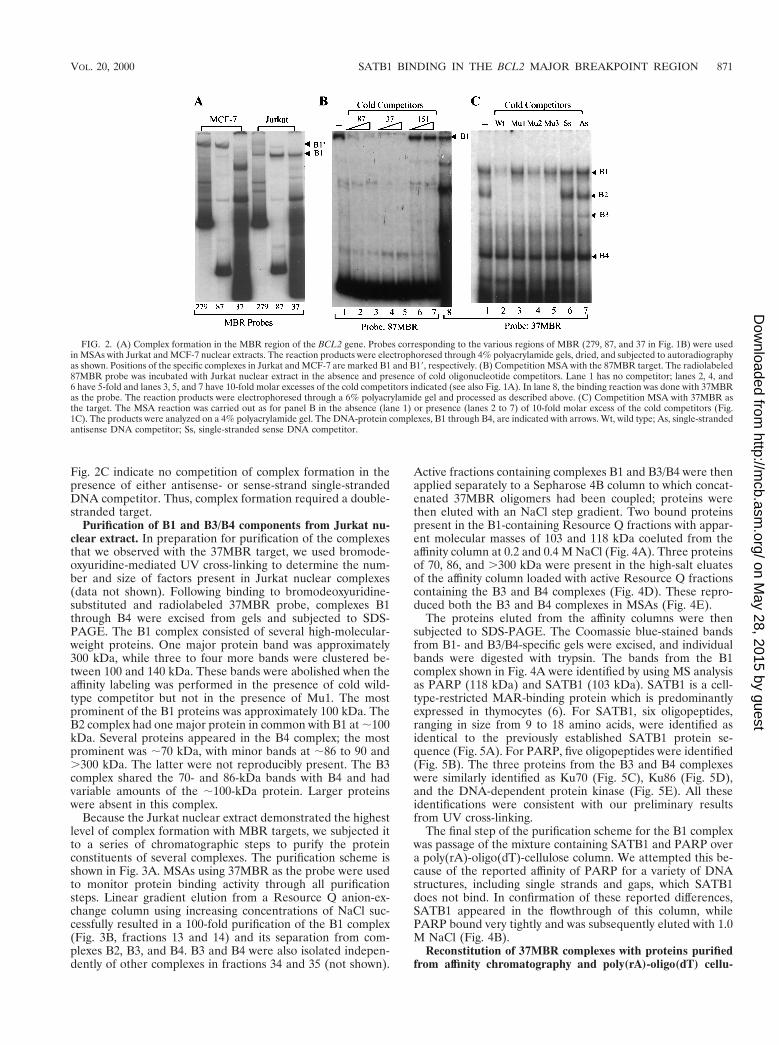
Fig. 2C indicate no competition of complex formation in thepresence of either antisense- or sense-strand single-strandedDNA competitor. Thus, complex formation required a double-stranded target.
Purification of B1 and B3/B4 components from Jurkat nu-clear extract. In preparation for purification of the complexesthat we observed with the 37MBR target, we used bromode-oxyuridine-mediated UV cross-linking to determine the num-ber and size of factors present in Jurkat nuclear complexes(data not shown). Following binding to bromodeoxyuridine-substituted and radiolabeled 37MBR probe, complexes B1through B4 were excised from gels and subjected to SDS-PAGE. The B1 complex consisted of several high-molecular-weight proteins. One major protein band was approximately300 kDa, while three to four more bands were clustered be-tween 100 and 140 kDa. These bands were abolished when theaffinity labeling was performed in the presence of cold wild-type competitor but not in the presence of Mu1. The mostprominent of the B1 proteins was approximately 100 kDa. TheB2 complex had one major protein in common with B1 at ;100kDa. Several proteins appeared in the B4 complex; the mostprominent was ;70 kDa, with minor bands at ;86 to 90 and.300 kDa. The latter were not reproducibly present. The B3complex shared the 70- and 86-kDa bands with B4 and hadvariable amounts of the ;100-kDa protein. Larger proteinswere absent in this complex.
Because the Jurkat nuclear extract demonstrated the highestlevel of complex formation with MBR targets, we subjected itto a series of chromatographic steps to purify the proteinconstituents of several complexes. The purification scheme isshown in Fig. 3A. MSAs using 37MBR as the probe were usedto monitor protein binding activity through all purificationsteps. Linear gradient elution from a Resource Q anion-ex-change column using increasing concentrations of NaCl suc-cessfully resulted in a 100-fold purification of the B1 complex(Fig. 3B, fractions 13 and 14) and its separation from com-plexes B2, B3, and B4. B3 and B4 were also isolated indepen-dently of other complexes in fractions 34 and 35 (not shown).
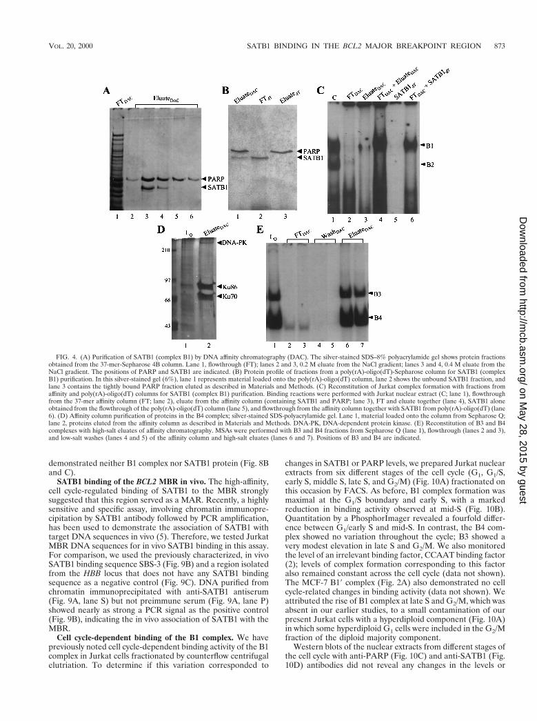
Active fractions containing complexes B1 and B3/B4 were thenapplied separately to a Sepharose 4B column to which concat-enated 37MBR oligomers had been coupled; proteins werethen eluted with an NaCl step gradient. Two bound proteinspresent in the B1-containing Resource Q fractions with appar-ent molecular masses of 103 and 118 kDa coeluted from theaffinity column at 0.2 and 0.4 M NaCl (Fig. 4A). Three proteinsof 70, 86, and .300 kDa were present in the high-salt eluatesof the affinity column loaded with active Resource Q fractionscontaining the B3 and B4 complexes (Fig. 4D). These repro-duced both the B3 and B4 complexes in MSAs (Fig. 4E).
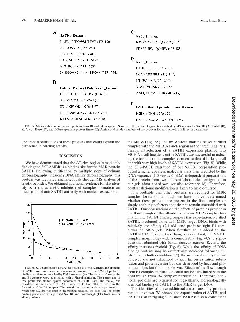
The proteins eluted from the affinity columns were thensubjected to SDS-PAGE. The Coomassie blue-stained bandsfrom B1- and B3/B4-specific gels were excised, and individualbands were digested with trypsin. The bands from the B1complex shown in Fig. 4A were identified by using MS analysisas PARP (118 kDa) and SATB1 (103 kDa). SATB1 is a cell-type-restricted MAR-binding protein which is predominantlyexpressed in thymocytes (6). For SATB1, six oligopeptides,ranging in size from 9 to 18 amino acids, were identified asidentical to the previously established SATB1 protein se-quence (Fig. 5A). For PARP, five oligopeptides were identified(Fig. 5B). The three proteins from the B3 and B4 complexeswere similarly identified as Ku70 (Fig. 5C), Ku86 (Fig. 5D),and the DNA-dependent protein kinase (Fig. 5E). All theseidentifications were consistent with our preliminary resultsfrom UV cross-linking.
The final step of the purification scheme for the B1 complexwas passage of the mixture containing SATB1 and PARP overa poly(rA)-oligo(dT)-cellulose column. We attempted this be-cause of the reported affinity of PARP for a variety of DNAstructures, including single strands and gaps, which SATB1does not bind. In confirmation of these reported differences,SATB1 appeared in the flowthrough of this column, whilePARP bound very tightly and was subsequently eluted with 1.0M NaCl (Fig. 4B).
Reconstitution of 37MBR complexes with proteins purifiedfrom affinity chromatography and poly(rA)-oligo(dT) cellu-
FIG. 2. (A) Complex formation in the MBR region of the BCL2 gene. Probes corresponding to the various regions of MBR (279, 87, and 37 in Fig. 1B) were usedin MSAs with Jurkat and MCF-7 nuclear extracts. The reaction products were electrophoresed through 4% polyacrylamide gels, dried, and subjected to autoradiographyas shown. Positions of the specific complexes in Jurkat and MCF-7 are marked B1 and B19, respectively. (B) Competition MSA with the 87MBR target. The radiolabeled87MBR probe was incubated with Jurkat nuclear extract in the absence and presence of cold oligonucleotide competitors. Lane 1 has no competitor; lanes 2, 4, and6 have 5-fold and lanes 3, 5, and 7 have 10-fold molar excesses of the cold competitors indicated (see also Fig. 1A). In lane 8, the binding reaction was done with 37MBRas the probe. The reaction products were electrophoresed through a 6% polyacrylamide gel and processed as described above. (C) Competition MSA with 37MBR asthe target. The MSA reaction was carried out as for panel B in the absence (lane 1) or presence (lanes 2 to 7) of 10-fold molar excess of the cold competitors (Fig.1C). The products were analyzed on a 4% polyacrylamide gel. The DNA-protein complexes, B1 through B4, are indicated with arrows. Wt, wild type; As, single-strandedantisense DNA competitor; Ss, single-stranded sense DNA competitor.
VOL. 20, 2000 SATB1 BINDING IN THE BCL2 MAJOR BREAKPOINT REGION 871
on May 28, 2015 by guest
http://mcb.asm
.org/D
ownloaded from
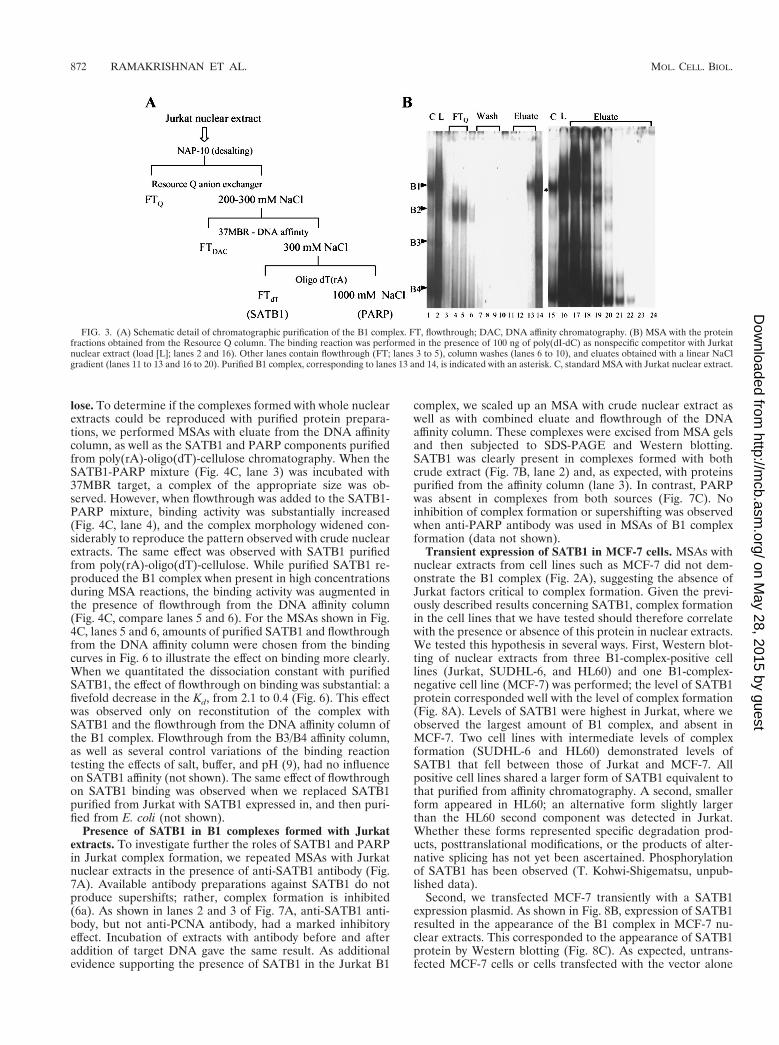
lose. To determine if the complexes formed with whole nuclearextracts could be reproduced with purified protein prepara-tions, we performed MSAs with eluate from the DNA affinitycolumn, as well as the SATB1 and PARP components purifiedfrom poly(rA)-oligo(dT)-cellulose chromatography. When theSATB1-PARP mixture (Fig. 4C, lane 3) was incubated with37MBR target, a complex of the appropriate size was ob-served. However, when flowthrough was added to the SATB1-PARP mixture, binding activity was substantially increased(Fig. 4C, lane 4), and the complex morphology widened con-siderably to reproduce the pattern observed with crude nuclearextracts. The same effect was observed with SATB1 purifiedfrom poly(rA)-oligo(dT)-cellulose. While purified SATB1 re-produced the B1 complex when present in high concentrationsduring MSA reactions, the binding activity was augmented inthe presence of flowthrough from the DNA affinity column(Fig. 4C, compare lanes 5 and 6). For the MSAs shown in Fig.4C, lanes 5 and 6, amounts of purified SATB1 and flowthroughfrom the DNA affinity column were chosen from the bindingcurves in Fig. 6 to illustrate the effect on binding more clearly.When we quantitated the dissociation constant with purifiedSATB1, the effect of flowthrough on binding was substantial: afivefold decrease in the Kd, from 2.1 to 0.4 (Fig. 6). This effectwas observed only on reconstitution of the complex withSATB1 and the flowthrough from the DNA affinity column ofthe B1 complex. Flowthrough from the B3/B4 affinity column,as well as several control variations of the binding reactiontesting the effects of salt, buffer, and pH (9), had no influenceon SATB1 affinity (not shown). The same effect of flowthroughon SATB1 binding was observed when we replaced SATB1purified from Jurkat with SATB1 expressed in, and then puri-fied from E. coli (not shown).
Presence of SATB1 in B1 complexes formed with Jurkatextracts. To investigate further the roles of SATB1 and PARPin Jurkat complex formation, we repeated MSAs with Jurkatnuclear extracts in the presence of anti-SATB1 antibody (Fig.7A). Available antibody preparations against SATB1 do notproduce supershifts; rather, complex formation is inhibited(6a). As shown in lanes 2 and 3 of Fig. 7A, anti-SATB1 anti-body, but not anti-PCNA antibody, had a marked inhibitoryeffect. Incubation of extracts with antibody before and afteraddition of target DNA gave the same result. As additionalevidence supporting the presence of SATB1 in the Jurkat B1
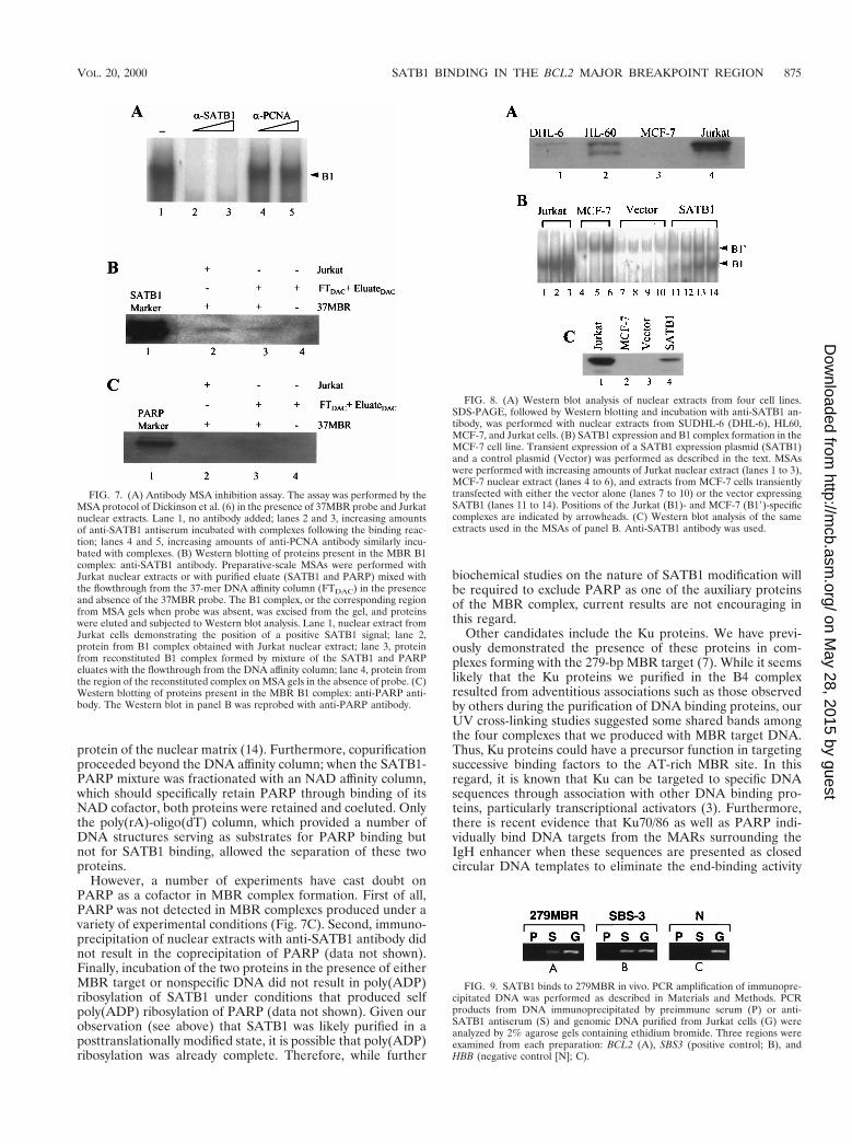
complex, we scaled up an MSA with crude nuclear extract aswell as with combined eluate and flowthrough of the DNAaffinity column. These complexes were excised from MSA gelsand then subjected to SDS-PAGE and Western blotting.SATB1 was clearly present in complexes formed with bothcrude extract (Fig. 7B, lane 2) and, as expected, with proteinspurified from the affinity column (lane 3). In contrast, PARPwas absent in complexes from both sources (Fig. 7C). Noinhibition of complex formation or supershifting was observedwhen anti-PARP antibody was used in MSAs of B1 complexformation (data not shown).
Transient expression of SATB1 in MCF-7 cells. MSAs withnuclear extracts from cell lines such as MCF-7 did not dem-onstrate the B1 complex (Fig. 2A), suggesting the absence ofJurkat factors critical to complex formation. Given the previ-ously described results concerning SATB1, complex formationin the cell lines that we have tested should therefore correlatewith the presence or absence of this protein in nuclear extracts.We tested this hypothesis in several ways. First, Western blot-ting of nuclear extracts from three B1-complex-positive celllines (Jurkat, SUDHL-6, and HL60) and one B1-complex-negative cell line (MCF-7) was performed; the level of SATB1protein corresponded well with the level of complex formation(Fig. 8A). Levels of SATB1 were highest in Jurkat, where weobserved the largest amount of B1 complex, and absent inMCF-7. Two cell lines with intermediate levels of complexformation (SUDHL-6 and HL60) demonstrated levels ofSATB1 that fell between those of Jurkat and MCF-7. Allpositive cell lines shared a larger form of SATB1 equivalent tothat purified from affinity chromatography. A second, smallerform appeared in HL60; an alternative form slightly largerthan the HL60 second component was detected in Jurkat.Whether these forms represented specific degradation prod-ucts, posttranslational modifications, or the products of alter-native splicing has not yet been ascertained. Phosphorylationof SATB1 has been observed (T. Kohwi-Shigematsu, unpub-lished data).
Second, we transfected MCF-7 transiently with a SATB1expression plasmid. As shown in Fig. 8B, expression of SATB1resulted in the appearance of the B1 complex in MCF-7 nu-clear extracts. This corresponded to the appearance of SATB1protein by Western blotting (Fig. 8C). As expected, untrans-fected MCF-7 cells or cells transfected with the vector alone
FIG. 3. (A) Schematic detail of chromatographic purification of the B1 complex. FT, flowthrough; DAC, DNA affinity chromatography. (B) MSA with the proteinfractions obtained from the Resource Q column. The binding reaction was performed in the presence of 100 ng of poly(dI-dC) as nonspecific competitor with Jurkatnuclear extract (load [L]; lanes 2 and 16). Other lanes contain flowthrough (FT; lanes 3 to 5), column washes (lanes 6 to 10), and eluates obtained with a linear NaClgradient (lanes 11 to 13 and 16 to 20). Purified B1 complex, corresponding to lanes 13 and 14, is indicated with an asterisk. C, standard MSA with Jurkat nuclear extract.
872 RAMAKRISHNAN ET AL. MOL. CELL. BIOL.
on May 28, 2015 by guest
http://mcb.asm
.org/D
ownloaded from
demonstrated neither B1 complex nor SATB1 protein (Fig. 8Band C).
SATB1 binding of the BCL2 MBR in vivo. The high-affinity,cell cycle-regulated binding of SATB1 to the MBR stronglysuggested that this region served as a MAR. Recently, a highlysensitive and specific assay, involving chromatin immunopre-cipitation by SATB1 antibody followed by PCR amplification,has been used to demonstrate the association of SATB1 withtarget DNA sequences in vivo (5). Therefore, we tested JurkatMBR DNA sequences for in vivo SATB1 binding in this assay.For comparison, we used the previously characterized, in vivoSATB1 binding sequence SBS-3 (Fig. 9B) and a region isolatedfrom the HBB locus that does not have any SATB1 bindingsequence as a negative control (Fig. 9C). DNA purified fromchromatin immunoprecipitated with anti-SATB1 antiserum(Fig. 9A, lane S) but not preimmune serum (Fig. 9A, lane P)showed nearly as strong a PCR signal as the positive control(Fig. 9B), indicating the in vivo association of SATB1 with theMBR.
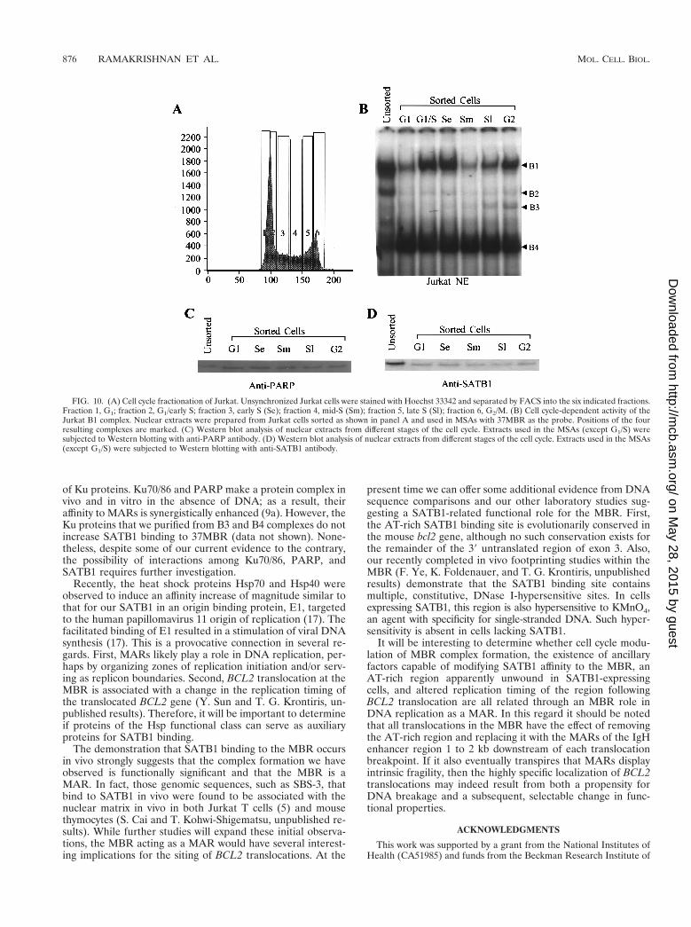
Cell cycle-dependent binding of the B1 complex. We havepreviously noted cell cycle-dependent binding activity of the B1complex in Jurkat cells fractionated by counterflow centrifugalelutriation. To determine if this variation corresponded to
changes in SATB1 or PARP levels, we prepared Jurkat nuclearextracts from six different stages of the cell cycle (G1, G1/S,early S, middle S, late S, and G2/M) (Fig. 10A) fractionated onthis occasion by FACS. As before, B1 complex formation wasmaximal at the G1/S boundary and early S, with a markedreduction in binding activity observed at mid-S (Fig. 10B).Quantitation by a PhosphorImager revealed a fourfold differ-ence between G1/early S and mid-S. In contrast, the B4 com-plex showed no variation throughout the cycle; B3 showed avery modest elevation in late S and G2/M. We also monitoredthe level of an irrelevant binding factor, CCAAT binding factor(2); levels of complex formation corresponding to this factoralso remained constant across the cell cycle (data not shown).The MCF-7 B19 complex (Fig. 2A) also demonstrated no cellcycle-related changes in binding activity (data not shown). Weattributed the rise of B1 complex at late S and G2/M, which wasabsent in our earlier studies, to a small contamination of ourpresent Jurkat cells with a hyperdiploid component (Fig. 10A)in which some hyperdiploid G1 cells were included in the G2/Mfraction of the diploid majority component.
Western blots of the nuclear extracts from different stages ofthe cell cycle with anti-PARP (Fig. 10C) and anti-SATB1 (Fig.10D) antibodies did not reveal any changes in the levels or
FIG. 4. (A) Purification of SATB1 (complex B1) by DNA affinity chromatography (DAC). The silver-stained SDS–8% polyacrylamide gel shows protein fractionsobtained from the 37-mer-Sepharose 4B column. Lane 1, flowthrough (FT); lanes 2 and 3, 0.2 M eluate from the NaCl gradient; lanes 3 and 4, 0.4 M eluate from theNaCl gradient. The positions of PARP and SATB1 are indicated. (B) Protein profile of fractions from a poly(rA)-oligo(dT)-Sepharose column for SATB1 (complexB1) purification. In this silver-stained gel (6%), lane 1 represents material loaded onto the poly(rA)-oligo(dT) column, lane 2 shows the unbound SATB1 fraction, andlane 3 contains the tightly bound PARP fraction eluted as described in Materials and Methods. (C) Reconstitution of Jurkat complex formation with fractions fromaffinity and poly(rA)-oligo(dT) columns for SATB1 (complex B1) purification. Binding reactions were performed with Jurkat nuclear extract (C; lane 1), flowthroughfrom the 37-mer affinity column (FT; lane 2), eluate from the affinity column (containing SATB1 and PARP; lane 3), FT and eluate together (lane 4), SATB1 aloneobtained from the flowthrough of the poly(rA)-oligo(dT) column (lane 5), and flowthrough from the affinity column together with SATB1 from poly(rA)-oligo(dT) (lane6). (D) Affinity column purification of proteins in the B4 complex; silver-stained SDS-polyacrylamide gel. Lane 1, material loaded onto the column from Sepharose Q;lane 2, proteins eluted from the affinity column as described in Materials and Methods. DNA-PK, DNA-dependent protein kinase. (E) Reconstitution of B3 and B4complexes with high-salt eluates of affinity chromatography. MSAs were performed with B3 and B4 fractions from Sepharose Q (lane 1), flowthrough (lanes 2 and 3),and low-salt washes (lanes 4 and 5) of the affinity column and high-salt eluates (lanes 6 and 7). Positions of B3 and B4 are indicated.
VOL. 20, 2000 SATB1 BINDING IN THE BCL2 MAJOR BREAKPOINT REGION 873
on May 28, 2015 by guest
http://mcb.asm
.org/D
ownloaded from
apparent modifications of these proteins that could explain thedifference in binding activity.
DISCUSSION
We have demonstrated that the AT-rich region immediatelyflanking the BCL2 MBR is a binding site for the MAR proteinSATB1. Following purification by multiple steps of columnchromatography, including DNA affinity chromatography, thisprotein was identified unambiguously through MS analysis oftryptic peptides. We obtained additional evidence for this iden-tity by a characteristic inhibition of complex formation onincubation of anti-SATB1 antibody with nuclear extracts dur-
ing MSAs (Fig. 7A) and by Western blotting of gel-purifiedcomplex with the MBR AT-rich region as the target (Fig. 7B).Finally, introduction of a SATB1 expression plasmid intoMCF-7, a cell line deficient in SATB1, was successful in induc-ing the formation of a complex identical to that of Jurkat, a cellline with very high levels of SATB1 expression (Fig. 8). Whilethe SDS-PAGE migration of our SATB1 preparation pro-duced a higher apparent molecular mass than predicted by theDNA sequence (103 versus 86 kDa), independent preparationsof the protein from two different laboratories comigrated onour gels (data not shown; see also reference 18). Therefore,posttranslational modification is likely to have occurred.
It is probable that other proteins are required for MBRcomplex formation, although we have not yet determinedwhether these proteins are present in the final complex orsimply enabling cofactors that do not remain assembled withSATB1. Our observations on the effects of proteins present inthe flowthrough of the affinity column on MBR complex for-mation and SATB1 binding support this expectation. PurifiedSATB1, incubated alone with MBR target DNA, binds withrelatively low affinity (2.1 nM) and produces tight B1 com-plexes on MSA gels. When flowthrough is added to theSATB1-DNA mixture, two changes occur. First, the SATB1complex morphology widens considerably (Fig. 4C) to repro-duce that obtained with Jurkat nuclear extracts. Second, theaffinity increases fivefold (Fig. 6). While the affinity of DNAbinding proteins may be artifactually increased following pu-rification by buffer conditions (9), the increased affinity that weobserved was not influenced by such factors as cation substi-tution and protein carrier but was destroyed by heat and pro-teinase digestion (data not shown). Effects of the flowthroughfrom B1 complex purification could not be substituted with theflowthrough from B4 complex purification. Therefore, addi-tional proteins are required for high-affinity, morphologicallyidentical binding of SATB1 to the MBR target DNA.
The identities of these additional and/or auxiliary proteinsremain unknown. We viewed the copurification of SATB1 andPARP as an intriguing clue, since PARP is also a constituent
FIG. 5. MS identification of purified proteins from B1 and B4 complexes. Shown are the peptide fragments identified by MS analysis for SATB1 (A), PARP (B),Ku70 (C), Ku86 (D), and DNA-dependent protein kinase (E). Amino acid residue numbers of the peptides for each protein are listed in parentheses.
FIG. 6. Kd determination for SATB1 binding to 37MBR. Increasing amountsof SATB1 were incubated with a constant amount of the 37MBR probe inbinding reactions as described by Dickinson et al. (6). The amount of free probeand B1 complex were quantitated with a PhosphorImager. The percentage offree probe was plotted against nanomoles of SATB1 used, and the Kd wascalculated as the amount of SATB1 required to bind 50% of probe in theformation of the B1 complex. The dotted line represents three experiments inwhich only SATB1 was used in the binding reaction; the solid line representsbinding performed with purified SATB1 and flowthrough (FT) from 37-meraffinity column.
874 RAMAKRISHNAN ET AL. MOL. CELL. BIOL.
on May 28, 2015 by guest
http://mcb.asm
.org/D
ownloaded from
protein of the nuclear matrix (14). Furthermore, copurificationproceeded beyond the DNA affinity column; when the SATB1-PARP mixture was fractionated with an NAD affinity column,which should specifically retain PARP through binding of itsNAD cofactor, both proteins were retained and coeluted. Onlythe poly(rA)-oligo(dT) column, which provided a number ofDNA structures serving as substrates for PARP binding butnot for SATB1 binding, allowed the separation of these twoproteins.
However, a number of experiments have cast doubt onPARP as a cofactor in MBR complex formation. First of all,PARP was not detected in MBR complexes produced under avariety of experimental conditions (Fig. 7C). Second, immuno-precipitation of nuclear extracts with anti-SATB1 antibody didnot result in the coprecipitation of PARP (data not shown).Finally, incubation of the two proteins in the presence of eitherMBR target or nonspecific DNA did not result in poly(ADP)ribosylation of SATB1 under conditions that produced selfpoly(ADP) ribosylation of PARP (data not shown). Given ourobservation (see above) that SATB1 was likely purified in aposttranslationally modified state, it is possible that poly(ADP)ribosylation was already complete. Therefore, while further
biochemical studies on the nature of SATB1 modification willbe required to exclude PARP as one of the auxiliary proteinsof the MBR complex, current results are not encouraging inthis regard.
Other candidates include the Ku proteins. We have previ-ously demonstrated the presence of these proteins in com-plexes forming with the 279-bp MBR target (7). While it seemslikely that the Ku proteins we purified in the B4 complexresulted from adventitious associations such as those observedby others during the purification of DNA binding proteins, ourUV cross-linking studies suggested some shared bands amongthe four complexes that we produced with MBR target DNA.Thus, Ku proteins could have a precursor function in targetingsuccessive binding factors to the AT-rich MBR site. In thisregard, it is known that Ku can be targeted to specific DNAsequences through association with other DNA binding pro-teins, particularly transcriptional activators (3). Furthermore,there is recent evidence that Ku70/86 as well as PARP indi-vidually bind DNA targets from the MARs surrounding theIgH enhancer when these sequences are presented as closedcircular DNA templates to eliminate the end-binding activity
FIG. 7. (A) Antibody MSA inhibition assay. The assay was performed by theMSA protocol of Dickinson et al. (6) in the presence of 37MBR probe and Jurkatnuclear extracts. Lane 1, no antibody added; lanes 2 and 3, increasing amountsof anti-SATB1 antiserum incubated with complexes following the binding reac-tion; lanes 4 and 5, increasing amounts of anti-PCNA antibody similarly incu-bated with complexes. (B) Western blotting of proteins present in the MBR B1complex: anti-SATB1 antibody. Preparative-scale MSAs were performed withJurkat nuclear extracts or with purified eluate (SATB1 and PARP) mixed withthe flowthrough from the 37-mer DNA affinity column (FTDAC) in the presenceand absence of the 37MBR probe. The B1 complex, or the corresponding regionfrom MSA gels when probe was absent, was excised from the gel, and proteinswere eluted and subjected to Western blot analysis. Lane 1, nuclear extract fromJurkat cells demonstrating the position of a positive SATB1 signal; lane 2,protein from B1 complex obtained with Jurkat nuclear extract; lane 3, proteinfrom reconstituted B1 complex formed by mixture of the SATB1 and PARPeluates with the flowthrough from the DNA affinity column; lane 4, protein fromthe region of the reconstituted complex on MSA gels in the absence of probe. (C)Western blotting of proteins present in the MBR B1 complex: anti-PARP anti-body. The Western blot in panel B was reprobed with anti-PARP antibody.
FIG. 8. (A) Western blot analysis of nuclear extracts from four cell lines.SDS-PAGE, followed by Western blotting and incubation with anti-SATB1 an-tibody, was performed with nuclear extracts from SUDHL-6 (DHL-6), HL60,MCF-7, and Jurkat cells. (B) SATB1 expression and B1 complex formation in theMCF-7 cell line. Transient expression of a SATB1 expression plasmid (SATB1)and a control plasmid (Vector) was performed as described in the text. MSAswere performed with increasing amounts of Jurkat nuclear extract (lanes 1 to 3),MCF-7 nuclear extract (lanes 4 to 6), and extracts from MCF-7 cells transientlytransfected with either the vector alone (lanes 7 to 10) or the vector expressingSATB1 (lanes 11 to 14). Positions of the Jurkat (B1)- and MCF-7 (B19)-specificcomplexes are indicated by arrowheads. (C) Western blot analysis of the sameextracts used in the MSAs of panel B. Anti-SATB1 antibody was used.
FIG. 9. SATB1 binds to 279MBR in vivo. PCR amplification of immunopre-cipitated DNA was performed as described in Materials and Methods. PCRproducts from DNA immunoprecipitated by preimmune serum (P) or anti-SATB1 antiserum (S) and genomic DNA purified from Jurkat cells (G) wereanalyzed by 2% agarose gels containing ethidium bromide. Three regions wereexamined from each preparation: BCL2 (A), SBS3 (positive control; B), andHBB (negative control [N]; C).
VOL. 20, 2000 SATB1 BINDING IN THE BCL2 MAJOR BREAKPOINT REGION 875
on May 28, 2015 by guest
http://mcb.asm
.org/D
ownloaded from
of Ku proteins. Ku70/86 and PARP make a protein complex invivo and in vitro in the absence of DNA; as a result, theiraffinity to MARs is synergistically enhanced (9a). However, theKu proteins that we purified from B3 and B4 complexes do notincrease SATB1 binding to 37MBR (data not shown). None-theless, despite some of our current evidence to the contrary,the possibility of interactions among Ku70/86, PARP, andSATB1 requires further investigation.
Recently, the heat shock proteins Hsp70 and Hsp40 wereobserved to induce an affinity increase of magnitude similar tothat for our SATB1 in an origin binding protein, E1, targetedto the human papillomavirus 11 origin of replication (17). Thefacilitated binding of E1 resulted in a stimulation of viral DNAsynthesis (17). This is a provocative connection in several re-gards. First, MARs likely play a role in DNA replication, per-haps by organizing zones of replication initiation and/or serv-ing as replicon boundaries. Second, BCL2 translocation at theMBR is associated with a change in the replication timing ofthe translocated BCL2 gene (Y. Sun and T. G. Krontiris, un-published results). Therefore, it will be important to determineif proteins of the Hsp functional class can serve as auxiliaryproteins for SATB1 binding.
The demonstration that SATB1 binding to the MBR occursin vivo strongly suggests that the complex formation we haveobserved is functionally significant and that the MBR is aMAR. In fact, those genomic sequences, such as SBS-3, thatbind to SATB1 in vivo were found to be associated with thenuclear matrix in vivo in both Jurkat T cells (5) and mousethymocytes (S. Cai and T. Kohwi-Shigematsu, unpublished re-sults). While further studies will expand these initial observa-tions, the MBR acting as a MAR would have several interest-ing implications for the siting of BCL2 translocations. At the
present time we can offer some additional evidence from DNAsequence comparisons and our other laboratory studies sug-gesting a SATB1-related functional role for the MBR. First,the AT-rich SATB1 binding site is evolutionarily conserved inthe mouse bcl2 gene, although no such conservation exists forthe remainder of the 39 untranslated region of exon 3. Also,our recently completed in vivo footprinting studies within theMBR (F. Ye, K. Foldenauer, and T. G. Krontiris, unpublishedresults) demonstrate that the SATB1 binding site containsmultiple, constitutive, DNase I-hypersensitive sites. In cellsexpressing SATB1, this region is also hypersensitive to KMnO4,an agent with specificity for single-stranded DNA. Such hyper-sensitivity is absent in cells lacking SATB1.
It will be interesting to determine whether cell cycle modu-lation of MBR complex formation, the existence of ancillaryfactors capable of modifying SATB1 affinity to the MBR, anAT-rich region apparently unwound in SATB1-expressingcells, and altered replication timing of the region followingBCL2 translocation are all related through an MBR role inDNA replication as a MAR. In this regard it should be notedthat all translocations in the MBR have the effect of removingthe AT-rich region and replacing it with the MARs of the IgHenhancer region 1 to 2 kb downstream of each translocationbreakpoint. If it also eventually transpires that MARs displayintrinsic fragility, then the highly specific localization of BCL2translocations may indeed result from both a propensity forDNA breakage and a subsequent, selectable change in func-tional properties.
ACKNOWLEDGMENTS
This work was supported by a grant from the National Institutes ofHealth (CA51985) and funds from the Beckman Research Institute of
FIG. 10. (A) Cell cycle fractionation of Jurkat. Unsynchronized Jurkat cells were stained with Hoechst 33342 and separated by FACS into the six indicated fractions.Fraction 1, G1; fraction 2, G1/early S; fraction 3, early S (Se); fraction 4, mid-S (Sm); fraction 5, late S (Sl); fraction 6, G2/M. (B) Cell cycle-dependent activity of theJurkat B1 complex. Nuclear extracts were prepared from Jurkat cells sorted as shown in panel A and used in MSAs with 37MBR as the probe. Positions of the fourresulting complexes are marked. (C) Western blot analysis of nuclear extracts from different stages of the cell cycle. Extracts used in the MSAs (except G1/S) weresubjected to Western blotting with anti-PARP antibody. (D) Western blot analysis of nuclear extracts from different stages of the cell cycle. Extracts used in the MSAs(except G1/S) were subjected to Western blotting with anti-SATB1 antibody.
876 RAMAKRISHNAN ET AL. MOL. CELL. BIOL.
on May 28, 2015 by guest
http://mcb.asm
.org/D
ownloaded from
the City of Hope to T. G. Krontiris and grants from the NationalInstitutes of Health (CA 39681) and the Department of Energy (DE-AC03-76SF00098) to T. Kohwi-Shigematsu.
REFERENCES
1. Andrews, N. C., and D. V. Faller. 1991. A rapid micropreparation techniquefor extraction of DNA-binding proteins from limiting numbers of mamma-lian cells. Nucleic Acids Res. 19:2499.
2. Bi, W., L. Wu, F. Coustry, B. de Crombrugghe, and S. N. Maity. 1997. DNAbinding specificity of the CCAAT-binding factor CBF/NF-Y. J. Biol. Chem.272:26562–26572.
3. Chung, U., T. Igarashi, T. Nishishita, H. Iwanari, A. Iwamatsu, A. Suwa, T.Mimori, K. Hata, S. Ebisu, E. Ogata, T. Fujita, and T. Okazaki. 1996. Theinteraction between Ku antigen and REF1 protein mediates negative generegulation by extracellular calcium. J. Biol. Chem. 271:8593–8598.
4. Cleary, M. L., and J. Sklar. 1985. Nucleotide sequence of a t(14;18) chro-mosomal breakpoint in follicular lymphoma and demonstration of a break-point-cluster region near a transcriptionally active locus on chromosome 18.Proc. Natl. Acad. Sci. USA 82:7439–7443.
5. de Belle, I., S. Cai, and T. Kohwi-Shigematsu. 1998. The genomic sequencesbound to special AT-rich sequence binding protein 1 (SATB1) in vivo inJurkat T cells are tightly associated with the nuclear matrix at the bases of thechromatin loops. J. Cell Biol. 141:335–348.
6. Dickinson, L. A., T. Joh, Y. Kohwi, and T. Kohwi-Shigematsu. 1992. Atissue-specific MAR/SAR DNA-binding protein with unusual binding siterecognition. Cell 70:631–645.
6a.Dickinson, L. A., and T. Kohwi-Shigematsu. 1995. Nucleolin is a MAR/SARDNA-binding protein specifically recognizing a region with high base-un-pairing potential. Mol. Cell. Biol. 15:456–465.
7. DiCroce, P. A., and T. G. Krontiris. 1995. The BCL2 major breakpointregion is a sequence- and cell-cycle-specific binding site of the Ku antigen.Proc. Natl. Acad. Sci. USA 92:10137–10141.
8. Dignam, J. D., R. M. Lebovitz, and R. G. Roeder. 1983. Accurate transcrip-tion initiation by RNA polymerase II in a soluble extract from isolatedmammalian nuclei. Nucleic Acids Res. 11:1475–1489.
9. Fried, M. G. 1989. Measurement of protein-DNA interaction parameters byelectrophoresis mobility shift assay. Electrophoresis 10:366–376.
9a.Galande, S., and T. Kohwi-Shigematsu. 1999. Poly(ADP-ribose) polymeraseand Ku antigen form a complex and synergistically bind to matrix attachmentregions. J. Biol. Chem. 274:20521–20528.
10. Hellman, U., C. Wernstedt, J. Gonez, and C.-H. Heldin. 1995. Improvementof an “in-gel” digestion procedure for the micropreparation of internalprotein fragments for amino acid sequencing. Biochemistry 224:451–455.
11. Hockenberry, D., G. Nunez, C. Milliman, R. D. Schreiber, and S. J. Kors-meyer. 1990. Bcl-2 is an inner mitochondrial membrane protein that blocksprogrammed cell death. Nature 348:334–336.
12. Hockenberry, D. M., M. Zutter, W. Hickley, M. Nahm, and S. A. Korsmeyer.1991. BCL2 protein is topographically restricted in tissues characterized byapoptotic cell death. Proc. Natl. Acad. Sci. USA 88:6961–6965.
13. Kadonaga, J. T., and R. Tjian. 1986. Affinity purification of sequence-specific
DNA binding proteins. Proc. Natl. Acad. Sci. USA 83:5889–5893.14. Kaufmann, S. H., G. Brunet, B. Talbot, D. Lamarr, C. Dumas, J. H. Shaper,
and G. Poirier. 1991. Association of poly(ADP-ribose) polymerase with thenuclear matrix: the role of intermolecular disulfide bond formation, RNAretention, and cell type. Exp. Cell Res. 192:524–535.
15. Kofler, B., E. Wallraff, H. Herzog, R. Schneider, B. Auer, and M. Schweiger.1993. Purification and characterization of NAD1:ADP-ribosyltransferase(polymerizing) from Dictyostelium discoideum. Biochem. J. 293:275–281.
15a.Kohwi-Shigematsu, T., I. deBelle, L. A. Dickinson, S. Galande, and Y. Ko-hwi. 1998. Identification of base-unpairing region-binding proteins and char-acterization of their in vivo binding sequences. Method. Cell Biol. 53:324–352.
16. Krowczynska, A. M., R. A. Rudders, and T. G. Krontiris. 1990. The humanminisatellite consensus at breakpoints of oncogene translocations. NucleicAcids Res. 18:1121–1127.
17. Liu, J.-S., S.-R. Kuo, A. M. Makhov, D. M. Cyr, J. D. Griffith, T. R. Broker,and L. T. Chow. 1998. Human Hsp70 and Hsp40 chaperone proteins facil-itate human papillomavirus-11 E1 protein binding to the origin and stimulatecell-free DNA replication. J. Biol. Chem. 273:30704–30712.
18. McDonnell, T. J., N. Deane, F. M. Platt, G. Nunez, U. Jaeger, J. P. McKearn,and S. J. Korsmeyer. 1989. Bcl-2-immunoglobulin transgenic mice demon-strate extended B cell survival and follicular lymphoproliferation. Cell 57:79–88.
19. McGuire, E. A., R. D. Hockett, K. M. Pollock, M. F. Bartholdi, S. J. O’Brien,and S. A. Korsmeyer. 1989. The t(11;14)(p15;q11) in a T-cell acute lympho-blastic leukemia cell line activates multiple transcripts, including Ttg-1, agene encoding a potential zinc finger protein. Mol. Cell. Biol. 9:2124–2132.
20. Nakagomi, K., Y. Kohwi, L. A. Dickinson, and T. Kohwi-Shigematsu. 1994.A novel DNA-binding motif in the nuclear matrix attachment DNA-bindingprotein SATB1. Mol. Cell. Biol. 14:1852–1860.
21. Petrovic, A. S., R. L. Young, B. Hilgarth, P. Ambros, S. J. Korsmeyer, and U.Jaeger. 1998. The Ig heavy chain 39 end confers a posttranscriptional pro-cessing advantage to Bcl-2-IgH fusion RNA in t(14;18) lymphoma. Blood91:3952–3961.
22. Seto, M., U. Jaeger, R. D. Hockett, W. Graniger, S. Bennett, P. Goldman,and S. J. Korsmeyer. 1988. Alternative promoters and exons, somatic mu-tation and deregulation of the bcl-2-lg fusion gene in lymphoma. EMBO J.7:123–131.
23. Smith, G. R. 1983. Chi hotspots of generalized recombination. Cell 34:709–710.
24. Stahl, D., K. M. Swiderek, M. T. Davis, and T. D. Lee. 1996. Data-controlledautomation of liquid chromatography/tandem mass spectrometry analysis ofpeptide mixtures. J. Am. Soc. Mass Spectrom. 7:532–540.
25. Tsujimoto, U., J. Cossman, E. Jaffe, and C. M. Croce. 1985. Involvement ofthe bcl-2 gene in human follicular lymphoma. Science 228:1440–1443.
26. Tsujimoto, Y., J. Gorham, J. Cossman, E. Jaffe, and C. M. Croce. 1985. Thet(14;18) chromosome translocations involved in B-cell neoplasms result frommistakes in VDJ joining. Science 229:1390–1393.
27. Wyatt, R. T., R. A. Rudders, R. Delellis, A. D. Zelenetz, and T. G. Krontiris.1992. BCL2 oncogene translocation is mediated by a chi-like consensus. J.Exp. Med. 175:1575–1588.
VOL. 20, 2000 SATB1 BINDING IN THE BCL2 MAJOR BREAKPOINT REGION 877
on May 28, 2015 by guest
http://mcb.asm
.org/D
ownloaded from