Modelling emergent order: from individual cell to tissue
19
Modelling emergent order: from individual cell to tissue Modelling emergent order revised.doc 1/19 4/12/03 Modelling emergent order: from individual cell to tissue R Smallwood 1 * , M Holcombe 2 , D Walker 3 , D R Hose 4 , Steven Wood 5 , S Mac Neil 6 , J Southgate 7 . 1 Professor Rod Smallwood, Department of Computer Science, University of Sheffield, Regent Court, 211 Portobello Street, Sheffield S1 4DP. *Corresponding author. [email protected]. Telephone +44 114 222 1840, fax +44 114 271 3403. 2 Professor Mike Holcombe, Department of Computer Science, University of Sheffield, Regent Court, 211 Portobello Street, Sheffield S1 4DP 3 Dr Dawn Walker, Medical Physics & Engineering, University of Sheffield, Royal Hallamshire Hospital, Sheffield S10 2JF 4 Dr D R Hose, Medical Physics & Engineering, University of Sheffield, Royal Hallamshire Hospital, Sheffield S10 2JF 5 Dr Steven Wood, Medical Physics & Engineering, University of Sheffield, Royal Hallamshire Hospital, Sheffield S10 2JF 6 Professor Sheila Mac Neil, Department of Engineering Materials, University of Sheffield, Sir Robert Hadfield Building, Mappin Street, Sheffield S1 3JD 7 Professor Jenny Southgate, Jack Birch Unit of Molecular Carcinogenesis, Department of Biology, University of York, York YO10 5YW
-
Upload
independent -
Category
Documents
-
view
0 -
download
0
Transcript of Modelling emergent order: from individual cell to tissue
Modelling emergent order: from individual cell to tissue
Modelling emergent order revised.doc 1/19 4/12/03
Modelling emergent order: from individual cell to tissue
R Smallwood1*, M Holcombe2, D Walker3, D R Hose4, StevenWood5, S Mac Neil6 , J Southgate7.
1Professor Rod Smallwood, Department of Computer Science, University of Sheffield, Regent Court, 211Portobello Street, Sheffield S1 4DP. *Corresponding author. [email protected]. Telephone +44 114 2221840, fax +44 114 271 3403.2Professor Mike Holcombe, Department of Computer Science, University of Sheffield, Regent Court, 211Portobello Street, Sheffield S1 4DP3Dr Dawn Walker, Medical Physics & Engineering, University of Sheffield, Royal Hallamshire Hospital,Sheffield S10 2JF4Dr D R Hose, Medical Physics & Engineering, University of Sheffield, Royal Hallamshire Hospital, SheffieldS10 2JF5Dr Steven Wood, Medical Physics & Engineering, University of Sheffield, Royal Hallamshire Hospital,Sheffield S10 2JF6Professor Sheila Mac Neil, Department of Engineering Materials, University of Sheffield, Sir Robert HadfieldBuilding, Mappin Street, Sheffield S1 3JD7Professor Jenny Southgate, Jack Birch Unit of Molecular Carcinogenesis, Department of Biology, University ofYork, York YO10 5YW
Modelling emergent order: from individual cell to tissue
Modelling emergent order revised.doc 2/19 4/12/03
AbstractThe emergence of order from highly complex systems without an over-arching explicit control structure is afundamental feature of biological processes. In this paper, we start by developing a model of a single cellorganism, the rice fungus Magnaporthe grisea, and show how gene activity can be introduced as part of themodel. We then develop a modelling paradigm for describing the development of a multi-cellular epithelialtissue, from the interaction of individual cells, which combines rule-based software agents and a physical model.We examine how information from genomics, proteomics, and molecular and cell biology in general could beincorporated into the model, and the problems associated with relating cellular-scale and tissue-scale properties.Finally, we consider how the computational model can be closely coupled to in vitro biological models ofepithelial tissue, providing a one-to-one mapping between in virtuo and in vitro worlds.
Modelling emergent order: from individual cell to tissue
Modelling emergent order revised.doc 3/19 4/12/03
Modelling emergent order: from individual cell to tissue
R Smallwood, M Holcombe, D Walker, D R Hose, Steven Wood, S MacNeil, J Southgate.
1 IntroductionModels of epithelial tissue either use postulated mechanisms in order to mimic cell growth (illustrative models)or build on known properties of the cells (explanatory models). The last three decades have seen thedevelopment of a number of models examining various aspects of cell culture and tissue behaviour. Models havevaried considerably in terms of implementation and underlying concepts and assumptions, but have tended toincrease in size and complexity in parallel with improvements in computer processing speed and capacity.Honda (1983) developed a graphical approach based on Dirichlet domains to model changes in 2D cellaggregates during morphogenesis. Ransom and Matela (1984) used Voronoi graphs to simulate colony growth.Odell et al. (1981) showed that the tightening of microfilament bundles at the apical surface of cells in responseto mechanical stress was sufficient to explain the folding characteristic of many embryonic tissues.Computational modelling has been used to examine the validity of hypotheses proposed to explain morphogenicprocesses, most notably the Differential Adhesion Hypothesis proposed by Steinberg (1970). Glazier and Graner(1993) used a model based on the minimisation of surface energy to demonstrate that differential adhesion withfluctuations was sufficient to explain sorting in cell aggregates, without the need to consider active migrationmechanisms.
Individual biological cells in a tissue or cell culture can be modelled as cellular automata, thus enabling theexecution of rule sets according to the internal properties or parameters of each cell, and possibly itsenvironment. Lim and Davies (1990) used a combination of cellular automata and Voronoi graphs to examinethe growth rate and shape of cell clusters, with stochastic cell growth and division and cell death. BothZygourakis et al (1991a,b) and Forestell et al (1992) used a purely automaton-based approach allowingstochastic selection of division. An important rule incorporated into these early automaton-type models is that ofcontact inhibition. Ruaan et al. (1993) introduced an extra element of complexity in considering the tendency ofcells to spread if sufficient space is available, and regain a spherical shape and hence generate additional spacefor growth and division at higher cell densities. Cells were given the ability to migrate in order to find space forgrowth and division. The idea of random migration was further developed by Lee et al. (1995).
In addition to providing an insight into patterns observed in tissue culture growth, modelling has been used as atool to simulate the formation of more complex tissue structures. Ryder et al (1999) simulated the developmentof the human cerebral cortex based on random migration, and the difference in cell cycle characteristics observedin cells of different ages. Stekel et al (1995) developed a model of the morphogenesis and homeostasis of thehuman epidermis. This is an illustrative model, with rules formulated to simulate observed behaviour, and not onthe basis of well understood mechanistic behaviour of individual cells. For instance, one of the rules stipulatesthat stem cells emit a substance called ‘stem cell factor’, the concentration of which can be sensed by other cellsin the model. Morel et al (2001) incorporated two distinct hierarchies in their model of epithelial tissue: akinematic model of cell cycle regulation, incorporating both intracellular components (e.g. cyclins) and responseto extracellular stimuli (e.g. growth factors); and a Voronoi graph-based tissue architecture model, withindividual cells represented by polygons. In addition to models of development and behaviour associated withspecific tissues such as epithelium or cortex, general embryonic morphogenesis has continued to be a field ofactive research. Hogeweg (2000) built on a more basic earlier model (Savill and Hogeweg, 1997) to construct atwo level hierarchical simulation of the effect of morphogenic evolution on cell differentiation and differentialadhesion.
One major factor absent in all the models discussed so far is the explicit consideration of forces acting on cells,and the resulting deformation and movement. In general, models that attempt to simulate the effect of physicalforces are continuum, rather than automaton or agent based. Examples include the work of Brodland and Chen(2000) and Chen and Brodland (2000) who used finite element models to simulate various morphogenic process,such as stretching and engulfment, for confluent sheets consisting of a pre-determined number of cells.Interestingly, in contrast with the earlier energy-based models of Glazier and Graner (1993), the results of these
Modelling emergent order: from individual cell to tissue
Modelling emergent order revised.doc 4/19 4/12/03
simulations suggested that differential adhesion alone is not sufficient to result in the sorting of a heterotypic cellpopulation.
Palsson (2001) proposed a model which simulates three dimensional morphogenic processes using an automatonrather than continuum based approach. Cells in this model are considered as individual entities that can respondto the environment according to the values of their internal parameters, and physically interact via contact forces.Each cell has viscoelastic properties, and moves and deforms according to the equations of motions anddeformation. The capacity exists to assign different properties or parameters according to designated cell type.Simulations produced using this model suggest that tissue structure and cell sorting can arise from differences incell adhesion properties, and movement in response to a chemotactic gradient. Three dimensional embryonicmodels have been produced consisting of up to 10,000 ‘cells’, each representing 4-16 actual biological cells. Incommon with many of the previous simulations of morphogenic behaviour, this model does not include thecapacity for cell division and differentiation.
In this paper, we start by developing a model of a single cell organism, the rice fungus Magnaporthe grisea, andshow how gene activity can be introduced as part of the model. We then develop a modelling paradigm fordescribing the development of a multi-cellular epithelial tissue, from the interaction of individual cells, whichcombines rule-based software agents and a physical model. We examine how information from genomics,proteomics, and molecular and cell biology in general could be incorporated into the model, and the problemsassociated with relating cellular-scale and tissue-scale properties. Finally, we consider how the computationalmodel can be closely coupled to in vitro biological models of epithelial tissue, providing a one-to-one mappingbetween in virtuo and in vitro worlds.
2 Why is cellular interaction importantWhat is considered to be fundamental depends on perspective, but few would argue that the transition fromsingle cell organisms to multi-cellular organisms is not fundamental to the development of complex life forms.Individual cells are autonomous entities containing the machinery for converting energy and substrates into morecomplex molecules and structures, for self-replication and differentiation, cell response and even self-destruction(apoptosis or programmed cell death). The structure and function of a multi-cellular organism is a result of theinteraction of these autonomous cells. There is no external plan for the structure – no deus ex machina. There is aone-to-many mapping between genes and proteins, so the genome cannot of itself provide a plan for the structureof an organism. If we want to understand the mechanisms of formation of normal tissue, of wound healing, ofprogression to malignancy, we need to understand how the interaction of autonomous cells can produce a socialentity, the tissue. Our current understanding is at a qualitative (descriptive) level, which differs between tissues,but nevertheless is very detailed even at the molecular level. For example, there is much known about adhesionmolecules, such as the cadherins, that bind cells together to maintain tissue integrity and whose presence orabsence can determine whether a tumour cell is benign or malignant.
The building of functional structures from autonomous cells is central to human biology, from embryology anddevelopment, to repair and even the development of cancer. We have started with epithelial tissue because it is(relatively) simple, consisting of only one cell type, is typically only a few cells (~500 µm) thick, there are goodin vitro models available, and there are important clinical problems (e.g. epithelial cancers account for more than90% of all adult malignancies and chronic non-healing skin wounds account for approximately 12% of theNational Health Service budget in the UK. The in vitro biological models that are being used to develop ourcomputational modelling encompass 2D and 3D models of uroepithelium (Southgate et al, 2002; Scriven et al,1997), oral mucosa (Bhargarv et al, 2003) and skin ( Chakrabarty et al, 1999; Ralston et al, 1999).
3 Convergence of autonomous software and cellular behaviourOur approach is based on the language of computational models. There are many different types ofcomputational model with varying processing capabilities which differ, mainly, in the way that they arerepresented. We choose to use a powerful model based on three key components; internal state, internal memoryand environmental interaction, since these seem to be the fundamental components of any reasonable model of aliving system at a variety of levels. One difficulty with classical theories of computation is that they areessentially discrete so that events occur in discrete time steps and there is no concept of continuum, such asprocesses of metabolism which proceed over a time period in a continuous manner. We propose to exploit a
Modelling emergent order: from individual cell to tissue
Modelling emergent order revised.doc 5/19 4/12/03
powerful computational approach based on communicating X-machines (Balanescu et al, 1999). X-machines areexamples of discrete computational models that operate in finite environments (finite input sets, finite output setsand finite memory variables). Firstly we identify an X-machine (Holcombe and Ipate, 1998; Kefalas et al,2003b) as a system which has internal states and an internal memory. The state transition functions will respondto events on the basis of both the environmental input as well as the current internal state. The system is in somestate, an input a is received, the initial contents of the memory are m and, depending on both a and m, thesystem changes state and produces an output x and updates the memory to m′. This provides a general modellingmechanism which enables many of the problems associated with state explosion, which bedevil many efforts atmodelling complex biological systems, to be dealt with sensibly. The memory can be used to abstract awaydetail in a way that does not prevent us from utilising it whenever necessary. These machine models are suitablefor modelling many types of system. However, they can only model instantaneous processing and hence onlyfinite discrete data is processed. Continuous functions and real valued data cannot be incorporated intotraditional finite state machine models. Such systems are problematic when trying to deal with the complexitiesof some biological models and the hybrid X-machine (Holcombe et al, 2003) overcomes some of these
problems.
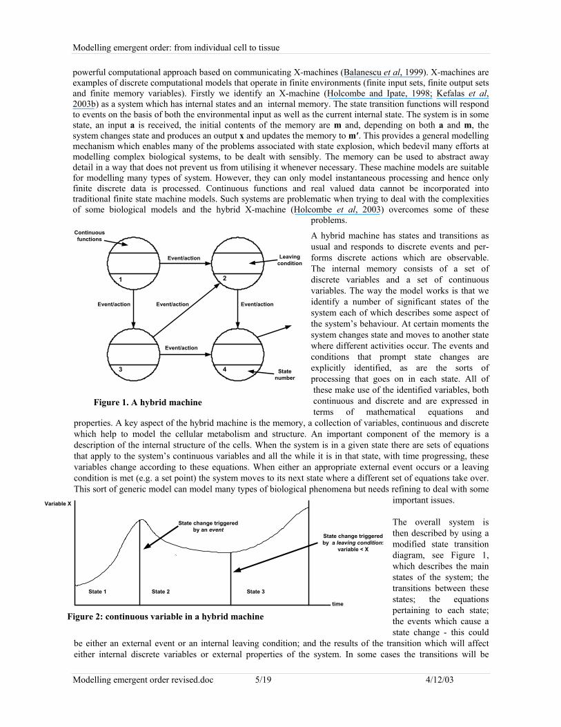
A hybrid machine has states and transitions asusual and responds to discrete events and per-forms discrete actions which are observable.The internal memory consists of a set ofdiscrete variables and a set of continuousvariables. The way the model works is that weidentify a number of significant states of thesystem each of which describes some aspect ofthe system’s behaviour. At certain moments thesystem changes state and moves to another statewhere different activities occur. The events andconditions that prompt state changes areexplicitly identified, as are the sorts ofprocessing that goes on in each state. All ofthese make use of the identified variables, bothcontinuous and discrete and are expressed interms of mathematical equations and
properties. A key aspect of the hybrid machine is the memory, a collection of variables, continuous and discretewhich help to model the cellular metabolism and structure. An important component of the memory is adescription of the internal structure of the cells. When the system is in a given state there are sets of equationsthat apply to the system’s continuous variables and all the while it is in that state, with time progressing, thesevariables change according to these equations. When either an appropriate external event occurs or a leavingcondition is met (e.g. a set point) the system moves to its next state where a different set of equations take over.This sort of generic model can model many types of biological phenomena but needs refining to deal with some
important issues.
The overall system isthen described by using amodified state transitiondiagram, see Figure 1,which describes the mainstates of the system; thetransitions between thesestates; the equationspertaining to each state;the events which cause astate change - this could
be either an external event or an internal leaving condition; and the results of the transition which will affecteither internal discrete variables or external properties of the system. In some cases the transitions will be
Figure 1. A hybrid machine
1 2
3 4
Event/action
Event/action Event/actionEvent/action
Event/action
Continuousfunctions
Leavingcondition
Statenumber
time
Variable X
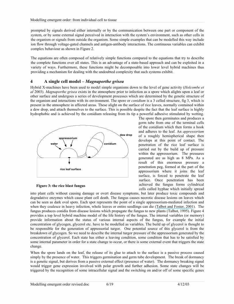
State 1 State 2 State 3
State change triggeredby an event
State change triggeredby a leaving condition:
variable < X
Figure 2: continuous variable in a hybrid machine
Modelling emergent order: from individual cell to tissue
Modelling emergent order revised.doc 6/19 4/12/03
prompted by signals derived either internally or by the communication between one part or component of thesystem, or by some external signal perceived in interaction with the system’s environment, such as other cells inthe organism or signals from outside the organism. Some simple examples that can be modelled this way includeion flow through voltage-gated channels and antigen-antibody interactions. The continuous variables can exhibitcomplex behaviour as shown in Figure 2.
The equations are often composed of relatively simple functions compared to the equations that try to describethe complete functions over all states. This is an advantage of a state-based approach and can be exploited in avariety of ways. Furthermore, these functions might be decomposable into lower level hybrid machines thusproviding a mechanism for dealing with the undoubted complexity that such systems exhibit.
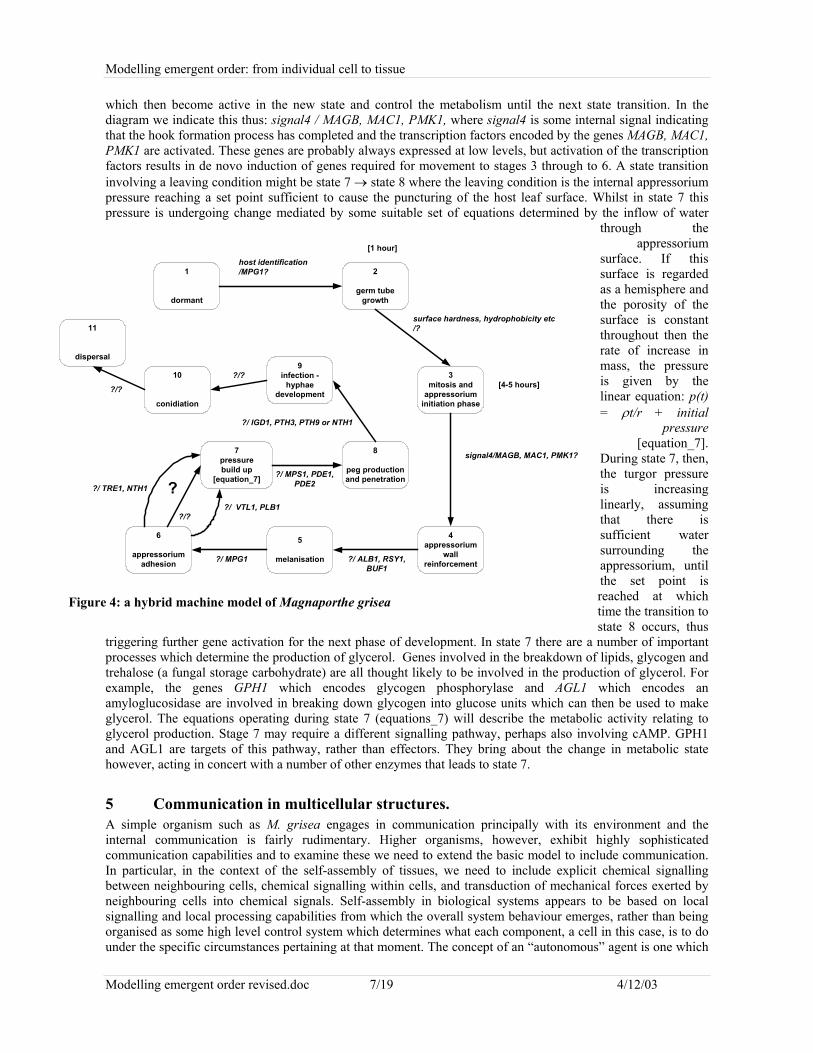
4 A single cell model – Magnaporthe griseaHybrid X-machines have been used to model simple organisms down to the level of gene activity (Holcombe etal 2003). Magnaporthe grisea exists in the atmosphere prior to infection as a spore which alights upon a leaf orother surface and undergoes a series of development processes which are determined by the genetic structure ofthe organism and interactions with its environment. The spore or conidium is a 3 celled structure, fig 3, which ispresent in the atmosphere in affected areas. These alight on the surface of rice leaves, normally contained withina dew drop, and attach themselves to the surface. This is possible despite the fact that the leaf surface is highlyhydrophobic and is achieved by the conidium releasing from its tip a powerful adhesive stimulated by wetting.
The spore then germinates and produces agerm tube from one of the terminal cellsof the conidium which then forms a hookand adheres to the leaf. An appressoriumof a roughly hemispherical shape thendevelops at this point of contact. Thepenetration of the rice leaf surface iscarried out by the build up of pressurewithin the appressorium. The pressuresgenerated are as high as 8 MPa. As aresult of this enormous pressure apenetration peg, formed at the part of theappressorium where it joins the leafsurface, is forced to penetrate the leafsurface. Once penetration has beenachieved the fungus forms cylindricalcells called hyphae which initially spread
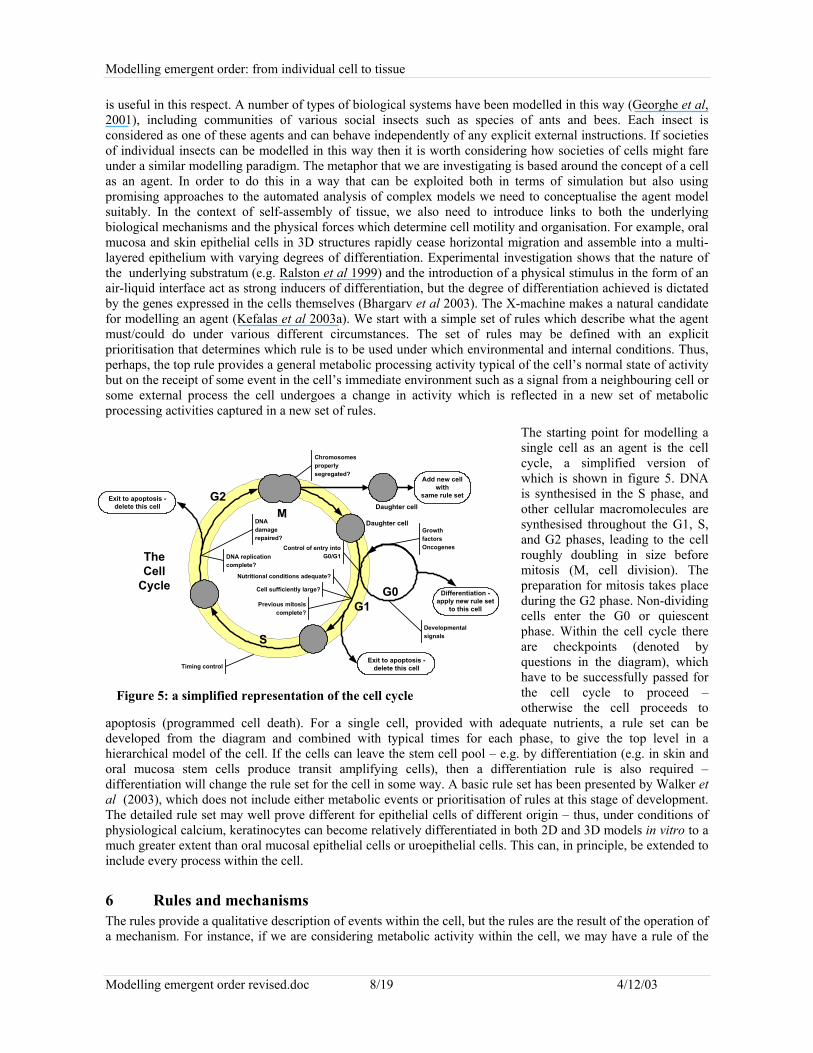
into plant cells without causing damage or overt disease symptoms, but later produce toxic compounds anddegradative enzymes which cause plant cell death. The fungus causes necrotic disease lesions on leaves whichcan be seen as dark oval spots. Each spot represents the point of a single appressorium-mediated infection andwhen they coalesce in heavy infection, whole leaves or entire seedlings can die (Talbot and Foster, 2001). Thefungus produces conidia from disease lesions which propagate the fungus to new plants (Talbot, 1995). Figure 4provides a top level hybrid machine model of the life history of the fungus. The internal variables (or memory)provide information about the status of various internal aspects of the fungus, for example the initialconcentration of glycogen, glycerol etc. have to be modelled as variables. The build up of glycerol is thought tobe responsible for the generation of appressorial turgor. One potential source of this glycerol is from thebreakdown of glycogen. So we need to describe the internal turgor pressure of the appressorium generated by theconcentration of glycerol. Each state has either a leaving condition, some condition that has to be satisfied bysome internal parameter in order for a state change to occur, or there is some external event that triggers the statechange.
When the spore lands on the leaf, the release of its glue to attach to the surface is a passive process causedsimply by the presence of water. This triggers germination and germ tube development. The break of dormancyis a genetic signal, but derives from a passive external effect (presence of water). The dormancy breaking signalwould trigger gene expression involved with polar growth and further adhesion. Some state changes will betriggered by the recognition of some intracellular signal and the switching on and/or off of some specific genes
Figure 3: the rice blast fungus
rice leaf surface
water
appressorium
conidium
dew drop
Modelling emergent order: from individual cell to tissue
Modelling emergent order revised.doc 7/19 4/12/03
which then become active in the new state and control the metabolism until the next state transition. In thediagram we indicate this thus: signal4 / MAGB, MAC1, PMK1, where signal4 is some internal signal indicatingthat the hook formation process has completed and the transcription factors encoded by the genes MAGB, MAC1,PMK1 are activated. These genes are probably always expressed at low levels, but activation of the transcriptionfactors results in de novo induction of genes required for movement to stages 3 through to 6. A state transitioninvolving a leaving condition might be state 7 → state 8 where the leaving condition is the internal appressoriumpressure reaching a set point sufficient to cause the puncturing of the host leaf surface. Whilst in state 7 thispressure is undergoing change mediated by some suitable set of equations determined by the inflow of water
through theappressorium
surface. If thissurface is regardedas a hemisphere andthe porosity of thesurface is constantthroughout then therate of increase inmass, the pressureis given by thelinear equation: p(t)= ρt/r + initial
pressure[equation_7].
During state 7, then,the turgor pressureis increasinglinearly, assumingthat there issufficient watersurrounding theappressorium, untilthe set point isreached at whichtime the transition tostate 8 occurs, thus
triggering further gene activation for the next phase of development. In state 7 there are a number of importantprocesses which determine the production of glycerol. Genes involved in the breakdown of lipids, glycogen andtrehalose (a fungal storage carbohydrate) are all thought likely to be involved in the production of glycerol. Forexample, the genes GPH1 which encodes glycogen phosphorylase and AGL1 which encodes anamyloglucosidase are involved in breaking down glycogen into glucose units which can then be used to makeglycerol. The equations operating during state 7 (equations_7) will describe the metabolic activity relating toglycerol production. Stage 7 may require a different signalling pathway, perhaps also involving cAMP. GPH1and AGL1 are targets of this pathway, rather than effectors. They bring about the change in metabolic statehowever, acting in concert with a number of other enzymes that leads to state 7.
5 Communication in multicellular structures.A simple organism such as M. grisea engages in communication principally with its environment and theinternal communication is fairly rudimentary. Higher organisms, however, exhibit highly sophisticatedcommunication capabilities and to examine these we need to extend the basic model to include communication.In particular, in the context of the self-assembly of tissues, we need to include explicit chemical signallingbetween neighbouring cells, chemical signalling within cells, and transduction of mechanical forces exerted byneighbouring cells into chemical signals. Self-assembly in biological systems appears to be based on localsignalling and local processing capabilities from which the overall system behaviour emerges, rather than beingorganised as some high level control system which determines what each component, a cell in this case, is to dounder the specific circumstances pertaining at that moment. The concept of an “autonomous” agent is one which
Figure 4: a hybrid machine model of Magnaporthe grisea
1
dormant
2
germ tubegrowth
3mitosis and
appressoriuminitiation phase
4appressorium
wallreinforcement
5
melanisation
6
appressoriumadhesion
7pressurebuild up
[equation_7]
8
peg productionand penetration
9infection -
hyphaedevelopment
10
conidiation
11
dispersal
host identification/MPG1?
surface hardness, hydrophobicity etc/?
[1 hour]
[4-5 hours]
signal4/MAGB, MAC1, PMK1?
?/ ALB1, RSY1,BUF1
?/ MPG1
?/ VTL1, PLB1?/?
??/ TRE1, NTH1
?/ MPS1, PDE1,PDE2
?/ IGD1, PTH3, PTH9 or NTH1
?/?
?/?
Modelling emergent order: from individual cell to tissue
Modelling emergent order revised.doc 8/19 4/12/03
is useful in this respect. A number of types of biological systems have been modelled in this way (Georghe et al,2001), including communities of various social insects such as species of ants and bees. Each insect isconsidered as one of these agents and can behave independently of any explicit external instructions. If societiesof individual insects can be modelled in this way then it is worth considering how societies of cells might fareunder a similar modelling paradigm. The metaphor that we are investigating is based around the concept of a cellas an agent. In order to do this in a way that can be exploited both in terms of simulation but also usingpromising approaches to the automated analysis of complex models we need to conceptualise the agent modelsuitably. In the context of self-assembly of tissue, we also need to introduce links to both the underlyingbiological mechanisms and the physical forces which determine cell motility and organisation. For example, oralmucosa and skin epithelial cells in 3D structures rapidly cease horizontal migration and assemble into a multi-layered epithelium with varying degrees of differentiation. Experimental investigation shows that the nature ofthe underlying substratum (e.g. Ralston et al 1999) and the introduction of a physical stimulus in the form of anair-liquid interface act as strong inducers of differentiation, but the degree of differentiation achieved is dictatedby the genes expressed in the cells themselves (Bhargarv et al 2003). The X-machine makes a natural candidatefor modelling an agent (Kefalas et al 2003a). We start with a simple set of rules which describe what the agentmust/could do under various different circumstances. The set of rules may be defined with an explicitprioritisation that determines which rule is to be used under which environmental and internal conditions. Thus,perhaps, the top rule provides a general metabolic processing activity typical of the cell’s normal state of activitybut on the receipt of some event in the cell’s immediate environment such as a signal from a neighbouring cell orsome external process the cell undergoes a change in activity which is reflected in a new set of metabolicprocessing activities captured in a new set of rules.
The starting point for modelling asingle cell as an agent is the cellcycle, a simplified version ofwhich is shown in figure 5. DNAis synthesised in the S phase, andother cellular macromolecules aresynthesised throughout the G1, S,and G2 phases, leading to the cellroughly doubling in size beforemitosis (M, cell division). Thepreparation for mitosis takes placeduring the G2 phase. Non-dividingcells enter the G0 or quiescentphase. Within the cell cycle thereare checkpoints (denoted byquestions in the diagram), whichhave to be successfully passed forthe cell cycle to proceed –otherwise the cell proceeds to
apoptosis (programmed cell death). For a single cell, provided with adequate nutrients, a rule set can bedeveloped from the diagram and combined with typical times for each phase, to give the top level in ahierarchical model of the cell. If the cells can leave the stem cell pool – e.g. by differentiation (e.g. in skin andoral mucosa stem cells produce transit amplifying cells), then a differentiation rule is also required –differentiation will change the rule set for the cell in some way. A basic rule set has been presented by Walker etal (2003), which does not include either metabolic events or prioritisation of rules at this stage of development.The detailed rule set may well prove different for epithelial cells of different origin – thus, under conditions ofphysiological calcium, keratinocytes can become relatively differentiated in both 2D and 3D models in vitro to amuch greater extent than oral mucosal epithelial cells or uroepithelial cells. This can, in principle, be extended toinclude every process within the cell.
6 Rules and mechanismsThe rules provide a qualitative description of events within the cell, but the rules are the result of the operation ofa mechanism. For instance, if we are considering metabolic activity within the cell, we may have a rule of the
MG2
S
G1G0
Daughter cell
Control of entry intoG0/G1
Nutritional conditions adequate?
DNAdamagerepaired?
Chromosomesproperlysegregated?
GrowthfactorsOncogenes
DNA replicationcomplete?
Cell sufficiently large?
Previous mitosiscomplete?
Developmentalsignals
TheCell
CycleDifferentiation -
apply new rule setto this cell
Add new cellwith
same rule set
Daughter cell
Exit to apoptosis -delete this cellTiming control
Exit to apoptosis -delete this cell
Figure 5: a simplified representation of the cell cycle
Modelling emergent order: from individual cell to tissue
Modelling emergent order revised.doc 9/19 4/12/03
form {if nutritional conditions adequate, then …., else ….}. Underlying this qualitative rule are the biochemicalpathways which produce the required proteins; diffusion of nutrients through the surrounding medium; transportacross the cell membrane; etc. So, in principle, it is possible to model the mechanisms which determine theoutput state of the rules. Mechanisms will be known to a greater or lesser extent, and can replace qualitativerules as more knowledge of the system becomes available. For instance, establishing adequate nutritionalconditions may require a rule {if [substrate x] > y, then …., else …..}, where the mechanism of manufacturing x,or the method by which its concentration y is measured, are both unknown. If this proves to be a critical path inthe model, then the mechanism will have to be determined experimentally. The model thus acts as a driver forexperiment.
When there is more than one cell present, the interaction between cells has to be introduced. The cells'communicate' (intercellular signalling), and the tissue as a whole (the continuum) 'communicates' withindividual cells through both chemical diffusion through the tissue and through mechano-transduction(mechanical strain eliciting chemical signals within the cell), and, possibly more importantly, integrin-mediatedbonding. The cells adhere to each other and can also be actively motile (in contrast to passive movement due tothe physical forces between adhesion molecules). In the agent-based model, the agents access a commonmemory area. In order to model this type of system we need to use some form of communication broker thatreflects how different agents can send and receive their messages. This is done by using a sparse matrix. Supposethat we have a collection of agents, in this case represented as individual X-machines. We now have to try toidentify the communication channels and how these might work. Suppose that there are N agents and each ispotentially able to communicate with any other. We thus have an N x N communication matrix. Agent i can senda message to agent j by placing the message in the (i, j)th place in the matrix during a write communicationprocess. When agent j is in a state to read the message from agent i it reads the message from this position in thematrix. Since most agents will only communicate with their immediate neighbours the matrix will be sparse withonly a small number of valid slots and with no diagonal elements.
Suppose that an agent can only communicate with other agents within a specific distance of it. There is a globalmemory that maintains the current position of each agent (an N-vector of co-ordinates). At any communicationstate an agent can interrogate this memory to ascertain which other agents are within communication distance. Anumber of strategies can be used to determine which and how many attempts at a communication can be made.The agent then puts data into the appropriate slots of the communication matrix and continues processing,moving or whatever.
The key thing is to define the rules of the agent including the inter-agent communication mechanism, to examinethe complete model to see what emergent behaviour is produced, and to verify by comparison with the 'realthing' – in this case, the cell culture systems.
7 The cell as software agentThe proof-of-concept model consists of a number of cells interacting with their environment, which comprises atwo-dimensional, square substrate with user-defined dimensions and modifiable exogenous calcium ionconcentration. The purpose of this stage of the model is to test the basic concept of cell-as-software agent, andthe link between the agent model and a physical model of the world. The user can select the number of cellsseeded and whether to place them randomly on the substrate, or in specified locations. The seed cell radii andepithelial type can also be selected by the user. All seeded cells are designated to be members of the stem ortransit amplifying (TA) cell classes. Model execution is based on an iterative process, with each tick or agentmodel iteration representing approximately 30 minutes in real time. This was achieved by selecting mean cellcycle duration and migration rates, so that the cell cycle step, or distance migrated by cells at each time step is,on average, similar to that undergone by a real cell during this time period. These data were based on a mean cellcycle duration of 60 hours for keratinocytes (Dover, 1983) and 15 hours for urothelial cells (Southgate, 1994)and a migration speed of approximately 1µm/ minute, as measured from time lapse sequences of urothelial cellculture. These parameters would be expected to vary as a result of culture conditions as well as cell type.
At each tick of the model clock, each cell is interrogated in turn, and depending on the state of its internalparameters (e.g. current position in cell cycle, flag indicating bond to substrate) and its environment (number andproximity of neighbouring cells, concentration of calcium ions) a number of rules are executed that may changethe state of the cell’s internal parameters. During this process, cells can receive messages from other cells in theirimmediate vicinity, or send messages to other cells by reading from and writing to a structure designated as a
Modelling emergent order: from individual cell to tissue
Modelling emergent order revised.doc 10/19 4/12/03
communication matrix. An additional global data structure, which can be accessed by all cells in the model,contains information relating to the exact position and dimensions of every cell. Each cell knows the location(distance and azimuth) of all neighbours which could come into contact with it (by growing or migrating) withinone time step. This distance is approximately 10*(initial cell radius) = 100 µm (cells can grow to 30 µm radiusand migrate up to 30 µm per time step). This information is updated every time a cell moves or changes shape. Ifa cell divides during a model iteration, one daughter cell overwrites the data structure of the parent, whilst thesecond daughter is added to end of the string of existing cell structures. Further details of rule sets pertaining toparticular cell behaviours are given below.
Each cell in our model is represented as assigned to a particular object class according to its designated cell type.The current permitted types are stem cells (specifically epithelial stem cells); transit amplifying cells; mitoticcells; post-mitotic cells; dead cells. Stem cells are predicted to exist in many types of epithelial tissue, although adefinitive biochemical/antigenic marker has yet to be found. The fundamental property of such cells is that theyhave an unlimited capacity for self-renewal, and thus are critical in maintaining a constant cell number in normalhomeostatic tissue. There is some evidence that in certain types of epithelium, stem cells may have a particularspatial distribution, may cycle more slowly or may express different integrins and thus exhibit distinct contactproperties to transit amplifying cells (Jensen, 1999; Potten, 2002). We have currently ignored suchcharacteristics, some of which remain speculative, and restrict our stem cells to differ from TA cells in terms oftheir self-renewal properties only. Ten percent of the initial seed cells are designated to be stem cells (Potten,1988). The permitted behaviour set for this cell type are: bonding (to substrate and other cells); spreading (oncebonded to substrate); lateral migration (once bonded to substrate); apoptosis (if failure to bond to substrate); cellgrowth and division. Transit Amplifying (TA) cells are designated to form approximately 90% of the basalcompartment of epithelial tissue. Unlike stem cells, they are capable of only a limited number of division cyclesbefore becoming committed to terminal differentiation. The number of rounds of division allowed appears tovary between different epithelial tissue types. For instance, as previously stated, it is not unusual forkeratinocytes (Chakrabarty et al, 1999) or oral mucosa cells (Bhargarv et al, 2003) in 3D culture to expresssome cytokeratin isotypes associated with differentiation, but this is not observed in the culture of urothelial cells(Southgate et al, 1994). It is, of course, quite possible that this disparity can be accounted for by the differentconditions used to culture these cell types, in contrast to being hard-coded for each cell type at an epi-geneticlevel. However, whilst this point remains in doubt, the number of TA cell divisions allowed are limited to 3 forkeratinocyte models and 30 for urothelial cell models. The permitted behaviour set for TA cells is identical tostem cells in all other respects. Mitotic cells are a subset of both stem and TA cells that are progressing throughthe M phase of the cell cycle. In cell culture, mitotic cells are observed to round-up, thus losing adhesion withtheir neighbours, and to some extent with the underlying substrate. For this reason, model cells assume arounded morphology, breaking all intercellular bonds on entering the M phase (defined as the final 20% of thecell cycle) and are not permitted to form new bonds or actively migrate until mitosis is complete. Post-mitoticcells are produced by the division of TA cells that have undergone the maximum permitted number of mitoticcycles. They are rarely observed in monolayer culture of urothelial cells, but are relatively common inkeratinocyte cultures which rapidly become enucleated keratin envelopes reflecting their barrier function in vivo.The extension of our model to be truly three dimensional will allow these cells to assume the characteristics ofcells committed to terminal differentiation, and migrate upwards to reside in the supra-basal layer. Currently,post-mitotic cells are permitted access to the same rule sets as stem and TA cells, with the obvious exception ofcell growth and division. Failure of normal cells in culture to adhere to the substrate results in the programmedcell death (apoptosis) of that cell. The dead cells are then fragmented and phagocytosed by their neighbours.Model cells that have failed to bond to the substrate before the end of their G1 phase become dead cells, shrinkand are removed from the model. In reality, most cells adhere very quickly after seeding, and hence dead cellsare a rare occurrence.
In addition to the internal parameters unique to the designated class of a cell, every cell structure in the modelcontains a set of parameters that defines the exact location of its centre in x, y and z Cartesian co-ordinates, andthe length of its axes in each direction. Hence, although the model remains essentially two dimensional at thisstage, each cell is defined as a three dimensional ellipsoid. This allows three dimensional changes in cellmorphology, such as cell spreading and rounding, to take place.
The central hinge of the model is the cell cycle. All cells capable of cycling (i.e. stem and TA cells) have aninternal clock, and provided that they are not inhibited, progress one tick through the cycle at each modeliteration. At model initiation, or when mitosis occurs to create new daughter cells, the new TA cells are
Modelling emergent order: from individual cell to tissue
Modelling emergent order revised.doc 11/19 4/12/03
designated a fixed S-G2-M phase length comprising approximately 50% of the total cell cycle durationaccording to the model cell type (keratinocytes = 60 hours or 120 model iterations, urothelial cells = 15 hours or30 iterations) and a G1 phase length that is selected randomly from a normal distribution with mean equal to halfthe total cycle length and standard deviation equal to a tenth of the mean. This reflects the situation that isobserved in real cells where the variation in the cell cycle duration between cells of the same lineage is derivedalmost entirely from variation in G1 duration.
The G1 phase of the cell cycle is the growth phase during which cells effectively double their volume bysynthesis of proteins within the cytoplasm. At the first tick of the G1 phase of a model cell, the cell’s volume iscalculated along with the volume increase required at each subsequent tick in order that the volume is exactlydoubled at the end of G1. This volume increment is added to the cell at each G1 tick.
Approximately half way through G1, a checkpoint has been introduced that, at present, governs whether a cellproceeds through the remainder of the cell cycle based on the number of contacts the cell has formed withneighbouring cells, and its morphology. In the culture of several epithelial cell types, cells situated at the centreof cell colonies grown in physiological or high calcium media withdraw from the cell cycle and becomequiescent. It is believed that this phenomenon, termed 'contact inhibition' is mediated via intercellular signalling– cells are aware of the existence of their nearest neighbours via the cadherin-mediated, and hence calcium-dependent physical bonds (St Croix et al 1998; Orford and Byers 1999). The rule for a model cell to becomecontact-inhibited is that it has four or more bonds with neighbouring cells (or, alternatively three bonds and beadjacent to the ‘edge’ of the substrate). Cells require a spread morphology in order to progress further in the cellcycle. For this reason, model cells also check that they are bonded to the substrate and sufficiently spread at thischeckpoint. Cells which have not attached at this stage undergo apoptosis.
Cells which do not fulfil the criteria to pass through the G1 checkpoint enter a quiescent, or G0 phase of the cellcycle. The cell will remain in this state until enough of its intercellular bonds are broken to allow it to re-enterthe cycle (this may occur if a neighbouring bonded cell reaches the mitotic phase of it cycle, rounds up, andbreaks its intercellular bonds), and the cell has spread sufficiently. A corollary of these rules is that they allowthe concept of wound repair to be introduced in a confluent monolayer model.
If a cell passes the checkpoint, or fulfils the requirements to leave the G0 state, it will then progress to the S andG2 phases. Biological cells synthesise new DNA during the S phase, and prepare for mitosis during the secondlag phase, or G2. However, as our model does not currently consider sub-cellular phenomena explicitly, duringthese phases model cells simply increment their cycle clock by a single tick at each model iteration. Furtherchecks relating to phenomena such as occupation of growth factor receptors (G1 phase) and successful DNAreplication (G2 phase) will be incorporated at a later stage of model development.
On approaching the final 20% of the cycle, defined to constitute the mitotic or M phase, the cell alters itsmorphology to become rounded, breaks any bonds it may have with neighbouring cells and is redefined as amember of the mitotic cell class. Cells in this class are defined as two distinct but overlapping spheres, whichmove apart over the duration of the M phase to produce two adjacent volumes that are eventually designated astwo separate daughter cells. The first daughter cell structure overwrites the position of the parent in the model,and the second is added to the end of the existing list of cells. Stem cells always undergo asymmetric division,i.e. produce one daughter that is also a stem cell, and a second that is a transit amplifying cell, whereas transitamplifying cells always produce transit amplifying daughters. New cells, whether seeded onto the substrate, orthe product of mitosis, have a rounded morphology and are not bonded to the substrate – i.e. they are notpolarised. At each subsequent model iteration, there is a probability-based calculation of the cell binding to thesubstrate. This calculation is biased to reflect the fact that it is extremely unusual for cells in culture not toadhere within the first hour or so after seeding. Once adherent, a cell is then permitted to engage in a number ofcell behaviours, including spreading and migration. The location of all cells in the model is continually updatedand stored in a global data structure, which allows every cell to know the relative positions of all its neighbours.For each cell, at every model iteration, a binding probability is calculated for each neighbour within a setdistance (defined to be half the specified radius of the original seeded cells, which in the case of the modelsdiscussed in this paper is 10µm). The probability of bonding for a pair of cells is inversely proportional to theseparation between the cell edges and related to the environmental calcium concentration via a sigmoid functionwith the inflection point at 1.0mM. This relationship is based on published data (Baumgartner et al 2000) onthe relationship between the binding activity of E-Cadherin-mediated bonds and exogenous calcium ionconcentration. This study used atomic force microscopy techniques to show that an extracellular calcium of
Modelling emergent order: from individual cell to tissue
Modelling emergent order revised.doc 12/19 4/12/03
greater than 1.0 mM is required for dimerisation of the E-Cadherin receptor. A random number between 0 and 1is generated and bonds will be formed with cells whose probability constant is greater than this number. All cellsthat form intercellular bonds automatically become bound to the substrate, if they are not already attached.
The first cell records the identity flags of the successfully bonded neighbouring cells within its data structure andsends a signal to these neighbours via the global communication matrix. On receiving the signal, a cell adds theflag of the sender to its own data structure. Hence every cell in the model that is able to form bonds will containa list of flags indicating the cells to which it is linked. Bonds between cells will be recognised when the data isassembled for the next physical correction and non-adjacent linked cells will feel an attractive force proportionalto their separation, resulting in them moving together until their edges are adjacent.
Cells seeded onto the substrate and new daughter cells produced by mitosis are not attached to the substrate andare spherical in shape. In order to be able to progress through the cell cycle, real biological cells spread byforming numerous attachments to the substrate, thus assuming a flattened morphology (Huang and Ingber 1999).This process is emulated in the model by increasing the surface area of attached cells by a small amount eachmodel iteration, whilst maintaining a constant volume. Cells continue to spread until their radius has increasedby a factor of approximately 1.5. Cells that are attached to the substrate but have no intercellular bonds are freeto migrate laterally on the substrate surface. The allowed migration distance per model iteration is fixed atapproximately 2.5 times its radius. This figure was obtained from measurement of urothelial cell movement fromsuccessive frames of a time-lapse recording of a scrape wound closure assay. Migration directions are randomlyassigned to new cells. At each subsequent iteration, a cell will tend to carry on migrating in the same direction,but may randomly choose another direction within 60° of its original trajectory. If the migration path is blockedby a neighbouring cell or the edge of the substrate, the cell will move by a smaller distance so that its edge isadjacent to that of its neighbour. If no movement at all is possible in the chosen direction, the cell will alter itstrajectory by 30 degrees and try again, continuing to do so up to 10 times, after which it will remain stationary.Such checks, and similar ones that are executed when selecting the direction of the separation in mitotic cells, forinstance, are essential to ensure that cells do not fall off the edge of the model, or overlap one another by morethan a single cell radius, as this could lead to errors during the physical correction. Stem and TA cells that havefailed to attach to the substrate by the end of their G1 phase are redefined as dead cells. These cells shrink, andare removed from the model.
After each time step of the agent model, the position and size of all the cells is passed to the physical model,which calculates the changes in position which occur as a result of growth, division, and physical interaction,and returns the new cell positions to the agent model for the next time step (figure 6).
8 The physical worldAt the current stage of development, the physical realisation of the cells and their interactions is very simplistic.The cells are rigid spheroids which can change the ratio of height to radius depending on the cell cycle stage(they are less disc-like in mitosis). They are confined to a plane i.e. only a monolayer of cells is modelled. Cellgrowth, movement and mitosis events during each agent-based iteration will result in forces betweenneighbouring cells. In addition, the edges of neighbouring cells that are bonded to one another may not becontiguous.
The acceleration induced in the ith cell, by the force exerted by its n neighbours is given by Newton’s secondlaw, with damping:
1
n
i i i ij
m u c u F=
+ =∑ (1.1)
where im is the mass of the cell, ic is its damping constant and u is its displacement. The mass of the cell iscalculated from the length of its two primary axes a and b, which is information retained in the computationaldata structure representing each cell. As the cells only interact in two dimensions, this is based on the planar areaof the cell, assuming a unit density:
2im aπ= (1.2)
Modelling emergent order: from individual cell to tissue
Modelling emergent order revised.doc 13/19 4/12/03
The damping constant, ic is proportional to the mass of the cell:
i ic mα= (1.3)
where the proportionality constant α is assigned according to the global location of the cell. Cells close to theedge of the model are given higher damping factors to inhibit their movement and prevent them being pushed offthe edge of the model. Damping factors in the x and y direction can be manipulated independently, thus a cellmay be more easily pushed in one direction than the other.
During each agent iteration, a cell determines the position of all other cells within approximately 10 cell radii. Inaddition, each cell contains information regarding its own position and radius. When the physical model isassembled, a check is made to identify whether the growth of these nearby cells would cause them to attempt tooccupy the same physical space. In addition, each data structure contains a list of the flags of other agents towhich it has formed an intercellular bond. Forces are only exerted by neighbours that are bonded or attempt tooccupy the same space. The force exerted the cell i by neighbour j is defined to be proportional to the ‘overlap’or edge separation ijO between the cells.
ij ij ijF k O= (1.4)
where ijk is an arbitrary ‘stiffness’ defined to be equal to the inverse of the separation of the centres of cells i
and j. If cells i and j have radii ir and jr and are located at ( ),i ix y and ( ),j jx y respectively, then:
( ) ( ) ( )2 2
ij j i j i i jO x x y y r r= − + − − + (1.5)
Note that the signs of the constants are constructed in such a way that cells experience a repulsive force fromneighbours attempting to occupy the same space, and an attractive force from bonded neighbours that may beseparated by a small distance.
Assuming the vector i j→ is orientated at angle θ with respect to the x axis, resolving into two orthogonaldirections and substituting into (1.4) gives:
( ) ( ) ( ){ }cosij j i i jij xF k x x r r θ= − − + (1.6a)
( ) ( ) ( ){ }sinij j i i jij yF k y y r r θ= − − + (1.6b)
The co-ordinates of cell i at time t are the co-ordinates ( ),Oi Oix y at the previous time step plus the
displacement ,xi yiu u during the current time step, so equation 1.6a can be written:
( ) ( ) ( ){ } ( )1 2ij Oi Oj xi xj xij i jij xF k x x u u k r r= − + − − + (1.7)
where 1k and 2xk are constants depending on the location of the two cells:
( ) ( )1 2 2
1ij
j i j i
kx x y y
=− + −
(1.8a)
( ) ( )2 2 2
cosxij
j i j i
kx x y y
θ=
− + −(1.8b)
Modelling emergent order: from individual cell to tissue
Modelling emergent order revised.doc 14/19 4/12/03
Equation 1.6b can be similarly rewritten, but in this case:
( ) ( )2 2 2
sinyij
j i j i
kx x y y
θ=
− + −(1.8c)
The value of the differential terms in equation (1.1) at time t, can be written as difference equations:
, , , 2, 2
2i t i t t i t ti t
u u uu
t−∆ − ∆− +
=∆
(1.9a)
, ,,
i t i t ti t
u uu
t−∆−
=∆
(1.9b)
where ,i t tu −∆ and , 2i t tu − ∆ denote the displacement of the ith cell evaluated at the two previous time
points. Substituting equations 1.7 and 1.9 a and b into 1.1 gives the complete equation of motion for cell i in the
x direction:
( ) ( ){ } ( ), , , 2 , ,1 22
1
2 ni t i t t i t t i t i t t
i i i ij Oi Oj xi xj xij i jj
u u u u um m k x x u u k r r
t tα−∆ − ∆ −∆
=
− + − + = − + − − + ∆ ∆ ∑
Similar equations can be assembled to describe the x and y displacements of every particle in the model.
Equation 1.1 can then be written in matrix form, with every row representing the displacement of one cell, and
rearranged:
[ ] [ ] [ ] { }
[ ]{ } [ ]{ } [ ]{ } [ ]{ } [ ]{ }
,2
22 2
1
21 2
x t
t t t t t tO
M MK u
t t
M u M u M uK x K r
t t t
α
α−∆ −∆ − ∆
+ − ∆ ∆
= − + + −∆ ∆ ∆
This equation is in the standard matrix equation form Ax B= and hence can be solved by commonly usednumerical analysis techniques such as Gaussian Elimination or conjugate gradients, to obtain the vectorcontaining the cell displacements at the current iteration. Displacements in x and y are solved independently andthe new positions of each cell obtained from its previous location and calculated displacements. The choice oftime step t∆ and damping constants α are critical in determining the time taken for a stable solution to bereached and were optimised empirically.
The solution at time t depends on the solutions at the previous two time points, so at the first two iterations ofeach solve an all zero vector is substituted for both ,i t tu −∆ and , 2i t tu − ∆ . The solution takes several iterations to
stabilise as a result. The constants in the [ ]1K and [ ]2K matrices depend on the current position of the cellsand are updated between every iteration. Also, it is possible that as the cells are displaced, they will lose contactwith old neighbours, or exert a force on other cells that were not previously in contact. This necessity to updatepositional information at each iteration means that this process of resolving forces is the limiting factor inoptimising the solution time of the whole model. Once a solution has been found the new position information isinserted into the individual cell structures, and the next iteration of the agent model commences. Hence thisphysical correction is an iterative process contained within the main iterative structure of the primary agentmodel. In high density culture conditions, it may be impossible to find a solution where there is no force betweenadjacent cells. In order to circumvent this problem, an error value, corresponding to the sum of any ‘overlaps’with adjacent cells is recorded for every cell at every physical iteration. This list of values is termed the errorvector. When the difference in the norm of this error vector between two consecutive iterations falls below athreshold value, the physical correction is terminated and the current solution is returned. Any remaining force is
Modelling emergent order: from individual cell to tissue
Modelling emergent order revised.doc 15/19 4/12/03
interpreted as squashing of cells, and during the next agent based iteration, cells will round up, reducing theirradius to minimise the force acting. Thus, at high cell density, cells will adopt an increasingly round morphologyand will eventually be prevented from passing the mid G1 checkpoint.
9 From individual cells to tissueA more sophisticated model of the physical environment has to recognise that tissue is three dimensional,contains cells which are not rigid, elastic bonds whose strength and density varies, and active movement of cells.All cell movement, and consequent sorting and self-assembly, is driven by physical forces, but these can be bothpassive – i.e. the result of cell growth and the formation of bonds between adjacent cells – and active – i.e. theresult of cells altering their size and shape in order to move. Forces acting on the cells will result in mechano-transduction through both integrin-mediated bonding and the cytoskeleton, which may alter cell behaviour. Ofparticular importance is the need to link mechano-transduction at the individual cell level to continuumdescriptions of tissue on a millimetre length scale. For example, an important feature of skin epithelial cells istheir ability to contract the underlying dermis (Chakrabarty et al, 2001) which almost certainly reflects part ofthe wound healing programme for skin which is seen to a much lesser extent with oral epithelial cells. Threetypes of models have been used to describe the mechanical properties of individual cells: continuum models (acontinuous elastic shell surrounding a continuous viscous or visco-elastic core); orthogonal non-linear elementswith a constant volume constraint; and a tensegrity structure (6 rigid struts plus 24 elastic cables in tension).Continuum models are, by their nature, unable to provide links to the internal cytoskeletal mechanics.Stamenović et al (1996) developed a tensegrity-based approach to model cytoskeletal mechanics in a generalsense i.e. to provide an adequate model of the mechanical properties of an individual cell without explicitlymodelling the complex architecture of actin filaments and microtubules within the cell. The mechanicalproperties of a 6 strut plus 24 cable tensegrity structure, when subjected to uniaxial stress, were similar to thosefound for shear stress applied to endothelial cells. Without further study, it is not clear how the cable tensioncould be related to mechano-transduction. Palsson (2001) modelled Dictyostelium discoideum as a viscoelasticellipsoid, with the three axes represented by a non-linear spring in parallel with a series spring and dashpot,under the constraint of constant volume. Chemotaxis and cell bonding were included in the model, and sortingdue to differential adhesion was demonstrated. Tensile and compressive forces applied to the cells will result in achange in length of the three orthogonal spring-dashpot elements, which could provide a mechanism forincorporating mechano-transduction.
In addition to compressive forces between contiguous cells resulting from cell growth, the distribution of bondsbetween cells will give rise to translational and rotational forces about all three axes, as will cell motility (whichis a result of bonds between an active extension of the cell and another cell or substrate). The physical model hasto resolve these forces. One possible approach to solving the physical problem is to make use of techniquesdeveloped for Distinct Element Methods (DEM). The initial physical model is conceptually related to DEM,which are widely used for modelling powders and grain structures in chemical engineering and geology. DEMhave been used for both linear and non-linear visco-elastic collisions. In particular, the techniques used to handlethe elastic response of cohesive aggregates (Jefferson et al 2002) are closely related could be used for simpleelastic cells with rigid bonds, but would require substantial development to handle visco-elastic cells which growand divide. Efficient proximity and contact detection algorithms are also needed (binding, and hence tension,between cells is a function of proximity, physical contact generates compressive forces). Williams et al (1999)have shown that altering the object representation scheme can reduce the collision detection problem in DEMfrom O(n) to O(√n).
It could be argued that developing a global model of the physical world is negating the original concept, whichwas to use agents to provide local processing because there is no over-arching explicit control structure. If thesame argument is applied to the physical forces – each individual cell moves a result of the forces applied by thecontiguous cells – then the resolution of the forces ought to take place at an individual cell level i.e. be part ofthe structure of the agents. We are currently exploring this option.
Modelling emergent order: from individual cell to tissue
Modelling emergent order revised.doc 16/19 4/12/03
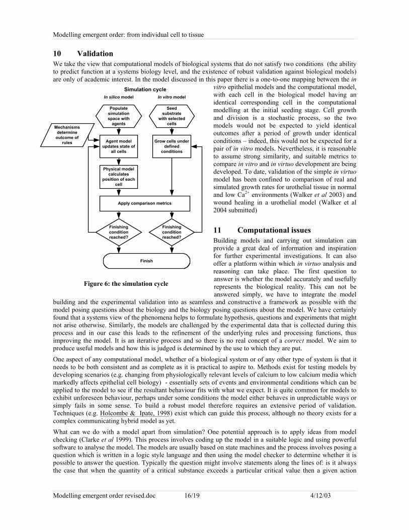
10 ValidationWe take the view that computational models of biological systems that do not satisfy two conditions (the abilityto predict function at a systems biology level, and the existence of robust validation against biological models)are only of academic interest. In the model discussed in this paper there is a one-to-one mapping between the in
vitro epithelial models and the computational model,with each cell in the biological model having anidentical corresponding cell in the computationalmodelling at the initial seeding stage. Cell growthand division is a stochastic process, so the twomodels would not be expected to yield identicaloutcomes after a period of growth under identicalconditions – indeed, this would not be expected for apair of in vitro models. Nevertheless, it is reasonableto assume strong similarity, and suitable metrics tocompare in vitro and in virtuo development are beingdeveloped. To date, validation of the simple in virtuomodel has been confined to comparison of real andsimulated growth rates for urothelial tissue in normaland low Ca2+ environments (Walker et al 2003) andwound healing in a urothelial model (Walker et al2004 submitted)
11 Computational issuesBuilding models and carrying out simulation canprovide a great deal of information and inspirationfor further experimental investigations. It can alsooffer a platform within which in virtuo analysis andreasoning can take place. The first question toanswer is whether the model accurately and usefullyrepresents the biological reality. This can not beanswered simply, we have to integrate the model
building and the experimental validation into as seamless and constructive a framework as possible with themodel posing questions about the biology and the biology posing questions about the model. We have certainlyfound that a systems view of the phenomena helps to formulate hypothesis, questions and experiments that mightnot arise otherwise. Similarly, the models are challenged by the experimental data that is collected during thisprocess and in our case this leads to the refinement of the underlying rules and processing functions, thusimproving the model. It is an iterative process and so there is no real concept of a correct model. We aim toproduce useful models and how this is judged is determined by the use to which they are put.
One aspect of any computational model, whether of a biological system or of any other type of system is that itneeds to be both consistent and as complete as it is practical to aspire to. Methods exist for testing models bydeveloping scenarios (e.g. changing from physiologically relevant levels of calcium to low calcium media whichmarkedly affects epithelial cell biology) - essentially sets of events and environmental conditions which can beapplied to the model to see if the resultant behaviour fits with what we expect. It is quite common for models toexhibit unforeseen behaviour, perhaps under some conditions the model either behaves in unpredictable ways orsimply fails in some sense. To build a robust model therefore requires an extensive period of validation.Techniques (e.g. Holcombe & Ipate, 1998) exist which can guide this process, although no theory exists for acomplex communicating hybrid model as yet.
What can we do with a model apart from simulation? One potential approach is to apply ideas from modelchecking (Clarke et al 1999). This process involves coding up the model in a suitable logic and using powerfulsoftware to analyse the model. The models are usually based on state machines and the process involves posing aquestion which is written in a logic style language and then using the model checker to determine whether it ispossible to answer the question. Typically the question might involve statements along the lines of: is it alwaysthe case that when the quantity of a critical substance exceeds a particular critical value then a given action
Seedsubstrate
with selectedcells
Populatesimulationspace with
agents
Grow cells underdefined
conditions
Agent modelupdates state of
all cells
Mechanismsdetermine
outcome ofrules
Physical modelcalculates
position of eachcell
Apply comparison metrics
Finishingconditionreached?
Finishingconditionreached?
Finish
In silico model In vitro modelSimulation cycle
Figure 6: the simulation cycle
Modelling emergent order: from individual cell to tissue
Modelling emergent order revised.doc 17/19 4/12/03
occurs or a given state is entered, is there a state in which a particular property of the model holds, is there a pathof behaviour in the model such that every state in that path has a certain property and so on. Model checkershave successfully explored models with very large numbers (billions) of states. Model checking forcommunicating X-machines has been developed by Eleftherakis using the XmCTL logic (Eleftherakis et al,2001). This provides an important basis for future analysis of large, complex biological models. It is not enoughthat we only rely on simulation for our understanding of the system, it may be that some highly critical sequenceof behaviour only occurs under conditions that we never get round to simulate, yet this knowledge might beimportant. Rather than trying things out to see what happens we try to identify interesting or undesirablephenomena and see whether the model can ever exhibit them (e.g. predicting what would happen if tissueengineered epithelia were constructed with a sub-optimal percentage of stem cells - this has been a long-standingand almost unanswerable concern for the clinical use of tissue engineered skin. Would one expect the skin tobreak down over a patient's life-time? A computational model could be used to predict the life-time wearcharacteristics of the tissue–engineered skin under both normal and wounded (regenerative) conditions. Thiskind of backwards reasoning is relatively novel and is still at an early stage but looking at the development ofcomputational models in the long term the ability to do this will be extremely powerful. Questions such as:whether there are wound healing advantages to enriching the skin stem cell population or lowering the woundbed calcium for patients with chronic wounds could be assessed initially in virtuo. The use of the model couldinform the design of in vitro (and possibly in vivo) experiments to then test these questions
12 The larger scheme: the Physiome ProjectThe long-range goal of the Physiome Project is to understand and describe the human organism, its physiologyand pathophysiology, and to use this understanding to improve human health. A major aim is to developcomputer models to integrate the observations from many laboratories into quantitative, self-consistent andcomprehensive descriptions (Hunter et al 2002). The models that have been hitherto been seen as central to thisendeavour fall into two classes – supra-cellular models (whole body system models; whole body continuummodels; and tissue and whole organ continuum models) and sub-cellular models (sub-cellular ODE models; sub-cellular Markov models; and molecular models). The development of a model which is specifically based on theindividual cell and its interaction with neighbouring cells provides an essential link between supra-cellular andsub-cellular models. In principle, the sub-cellular models provide the mechanisms to replace the rules in theagent model. An important goal for the cellular model is to provide a means for describing the parameters inconstitutive equations of tissue (continuum models of the macro world) in terms of events at a sub-cellular level.
13 ConclusionModelling biological systems such as epithelial organisation will immediately necessitate a more quantitativeapproach to describing cell/cell social behaviour. When used in parallel with well characterised 2D and 3Depithelial culture systems then we anticipate that descriptive rule sets will be the first “ deliverable” and thatthese will inform our understanding of some of the biological rules implicit in tissue organisation. It seems asafe prediction that the next “deliverable” will be the ability to run experiments in virtuo to test concepts andunderstanding. In the long term, we propose that the in virtuo model will function as a predictive tool of cellbehaviour in vitro and in vivo.
14 ReferencesBalanescu, T., Cowling, A.J., Gheorgescu, H., Gheorghe, M., Holcombe, M., & Vertan, C. 1999. Communicating stream X-machinessystems are no more than X-machines. Journal of Universal Computer Science, 5, 494-507Bhargava S, Chapple C R, Layton C, Bullock A J and MacNeil S 2003 Tissue-engineered buccal mucosa for substitution urethroplasty.British Journal of Urology. In pressBassingthwaighte, J.B. 1995 Towards modelling the human physiome. Adv. Expt. Med. Biol. 382, 331-339.Bassingthwaighte, J.B. 2000a Symposium on Integrative Biology of the Heart. Ann. Biomed Eng 28, 835-1058.Bassingthwaighte, J.B. 2000b Strategies for the Physiome Project. Ann. Biomed. Eng. 28, 1043-1058.Baumgarter W, Hinterdorfer P, Ness W, Raab A, Vestweber D, Schindler H and Drenckhahn D. 2000.Cadherin interaction probed by atomic force microscopy. PNAS 97 4005-4010.Booth C, Harnden P, Trejdosiewicz LK, Scriven S, Selby PJ, Southgate J 1997 Stromal and vascular invasion in an human in vitro bladdercancer model. Lab Invest 76, 843-857
Modelling emergent order: from individual cell to tissue
Modelling emergent order revised.doc 18/19 4/12/03
Brodland G W, Chen H. H. 2000. The Mechanics of Heterotypic Cell Aggregates:Insights from Computer Simulations. Journal ofBiomechanical Engineering 122, 402-407.Chakrabarty K H, Dawson R A, Harris P, Layton C, Babu M, Gould L, Phillips J, Leigh I, Green C, Freedlander E, Mac Neil S 1999Development of autologous human dermal-epidermal composites based on sterilised human allodermis for clinical use. British Journal ofDermatology 141, 811-823Chakrabarty, K.H., Heaton, J., Dalley, A.J., Dawson, R.A., Freedlander, E., Khaw, P.T. and Mac Neil, S 2001. Keratinocyte-drivencontraction of reconstructed human skin. Wound Repair and Regeneration, 9, 95-106Chen H H, Brodland G. W. 2000. Cell-level Finite Element Studies of Viscous Cells in Planar Aggregates. Journal of BiomechanicalEngineering 122, 394-401.Clarke E M, Grumberg O, Peled D, 1999 Model checking, MIT Press.Dover R and Potten C S 1983. Cell Cycle Kinetics of Cultured Human Epidermal Keratinocytes. Journal of Investigative Dermatology 80423-429Eleftherakis, G., & Kefalas P. 2001. Towards model checking of finite state machines extended with memory through refinement. InG.Antoniou, N.Mastorakis, & O.Panfilov (Eds.). (pp.321-326). Advances in Signal Processing and Computer Technologies, World Scientificand Engineering Society Press.Eleftherakis G, Kefalas P, Sotiriadou A, 2002 Formal verification of agent models, in Proc. 2nd Hellenic Conf. On AI (SETN02), 25-435Eleftherakis G, Kefalas P, Sotiriadou A, 2001 XmCTL: extending temporal logic to facilitate formal verification of X-machine models.Analel Uni. Bucuresti, Math.-Inf. 50, 79-95Eves P, Layton C, Hedley S, Dawson R A, Wagner M, Morandini R, Ghanem G, Mac Neil S 2000 Characterisation of an in vitro model ofhuman melanoma invasion based on reconstructed human skin. British Journal of Dermatology 142, 210-222Forestell S P, M. B. J., Kalogerakis N, Behie L A 1992. A Cellular Automaton Model for the Growth of Anchorage-dependent MammalianCells used in Vaccine Production. Chemical Engineering Science 47, 2381-2386.Georghe, M., Holcombe, M., & Kefalas, P. 2001. Computational models for collective foraging, 4th International Workshop on InformationProcessing in Cells and Tissues, Leuven, BelgiumGlazier J A and Graner F. 1993. Simulation of the Differential Adhesion driven Rearrangement of Biological Cells. Physical Review E 47,2128-2153.Hill G, Varley C, PellegrinS, Trejdosiewicz L K and Southgate J 2003 Human urothelial cell proliferation andmigration isdifferentiallyregulated by autocrine EGFR signalling during re-epithelialisation in vitro. In preparationHogeweg, P. 2000. Evolving Mechanisms of Morphogenesis: on the Interplay between Differential Adhesion and Cell Differentiation.Journal of Theoretical Biology 203, 317-333.Holcombe M, Holcombe L, Gheorghe M, Talbot N 2003 A hybrid machine model of rice blast fungus, Magnaporthe grisea. BioSystems, 68,223-228Holcombe, M., & Ipate, F. 1998. Correct systems: Building a business process Solution. Springer Verlag, LondonHonda, H. 1983. Geometrical Models for Cells in Tissues. International Review of Cytology 81, 191-248.Huang S and Ingber D E 1999. The structural and mechanical complexity of cell-growth control. Nature Cell Biology 1 E131-E138.Hunter P, Robbins Pand Noble D, 2002, The IUPS human physiome project. Pflugers Arch - Eur J Physiol 445:1–9Jefferson G, Haritos G K and McMeeking R M 2002 The elastic response of a cohesive aggregate – a discrete element model with coupledparticle interaction. Journal of the Physics and Mechanics of Solids 50 2539-2575Jensen U B, Lowell S and Watt F M 1999. The spatial relationship between stem cells and their progeny in the basal layer of humanepidermis: a new view based on whole-mount labelling and lineage analysis. Development 126 2409-2418.International Human Genome Mapping Consortium 2001 A physical map of the human genome. Nature 409, 934-941Kefalas P, Holcombe M & Gheorghe M, 2003a, “A formal method for the development of agent-based systems”, in Intelligent AgentSoftware Engineering, V. Plekhanova,,ed. Idea Group, 68-98Kefalas, P., Eleftherakis, G., & Kehris, E. 2003b. Modular modeling of large-scale systems using communicating X-machines, Proceedingsof the 8th Panhellenic Conference in Informatics, Greek Computer Society. In pressLee Y, K. S., McIntire L V, Zygourakis K 1995. A Cellular Automaton Model for the Proliferation of Migrating Contact-Inhibited Cells.Biophysical Journal 69, 1284-1298.Lim J H F and Davies G. A. 1990. A Stochastic Model to Simulate the Growth of Anchorage Dependent Cells on Flat Surfaces.Biotechnology and Bioengineering 36, 547-562.Loew L M and Schaff J C 2001 The virtual cell: a software environment for computational cell biology. Trends in Biotechnology 19, 401-406Morel D, Marcelpoil R., Brugal G 2001. A proliferation control network model:the simulation of two-dimensional epithelial homeostasis.Acta Biotheoretica 49, 219-234.Morton-Firth C J and Bray D 1998 Predicting temporal fluctuations in an intracellular signalling pathway. J Theor Biol 192, 117-128Odell G M, O. G., Alberch P, Burnside B 1981. The Mechanical Basis of Morphogenesis. Developmental Biology 85, 446-462.Orford K, Orford C C and Byers S W 1999. Exogenous expresion of Beta-Catenin regulates contact inhibition, anchorage-independentgrowth, anoikis and radiation-induced cell cycle arrest. J Cell Biol 146 855-867.Palsson, E. 2001. A three-dimensional model of cell movement in multi-cellular systems. Future Generation Computer Systems 17, 835-852.Potten C S and Morris R J 1988. Epithelial stem cells in vivo. J Cell Science Suppl. 10 45-62.
Modelling emergent order: from individual cell to tissue
Modelling emergent order revised.doc 19/19 4/12/03
Potten C S and Booth C 2002. Keratinocyte Stem Cells: a Commentary. J Investigative Dermatology 119 888-899.Ralston D R, Layton C, Dalley A J, Boyce S G, Freedlander E, Mac Neil S 1999 The requirement for basement membrane antigens in theproduction of human epidermal/dermal composites in vitro. British Journal of Dermatology 140, 605-615Ransom R and Matela R. J. 1984. Computer Modelling of Cell Division during Development using a Topological Approach. Journal ofEmbryology and Experimental Morphology 83 Suppl.: 233-259.Ruaan R C, Tsai. G. J., Tsao G T 1993. Monitoring and Modelling of Density-Dependent Growth of Anchorage-Dependent Cells.Biotechnology and Bioengineering 41, 380-389.Ryder E F, Bullard L, Hone J, Olmstead J, Ward M O 1999. Graphical simulation of early development of the cerebral cortex. Computermethods and Progress in Biomedicine 59, 107-114.Savill N J, Hogeweg P. 1997. Modelling Morphogenesis: From Single Cells to Crawling Slugs. Journal of Theoretical Biology 184, 229-235.Scriven SD, Booth C, Thomas DF, Trejdosiewicz LK, Southgate J 1997 Reconstitution of human urothelium from monolayer cultures. JUrol 158, 1147-1152Southgate J, Hutton KA, Thomas D F and Trejdosiewicz L K 1994 Normal human urothelial cells in vitro: proliferation and induction ofstratification. Lab Invest 71, 583-594Southgate J, Masters JR, Trejdosiewicz LK 2002 Culture of Human Urothelium. In: Freshney RI, Freshney MG, eds. Culture of EpithelialCells. 2nd Ed. New York: J Wiley and Sons, Inc.St Croix B, Sheehan C, Rak J W, Florenes V A, Slingerland J M and Kerbel R S 1998. E-Cadherin dependent growth suppression ismediated by the cyclin-dependent kinase inhibitor p27 KIPI. J Cell Biol 142 557-571.Stamenović D, Fredberg J J, Wang N, Butler J P and Ingber D E 1996 A Microstructural Approach to Cytoskeletal Mechanics based onTensegrity. J Theor Biol 181, 125-136Steinberg, M. S. 1970. Does differential adhesion govern cell self-assembly processes in histogenesis? Equilibrium configurations and theemergence of a hierarchy among populations of embryonice cells. Journal of Experimental Zoology 173, 395-433.Stekel D, Rashbass. J., Williams E D 1995. A computer graphic simulation of squamous epithelium. Journal of theoretical biology 175, 283-293.Talbot N J, 1995 Having a blast: exploring the pathogenicity of Magnaportha grisea. Trends in Microbiology, 3, 9-16.Talbot N J & Foster A J, 2001 Genetics and genomics of the rice blast fungus Magnaporthe grisea: developing an experimental model forunderstanding fungal diseases of cereals, Adv. Bot. Res., 34 , 263- 287.Tomita M, Hashimoto K, Takahashi K, Shimuzu T S, Matsuzaki Y, Miyoshi F, Saito K, Tanida S, Yugi K, Venter J C and Hutchison C A1999 E-CELL: software environment for whole-cell simulation. Bioinformatics 15, 72-84Venter C et al 2001 The sequence of the human genome. Science 291, 1304-1351Walker D C, Southgate J, Hill G, Holcombe M, Hose D R, Wood S M, Mac Neil S, Smallwood R H 2003 Agent-based modelling of thesocial behaviour of cells. Biosystems. In press.Williams J R and O'Connor R (1999) Discrete element simulation and the contact problem. Archives of Computational Methods inEngineeringZygourakis K, Bizios R., Markenscoff P 1991a. Proliferation of Anchorage-Dependent Contact-Inhibited Cells: I. Development ofTheoretical Models based on Cellular Automata. Biotechnology and Bioengineering 38, 459-470.Zygourakis K, Markenscoff P., Bizios R, 1991b. Proliferation of Anchorage-Dependent Contact-Inhibited Cells: II. Experimental Resultsand Validation of the Theoretical Models. Biotechnology and Bioengineering 38, 471-479.Walker D C, Hill G, Wood S M, Smallwood R, Southgate J 2004. Agent-based computational modeling of epithelial cell monolayers:predicting the effect of exogenous calcium concentration on the rate of wound closure. Submitted
.