miR29a and miR29b contribute to pancreatic beta-cell-specific silencing of monocarboxylate...
13
MOLECULAR AND CELLULAR BIOLOGY, Aug. 2011, p. 3182–3194 Vol. 31, No. 15 0270-7306/11/$12.00 doi:10.1128/MCB.01433-10 Copyright © 2011, American Society for Microbiology. All Rights Reserved. miR-29a and miR-29b Contribute to Pancreatic -Cell-Specific Silencing of Monocarboxylate Transporter 1 (Mct1) † Timothy J. Pullen, 1 Gabriela da Silva Xavier, 1 Gavin Kelsey, 2,3 and Guy A. Rutter 1 * Department of Cell Biology, Division of Medicine, Sir Alexander Fleming Building, Imperial College London, Exhibition Road, London, United Kingdom 1 ; Developmental Genetics and Imprinting, Babraham Institute, Cambridge, United Kingdom 2 ; and Centre for Trophoblast Research, University of Cambridge, Cambridge CB2 3EG, United Kingdom 3 Received 17 December 2010/Returned for modification 1 March 2011/Accepted 24 May 2011 In pancreatic cells, elevated glucose concentrations stimulate mitochondrial oxidative metabolism to raise intracellular ATP/ADP levels, prompting insulin secretion. Unusually low levels of expression of genes encod- ing the plasma membrane monocarboxylate transporter, MCT1 (SLC16A1), as well as lactate dehydrogenase A (LDHA) ensure that glucose-derived pyruvate is efficiently metabolized by mitochondria, while exogenous lactate or pyruvate is unable to stimulate metabolism and hence insulin secretion inappropriately. We show here that whereas DNA methylation at the Mct1 promoter is unlikely to be involved in cell-type-specific transcriptional repression, three microRNAs (miRNAs), miR-29a, miR-29b, and miR-124, selectively target both human and mouse MCT1 3 untranslated regions. Mutation of the cognate miR-29 or miR-124 binding sites abolishes the effects of the corresponding miRNAs, demonstrating a direct action of these miRNAs on the MCT1 message. However, despite reports of its expression in the mouse -cell line MIN6, miR-124 was not detectably expressed in mature mouse islets. In contrast, the three isoforms of miR-29 are highly expressed and enriched in mouse islets. We show that inhibition of miR-29a in primary mouse islets increases Mct1 mRNA levels, demonstrating that miR-29 isoforms contribute to the -cell-specific silencing of the MCT1 transporter and may thus affect insulin release. Glucose metabolism in pancreatic cells is specialized to efficiently couple glucose oxidation to an increase in ATP/ADP ratio, critical for stimulating insulin secretion (37). Alternative metabolic pathways that could interfere with glucose sensing are suppressed by specifically “disallowing” expression of cer- tain genes in cells. These disallowed genes include those encoding lactate dehydrogenase A (LDHA), which converts pyruvate to lactate (25, 39, 40), and MCT1 (SLC16A1) (14, 15, 17, 40, 45, 46), a plasma membrane monocarboxylate trans- porter. Both of these genes are widely expressed in other tissues but display very low expression levels in cells (32, 40). This modification seems likely to serve a 2-fold role: first, to avoid inappropriate stimulation of oxidative metabolism, and hence insulin release, in response to circulating pyruvate or lactate; and second, to prevent the loss of glucose-derived pyruvate from cells. The effects of inappropriate overexpression of MCT1 are observed in the rare genetic disorder physical exercise-induced hypoglycemia (32). In this condition, autosomal dominant mu- tations in the MCT1 (SLC16A1) promoter lead to increased transcription of the MCT1 gene sufficient to overcome the -cell-specific block on expression (31). During strenuous physical exercise, pyruvate and lactate produced by anaerobic metabolism in skeletal muscle are released into the blood- stream. The presence of MCT1 then appears to allow the circulating pyruvate/lactate to enter cells, where it acts as a substrate for mitochondrial oxidation leading to an increased cytosolic ATP/ADP ratio. This triggers insulin release despite the absence of elevated blood glucose levels, resulting in hy- poglycemia. Given the critical importance of disallowing MCT1 expres- sion in cells, we were interested in the mechanism by which this widely expressed gene is so specifically silenced. Although mouse cells express very low levels Mct1 mRNA, luciferase assays have demonstrated low but significant activity of exog- enous Mct1 promoter sequences when transfected into these cells (31). This suggests that additional epigenetic or posttran- scriptional mechanisms are responsible for further suppressing Mct1 expression in the cell. DNA methylation is an epigenetic modification of DNA which can regulate gene expression. In eukaryotes, DNA meth- ylation occurs on cytidine residues of CG dinucleotides (CpG) (29). High levels of DNA methylation at gene promoters are associated with gene silencing. DNA methylation may contrib- ute to silencing genes both in a tissue-specific manner and also for aberrantly silencing tumor suppressor genes in cancer (3). MicroRNAs (miRNAs) are short 19- to 21-nucleotide (nt) RNAs expressed as hairpin precursors which, following pro- cessing by Dicer, can bind to sites mainly within the 3 untrans- lated region (UTR) of target genes. This interaction can either block translation or can destabilize the mRNA leading to de- struction of the message (6, 30, 42). A number of miRNAs have previously been implicated in cell function. miR-375 is specifically expressed in islets and is reportedly the most abun- dant miRNA in cells (35). This miRNA plays roles both in regulating insulin secretion (35) and in islet development (19, * Corresponding author. Mailing address: Department of Cell Biol- ogy, Division of Medicine, Sir Alexander Fleming Building, Imperial College London, Exhibition Road, South Kensington, London SW7 2AZ, United Kingdom. Phone: 44 20 7594 3391. Fax: 44 20 7594 3351. E-mail: [email protected]. † Supplemental material for this article may be found at http://mcb .asm.org/. Published ahead of print on 6 June 2011. 3182
Transcript of miR29a and miR29b contribute to pancreatic beta-cell-specific silencing of monocarboxylate...
MOLECULAR AND CELLULAR BIOLOGY, Aug. 2011, p. 3182–3194 Vol. 31, No. 150270-7306/11/$12.00 doi:10.1128/MCB.01433-10Copyright © 2011, American Society for Microbiology. All Rights Reserved.
miR-29a and miR-29b Contribute to Pancreatic �-Cell-SpecificSilencing of Monocarboxylate Transporter 1 (Mct1)�†
Timothy J. Pullen,1 Gabriela da Silva Xavier,1 Gavin Kelsey,2,3 and Guy A. Rutter1*Department of Cell Biology, Division of Medicine, Sir Alexander Fleming Building, Imperial College London, Exhibition Road,
London, United Kingdom1; Developmental Genetics and Imprinting, Babraham Institute, Cambridge, United Kingdom2;and Centre for Trophoblast Research, University of Cambridge, Cambridge CB2 3EG, United Kingdom3
Received 17 December 2010/Returned for modification 1 March 2011/Accepted 24 May 2011
In pancreatic � cells, elevated glucose concentrations stimulate mitochondrial oxidative metabolism to raiseintracellular ATP/ADP levels, prompting insulin secretion. Unusually low levels of expression of genes encod-ing the plasma membrane monocarboxylate transporter, MCT1 (SLC16A1), as well as lactate dehydrogenaseA (LDHA) ensure that glucose-derived pyruvate is efficiently metabolized by mitochondria, while exogenouslactate or pyruvate is unable to stimulate metabolism and hence insulin secretion inappropriately. We showhere that whereas DNA methylation at the Mct1 promoter is unlikely to be involved in cell-type-specifictranscriptional repression, three microRNAs (miRNAs), miR-29a, miR-29b, and miR-124, selectively targetboth human and mouse MCT1 3� untranslated regions. Mutation of the cognate miR-29 or miR-124 bindingsites abolishes the effects of the corresponding miRNAs, demonstrating a direct action of these miRNAs on theMCT1 message. However, despite reports of its expression in the mouse �-cell line MIN6, miR-124 was notdetectably expressed in mature mouse islets. In contrast, the three isoforms of miR-29 are highly expressed andenriched in mouse islets. We show that inhibition of miR-29a in primary mouse islets increases Mct1 mRNAlevels, demonstrating that miR-29 isoforms contribute to the �-cell-specific silencing of the MCT1 transporterand may thus affect insulin release.
Glucose metabolism in pancreatic � cells is specialized toefficiently couple glucose oxidation to an increase in ATP/ADPratio, critical for stimulating insulin secretion (37). Alternativemetabolic pathways that could interfere with glucose sensingare suppressed by specifically “disallowing” expression of cer-tain genes in � cells. These disallowed genes include thoseencoding lactate dehydrogenase A (LDHA), which convertspyruvate to lactate (25, 39, 40), and MCT1 (SLC16A1) (14, 15,17, 40, 45, 46), a plasma membrane monocarboxylate trans-porter. Both of these genes are widely expressed in othertissues but display very low expression levels in � cells (32, 40).This modification seems likely to serve a 2-fold role: first, toavoid inappropriate stimulation of oxidative metabolism, andhence insulin release, in response to circulating pyruvate orlactate; and second, to prevent the loss of glucose-derivedpyruvate from � cells.
The effects of inappropriate overexpression of MCT1 areobserved in the rare genetic disorder physical exercise-inducedhypoglycemia (32). In this condition, autosomal dominant mu-tations in the MCT1 (SLC16A1) promoter lead to increasedtranscription of the MCT1 gene sufficient to overcome the�-cell-specific block on expression (31). During strenuousphysical exercise, pyruvate and lactate produced by anaerobicmetabolism in skeletal muscle are released into the blood-
stream. The presence of MCT1 then appears to allow thecirculating pyruvate/lactate to enter � cells, where it acts as asubstrate for mitochondrial oxidation leading to an increasedcytosolic ATP/ADP ratio. This triggers insulin release despitethe absence of elevated blood glucose levels, resulting in hy-poglycemia.
Given the critical importance of disallowing MCT1 expres-sion in � cells, we were interested in the mechanism by whichthis widely expressed gene is so specifically silenced. Althoughmouse � cells express very low levels Mct1 mRNA, luciferaseassays have demonstrated low but significant activity of exog-enous Mct1 promoter sequences when transfected into thesecells (31). This suggests that additional epigenetic or posttran-scriptional mechanisms are responsible for further suppressingMct1 expression in the � cell.
DNA methylation is an epigenetic modification of DNAwhich can regulate gene expression. In eukaryotes, DNA meth-ylation occurs on cytidine residues of CG dinucleotides (CpG)(29). High levels of DNA methylation at gene promoters areassociated with gene silencing. DNA methylation may contrib-ute to silencing genes both in a tissue-specific manner and alsofor aberrantly silencing tumor suppressor genes in cancer (3).
MicroRNAs (miRNAs) are short 19- to 21-nucleotide (nt)RNAs expressed as hairpin precursors which, following pro-cessing by Dicer, can bind to sites mainly within the 3� untrans-lated region (UTR) of target genes. This interaction can eitherblock translation or can destabilize the mRNA leading to de-struction of the message (6, 30, 42). A number of miRNAshave previously been implicated in � cell function. miR-375 isspecifically expressed in islets and is reportedly the most abun-dant miRNA in � cells (35). This miRNA plays roles both inregulating insulin secretion (35) and in islet development (19,
* Corresponding author. Mailing address: Department of Cell Biol-ogy, Division of Medicine, Sir Alexander Fleming Building, ImperialCollege London, Exhibition Road, South Kensington, London SW72AZ, United Kingdom. Phone: 44 20 7594 3391. Fax: 44 20 7594 3351.E-mail: [email protected].
† Supplemental material for this article may be found at http://mcb.asm.org/.
� Published ahead of print on 6 June 2011.
3182
36). miR-7 is also abundantly and specifically expressed in �cells (5, 9). miR-124 is reportedly expressed both in � cell lines(35) and in mouse islets (1) and is thought to regulate both thedevelopment (1) and the secretory function (26) of islets.miR-9 has also been shown to regulate insulin secretion bycontrolling the expression of granuphilin (34).
Here, we have investigated whether DNA methylation ormicroRNAs contribute to �-cell-specific silencing of Mct1. Weshow that whereas DNA methylation is unlikely to be respon-sible for regulating Mct1 expression in � cells, miR-29a, miR-29b, and miR-124 all target Mct1. Of these, the miR-29 iso-forms are highly expressed in islets and contribute to silencingMct1 in � cells. The maintained expression of miR-29 isoformsin the � cell thus seems likely to be required for the normal andselective stimulation of insulin secretion by glucose.
MATERIALS AND METHODS
Cell culture. The cell lines mhAT3F (22), MIN6 (28), and HEK293 (13) andisolated mouse islets were cultured as described previously (1).
DNA methylation analysis. MIN6 cells previously grown in medium containing25 mM glucose were maintained at 3 mM glucose for 16 h prior to culture at 3or 30 mM glucose in the presence or absence of 100 �M 5-azacytidine (ACT) for6 h. Cells were harvested in Trizol, and RNA was purified according to themanufacturer’s instructions. RNA was quantified using a Nanodrop-1000 spec-trophotometer and then reverse transcribed using a high-capacity cDNA reversetranscription (RT) kit (Applied Biosystems) with random primers. Real-timePCR was performed using Power SYBR green PCR Mastermix (Applied Bio-systems) on a 7500 fast real-time PCR machine (Applied Biosystems). Mct1mRNA was quantified relative to cyclophilin A by using the threshold cycle(��CT) method. Results are the means of four experiments, analyzed for sig-nificant differences by Student’s t test with Bonferroni correction for multipletesting.
Genomic DNA (gDNA) was prepared from MIN6 and mhAT3F cells usingproteinase K digestion, phenol-chloroform extraction, and isopropanol precipi-tation (38). Purified gDNA was subjected to bisulfite conversion using theEpiTect bisulfite kit (Qiagen) and then amplified by PCR using primers designedwith MethPrimer (http://www.urogene.org/methprimer/) (24). (All primer se-quences are given in a table in the supplemental material.) Products were ligatedinto pCR2.1 by TA cloning, and the inserts were sequenced. Sequence readswere analyzed using BiQ Analyzer (4) (http://biq-analyzer.bioinf.mpi-inf.mpg.de)to exclude poorly converted sequences and multiple copies of a single clone. Fiveindependent clones were included for each analysis. For bisulfite sequencing ofislets, DNA was prepared using a protocol based on the method of Millar et al.(27). Ten islets isolated from a CD1 mouse were placed in 10 �l lysis buffer (10mM Tris-Cl [pH 8.0], 1 mM EDTA, 1 mM SDS, 280 �g/ml proteinase K), heatedto 37°C for 90 min and 98°C for 15 min, and then cooled on ice. The whole 20-�lincubation was used as the input for the bisulfite conversion reaction, resulting inapproximately 750 cell equivalents per PCR.
Bioinformatic analysis of microRNAs. Data containing the raw counts formature miRNA detected in various mouse and human tissues and cell lines bylarge-scale cloning and sequencing were obtained from the supplemental data ofLandgraf et al. (21). To exclude cancerous samples, libraries were only includedwith malignancy code 1. Libraries containing fewer than 100 counts were alsoexcluded. The miRNA expression profile of the mouse MIN6 � cell line wascompared to that of other mouse samples. Islet � and � cells have relativelysimilar miRNA profiles: for example, both highly express miR-375. As we aimedto assess the degree to which miRNAs expressed in � cells are also expressed innonislet cells, the �-TC1 library was also excluded from the analysis. The fol-lowing libraries were therefore included: MIN6 pancreatic � cells, brain, glo-meruli, heart, kidney, kidney-collecting duct, liver, lung, midbrain, ovary, pla-centa, skin, spermatogonia-C18, and testis. The miRNA expression profile ofhuman islets was also compared to those of all tissue samples fulfilling thecriteria described above, which comprised cerebellum, frontal cortex, midbrain,hippocampus, liver, heart, spleen, pituitary, thyroid, ovary, testis, uterus, pla-centa, epididymis, and prostata.
Counts for each miRNA were converted to counts per million (CPM) miRNAsdetected in each library. The abundance of miRNAs in � cells/islets was calcu-lated as a percentage of the abundance of miR-375 as this has been widelyreported to be the most abundant miRNA in � cells and islets (5, 35). Specificity
of expression to � cells was calculated by dividing the CPM for an miRNA in the� cell/islet library by the total CPM for that miRNA in all libraries included fromthat species. A combined score for abundance and specificity was calculated bymultiplying the percentage scores for each. Lines dividing the quartiles of theranked distribution of this combined score are marked on the graphs in Fig. 2.
DNA constructs. miRNA expression vectors were created by first amplifyingthe coding sequence of selected miRNAs along with 230- to 300-bp flankingsequences on either side by PCR using mouse genomic DNA as template. A listof primers used is given in the supplemental material. The resulting PCR prod-ucts were inserted between the BamHI and XbaI sites of pcDNA3.1(�).
Firefly luciferase-expressing vectors were constructed as follows. A BglII-to-NheI fragment containing the herpes simplex virus thymidine kinase (TK) pro-moter was inserted between BglII and HindIII sites of pGL3 Basic to createpGL3-TK, in which firefly luciferase cDNA, followed by a simian virus 40(SV40)-derived polyadenylation signal, is expressed from the TK promoter.Regions covering the entire human and mouse MCT1 3� UTR were amplified byPCR and inserted between XbaI and BamHI sites of pGL3-TK to replace theSV40 polyadenylation signal and create pGL3-TK-HsM3 and pGL3-TK-MmM3,respectively. Putative microRNA binding sites were mutated by amplifying thewhole vector excluding the 8-bp seed region from a pair of primers containing anadditional restriction enzyme site, using high-fidelity Phusion polymerase(Finnzyme). The product was digested using that restriction enzyme and ligatedto recircularize the plasmid such that the seed region was replaced with therestriction enzyme site.
Luciferase assay of microRNA binding. The effect of overexpressed miRNAson the Mct1 3� UTRs was assessed in HEK293 cells. Cells grown in 24-well plateswere transfected with the following plasmids using Lipofectamine 2000 (Invitro-gen): 50 ng firefly luciferase reporter plasmid, 50 ng pRL-TK control plasmid,and 300 ng miRNA expression plasmid. In tests of multiple miRNAs, the fol-lowing mixture of DNA was used: 25 ng pRL-TK, 25 ng firefly luciferase reporterplasmid, 250 ng each miRNA expression plasmid, and sufficient empty miRNAexpression vector (pcDNA3.1) to make the total up to 800 ng. Cells wereharvested 48 h later and assayed with the dual-luciferase assay system (Promega).Two levels of controls were used to exclude from the results variation due totransfection efficiency and nonspecific effects of miRNAs. First, the firefly lucif-erase signal was normalized to the Renilla luciferase signal for each reading.Second, each miRNA was assessed in parallel with firefly plasmids containing a3� UTR (pGL3-TK-HsM3 or pGL3-TK-MmM3) or just the SV40 polyadenyla-tion signal (pGL3-TK) and the signal from the former normalized to the latter.All results were then presented as a percentage of the signal obtained with anempty miRNA expression vector. All results are the mean of at least threeindependent experiments. Significant downregulation of normalized luciferaseexpression was identified using one-tailed Student’s t test with Bonferroni cor-rection for multiple tests.
The effect of endogenous � cell miRNAs on the Mct1 3� UTR were assessedby luciferase assay in MIN6 cells. Cells grown in 24-well plates were transfectedwith 50 ng pRL-TK control plasmid along with 50 ng of firefly luciferase plasmidcontaining either the wild-type mouse Mct1 3� UTR (pGL3-TK-MmM3) or aplasmid in which the miR-29 or miR-124 binding sites had been mutated bysite-directed mutagenesis.
Effects of miRNA overexpression on endogenous Mct1. Mouse hepatoma-derived mhAT3F cells, grown in 12-well plates, were transiently transfected with1.6 �g of various miRNA expression vectors or an empty vector control, usingLipofectamine 2000. Cells were harvested 72 h later in TRIzol (Invitrogen), andtotal RNA was prepared according to the manufacturer’s instructions. Mct1mRNA levels were quantified relative to cyclophilin A using an Applied Biosys-tems high-capacity reverse transcription kit followed by quantitative PCR(qPCR) using Power SYBR green master mix (Applied Biosystems) running ona 7500 fast real-time PCR system (Applied Biosystems). Data are means of threeindependent experiments, and significant downregulation of the Mct1 level wasidentified using one-tailed Student’s t test with Bonferroni’s correction for mul-tiple tests.
mhAT3F cells grown in 6-well plates were stably transfected with 4 �g linear-ized miRNA expression vector or empty expression vector, using Lipofectamine2000. Twenty-four hours after transfection, cells were passaged into 75-cm2 flasksand then transferred to selective medium containing 250 �g/ml G418 24 h later.Stably transfected cells were kept as a polyclonal mixture resulting from presum-ably many individual stable transfection events and were maintained in 125 �g/mlG418.
Protein samples were separated by SDS-PAGE, transferred to nitrocellulosemembrane, and then probed with a rabbit anti-rat Mct1 antibody followed by ahorseradish peroxidase (HRP)-conjugated secondary antibody. The signal wasdetected using Supersignal West Dura substrate (Pierce) on a Chemidoc XRS
VOL. 31, 2011 microRNAs AND Mct1 SILENCING IN � CELLS 3183
imager (Bio-Rad). Blots were reprobed with mouse anti-�-tubulin antibodyfollowed by an HRP-conjugated secondary antibody.
Detection of microRNA expression by stem-loop reverse transcription-PCR.Mature microRNAs were specifically detected using a modified version of thestem-loop RT-PCR protocol described by Chen et al (7). miRNA expressionduring luciferase assays in HEK293 cells was confirmed by performing RT-PCRon heat-treated cell lysates. Cells grown in 24-well plates were transfected as forthe luciferase assay and harvested 48 h later in 100 �l phosphate-buffered saline(PBS) supplemented with 10 mM EDTA and then heated to 95°C for 5 min,diluted with an equal volume of water. For mhAT3F cells, total RNA waspurified using TRIzol as described above and then diluted to 2 ng/�l. RNAsamples were reverse transcribed using stem-loop primers designed to specifi-cally transcribe mature miRNA molecules at a concentration of 50 mM in 15-�lreaction mixtures also containing 1� RT buffer (P/N; 4319981 [Applied Biosys-tems]), 1 mM deoxynucleoside triphosphate (dNTP) mix, 2.67 U/�l Multiscribe(Applied Biosystems), and 0.63 U/�l RNase inhibitor (Applied Biosystems),which were incubated at 16°C for 30 min, 42°C for 30 min, and then 85°C for 5min. The cDNA was amplified by PCR with a pair of primers, one of which wascomplementary to part of the reverse transcription stem-loop primer and theother of which was complementary to the miRNA in 50-�l reaction mixturescontaining 1 � Phire reaction buffer (Finnzyme), 0.2 mM dNTP mix, 0.5 �M(each) forward and reverse primers, 3.5 �l reverse transcription reaction mixture,and 1 �l Phire hot-start polymerase (Finnzyme). The products were visualized byagarose gel electrophoresis. Controls without the reverse transcriptase enzymeconfirmed that the primers were specific to miRNAs and could not amplify theexpression vectors (data not shown). As the recombinant Moloney murine leu-kemia virus (rMo-MuLV) reverse transcriptase recognizes either RNA or DNAas a template, DNA oligonucleotides corresponding to the mature miRNA wereused a positive control for each miRNA assay at a concentration of 1 pM in thereverse transcription reaction. As a loading control, the U6 small nuclear RNAwas also amplified using stem-loop RT-PCR from each sample.
Measurement of miRNAs using quantitative stem-loop RT-PCR. Levels ofendogenous miRNAs were measured in total RNA samples with relative quan-tification by quantitative RT-PCR (qRT-PCR) using standard curves. TotalRNA was prepared from three cultures of MIN6 cells and from islets isolatedfrom three male C57BL/6 mice aged 26 weeks. Five-point standard curves wereprepared from DNA oligonucleotides corresponding to mature miRNAs in therange of 200 pM to 20 fM (miR-375), 100 pM to 10 fM (miR-29a, miR-29b,miR-29c, and miR-7a), or 1 pM to 100 aM (miR-124) (final concentration inreverse transcription reaction). DNA oligonucleotides were used in place ofRNA oligonucleotides for the standard curve as they are both less expensive andmore stable. In test experiments with miRNA-29a and miR-124 sequences, thetwo template types produced very similar results (see the figure in the supple-mental material). RNA samples were diluted to 10 ng/�l, and separate reversetranscription reactions, as described above, were performed for each sample andstandard curve point. Reverse transcription reaction mixtures were diluted4-fold, and 5 �l was used in 20-�l qPCRs using Power SYBR green master mix(Applied Biosystems) with forward and reverse primers at 0.2 �M. Reactionswere run on a 7500 fast real-time PCR system (Applied Biosystems), and indi-vidual miRNAs were first quantified by using the standard curves and thenpresented as a percentage of the miR-375 expression of that sample to permitcomparison with the miRNA quantification by large-scale cloning. To test thespecificity of the miR-29 isoform assays, 1 pM DNA oligonucleotide template foreach miR-29 isoform was tested in each miR-29 isoform assay. miR-29a andmiR-29b assays failed to detect either of the other two isoforms; however, themiR-29c assay detected the miR-29a oligonucleotide at 22% � 1.19% (mean �standard deviation [SD]) of the level at which it detected the miR-29a oligonu-cleotide.
miRNA knockdown with LNA inhibitors. Pancreatic islets were isolated fromC57BL/6 mice, cultured overnight, and then transfected with locked nucleic acid(LNA) miRNA inhibitors (Exiqon) targeting either miR-29a or miR-29b. Isletsgrown in 3 ml complete medium in 60-mm petri dishes were transfected with 25nM LNA using 12 �l TransIT-TKO (Mirus) according to the manufacturer’sinstructions. Islets were harvested 48 h later, and total RNA was purified usingTRIzol. Data are the means of three independent experiments performed onislets isolated from three separate mice.
RESULTS
DNA methylation at the promoter CpG island of Mct1 is notresponsible for �-cell-specific silencing. Initially, the DNAmethylation inhibitor 5-azacytidine (ACT) was used to inves-
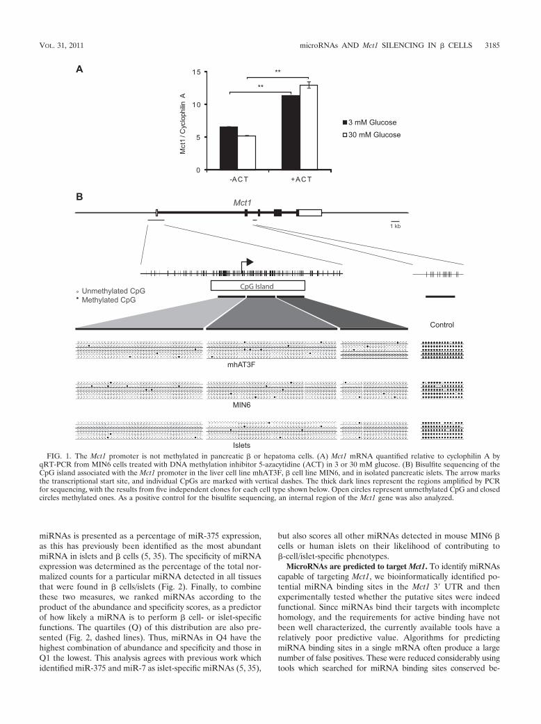
tigate the potential involvement of DNA methylation in Mct1expression in the mouse � cell line MIN6 (28). Cells were“starved” for 16 h in culture medium containing 3 mM glucoseand then treated with 1 mM ACT for 6 h in medium containing3 or 30 mM glucose. Mct1 mRNA was quantified by quantita-tive reverse transcription-PCR (qRT-PCR) using SYBR green.In both high- and low-glucose media, ACT treatment in-creased Mct1 mRNA levels (P 0.01) (Fig. 1A). These dataindicate that, either through direct or indirect mechanisms,DNA methylation may be acting to suppress Mct1 expressionin � cells. To identify whether this was a direct effect of DNAmethylation of the Mct1 promoter, bisulfite sequencing (8) wasperformed and the methylation status at this locus was as-sessed.
Unmethylated cytidines were converted to uracil by bisulfiteconversion of genomic DNA from both MIN6 cells and amouse liver cell line, mhAT3F, which expresses a high level ofMct1. Three contiguous regions of the Mct1 promoter coveringalmost the entire CpG island were amplified by PCR, and themethylation status of individual CpGs was determined by se-quencing (Fig. 1B).
The promoter region displayed a very low level of methyl-ation in both cell lines, with no differences apparent betweenthe two (Fig. 1B). To determine whether the methylation sta-tus of the � cell line MIN6 accurately represented the in vivosituation in primary � cells, the same analysis was also per-formed on isolated mouse islet tissue. The Mct1 promoter wasexamined in freshly isolated pancreatic islets and was againfound to display a very low level of DNA methylation. Tocontrol for the possibility that demethylation might occur dur-ing the procedure, another region of the Mct1 gene, expectedto be highly methylated, was also analyzed. A region was cho-sen which was distant from a CpG island such that its CpGswere likely to be methylated. In both cell lines and in isolatedislets, this region was indeed found to be highly methylated,confirming the validity of the analysis of the Mct1 promoter(Fig. 1B). We therefore conclude that the CpG island associ-ated with the Mct1 promoter is not methylated in � cells andtherefore that DNA methylation at this locus is not responsiblefor the �-cell-specific silencing of Mct1.
miRNA expression analysis in mouse � cells and humanislets. We next investigated whether miRNAs may contributeto silencing Mct1 expression in � cells. To this end, we neededto identify both miRNAs that specifically target Mct1 and alsodetermine which miRNAs are expressed in � cells or islets.Both the abundance and specificity of expression are expectedto affect the ability of a particular miRNA to perform �-cell-specific functions. While previous analyses have described themost abundant miRNAs in � cells (35) or those most differ-entially expressed between two tissues (5), a comprehensiveanalysis comparing the � cell miRNA complement with thoseof a range of other tissues has not yet been published. Wetherefore aimed to generate in silico a measure of both abun-dance and specificity of miRNA expression in � cells.
This analysis was performed using publically available datafrom large-scale cloning and sequencing of miRNAs in varioustissues from both humans and mice (21). Data from humanislets or mouse MIN6 � cells were compared to miRNA ex-pression in other nonmalignant, differentiated, adult tissues ofthe same species. In Fig. 2, the abundance of individual
3184 PULLEN ET AL. MOL. CELL. BIOL.
miRNAs is presented as a percentage of miR-375 expression,as this has previously been identified as the most abundantmiRNA in islets and � cells (5, 35). The specificity of miRNAexpression was determined as the percentage of the total nor-malized counts for a particular miRNA detected in all tissuesthat were found in � cells/islets (Fig. 2). Finally, to combinethese two measures, we ranked miRNAs according to theproduct of the abundance and specificity scores, as a predictorof how likely a miRNA is to perform � cell- or islet-specificfunctions. The quartiles (Q) of this distribution are also pre-sented (Fig. 2, dashed lines). Thus, miRNAs in Q4 have thehighest combination of abundance and specificity and those inQ1 the lowest. This analysis agrees with previous work whichidentified miR-375 and miR-7 as islet-specific miRNAs (5, 35),
but also scores all other miRNAs detected in mouse MIN6 �cells or human islets on their likelihood of contributing to�-cell/islet-specific phenotypes.
MicroRNAs are predicted to target Mct1. To identify miRNAscapable of targeting Mct1, we bioinformatically identified po-tential miRNA binding sites in the Mct1 3� UTR and thenexperimentally tested whether the putative sites were indeedfunctional. Since miRNAs bind their targets with incompletehomology, and the requirements for active binding have notbeen well characterized, the currently available tools have arelatively poor predictive value. Algorithms for predictingmiRNA binding sites in a single mRNA often produce a largenumber of false positives. These were reduced considerably usingtools which searched for miRNA binding sites conserved be-
FIG. 1. The Mct1 promoter is not methylated in pancreatic � or hepatoma cells. (A) Mct1 mRNA quantified relative to cyclophilin A byqRT-PCR from MIN6 cells treated with DNA methylation inhibitor 5-azacytidine (ACT) in 3 or 30 mM glucose. (B) Bisulfite sequencing of theCpG island associated with the Mct1 promoter in the liver cell line mhAT3F, � cell line MIN6, and in isolated pancreatic islets. The arrow marksthe transcriptional start site, and individual CpGs are marked with vertical dashes. The thick dark lines represent the regions amplified by PCRfor sequencing, with the results from five independent clones for each cell type shown below. Open circles represent unmethylated CpG and closedcircles methylated ones. As a positive control for the bisulfite sequencing, an internal region of the Mct1 gene was also analyzed.
VOL. 31, 2011 microRNAs AND Mct1 SILENCING IN � CELLS 3185
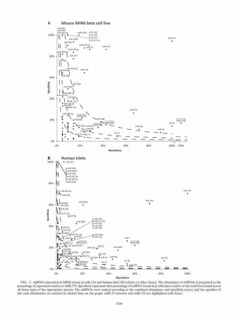
FIG. 2. miRNA expression in MIN6 mouse � cells (A) and human islets (B) relative to other tissues. The abundance of miRNAs is presented as thepercentage of expression relative to miR-375. Specificity represents that percentage of miRNA found in � cells/islets relative to the total level found acrossall tissue types of the appropriate species. The miRNAs were ranked according to the combined abundance and specificity scores, and the quartiles ofthis rank distribution are marked by dashed lines on the graph. miR-29 isoforms and miR-124 are highlighted with boxes.
3186
tween orthologous genes in several species. The Human Micro-RNA Targets tool (http://cbio.mskcc.org/cgi-bin/mirnaviewer/mirnaviewer.pl) (18), which uses the miRanda algorithm,identified five miRNAs with binding sites conserved betweenthe human, mouse, and rat Mct1 3� UTRs. The PicTar algo-rithm (http://pictar.mdc-berlin.de) (20), searching for bindingsites conserved across vertebrate orthologues, identified sixmiRNAs showing considerable overlap with the previous algo-rithm (Table 1).
A different version of the miRanda algorithm (http://www.microrna.org) (2), which includes sites not highly conservedbetween species, predicted 160 miRNAs targeting humanMCT1 and 83 targeting mouse Mct1. While this number ofmiRNAs was unfeasible to validate experimentally, we notedthat both miR-375 and miR-7 were predicted to target humanMCT1. Since these have previously both been identified asspecifically and highly expressed in pancreatic islets (5, 35),they were included in the list of miRNAs which we went on toexamine experimentally.
All three miR-29 isoforms and miR-124 target Mct1. Toidentify miRNAs capable of directly targeting the Mct1 3�UTR, we assessed our candidate miRNAs by luciferase assay.This assay quantified the ability of overexpressed miRNAs toreduce expression of firefly (Photinus pyralis) luciferase froman mRNA bearing the Mct1 3� UTR.
miRNA expression plasmids were constructed by amplifyingthe corresponding miRNA gene with short flanking sequencesfrom mouse genomic DNA and then cloning this downstreamof a polymerase II promoter in a mammalian expression vector(pcDNA3.1). Expression of the correct mature miRNA fromthese vectors was confirmed by RT-PCR using stem-loop prim-ers (Fig. 3C). miR-124 and miR-375 were also expressed bycloning annealed oligonucleotides into the miRNA expressionvector pcDNA 6.2 EmGFP-miR. In both cases, very similarresults were produced from the different types of vector, soresults from different vectors expressing the same miRNAwere averaged.
The whole 3� UTR of mouse Mct1 was cloned downstreamof the firefly luciferase coding sequence, whose expression wasdriven by the herpes simplex virus thymidine kinase promoter(TK). A second vector used the same TK promoter to expressRenilla reniformis luciferase, followed by an SV40 polyadenyl-
ation signal. The two vectors were cotransfected into HEK293cells along with various miRNA expression vectors, and theratio of the two luciferases was detected by dual-luciferaseassay. To exclude any confounding effects of miRNAs bindingto the luciferase open reading frames (ORFs), data were nor-malized to a parallel experiment that examined the effect ofmiRNA expression on firefly luciferase followed by an SV40polyadenylation signal rather than the Mct1 3� UTR (Fig. 3A).We also performed the same analysis using the human MCT13� UTR on the basis that evolutionary conserved interactionsare more likely to be biologically meaningful (Fig. 3B).
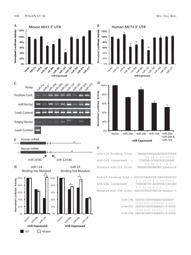
A one-tailed Student’s t test identified five miRNAs whichsignificantly reduced expression of firefly luciferase by target-ing the mouse Mct1 3� UTR (Fig. 3A). Four of these alsosignificantly targeted the human MCT1 3� UTR (Fig. 3B).miR-124 exerted the largest effect, decreasing luciferase ex-pression by at least 50% in assays of both mouse and humansequences (P 0.00001). miR-29a reduced expression by�33% (P 0.01), and miR-29b reduced expression by �27%(P 0.01) in both cases. A smaller effect was produced bymiR-29c which reduced expression by �7% (P 0.05). Trans-fection with miR-7 produced a small but nonsignificant de-crease in luciferase activity for both human and mouse Mct1sequences. Thus, 4 out of the 10 miRNAs tested significantlytarget both human and mouse Mct1 3� UTRs: miR-124 and thethree isoforms of miR-29. Of these, miR-29a, miR-29b, andmiR-124 were capable of reducing expression by over one-quarter.
To test the combined effect of multiple miRNAs on themouse Mct1 3� UTR, miR-29a, miR-29b, and miR-124 weretested individually and together by luciferase assay (Fig. 3D).This confirmed that the combined effect of the three miRNAswas greater than the effect of each miRNA individually.
miR-29a, miR-29b, and miR-124 target Mct1 directly: mu-tational analysis of binding. The controls used in the aboveluciferase assays excluded from the analysis any effects ofmiRNA expression on the promoter or coding sequence ofeither luciferase variant. The assay therefore identified directinteractions between the miRNA and Mct1 3� UTR. To pro-vide further confirmation of the specificity of the interactionand to confirm the precise binding site, we mutated the pre-dicted binding sites and repeated the luciferase assay.
The predicted binding sites for both miR-29b and miR-124are found in the first 150 bp of the 3� UTRs (Fig. 3E). PicTarpredicted that miR-29a and miR-29c may also potentially tar-get the miR-29b site. The 6 to 8 nucleotides at the 3� end ofmiRNA binding sites, termed the “seed region” (10), arehighly complementary to the miRNA sequence and critical forbinding. In both of the putative miR-29 and miR-124 bindingsites, the 8 bp of the seed region were replaced with a restric-tion enzyme site with minimal complementarity to the miRNAsequence (Fig. 3F). Mutation of the putative miR-124 siteabolished the effect of miR-124 while leaving the action miR-29a unaffected (Fig. 3G). This confirmed that the former re-gion is an miR-124 binding site.
Mutation of the putative miR-29 binding site led to theunexpected finding that while this virtually abolished the actionof miR-29b, miR-29a was still able to target the mutated 3�UTR albeit with an efficacy reduced by 25% (P 0.05) (Fig.3G). This finding confirmed that this region forms an miR-29b
TABLE 1. miRNAs predicted to target Mct1a
miRNA
Result withprediction tool: Quartile in:
miRanda PicTar Mouse MIN6� cell line Human islets
miR-9* � �miR-29a � � Q3 Q3miR-29b � � Q3 Q4miR-29c � � Q3 Q4miR-124 � � Q4miR-183 � � Q4miR-320 � � Q1miR-377 � � Q4
a Listed are miRNAs predicted have conserved binding sites in the Mct1 3�UTR by the miRanda and PicTar algorithms. The quartile within the distributionof the combined abundance and specificity score of the individual miRNAs inboth MIN6 cells and human islets are also given.
VOL. 31, 2011 microRNAs AND Mct1 SILENCING IN � CELLS 3187
3188 PULLEN ET AL. MOL. CELL. BIOL.
binding site, and while miR-29a does bind this site, it also actsthrough binding at other sites within the Mct1 3� UTR.
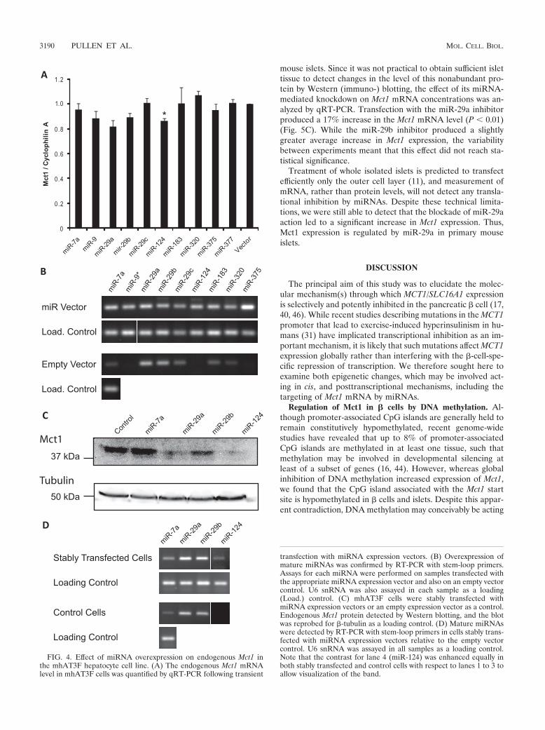
miR-29a, miR-29b, and miR-124 target endogenous Mct1mRNA. As an independent assay of miRNA function, we alsoassessed the ability of each of the above miRNAs to targetendogenous Mct1 mRNA. miRNAs predicted to target Mct1were overexpressed in a mouse liver cell line, mhAT3F, whichexpresses a high level of Mct1 consistent with high levels ofMct1 protein expression in this tissue (15, 31). The effect onendogenous Mct1 mRNA abundance was quantified by qRT-PCR (Fig. 4A). Overexpression of miR-124 produced a statis-tically significant decrease in the endogenous Mct1 mRNAlevel. This confirms that miR-124 is able to target naturallyoccurring Mct1 mRNA and that this results in mRNA degra-dation. Although the results for miR-29a and -29b fell belowthe level of statistical significance, the pattern of effects ob-served in the luciferase assay (Fig. 3A and B) were broadlyreplicated. miR-29a and miR-29b thus both tended to causedecreases in Mct1 mRNA levels, with miR-29a exerting alarger effect than miR-29b. This suggests that miR-29a andmiR-29b may target Mct1 at least partially through mRNAdegradation.
To overcome problems caused by limited transfection effi-ciency, mhAT3F cells were stably transfected with expressionvectors for miRNAs shown to target Mct1 (miR-29a, miR-29b,and miR-124), miRNAs shown not to target Mct1 (miR-7a), oran empty vector control. Expression of mature miRNAs in thestably transfected cell lines was confirmed by RT-PCR usingstem-loop primers (Fig. 4D). As in the transient transfectionexperiments some endogenous expression of miR-29a andmiR-29b was detected in the control cell line consistent withlow expression of miR-29 detected in human liver tissue. Stabletransfection with each of the miRNA expression vectors led toa moderate increase in expression of that miRNA. The level ofMct1 protein in the stable cell lines was detected by Westernblotting of whole-cell lysates (Fig. 4C). The anti-Mct1 antibodydetected a single band of 43 kDa that was greatly reduced insamples expressing miR-29b and virtually abolished in samplesexpressing miR-29a or miR-124. It is interesting to note thatthese dramatic changes in Mct1 protein level are achieved byrelatively modest expression of miRNAs, partly because con-tinued drug selection presumably ensured consistent miRNAexpression across the whole-cell population.
miR-29 downregulates Mct1 in primary mouse islets. Havingconfirmed that miR-29a, miR-29b, and miR-124 can specifi-cally and significantly target Mct1, we examined which of these
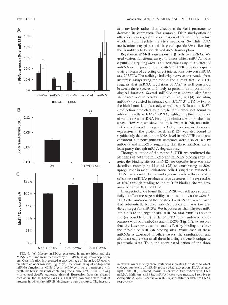
may act in primary � cells within islets. All three members ofthe miR-29 family fall within the top two quartiles in bothMIN6 cells and human islets. Indeed, miR-29b is the fourthmost abundant miRNA in human islets. This indicates thatmiR-29 isoforms are both abundant and enriched in islets andare therefore highly likely to contribute to the islet phenotype.Although miR-124 was abundantly present in MIN6 cells, itwas not detected in human islets. To confirm these data fromlarge-scale sequencing, we measured expression levels ofmiR-29 isoforms and miR-124 in both MIN6 cells and mousepancreatic islets by qRT-PCR with stem-loop primers, andtheir expression was calculated relative to that of miR-375(Fig. 5A). Expression of all three miR-29 isoforms was con-firmed in both mouse islets and MIN6 cells. In islets, miR-29aand miR-29c were detected at �15% of the miR-375 level, andmiR-29b was detected at �10%. The levels of the three iso-forms were more variable in MIN6 cells, ranging from miR-29aat 24% to miR-29b at 4%. While miR-124 was abundantlydetected in the MIN6 cells used for large-scale cloning (21),the level detected in MIN6 cells grown for these experimentsby qRT-PCR was substantially lower at just 0.24% � 0.032%(mean � standard error [SE]) of the miR-375 level. Indeed, inthe study that provided the data for the bioinformatic analysis(21), Landgraf et al. reported failing to detect mature miR-124in mouse or human islets and considered the presence ofmiR-124 in MIN6 cells to be an artifact of cell immortalization.In agreement with this finding, levels of miR-124 in islets, asgiven by our qRT-PCR analysis, were below the level ofdetection. Thus, the lowest point of the standard curve formiR-124 which detected a higher signal than the no-templatecontrol was 1 fM, equivalent to 0.19% of the mean level ofmiR-375 detected in the islet samples. We therefore concludethat, in the mouse islets studied here, the miR-124 level is0.19% of the miR-375 level, and at this level we considermiR-124 unlikely to play a significant role in � cells, by far themost abundant islet cell type.
To assess the effect of endogenous � cell miRNAs on theMct1 3� UTR, we compared levels of luciferase expression inMIN6 � cells from constructs containing the wild-type Mct1 3�UTR with those containing mutated miR-29b binding sites(Fig. 5B). Destruction of the miR-29 binding site doubledexpression of luciferase (P 0.01), showing that the levels ofmiR-29 isoforms in � cells are sufficient to downregulate Mct1.
Finally, locked nucleic acid (LNA) miRNA inhibitors de-signed against miR-29a and miR-29b were used to investigatethe effect of miR-29 isoforms on Mct1 expression in isolated
FIG. 3. Reporter-based assays of the impact of miRNAs targeting the Mct1 3� UTR in HEK293 cells. Luciferase assays were performed toassess the effect of miRNA overexpression on mouse (A) or human (B) Mct1 3� UTR. The level of firefly luciferase activity, normalized againstRenilla luciferase, is presented as a percentage of signal obtained with an empty miRNA expression vector. (C) Mature miRNAs expressed insamples from a parallel transfection were detected by reverse transcription using stem-loop primers followed by PCR. Assays for each miRNA wereperformed on samples transfected with the appropriate miRNA expression vector and on an empty vector control. U6 snRNA was assayed in allsamples as a loading (load.) control. As a positive control for each miRNA assay, 1 pM synthetic DNA oligonucleotide corresponding to the maturemiRNA sequence was assayed in parallel. (D) Luciferase assay of the combined effects of miR-29a, miR-29b, and miR-124 expression on mouseMct1 3� UTR. (E) Mct1 mRNA from humans and mice with open reading frames marked with solid boxes. The locations of putative miR-29 andmiR-124 binding sites (BS) are also indicated. In both species, cDNAs with shorter 3� UTRs are reported in databases, and the sites of alternativetranscription termination sites are marked with asterisks. (F) Base pair binding between miRNAs and putative miRNA binding sites in the mouseMct1 3� UTR. The binding sites were mutated by replacing the 8-bp seed region with a restriction enzyme site. An alignment of the three miR-29isoforms is also shown. (G) Luciferase assay of mouse Mct1 3� UTR with the putative miRNA binding sites mutated.
VOL. 31, 2011 microRNAs AND Mct1 SILENCING IN � CELLS 3189
mouse islets. Since it was not practical to obtain sufficient islettissue to detect changes in the level of this nonabundant pro-tein by Western (immuno-) blotting, the effect of its miRNA-mediated knockdown on Mct1 mRNA concentrations was an-alyzed by qRT-PCR. Transfection with the miR-29a inhibitorproduced a 17% increase in the Mct1 mRNA level (P 0.01)(Fig. 5C). While the miR-29b inhibitor produced a slightlygreater average increase in Mct1 expression, the variabilitybetween experiments meant that this effect did not reach sta-tistical significance.
Treatment of whole isolated islets is predicted to transfectefficiently only the outer cell layer (11), and measurement ofmRNA, rather than protein levels, will not detect any transla-tional inhibition by miRNAs. Despite these technical limita-tions, we were still able to detect that the blockade of miR-29aaction led to a significant increase in Mct1 expression. Thus,Mct1 expression is regulated by miR-29a in primary mouseislets.
DISCUSSION
The principal aim of this study was to elucidate the molec-ular mechanism(s) through which MCT1/SLC16A1 expressionis selectively and potently inhibited in the pancreatic � cell (17,40, 46). While recent studies describing mutations in the MCT1promoter that lead to exercise-induced hyperinsulinism in hu-mans (31) have implicated transcriptional inhibition as an im-portant mechanism, it is likely that such mutations affect MCT1expression globally rather than interfering with the �-cell-spe-cific repression of transcription. We therefore sought here toexamine both epigenetic changes, which may be involved act-ing in cis, and posttranscriptional mechanisms, including thetargeting of Mct1 mRNA by miRNAs.
Regulation of Mct1 in � cells by DNA methylation. Al-though promoter-associated CpG islands are generally held toremain constitutively hypomethylated, recent genome-widestudies have revealed that up to 8% of promoter-associatedCpG islands are methylated in at least one tissue, such thatmethylation may be involved in developmental silencing atleast of a subset of genes (16, 44). However, whereas globalinhibition of DNA methylation increased expression of Mct1,we found that the CpG island associated with the Mct1 startsite is hypomethylated in � cells and islets. Despite this appar-ent contradiction, DNA methylation may conceivably be acting
FIG. 4. Effect of miRNA overexpression on endogenous Mct1 inthe mhAT3F hepatocyte cell line. (A) The endogenous Mct1 mRNAlevel in mhAT3F cells was quantified by qRT-PCR following transient
transfection with miRNA expression vectors. (B) Overexpression ofmature miRNAs was confirmed by RT-PCR with stem-loop primers.Assays for each miRNA were performed on samples transfected withthe appropriate miRNA expression vector and also on an empty vectorcontrol. U6 snRNA was also assayed in each sample as a loading(Load.) control. (C) mhAT3F cells were stably transfected withmiRNA expression vectors or an empty expression vector as a control.Endogenous Mct1 protein detected by Western blotting, and the blotwas reprobed for �-tubulin as a loading control. (D) Mature miRNAswere detected by RT-PCR with stem-loop primers in cells stably trans-fected with miRNA expression vectors relative to the empty vectorcontrol. U6 snRNA was assayed in all samples as a loading control.Note that the contrast for lane 4 (miR-124) was enhanced equally inboth stably transfected and control cells with respect to lanes 1 to 3 toallow visualization of the band.
3190 PULLEN ET AL. MOL. CELL. BIOL.
at many levels rather than directly at the Mct1 promoter todecrease its expression. For example, DNA methylation atother loci may regulate the expression of transcription factorswhich in turn regulate the Mct1 promoter. So while DNAmethylation may play a role in �-cell-specific Mct1 silencing,this is unlikely to be via altered Mct1 transcription.
Regulation of Mct1 expression in � cells by miRNAs. Weused various functional assays to assess which miRNAs werecapable of targeting Mct1. The luciferase assay of the effect ofmiRNA overexpression on the Mct1 3� UTR provides a quan-titative means of detecting direct interactions between miRNAand 3� UTR. The striking similarity between the results fromluciferase assays using the mouse and human Mct1 3� UTRssuggests that miRNA regulation of Mct1 is well conservedbetween these species and likely to perform an important bi-ological function. Several miRNAs that showed significantabundance and selectivity in � cells (i.e., in Q4), includingmiR-377 (predicted to interact with MCT1 3� UTR by two ofthe bioinformatic tools used), as well as miR-7a and miR-375(interaction predicted by a single tool), were not found tointeract directly with Mct1 mRNA, highlighting the importanceof validating all miRNA-binding predictions with biochemicalassays. However, we show that miR-29a, miR-29b, and miR-124 can all target endogenous Mct1, resulting in decreasedexpression at the protein level. miR-124 was also found tosignificantly decrease the mRNA level in mhAT3F cells, andconsistent but nonsignificant decreases were also caused bymiR-29a and miR-29b, suggesting that these miRNAs act atleast partly through mRNA degradation.
Through mutation of the mouse 3� UTR, we confirmed theidentities of both the miR-29b and miR-124 binding sites. Ofnote, the binding site for miR-124 we describe here was alsodescribed recently by Li et al. (23) as contributing to Mct1upregulation in medulloblastoma cells. Using these mutated 3�UTRs, we showed that at endogenous levels within clonal �cells, these miRNAs produce a large decrease in the expressionof Mct1 through binding to the miR-29 binding site we havemapped in the Mct1 3� UTR.
Unexpectedly, we found that miR-29a was still able substan-tially to affect message stability or translation via the Mct1 3�UTR after mutation of the identified miR-29 site, a maneuverthat substantially blocked miR-29b action and was the pre-dicted target for miR-29a. We hypothesize that whereas miR-29b binds to the cognate site, miR-29a also binds to anothersite (or possibly sites) in the 3� UTR. Since miR-29c sharesfeatures with both miR-29a and miR-29b (Fig. 3F), we suspectthat the latter produces its small effect by binding to eitherthe mir-29a or miR-29b binding sites. While each of thesemiRNAs is expressed in other tissues, the simultaneous andabundant expression of all three in a single tissue is unique topancreatic islets. Thus, the coordinated action of the three
FIG. 5. (A) Mature miRNAs expressed in mouse islets and theMIN6 � cell line were measured by qRT-PCR using stem-loop prim-ers. Quantification is presented as a percentage of the miR-375 level tofacilitate comparison with Fig. 2. (B) Luciferase assay of endogenousmiRNA function in MIN6 � cells. MIN6 cells were transfected withfirefly luciferase plasmids containing the mouse Mct1 3� UTR alongwith control Renilla luciferase plasmid. Expression from the plasmidcontaining the wild-type (WT) 3� UTR was compared with that ofmutants in which the miR-29 binding site was disrupted. The increase
in expression caused by these mutations indicates the extent to whichendogenous levels of miR-29 reduce Mct1 expression. RLU, relativelight units. (C) Isolated mouse islets were transfected with LNAmiRNA inhibitors, and Mct1 mRNA levels were measured relative tocyclophilin A. �-miR-29 and �-miR-29b, anti-miR-29a and -29b LNAs,respectively.
VOL. 31, 2011 microRNAs AND Mct1 SILENCING IN � CELLS 3191
miR-29 isoforms is likely to contribute to the highly efficient�-cell-specific silencing of Mct1.
The present investigation as to whether miRNAs contributeto �-cell-specific silencing of Mct1 required an understandingof miRNA expression in the � cell. The likelihood that aparticular miRNA will contribute to a �-cell-specific pheno-type is determined by both the magnitude and specificity of itsexpression in � cells. While there have been several reports ofthe relative abundance of miRNAs in � cells, there has beenonly limited analysis of specificity. However, the large-scalesequencing of miRNAs purified from a range of tissues (21)has provided a large amount of publically available data allow-ing us to perform a more comprehensive analysis of � cellmiRNA expression. Our analyses covered 14 mouse tissues,including the mouse � cell line MIN6 and 16 human tissues,including pancreatic islets.
The miRNAs with high combined specificity and abundanceshow many similarities between the two samples. In addition tomiR-375 and miR-7a/miR-7, members of the miR-8 family(including miR-200a, miR-200b, miR-200c, and miR-141) andmiR-29 family (miR-29a, miR-29b, and miR-29c) have highcombined specificity and abundance in both samples. In total,there are 15 miRNAs found in the top two quartiles of thecombined abundance and specificity distribution in both MIN6cells and human islets: let-7b, miR-7/miR-7a, miR-16, miR-24,miR-26a, miR-27b, miR-29a, miR-29b, miR-29c, miR-30b,miR-30d, miR-127-3p, miR-200c, miR-368, and miR-375.
Whereas the � cell line used here represents a single definedcell type, it is nonetheless a transformed cell line whosemiRNA expression profile may have altered with time. In con-trast, human and mouse islets contain a mixture of the five celltypes. While � cells make up the majority of islet cells, and thus� cell miRNAs will be well represented, the expression profileobtained will be influenced by the other cell types present.Given the different natures of the samples, it is not possible todraw detailed conclusions about species differences in miRNAexpression, yet similarities between the samples particularly inthe region of high abundance and specificity suggest thatmiRNA expression is likely to be well conserved between thespecies. This view is supported by the demonstration that themiR-29 and miR-124 binding sites are well conserved betweenhuman and mouse Mct1.
Although miR-29 isoforms are highly expressed in humanislets, they are also expressed in other tissues, so the questionof whether they are likely to contribute to �-cell-specific si-lencing must be addressed. First, the level of expression inislets is very high, where miR-29b is the fourth most abundantmiRNA detected. Indeed the combined expression of all threemiR-29 isoforms is close (92%) to that of miR-375. For thehuman tissues for which data were obtained, the expressionlevel of both miR-29b alone and the combined expression of allmiR-29 isoforms is second highest in islets, being more abun-dant only in pituitary cells. While the levels of miR-29 isoformsmeasured by qRT-PCR in this study were lower than thosefrom the large-scale sequencing study, all three isoforms stillshowed significant levels of expression, with a combined ex-pression substantially greater than that of miR-7a, previouslyidentified as a major islet miRNA (5).
While miR-124 was confirmed to significantly and specifi-cally target Mct1, we failed to detect any expression of this
miRNA in primary mouse islets and only very low levels inMIN6 cells. These data contrast strongly with the report ofhigh levels of miR-124 in MIN6 cells by Landgraf et al (21). Itseems likely that clonal differences between the MIN6 cellsused in the two studies are most likely to account for thedifference in miR-124 levels measured. miR-124 levels inmouse islets were below the level of detection of our assay,producing a similar signal to the no-template control. Thiscontrasts with previous reports, which did detect miR-124 ex-pression in mouse (1) and human (12) islets. Baroukh et al (1).used RT-PCR with conventional primers (as opposed to stem-loop primers) which would detect miRNA primary transcriptsand pre-miRNAs but not mature miRNA. The signal obtainedin this earlier study may therefore represent immature miR-124, which may not be fully processed into mature miR-124.However, as miR-124 expression was not quantified in mouseislets, it is also possible that the earlier results correspond to alow level of mature miR-124. Fred et al. (12) quantify miR-124in human islets by qRT-PCR using TaqMan probes. However,as these authors only quantified relative changes in miR-124expression upon various treatments, this study gives little indi-cation of the amount of miR-124 relative to other miRNAs.Importantly, the high average CTs obtained for miR-124 indi-cated “a low abundance for miR-124a” (12), consistent withour own findings. It should be emphasized that, in the presentstudy, we performed relative quantification of mature miR-124and miR-375 in mouse islets by qRT-PCR using stem-loopprimers with DNA oligonucleotide standard curves, allowingus to quantify miRNAs relative to each other. This revealedthat the abundance of miR-124 is 0.19% of the miR-375level. This measurement does, however, represent a single timepoint, and miRNA levels may be affected by a variety of stimuli(e.g., glucose). At this level, we conclude that miR-124 is un-likely to affect Mct1 expression significantly in � cells, butcannot exclude the possibility that under conditions wheremiR-124 levels were increased, it may also regulate Mct1 ex-pression.
We also show here that miR-29a is able to significantlyregulate Mct1 expression in a hepatocyte cell line, in MIN6cells, and, importantly, primary mouse islets. The latter obser-vation demonstrates the likely functional relevance of this in-teraction in mature � cells. We have previously predicted (40,46) that low levels of MCT1 are a prerequisite in the � cell toensure that exogenous pyruvate does not inappropriately stim-ulate insulin secretion and also to prevent the possibility ofglucose carbons being lost as pyruvate. Changes in miR-29 thatimpact MCT1 level would therefore be expected to affect ei-ther or both of these parameters. In the present study, we wereunable to detect a significant impact on basal or glucose-stim-ulated insulin secretion of miR-29a or miR-29b suppressioneither in clonal � cells (which show abnormal stimulation ofsecretion) (40) or in primary islets despite, in the latter case,observing significant changes in Mct1 mRNA (Fig. 5C). Wesuspect, however, that the extent of the changes in Mct1 levelwe were able to induce using the tools currently available to usto modulate miR-29a or miR-29b are below the limits likely toimpact secretion from the intact islet, given that only a smallpercentage of cells are likely to be affected. Furthermore, wecannot exclude the possibility that these miRNAs target otherproteins whose expression may influence, and in part compen-
3192 PULLEN ET AL. MOL. CELL. BIOL.
sate for, the increase in MCT1 level. Importantly, in unpub-lished studies in which we have induced MCT1 expressionselectively in the � cells of adult transgenic mice, we haveobserved only modest effects on glucose-stimulated insulin se-cretion and a 2- to 3-fold stimulation of insulin secretion byexogenous pyruvate (T. J. Pullen and G. A. Rutter, unpub-lished data). It seems likely, therefore, that a full exploration ofthe impact of miR-29a and miR-29b levels will await the gen-eration of analogous cell-type-specific knockout or transgenicmice.
While the present studies were under revision, Thorrez et al(41). also provided a bioinformatic prediction that severalmiRNAs may regulate Mct1, although no direct experimentalevidence of interactions was given. This recent report alsosupported previous findings from van Arensbergen et al. (43)demonstrating that the Mct1 promoter from islets or purified �cells is enriched in repressive histone modifications. It there-fore appears that miRNA action is one of multiple indepen-dent mechanisms that contribute to the deep repression ofMct1 expression in � cells.
Conclusion. We demonstrate that Mct1 expression is si-lenced in pancreatic � cells at least in part by miRNAs selec-tively present in this cell type. The presence of these miRNAsseems likely to complement other, nonredundant transcrip-tional mechanisms which must also play an important rolegiven the impact of mutations in the MCT1 promoter thatresult in exercise-induced hyperinsulinism. Given the impor-tance of Mct1 in controlling insulin secretion, and its dysregu-lation in some forms of type 2 diabetes (33), regulation of theexpression of the latter gene by exogenous miRNAs may pro-vide a new therapeutic strategy for some forms of type 2 dia-betes.
ACKNOWLEDGMENTS
This study was supported by grants to G.A.R. from the WellcomeTrust (Programme 081958/Z/07/Z), MRC (G0401641), National Insti-tutes of Health (ROI DKO71962-01), and EU FP6 grant Save beta(G.A.R. and G.K.).
We thank Maz Wilson (University of Bristol) for the kind gift of theanti-MCT1 antibody and Andrew P. Halestrap (University of Bristol)for helpful discussions.
REFERENCES
1. Baroukh, N., et al. 2007. MicroRNA-124a regulates Foxa2 expression andintracellular signaling in pancreatic beta-cell lines. J. Biol. Chem. 282:19575–19588.
2. Betel, D., M. Wilson, A. Gabow, D. S. Marks, and C. Sander. 2008. ThemicroRNA.org resource: targets and expression. Nucleic Acids Res. 36:D149–D153.
3. Bird, A. 2002. DNA methylation patterns and epigenetic memory. GenesDev. 16:6–21.
4. Bock, C., et al. 2005. BiQ Analyzer: visualization and quality control forDNA methylation data from bisulfite sequencing. Bioinformatics 21:4067–4068.
5. Bravo-Egana, V., et al. 2008. Quantitative differential expression analysisreveals miR-7 as major islet microRNA. Biochem. Biophys. Res. Commun.366:922–926.
6. Carthew, R. W., and E. J. Sontheimer. 2009. Origins and mechanisms ofmiRNAs and siRNAs. Cell 136:642–655.
7. Chen, C., et al. 2005. Real-time quantification of microRNAs by stem-loopRT-PCR. Nucleic Acids Res. 33:e179.
8. Clark, S. J., A. Statham, C. Stirzaker, P. L. Molloy, and M. Frommer. 2006.DNA methylation: bisulphite modification and analysis. Nat. Protoc. 1:2353–2364.
9. Correa-Medina, M., et al. 2009. MicroRNA miR-7 is preferentially ex-pressed in endocrine cells of the developing and adult human pancreas.Gene Expr. Patterns 9:193–199.
10. Didiano, D., and O. Hobert. 2006. Perfect seed pairing is not a generallyreliable predictor for miRNA-target interactions. Nat. Struct. Mol. Biol.13:849–851.
11. Diraison, F., et al. 2004. Over-expression of sterol-regulatory-element-binding protein-1c (SREBP1c) in rat pancreatic islets induces lipogenesisand decreases glucose-stimulated insulin release: modulation by 5-aminoimid-azole-4-carboxamide ribonucleoside (AICAR). Biochem. J. 378:769–778.
12. Fred, R. G., C. H. Bang-Berthelsen, T. Mandrup-Poulsen, L. G. Grunnet,and N. Welsh. 2010. High glucose suppresses human islet insulin biosynthesisby inducing miR-133a leading to decreased polypyrimidine tract bindingprotein-expression. PLoS One 5:e10843.
13. Graham, F. L., J. Smiley, W. C. Russell, and R. Nairn. 1977. Characteristicsof a human cell line transformed by DNA from human adenovirus type 5.J. Gen. Virol. 36:59–72.
14. Halestrap, A. P., and D. Meredith. 2004. The SLC16 gene family-frommonocarboxylate transporters (MCTs) to aromatic amino acid transportersand beyond. Pflugers Arch. 447:619–628.
15. Halestrap, A. P., and N. T. Price. 1999. The proton-linked monocarboxylatetransporter (MCT) family: structure, function and regulation. Biochem. J.343:281–299.
16. Illingworth, R., et al. 2008. A novel CpG island set identifies tissue-specificmethylation at developmental gene loci. PLoS Biol. 6:e22.
17. Ishihara, H., H. Wang, L. R. Drewes, and C. B. Wollheim. 1999. Overex-pression of monocarboxylate transporter and lactate dehydrogenase altersinsulin secretory responses to pyruvate and lactate in beta cells. J. Clin.Invest. 104:1621–1629.
18. John, B., et al. 2004. Human microRNA targets. PLoS Biol. 2:e363.19. Kloosterman, W. P., A. K. Lagendijk, R. F. Ketting, J. D. Moulton, and R. H.
Plasterk. 2007. Targeted inhibition of miRNA maturation with morpholinosreveals a role for miR-375 in pancreatic islet development. PLoS Biol.5:e203.
20. Krek, A., et al. 2005. Combinatorial microRNA target predictions. Nat.Genet. 37:495–500.
21. Landgraf, P., et al. 2007. A mammalian microRNA expression atlas based onsmall RNA library sequencing. Cell 129:1401–1414.
22. Levrat, F., et al. 1993. Influence of the content in transcription factors on thephenotype of mouse hepatocyte-like cell lines (mhAT). Exp. Cell Res. 209:307–316.
23. Li, K. K., et al. 2009. miR-124 is frequently down-regulated in medulloblas-toma and is a negative regulator of SLC16A1. Hum. Pathol. 40:1234–1243.
24. Li, L. C., and R. Dahiya. 2002. MethPrimer: designing primers for methyl-ation PCRs. Bioinformatics 18:1427–1431.
25. Liang, Y., et al. 1996. Glucose metabolism and insulin release in mouse betaHC9 cells, as model for wild-type pancreatic beta-cells. Am. J. Physiol.270:E846–E857.
26. Lovis, P., S. Gattesco, and R. Regazzi. 2008. Regulation of the expression ofcomponents of the exocytotic machinery of insulin-secreting cells by micro-RNAs. Biol. Chem. 389:305–312.
27. Millar, D. S., P. M. Warnecke, J. R. Melki, and S. J. Clark. 2002. Methyl-ation sequencing from limiting DNA: embryonic, fixed, and microdissectedcells. Methods 27:108–113.
28. Miyazaki, J., et al. 1990. Establishment of a pancreatic beta cell line thatretains glucose-inducible insulin secretion: special reference to expression ofglucose transporter isoforms. Endocrinology 127:126–132.
29. Mueller, W. C., and A. von Deimling. 2009. Gene regulation by methylation.Recent Results Cancer Res. 171:217–239.
30. Murchison, E. P., and G. J. Hannon. 2004. miRNAs on the move: miRNAbiogenesis and the RNAi machinery. Curr. Opin. Cell Biol. 16:223–229.
31. Otonkoski, T., et al. 2007. Physical exercise-induced hypoglycemia caused byfailed silencing of monocarboxylate transporter 1 in pancreatic beta cells.Am. J. Hum. Genet. 81:467–474.
32. Otonkoski, T., et al. 2003. Physical exercise-induced hyperinsulinemic hypo-glycemia is an autosomal-dominant trait characterized by abnormal pyru-vate-induced insulin release. Diabetes 52:199–204.
33. Parton, L. E., et al. 2006. Limited role for SREBP-1c in defective glucose-induced insulin secretion from Zucker diabetic fatty rat islets: a functionaland gene profiling analysis. Am. J. Physiol. Endocrinol. Metab. 291:E982–E994.
34. Plaisance, V., et al. 2006. MicroRNA-9 controls the expression of Granuphi-lin/Slp4 and the secretory response of insulin-producing cells. J. Biol. Chem.281:26932–26942.
35. Poy, M. N., et al. 2004. A pancreatic islet-specific microRNA regulatesinsulin secretion. Nature 432:226–230.
36. Poy, M. N., et al. 2009. miR-375 maintains normal pancreatic alpha- andbeta-cell mass. Proc. Natl. Acad. Sci. U. S. A. 106:5813–5818.
37. Rutter, G. A. 2004. Visualising insulin secretion. The Minkowski Lecture2004. Diabetologia 47:1861–1872.
38. Sambrook, J., and D. W. Russell. 2001. Molecular cloning: a laboratorymanual, 3rd ed. Cold Spring Harbor Laboratory Press, Cold Spring Har-bor, NY.
39. Schuit, F., et al. 1997. Metabolic fate of glucose in purified islet cells.Glucose-regulated anaplerosis in beta cells. J. Biol. Chem. 272:18572–18579.
VOL. 31, 2011 microRNAs AND Mct1 SILENCING IN � CELLS 3193

40. Sekine, N., et al. 1994. Low lactate dehydrogenase and high mitochondrialglycerol phosphate dehydrogenase in pancreatic beta-cells. Potential role innutrient sensing. J. Biol. Chem. 269:4895–4902.
41. Thorrez, L., et al. 2011. Tissue-specific disallowance of housekeeping genes:the other face of cell differentiation. Genome Res. 21:95–105.
42. Valencia-Sanchez, M. A., J. Liu, G. J. Hannon, and R. Parker. 2006. Controlof translation and mRNA degradation by miRNAs and siRNAs. Genes Dev.20:515–524.
43. van Arensbergen, J., et al. 2010. Derepression of Polycomb targets duringpancreatic organogenesis allows insulin-producing beta-cells to adopt a neu-ral gene activity program. Genome Res. 20:722–732.
44. Weber, M., et al. 2005. Chromosome-wide and promoter-specific analysesidentify sites of differential DNA methylation in normal and transformedhuman cells. Nat. Genet. 37:853–862.
45. Zhao, C., and G. A. Rutter. 1998. Overexpression of lactate dehydrogenaseA attenuates glucose-induced insulin secretion in stable MIN-6 beta-celllines. FEBS Lett. 430:213–216.
46. Zhao, C., M. C. Wilson, F. Schuit, A. P. Halestrap, and G. A. Rutter.2001. Expression and distribution of lactate/monocarboxylate transporterisoforms in pancreatic islets and the exocrine pancreas. Diabetes 50:361–366.
3194 PULLEN ET AL. MOL. CELL. BIOL.