Enabling Dependable Data Storage for Miniaturized Satellites
Upload
independentCategory
view
0download
0
Miniaturized Ultrasound Imaging Probes Enabled by CMUTArrays with Integrated Frontend Electronic Circuits
B. (Pierre) T. Khuri-Yakub,E. L. Ginzton Laboratory, Stanford University, Stanford, CA 94305, USA.
Ömer Oralkan,E. L. Ginzton Laboratory, Stanford University, Stanford, CA 94305, USA([email protected]).
Amin Nikoozadeh,E. L. Ginzton Laboratory, Stanford University, Stanford, CA 94305, USA.
Ira O. Wygant,E. L. Ginzton Laboratory, Stanford University, Stanford, CA 94305, USA.
Steve Zhuang,E. L. Ginzton Laboratory, Stanford University, Stanford, CA 94305, USA.
Mustafa Gencel,E. L. Ginzton Laboratory, Stanford University, Stanford, CA 94305, USA.
Jung Woo Choe,E. L. Ginzton Laboratory, Stanford University, Stanford, CA 94305, USA.
Douglas N. Stephens,University of California, Davis, CA 95616, USA.
Alan de la Rama,St. Jude Medical, Irvine, CA 92618, USA.
Peter Chen,St. Jude Medical, Irvine, CA 92618, USA.
Feng Lin,General Electric Global Research Center, Niskayuna, NY 12309, USA.
Aaron Dentinger,General Electric Global Research Center, Niskayuna, NY 12309, USA.
Douglas Wildes,General Electric Global Research Center, Niskayuna, NY 12309, USA.
Kai Thomenius,General Electric Global Research Center, Niskayuna, NY 12309, USA.
Kalyanam Shivkumar,University of California, Los Angeles, CA 90095, USA.
Aman Mahajan,University of California, Los Angeles, CA 90095, USA.
Chi Hyung Seo,University of Washington, Seattle, WA 98101, USA.
Matthew O’Donnell,University of Washington, Seattle, WA 98101, USA.
NIH Public AccessAuthor ManuscriptConf Proc IEEE Eng Med Biol Soc. Author manuscript; available in PMC 2010 December 6.
Published in final edited form as:Conf Proc IEEE Eng Med Biol Soc. 2010 ; 1: 5987–5990. doi:10.1109/IEMBS.2010.5627580.
NIH
-PA Author Manuscript
NIH
-PA Author Manuscript
NIH
-PA Author Manuscript

Uyen Truong, andOregon Health and Science University, Portland, OR 97239, USA.
David J. SahnOregon Health and Science University, Portland, OR 97239, USA.
AbstractCapacitive micromachined ultrasonic transducer (CMUT) arrays are conveniently integrated withfrontend integrated circuits either monolithically or in a hybrid multichip form. This integrationhelps with reducing the number of active data processing channels for 2D arrays. This approachalso preserves the signal integrity for arrays with small elements. Therefore CMUT arraysintegrated with electronic circuits are most suitable to implement miniaturized probes required formany intravascular, intracardiac, and endoscopic applications. This paper presents examples ofminiaturized CMUT probes utilizing 1D, 2D, and ring arrays with integrated electronics.
I. INTRODUCTIONIn conventional ultrasound imaging systems that utilize 1D transducer arrays for 2D cross-sectional imaging the frontend transmit/receive (T/R) electronic circuits are typically locatedin the main processing unit. The transducer array is packaged in the form of a hand-heldprobe. Each element in the array is connected to a dedicated frontend channel through a long(~2 m) coaxial cable.
This conventional scheme has not been practical for transducer arrays with increasednumber of elements such as the 2D arrays with over 2000 elements. All major ultrasoundsystem providers today offer 2D matrix arrays for 3D imaging. All these 2D matrix probeshouse a portion of the beamforming electronics next to the array so that the number ofcables between the system and the transducer can be minimized and the acquired data can beprocessed by the existing imaging systems without having the need for thousands of activeelectronic channels. Placing the frontend electronics next to the array also improves thesignal integrity.
Ultrasound imaging catheters for intracardiac and intravascular applications also benefitfrom close integration of frontend electronic circuits with transducer arrays. This approachhelps with preserving the signal integrity and in some cases also to minimize the number ofcables. Catheter-based applications impose additional constraints on the physical size of thetransducer-electronics assembly and the total power consumption of the electronic circuits.
One of the great promises of the capacitive micromachined ultrasonic transducer technologyhas been the convenient and compact integration of electronic circuits with transducer arraysfor both large-area 2D arrays and miniature probes.
This paper reviews different approaches that enable close integration of CMUT arrays withsupporting electronic circuits. Following this brief review we present integration examplesincluding 2D arrays designed for intracavital imaging and 1D and ring arrays forintracardiac imaging.
II. INTEGRATION OF CMUT ARRAYS WITH SUPPORTING FRONTENDELECTRONIC CIRCUITS
Several methods developed for a variety of micro-electromechanical systems (MEMS)applications have also been used to integrate CMUT arrays with electronic circuits. These
Khuri-Yakub et al. Page 2
Conf Proc IEEE Eng Med Biol Soc. Author manuscript; available in PMC 2010 December 6.
NIH
-PA Author Manuscript
NIH
-PA Author Manuscript
NIH
-PA Author Manuscript

methods can be categorized in two main groups: monolithic and multi-chip (hybrid)approaches. As summarized in Table I, monolithic integration can be realized by buildingCMUTs and electronics at the same time or building CMUTs on finished electronics wafers.For multichip (hybrid) integration an intermediate substrate may or may not be used.
A. Co-ProcessingA BiCMOS process using 16 masks has been used with only minor modifications includingan additional photolithography step and sacrificial layer etching to fabricate CMUTs side-by-side with electronic circuits on the same substrate [1]. Although this method offers acost-effective means of integration, it has two major limitations: 1) The transducer elementarea is shared by electronic circuits or interconnects. 2) The device dimensions in thevertical direction are limited by the film thicknesses available in the process used to makeelectronic circuits.
B. Post-ProcessingAnother monolithic integration technique is to make the electronic circuits first using afoundry process and then build CMUTs on top of finished electronics by further processing,which includes surface passivation, planarization, opening of contacts to electronics, andfurther successive thin film deposition and etching steps to define the CMUT membranes[2,3,4]. This approach has good area utilization. However, processing techniques for makingCMUTs are still limited, mainly due to temperature constraints set by the existing metallines on the electronics.
C. Independently ProcessingBy making CMUTs and electronic circuits on two separate substrates one can use optimizedprocess flows for each component. CMUTs, in this case, need through-wafer viainterconnections from the front side of the substrate to the back side. Deep reactive ionetching (DRIE) enables these vias in silicon. CMUT arrays can then be bonded directly onelectronics [5] or on an intermediate substrate, which can be flexible [6] or rigid [7]. Directbonding usually requires that the electronics die area to be greater than the CMUT die area.Using an intermediate substrate makes the size of the CMUT and electronics diceindependent. Both components can be tiled to obtain larger arrays. Flexible substrates arepreferred for catheter applications because of the smaller form factor they offer throughbending.
III. 2D CMUT ARRAYS WITH INTEGRATED FRONTEND ELECTRONICCIRCUITS
We developed two generations of frontend integrated circuits for 16×16-element 2D CMUTarrays for 3D intracavital imaging applications such as endoscopic, endorectal, orendovaginal imaging.
A. Synthetic Aperture SystemThe first generation frontend integrated circuit (IC) we designed comprised a 25-V pulser, aT/R switch and a low-noise preamplifier for each transducer element in the array [Fig.1(a)][5]. Both the 2D CMUT array and the frontend IC had a pitch of 250 µm. Using Sn-Pbeutectic solder bumps the two components were directly bonded together [Fig.1(b)].Although each transducer element had their dedicated T/R frontend circuitry, only onechannel was programmed to be active at a time for simplicity and hence classical syntheticaperture imaging was performed with this first-generation prototype.
Khuri-Yakub et al. Page 3
Conf Proc IEEE Eng Med Biol Soc. Author manuscript; available in PMC 2010 December 6.
NIH
-PA Author Manuscript
NIH
-PA Author Manuscript
NIH
-PA Author Manuscript

B. Full-Transmit X-Receive System with Integrated Beamforming HardwareThe second-generation frontend IC we designed for the 16×16-element 2D array includeddigital circuitry to perform the functions of a transmit beamformer on the chip [Fig.2(a)] [8].With this IC 224 elements can be simultaneously fired in transmit and 16 elements can beused at a time for receive. We use a beamforming scheme, which we developed to reducethe frontend hardware complexity with minimum compromise from image quality. In thisscheme the 32 diagonal elements in the array constitute the receive aperture and theremainder of the array constitutes the transmit aperture. A preamplifier is provided for eachof the 32 receive elements. To interface to a 16-channel data acquisition system, 16 of the 32receive elements are active at a time. Sixteen buffers drive the cable capacitance in receive.A high-voltage pulser and an 8-bit shift register are provided for each of the 224 transmittingelements. The timing of each pulser is determined by delay information stored in the shiftregister. When transmitting, a particular pulser fires when a global counter equals thepulser’s stored delay value. For each new beam, delay information is loaded into the delayshift registers by 16 parallel lines. This scheme also reduces the number of cables betweenthe backend system and the frontend. We bonded a 16×16-element 2D CMUT arrayoperating at 2.5 MHz directly on this frontend IC and used this prototype for imagingseveral phantoms in 3D (Fig.3).
IV. INTRACARDIAC ECHOGRAPHY CATHETERS WITH CMUT ARRAYSWITH INTEGRATED FRONTEND ELECTRONIC CIRCUITS
We developed two types of forward looking intracardiac echography (ICE) catheters toprovide guidance during electrophysiological (EP) interventions.
A. Forward-Looking Microlinear Arrays for Real-Time Cross-Sectional ImagingThe first device we developed for forward-looking ICE applications is a 24-elementmicrolinear array operating at 10 MHz center frequency with a fractional bandwidth of100%. The element pitch is 63 µm. The array measures 1.7 mm by 1.3 mm and employsdoped-polysilicon filled through-wafer via interconnects [Fig.4(a)]. This array was bondedon one side of a custom flexible printed circuit board (PCB). The 24-channel electronics dieproviding each transducer element with a T/R switch, a low-noise preamplifier and anoutput buffer was bonded on the back side of the flexible PCB [Fig.4(b)]. With this IC thetransmit pulses with amplitudes up to −/+ 50 V were provided from the imaging system (GEVivid7), which is connected to the flex assembly by microcoaxial cables. A thin layer ofPolydimethylsiloxane (PDMS) provided electrical insulation on the front side of the array.
The final catheter packaged in a 9-F shaft [Fig.4(c)] was tested successfully in an animalstudy recently providing forward-looking cross-sectional images up to 4-cm depth (Fig.5).
B. Forward-Looking Ring Arrays for Volumetric ImagingThe second type of the ICE catheters we developed is based on a 10-MHz, 64-elementCMUT ring array [Fig.6(a)]. The electronics we used along with this array has the samecircuit topology as the one used with the microlinear array. However, in this case 8 channelsare grouped on one die and 8 ICs were used to address the entire array. The CMUT ringarray and the 8 ICs were bonded on an 8-legged flexible printed circuit board. A compactassembly was achieved by folding the legs of the flex circuit on a support structure [Fig.6(b)]. Microcoaxial cables connected this assembly to the imaging system. A programmableimaging platform (Verasonics, Redmond, WA) was used to reconstruct real-time volumeimages on multi-plane display with the option of off-line 3D rendering.
Khuri-Yakub et al. Page 4
Conf Proc IEEE Eng Med Biol Soc. Author manuscript; available in PMC 2010 December 6.
NIH
-PA Author Manuscript
NIH
-PA Author Manuscript
NIH
-PA Author Manuscript

The initial prototype was packaged in a 12-F catheter shaft [Fig.6(c)] and was used forimaging several phantoms (Fig. 7).
The ring catheter provides a 4.5-F inner lumen that can be used to introduce therapeuticdevices such as RF ablation catheters. Optical fibers can be inserted into the inner lumen toenable photoacoustic imaging.
V. CONCLUSIONWe have demonstrated several prototypes of miniaturized ultrasound probes using CMUTarrays with integrated electronics. The use of standard silicon microfabrication techniques inCMUT manufacturing enables densely populated transducer arrays with arbitrarygeometries. These arrays can be conveniently integrated with supporting electronic circuitsmonolithically or in the form of a multichip module to reduce the number of cables betweenthe probe and the system and also to preserve signal integrity. Results presented in this papershow that CMUT technology has the potential to enable many applications in ultrasoundimaging and therapy that were not possible with the conventional piezoelectric transducers.
AcknowledgmentsAuthors thank National Semiconductor for manufacturing the frontend integrated circuits, Tyco Electronics forcabling, and PacTech USA for their support with packaging.
This work was supported by the National Institutes of Health under grants CA99059, CA134720, and HL67647.
REFERENCES1. Eccardt PC, Niederer K, Fischer B. Micromachined transducers for ultrasound applications. Proc.
IEEE Ultrason. Symp 1997:1609–1618.2. Noble RA, Davies RR, King DO, Day MM, Jones ARD, McIntosh JS, Hutchins DA, Saul P. Low
temperature micromachined cMUTs with fully-integrated analogue front-end electronics. Proc.IEEE Ultrason. Symp 2002:1045–1050.
3. Daft C, Calmes S, da Graca D, Patel K, Wagner P, Ladabaum I. Microfabricated ultrasonictransducers monolithically integrated with high voltage electronics. Proc. IEEE Ultrason. Symp2004:493–496.
4. Gurun G, Qureshi MS, Balantekin M, Guldiken R, Zahorian J, Sheng-Yu P, Basu A, Karaman M,Hasler P, Degertekin L. Front-end CMOS electronics for monolithic integration with CMUT arrays:circuit design and initial experimental results. Proc. IEEE Ultrason. Symp 2008:390–393.
5. Wygant I, Zhuang X, Yeh D, Oralkan Ö, Ergun AS, Karaman M, Khuri-Yakub BT. Integration of2D CMUT arrays with front-end electronics for volumetric ultrasound imaging. IEEE Trans.Ultrason., Ferroelect., Freq. Contr 2008 Feb;vol. 55(no. 2):327–342.
6. Nikoozadeh, A.; Oralkan, Ö.; Gencel, M.; Choe, JW.; Stephens, DN.; de la Rama, A.; Chen, P.;Thomenius, K.; Dentinger, A.; Wildes, D.; Shivkumar, K.; Mahajan, A.; O’Donnell, M.; Sahn, D.;Khuri-Yakub, PT. Forward-Looking Volumetric Intracardiac Imaging Using a Fully IntegratedCMUT Ring Array. presented at the IEEE Ultrason. Symp.; Sep. 2009; Rome, Italy.
7. Wodnicki, R.; Woychik, CG.; Byun, AT.; Fisher, R.; Thomenius, K.; Lin, D-S.; Zhuang, X.;Oralkan, O.; Vaithilingam, S.; Khuri-Yakub, BT. Multi-row linear cMUT array using cMUTs andmultiplexing electronics. presented at the IEEE Ultrason. Symp.; Sep. 2009; Rome, Italy.
8. Wygant IO, Jamal NS, Lee HJ, Nikoozadeh A, Oralkan Ö, Karaman M, Khuri-Yakub BT. Anintegrated circuit with transmit beamforming flip-chip bonded to a 2-D CMUT array for 3-Dultrasound imaging. IEEE Trans. Ultrason., Ferroelect., Freq. Contr 2009 Oct;vol. 56(no. 10):2145–2156.
Khuri-Yakub et al. Page 5
Conf Proc IEEE Eng Med Biol Soc. Author manuscript; available in PMC 2010 December 6.
NIH
-PA Author Manuscript
NIH
-PA Author Manuscript
NIH
-PA Author Manuscript

Fig. 1.(a) Custom-designed frontend IC for the 3D synthetic aperture system. (b) A 16×16-element, 250-µm pitch, 2D CMUT array bonded on frontend electronics.
Khuri-Yakub et al. Page 6
Conf Proc IEEE Eng Med Biol Soc. Author manuscript; available in PMC 2010 December 6.
NIH
-PA Author Manuscript
NIH
-PA Author Manuscript
NIH
-PA Author Manuscript

Fig. 2.(a) Custom-designed frontend IC for the full-transmit X-receive imaging system. (b) A16×16-element, 250-µm pitch, 2D CMUT array bonded on frontend electronics.
Khuri-Yakub et al. Page 7
Conf Proc IEEE Eng Med Biol Soc. Author manuscript; available in PMC 2010 December 6.
NIH
-PA Author Manuscript
NIH
-PA Author Manuscript
NIH
-PA Author Manuscript

Fig. 3.3D renedered image of a nylon wire phantom obtained with a 2.5-MHz 2D CMUT array
Khuri-Yakub et al. Page 8
Conf Proc IEEE Eng Med Biol Soc. Author manuscript; available in PMC 2010 December 6.
NIH
-PA Author Manuscript
NIH
-PA Author Manuscript
NIH
-PA Author Manuscript
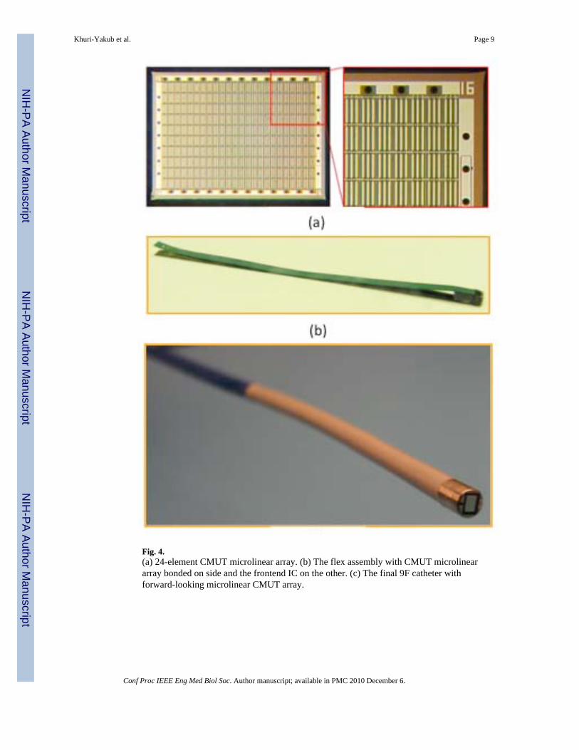
Fig. 4.(a) 24-element CMUT microlinear array. (b) The flex assembly with CMUT microlineararray bonded on side and the frontend IC on the other. (c) The final 9F catheter withforward-looking microlinear CMUT array.
Khuri-Yakub et al. Page 9
Conf Proc IEEE Eng Med Biol Soc. Author manuscript; available in PMC 2010 December 6.
NIH
-PA Author Manuscript
NIH
-PA Author Manuscript
NIH
-PA Author Manuscript

Fig. 5.A 2D image obtained with the CMUT ML array in-vivo in a pig heart.
Khuri-Yakub et al. Page 10
Conf Proc IEEE Eng Med Biol Soc. Author manuscript; available in PMC 2010 December 6.
NIH
-PA Author Manuscript
NIH
-PA Author Manuscript
NIH
-PA Author Manuscript

Fig. 6.(a) 64-element CMUT ring array. (b) Flex assembly with the CMUT array and eight 8-channel frontend ICs. (c) The final 12-F catheter.
Khuri-Yakub et al. Page 11
Conf Proc IEEE Eng Med Biol Soc. Author manuscript; available in PMC 2010 December 6.
NIH
-PA Author Manuscript
NIH
-PA Author Manuscript
NIH
-PA Author Manuscript

Fig. 7.3D rendered image of a metal spring obtained with the CMUT ring array.
Khuri-Yakub et al. Page 12
Conf Proc IEEE Eng Med Biol Soc. Author manuscript; available in PMC 2010 December 6.
NIH
-PA Author Manuscript
NIH
-PA Author Manuscript
NIH
-PA Author Manuscript

NIH
-PA Author Manuscript
NIH
-PA Author Manuscript
NIH
-PA Author Manuscript
Khuri-Yakub et al. Page 13
TABLE I
Summary of Integration Methods of CMUTs with Supporting Frontend Circuits
Monolithic Co-processing (e.g., Eccardt et al. [1]) Post-processing (e.g., Noble et al. [2], Daft et al. [3], Gurun et al. [4])
Multichip Chip-to-chip bonding (e.g., Wygant et al. [5])Bonding on a flexible intermediate substrate (e.g., Nikoozadeh et al. [6])
Bonding on a rigid intermediate substrate (e.g., Wodnicki et al. [7]
Conf Proc IEEE Eng Med Biol Soc. Author manuscript; available in PMC 2010 December 6.
Copyright © 2022 FDOKUMEN