Migration of mitochondrial DNA in the nuclear genome of ...
15
RESEARCH Open Access Migration of mitochondrial DNA in the nuclear genome of colorectal adenocarcinoma Vinodh Srinivasainagendra 1 , Michael W. Sandel 1,7† , Bhupendra Singh 2† , Aishwarya Sundaresan 1 , Ved P. Mooga 2 , Prachi Bajpai 2 , Hemant K. Tiwari 1* and Keshav K. Singh 3,4,5,6* Abstract Background: Colorectal adenocarcinomas are characterized by abnormal mitochondrial DNA (mtDNA) copy number and genomic instability, but a molecular interaction between mitochondrial and nuclear genome remains unknown. Here we report the discovery of increased copies of nuclear mtDNA (NUMT) in colorectal adenocarcinomas, which supports link between mtDNA and genomic instability in the nucleus. We name this phenomenon of nuclear occurrence of mitochondrial component as numtogenesis. We provide a description of NUMT abundance and distribution in tumor versus matched blood-derived normal genomes. Methods: Whole-genome sequence data were obtained for colon adenocarcinoma and rectum adenocarcinoma patients participating in The Cancer Genome Atlas, via the Cancer Genomics Hub, using the GeneTorrent file acquisition tool. Data were analyzed to determine NUMT proportion and distribution on a genome-wide scale. A NUMT suppressor gene was identified by comparing numtogenesis in other organisms. Results: Our study reveals that colorectal adenocarcinoma genomes, on average, contains up to 4.2-fold more somatic NUMTs than matched normal genomes. Women colorectal tumors contained more NUMT than men. NUMT abundance in tumor predicted parallel abundance in blood. NUMT abundance positively correlated with GC content and gene density. Increased numtogenesis was observed with higher mortality. We identified YME1L1, a human homolog of yeast YME1 (yeast mitochondrial DNA escape 1) to be frequently mutated in colorectal tumors. YME1L1 was also mutated in tumors derived from other tissues. We show that inactivation of YME1L1 results in increased transfer of mtDNA in the nuclear genome. Conclusions: Our study demonstrates increased somatic transfer of mtDNA in colorectal tumors. Our study also reveals sex-based differences in frequency of NUMT occurrence and that NUMT in blood reflects NUMT in tumors, suggesting NUMT may be used as a biomarker for tumorigenesis. We identify YME1L1 as the first NUMT suppressor gene in human and demonstrate that inactivation of YME1L1 induces migration of mtDNA to the nuclear genome. Our study reveals that numtogenesis plays an important role in the development of cancer. Keywords: Cancer, Tumor, Colorectal cancer, Mitochondria, Mitochondrial DNA, YME1L1, NUMT, Numtogenesis, mtDNA transfer, Genetic instability * Correspondence: [email protected]; [email protected] † Equal contributors 1 Department of Biostatistics, School of Public Health, University of Alabama at Birmingham, Birmingham, Alabama 35294, USA 3 Departments of Genetics, Environmental Health, Center for Free Radical Biology, Center for Aging and UAB Comprehensive Cancer Center, University of Alabama at Birmingham, Birmingham, Alabama 35294, USA Full list of author information is available at the end of the article © The Author(s). 2017 Open Access This article is distributed under the terms of the Creative Commons Attribution 4.0 International License (http://creativecommons.org/licenses/by/4.0/), which permits unrestricted use, distribution, and reproduction in any medium, provided you give appropriate credit to the original author(s) and the source, provide a link to the Creative Commons license, and indicate if changes were made. The Creative Commons Public Domain Dedication waiver (http://creativecommons.org/publicdomain/zero/1.0/) applies to the data made available in this article, unless otherwise stated. Srinivasainagendra et al. Genome Medicine (2017) 9:31 DOI 10.1186/s13073-017-0420-6
-
Upload
khangminh22 -
Category
Documents
-
view
2 -
download
0
Transcript of Migration of mitochondrial DNA in the nuclear genome of ...
Srinivasainagendra et al. Genome Medicine (2017) 9:31 DOI 10.1186/s13073-017-0420-6
RESEARCH Open Access
Migration of mitochondrial DNA in thenuclear genome of colorectaladenocarcinoma
Vinodh Srinivasainagendra1, Michael W. Sandel1,7†, Bhupendra Singh2†, Aishwarya Sundaresan1, Ved P. Mooga2,Prachi Bajpai2, Hemant K. Tiwari1* and Keshav K. Singh3,4,5,6*Abstract
Background: Colorectal adenocarcinomas are characterized by abnormal mitochondrial DNA (mtDNA) copy numberand genomic instability, but a molecular interaction between mitochondrial and nuclear genome remains unknown.Here we report the discovery of increased copies of nuclear mtDNA (NUMT) in colorectal adenocarcinomas, whichsupports link between mtDNA and genomic instability in the nucleus. We name this phenomenon of nuclearoccurrence of mitochondrial component as numtogenesis. We provide a description of NUMT abundance anddistribution in tumor versus matched blood-derived normal genomes.
Methods: Whole-genome sequence data were obtained for colon adenocarcinoma and rectum adenocarcinomapatients participating in The Cancer Genome Atlas, via the Cancer Genomics Hub, using the GeneTorrent fileacquisition tool. Data were analyzed to determine NUMT proportion and distribution on a genome-wide scale.A NUMT suppressor gene was identified by comparing numtogenesis in other organisms.
Results: Our study reveals that colorectal adenocarcinoma genomes, on average, contains up to 4.2-fold moresomatic NUMTs than matched normal genomes. Women colorectal tumors contained more NUMT than men.NUMT abundance in tumor predicted parallel abundance in blood. NUMT abundance positively correlated withGC content and gene density. Increased numtogenesis was observed with higher mortality. We identified YME1L1,a human homolog of yeast YME1 (yeast mitochondrial DNA escape 1) to be frequently mutated in colorectaltumors. YME1L1 was also mutated in tumors derived from other tissues. We show that inactivation of YME1L1results in increased transfer of mtDNA in the nuclear genome.
Conclusions: Our study demonstrates increased somatic transfer of mtDNA in colorectal tumors. Our study alsoreveals sex-based differences in frequency of NUMT occurrence and that NUMT in blood reflects NUMT in tumors,suggesting NUMT may be used as a biomarker for tumorigenesis. We identify YME1L1 as the first NUMT suppressorgene in human and demonstrate that inactivation of YME1L1 induces migration of mtDNA to the nuclear genome.Our study reveals that numtogenesis plays an important role in the development of cancer.
Keywords: Cancer, Tumor, Colorectal cancer, Mitochondria, Mitochondrial DNA, YME1L1, NUMT, Numtogenesis,mtDNA transfer, Genetic instability
* Correspondence: [email protected]; [email protected]†Equal contributors1Department of Biostatistics, School of Public Health, University of Alabamaat Birmingham, Birmingham, Alabama 35294, USA3Departments of Genetics, Environmental Health, Center for Free RadicalBiology, Center for Aging and UAB Comprehensive Cancer Center, Universityof Alabama at Birmingham, Birmingham, Alabama 35294, USAFull list of author information is available at the end of the article
© The Author(s). 2017 Open Access This article is distributed under the terms of the Creative Commons Attribution 4.0International License (http://creativecommons.org/licenses/by/4.0/), which permits unrestricted use, distribution, andreproduction in any medium, provided you give appropriate credit to the original author(s) and the source, provide a link tothe Creative Commons license, and indicate if changes were made. The Creative Commons Public Domain Dedication waiver(http://creativecommons.org/publicdomain/zero/1.0/) applies to the data made available in this article, unless otherwise stated.

Srinivasainagendra et al. Genome Medicine (2017) 9:31 Page 2 of 15
BackgroundNatural transfer of mitochondrial DNA (mtDNA) into thenuclear genomes of eukaryotic cells is a well-establishedand evolutionarily ongoing process. The nuclear copies ofmtDNA are described as NUMTs (nuclear mtDNA se-quences) [1]. Frequently, intact mitochondria containingmtDNA, mitochondrial RNA (mtRNA), and mitochon-drial proteins are also reported to localize into the nucleus[2–9]. We have named this phenomenon of occurrence ofnuclear mitochondria as numtogenesis. We define numto-genesis as the occurrence of any mitochondrial compo-nents into the nucleus or nuclear genome. Numtogenesisis reported in at least 85 sequenced eukaryotic genomes[1]. These include human, plant, yeast, fruit fly, Plasmo-dium, Caenorhabditis, and other species [1, 10, 11]. Inhuman, NUMT insertions are estimated to occur at a rateof ~5 × 10⁻⁶ per germ cell per generation [12].Evolutionary studies suggest that the origin and inser-
tion of germline NUMTs are distributed non-randomlyin humans and other mammals [13–15]. GermlineNUMTs tend not to originate from the mtDNA dis-placement loop (“d-loop”), and they tend to be locatedin damage-prone regions of the nuclear genome, such asopen chromatin and fragile sites [13–15]. These studiesimplicate NUMTs in double-strand break repair [13]. Themechanism(s) of NUMT accumulation is not well under-stood. It is suggested that mitochondria migrate towardsthe nucleus and accumulate near the nuclear membrane[16–18]. The most parsimonious mechanism explainingNUMT accumulation involves de novo transposition fromthe mitochondrion to the nucleus; however, NUMTs arealso known to accumulate via segmental duplication(sometimes within repetitive elements), and possibly RNAretro-transposition [19–21]. The human genome containsbetween 755 and 1105 germline NUMTs, with mtDNAidentities ranging from 64–100% [12, 22]. GermlineNUMTs with the lowest similarity to mtDNA have beenevolutionarily conserved for tens of millions of years,while the most recent insertions occurred after certainHomo sapiens populations migrated to Eurasia [22–26].Human germline NUMTs are relatively well described butlittle is known about the somatic NUMT and its role inhuman pathology [27].We and others have demonstrated that mito-nuclear
interactions play a key role in tumorigenesis [28–35],but a role of mtDNA integration within the nucleargenome remains relatively unexplored. In this study, weanalyzed the prevalence of NUMT in colorectal cancer(CRC) because a number of mitochondrial associationsare relatively well characterized in CRC. There is a re-ported relationship between CRC risk and mtDNA copynumber [36–38], and there are associations betweengermline mtDNA variants and CRC risk and mortality[39, 40]. Similarly, colorectal adenocarcinomas tend to
have aberrant mtDNA copy number and somatic variantfrequencies compared to matched blood-derived normalgenomes [41–44]. We differentiated two classes ofNUMTs with distinct characteristics, those that are inher-ited in the germline and those that are somatic NUMTsacquired during tumorigenesis. We present the first quan-titative analysis on the abundance of somatic NUMTs inhuman colorectal adenocarcinoma genomes relative tomatched blood-derived normal samples. Further, we com-pare the distributions of somatic NUMTs and germlineNUMTs, describe sex-based differences in numtogenesis,and demonstrate that NUMT abundance in blood reflectsNUMT abundance in tumor. In addition, we identifyYME1L1 as the first “NUMT suppressor” gene in humanswhose inactivation leads to increased numtogenesis.
MethodsData harvestingWhole-genome sequence data were obtained for colonadenocarcinoma (COAD) and rectum adenocarcinoma(READ) patients participating in The Cancer GenomeAtlas (TCGA), via the Cancer Genomics Hub (CGHub)[45], using the GeneTorrent file acquisition tool. Inorder to appraise alternative protocols for transactionson big-data endpoints, we evaluated GTFuse, aninnovate software that offers faster access to DNA se-quence data without the need for staging the data lo-cally. Although data bandwidth hungry, GTFuse stillenabled us to rapidly prototype our research on selectedregions of the genome, thereby indicating signals ofNUMT deposition in the nuclear genome. A symbioticcombination of GeneTorrent and GTFuse was used tochoreograph the downstream data analysis pipeline. Ourdata harvester robots adopt a high-throughput analysismodel by spreading GTFuse across a computational clus-ter fabric available locally on-campus At the time ofmanuscript generation, although the GTFuse web-linkwithin AnnaiSystems webportal was unavailable, analternate web URL https://annaisystems.zendesk.com/hc/en-us was found through web search means.
Quality controlTCGA sequence data for individuals with matched colo-rectal tumor and blood-derived normal samples wereharvested from CGHub. Downloaded data went throughan intense quality control (QC) pipeline, which involvedthe use of (a) clinical information downloaded fromTCGA Data Matrix, (b) short-reads alignment statisticsderived from the Binary AlignMent (BAM) sequencedata downloaded from CGHub, and (c) tagging andelimination of duplicate reads in the alignment data(BAM) using a popular next-generation sequencing tool,Picard 2.5. All sequence data used in this study weregenerated at Harvard Medical School using Illumina
Srinivasainagendra et al. Genome Medicine (2017) 9:31 Page 3 of 15
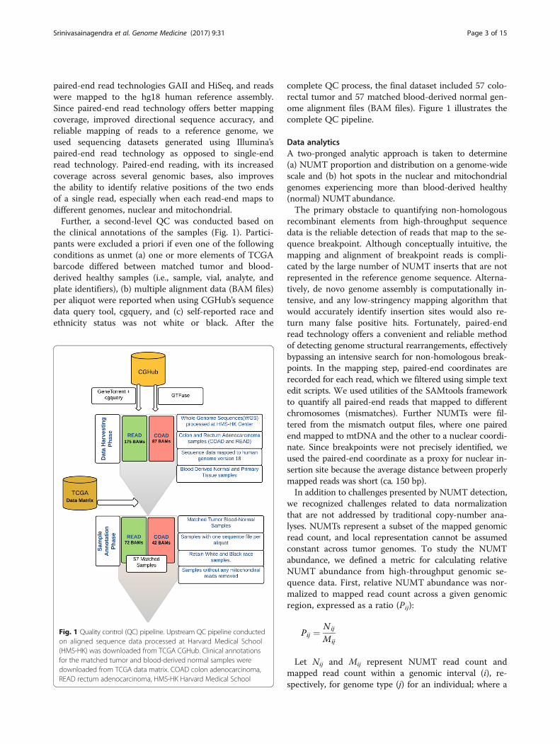
paired-end read technologies GAII and HiSeq, and readswere mapped to the hg18 human reference assembly.Since paired-end read technology offers better mappingcoverage, improved directional sequence accuracy, andreliable mapping of reads to a reference genome, weused sequencing datasets generated using Illumina’spaired-end read technology as opposed to single-endread technology. Paired-end reading, with its increasedcoverage across several genomic bases, also improvesthe ability to identify relative positions of the two endsof a single read, especially when each read-end maps todifferent genomes, nuclear and mitochondrial.Further, a second-level QC was conducted based on
the clinical annotations of the samples (Fig. 1). Partici-pants were excluded a priori if even one of the followingconditions as unmet (a) one or more elements of TCGAbarcode differed between matched tumor and blood-derived healthy samples (i.e., sample, vial, analyte, andplate identifiers), (b) multiple alignment data (BAM files)per aliquot were reported when using CGHub’s sequencedata query tool, cgquery, and (c) self-reported race andethnicity status was not white or black. After the
Fig. 1 Quality control (QC) pipeline. Upstream QC pipeline conductedon aligned sequence data processed at Harvard Medical School(HMS-HK) was downloaded from TCGA CGHub. Clinical annotationsfor the matched tumor and blood-derived normal samples weredownloaded from TCGA data matrix. COAD colon adenocarcinoma,READ rectum adenocarcinoma, HMS-HK Harvard Medical School
complete QC process, the final dataset included 57 colo-rectal tumor and 57 matched blood-derived normal gen-ome alignment files (BAM files). Figure 1 illustrates thecomplete QC pipeline.
Data analyticsA two-pronged analytic approach is taken to determine(a) NUMT proportion and distribution on a genome-widescale and (b) hot spots in the nuclear and mitochondrialgenomes experiencing more than blood-derived healthy(normal) NUMTabundance.The primary obstacle to quantifying non-homologous
recombinant elements from high-throughput sequencedata is the reliable detection of reads that map to the se-quence breakpoint. Although conceptually intuitive, themapping and alignment of breakpoint reads is compli-cated by the large number of NUMT inserts that are notrepresented in the reference genome sequence. Alterna-tively, de novo genome assembly is computationally in-tensive, and any low-stringency mapping algorithm thatwould accurately identify insertion sites would also re-turn many false positive hits. Fortunately, paired-endread technology offers a convenient and reliable methodof detecting genome structural rearrangements, effectivelybypassing an intensive search for non-homologous break-points. In the mapping step, paired-end coordinates arerecorded for each read, which we filtered using simple textedit scripts. We used utilities of the SAMtools frameworkto quantify all paired-end reads that mapped to differentchromosomes (mismatches). Further NUMTs were fil-tered from the mismatch output files, where one pairedend mapped to mtDNA and the other to a nuclear coordi-nate. Since breakpoints were not precisely identified, weused the paired-end coordinate as a proxy for nuclear in-sertion site because the average distance between properlymapped reads was short (ca. 150 bp).In addition to challenges presented by NUMT detection,
we recognized challenges related to data normalizationthat are not addressed by traditional copy-number ana-lyses. NUMTs represent a subset of the mapped genomicread count, and local representation cannot be assumedconstant across tumor genomes. To study the NUMTabundance, we defined a metric for calculating relativeNUMT abundance from high-throughput genomic se-quence data. First, relative NUMT abundance was nor-malized to mapped read count across a given genomicregion, expressed as a ratio (Pij):
Pij ¼ Nij
Mij
Let Nij and Mij represent NUMT read count andmapped read count within a genomic interval (i), re-spectively, for genome type (j) for an individual; where a
Srinivasainagendra et al. Genome Medicine (2017) 9:31 Page 4 of 15
genomic interval is defined as whole genome (gen),chromosome (chr), chromosome arm (arm), or 2.5-mbpsliding window (win); and where genome type (j) is eithertumor (t) or matched blood-derived healthy (h) genome ofan individual. The change in tumor and matched healthyNUMT proportions is defined as ΔPi = Pit − Pih and the ra-tio is given as:
Ri ¼ Pit � Pihð Þ−1
where Ri represents the proportional fold-change inNUMT abundance within a genomic region (i) for tumor(t) versus matched blood-derived healthy (h) genomes.When sub-genomic regions (e.g., sliding windows andcytobands) were compared, Pih was replaced with Pih ,where Pih is the average NUMT proportion within thegenomic interval for all blood-derived healthy samplessharing the same plate barcode to avoid zero denomin-ator because blood-derived healthy samples may nothave any NUMTs within the short regions.The Ri derived earlier can be less than or greater than
1 due to the asymmetric property of ratios (e.g., whereRi = 0.5 represents a change in the opposite direction,i.e., there are twice as many NUMTs in healthy samplescompared to tumor; and similarly, Ri = 2.0 represents achange in the positive direction, i.e., there are twice asmany NUMTs in tumor samples compared to theirmatched blood-derived normal samples). In order tocompare NUMTs between tumor and blood-derivedhealthy samples within the genomic regions, it is neces-sary to rescale Ri values less than one and to collapsevalues of one and negative one to zero. Further, we de-fined an indicator value Si to denote the direction ofchange between tumor and matched blood-derived nor-mal NUMT abundance:
Si ¼ ΔPi
ΔPij jand:
R=i ¼ RSi
i � SiLet the Ri
/represent the rescaled proportional fold-change in relative NUMT abundance between tumorand matched blood-derived normal samples. Ri
/was cal-culated for the nuclear genome using five nested datapartitions, where i represents whole genomes (gen),chromosomes (chr), chromosome arms (arm), cytobands(cyt), and sliding windows (win). For nuclear genome-scale comparisons, normalization to mapped reads (Mij)did not include sex chromosomes due to sequence repre-sentation bias associated with differences in chromosomelength. For small-partition comparisons (e.g., sub-band),samples were pooled before normalization to avoid biasassociated with zero denominators. For the mitochondrial
genome, Ri/was calculated for three nested data partitions,
where i represents genome (mtg), replication strand (mst),and gene (mgn).The relationship between Rarm
/ and mapped read countwas evaluated with linear regression to assess whetherNUMT transposition is coincident with aneuploidy.The relationships between Rwin
/ were evaluated betweenGC content to assess whether transcriptional activitypredisposes the nuclear genome to integration ofnon-homologous DNA. All statistical analyses wereconducted in the R statistical environment (http://www.R-project.org/). The NUMT proportions and abun-dance in blood-derived normal and primary tumor sitesare quantified in Additional file 1: Table S1.
YME1L1 gene knockout and NUMT analyses in isolatednuclear DNAUsing the CRISPR-Cas9 method, we knocked out thehuman YME1L1 gene in human breast epithelial MCF-7cells as described earlier [46]. To avoid any mtDNA con-tamination in NUMT analyses, we isolated nuclear frac-tions free of mitochondrial contamination. Briefly,YME1L1 knockout and wild-type MCF-7 cells were lysedusing lysis buffer (10 mM HEPES, pH 7.9, 10 mM KCl,0.1 mM EDTA) containing 10% IGEPAL detergent for10 min at room temperature and centrifugation at15,000xg for 3 minutes was carried out to pellet the in-tact nuclei. To completely avoid cytoplasmic fractioncontamination in the nuclear pellet, the lysis step was re-peated one more time. The purity of the nuclear fractionwas ascertained by performing western blotting formitochondrial encoded cytchrome oxidase II (COXII)protein. Nuclear DNA was prepared from the nuclearpellet using the NAOH boiling method [47]. Mitochon-drial DNA content in the nuclear fraction was analyzedby real-time PCR by absolute quantification usingprimers for COXII (mtDNA-encoded gene) and Beta-2microglobulin (B2M; nuclear DNA-encoded gene). B2Mserved as an internal control.
Yeast transformation and genetic selectionYeast expression vectors (pYX113, pPT31-yYme1, andpYX113-hYme1L) were transformed using the lithiumacetate, single-stranded DNA, polyethylene glycolmethod [48–50]. Following transformation, yeast harbor-ing the desired vector was selected using synthetic drop-out (SD) medium (0.67% [w/v] nitrogen base withoutamino acids, 0.07% [v/v] drop-out amino acid mix(-His/-Trp/-Ura), 0.02% [w/v] L-histidine and excludingthe amino acid that is a selectable marker, 2% [w/v] dex-trose, and 1.5% agar for agar plates). Single cell coloniesfrom plates lacking uracil were cultured in glucosemedium and 1 × 10⁴ and 5 × 10⁷ or 5 × 10⁸ cells were
Srinivasainagendra et al. Genome Medicine (2017) 9:31 Page 5 of 15
plated in triplicate onto YPD and SD media lackingtryptophan.The yeast strains throughout this study were grown in
YPD medium (1% [w/v] yeast extract, 2% [w/v] bacto-peptone, 2% [w/v] dextrose) or YPG medium (1% [w/v]yeast extract, 2% [w/v] bacto-peptone, 3% [w/v] glycerol,pH 4.9) at 30 °C. The yeast strains constructed and usedin this study are detailed in Table 1. The yeast Yme1-1strain (PTY62) was transformed with a plasmid expressingthe yeast YME1 (yYme1) or human YME1L1 (hYme1L1)gene under the alcohol dehydrogenase (ADH) promoter.
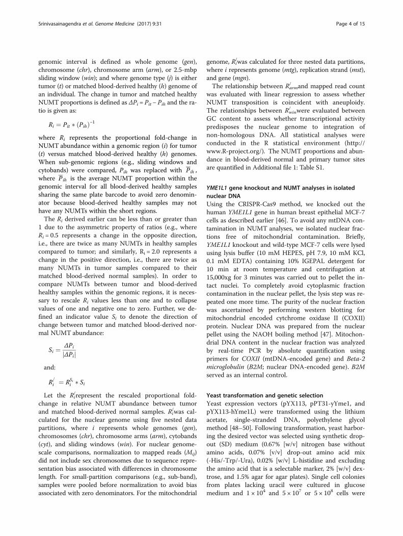
ResultsAdenocarcinoma genomes contain increased NUMTscompared to healthy genomesSince mitochondrial abnormalities are frequently de-scribed in cancer, we asked whether mitochondrial dys-function results in increased prevalence of NUMTs intumors. Constitutively, tumor genomes contained moreNUMTs than matched blood-derived healthy genomeswhen normalized to mapped read pairs. When scannedfor NUMT proportion and distribution on a genome-wide scale across 57 samples, tumor genomes contained,on average, 4.42-fold more NUMTs than healthy normalgenomes. Box plots (Fig. 2a, b) indicate the mean, dis-persion, and skewness of NUMT density for the genomegroups. Although both tumor and matched normal ge-nomes show right-skewed distributions (Fig. 2c), the thirdquartile (3.43 × 10⁻⁶) of the normal blood genome groupis less than the first quartile (3.13E-6) of the tumorgenomes, indicating significant difference in the NUMTdensity. A one-tailed, paired t-test was performed ontumor and matched blood-derived healthy samples todetermine the statistical significance (P value 8.79 × 10⁻¹³)of the log transformed NUMT abundance levels. In onecase, however, 22-fold more tumor NUMTs were observedin a Caucasian woman. This individual’s vital status waspronounced as deceased after reporting her pathologictumor stage as I. These studies suggest that colorectaladenocarcinomas contain increased NUMTs compared toblood-derived healthy NUMTs.
NUMT abundance in blood correlates with NUMTabundance in tumorIncreasingly, molecular characteristics of tumors aredemonstrated to be reflected in the blood of cancer
Table 1 Sacharomyces cerevisiae strains used in the study
Strain Genotype Mitochondrial genotype
PTY62 Yme1-1 MATa ura3-52 lys2 leu2-3,112 trp1-Δ1 yme1-1
ρ + (TRP1)
PTY62- yYme1
PTY62-hYme1L1
patients. We therefore envision that increased NUMTincidence observed in tumors may be found in thematched blood of cancer patients. In order to determinethe predictability of tumor NUMT density based on theNUMT frequency in matched blood-derived healthysamples, a simple linear regression was performed onthe log base 2-transformed proportions of NUMTs inblood-derived healthy genomes (Pgenh) and tumor ge-nomes (Pgent). Linear regression revealed a positive rela-tionship between Pgent and Pgenh with an R2 = 0.17, Pvalue = 0.0016 (Fig. 3a, b). These results indicate that thereis a positive correlation between proportions of NUMTsin blood-derived and primary tumor in colorectal cancersamples.
Colorectal tumors in women harbor more NUMTsRecent studies suggest hormonal regulation of mitochon-drial functions [51]. Therefore, we delineated NUMT dis-tribution among males and females. Colorectal tumors ofwomen harbored more NUMTs than those of men (Fig. 4).Women had a median NUMT fold change proportion of4.52 compared 3.1 for men. We conclude that tumorsfrom women contained more NUMTs than those frommen.
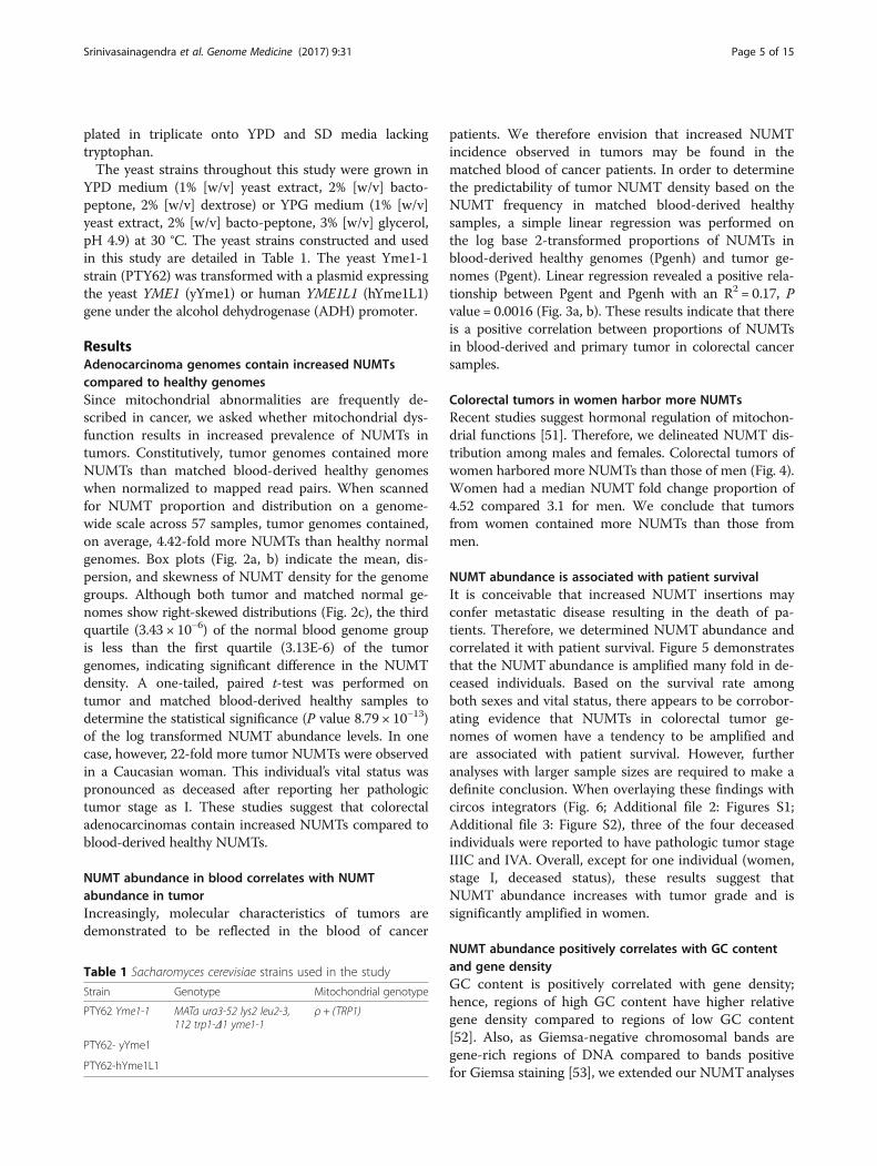
NUMT abundance is associated with patient survivalIt is conceivable that increased NUMT insertions mayconfer metastatic disease resulting in the death of pa-tients. Therefore, we determined NUMT abundance andcorrelated it with patient survival. Figure 5 demonstratesthat the NUMT abundance is amplified many fold in de-ceased individuals. Based on the survival rate amongboth sexes and vital status, there appears to be corrobor-ating evidence that NUMTs in colorectal tumor ge-nomes of women have a tendency to be amplified andare associated with patient survival. However, furtheranalyses with larger sample sizes are required to make adefinite conclusion. When overlaying these findings withcircos integrators (Fig. 6; Additional file 2: Figures S1;Additional file 3: Figure S2), three of the four deceasedindividuals were reported to have pathologic tumor stageIIIC and IVA. Overall, except for one individual (women,stage I, deceased status), these results suggest thatNUMT abundance increases with tumor grade and issignificantly amplified in women.
NUMT abundance positively correlates with GC contentand gene densityGC content is positively correlated with gene density;hence, regions of high GC content have higher relativegene density compared to regions of low GC content[52]. Also, as Giemsa-negative chromosomal bands aregene-rich regions of DNA compared to bands positivefor Giemsa staining [53], we extended our NUMTanalyses
Fig. 2 Distribution of NUMT proportions in tumor and normal genomes. a Distribution of NUMT proportions in 57 samples with matched tumorand blood-derived normal genomes. NUMT proportion is defined as the ratio between NUMT read count and total mapped read count on agenome-wide scale. The mean and standard deviation, respectively, across the 57 samples are 8.31 × 10⁻⁶ and 7.11 × 10⁻⁶ for tumor genomesand 2.65 × 10⁻⁶ and 2.49 × 10⁻⁶ for normal genomes. A two-tailed paired t-test conducted between the NUMT proportions for 57 (COAD + READ)samples revealed a P value of 1.63 × 10⁻⁵. When comparing the NUMT proportions between the cancer site group versus blood-derived groupusing a two-tailed unequal variance t-test, a P value of 1.43 × 10⁻⁵ was observed for COAD samples (N = 36) and 3.82 × 10⁻³ for READ samples(N = 21). b Fold change in the tumor NUMT proportions compared to blood-derived normal genomes across colon (COAD) and rectum (READ)cancer samples. Tumor genomes contained 4.42-fold more NUMTs than blood-derived normal genomes. A two-tailed unequal variance t-testconducted between the NUMT abundance of colon cancer samples (N = 36) and rectal cancer samples (N = 21) revealed a P value of 0.91, indicatingno difference in the NUMT abundance between the two cancer sites, colon and rectum. c Right-skewed distribution, log transformed, and one-tailedpaired t-test performed on tumor and matched blood-derived normal samples to determine the statistical significance (P value 8.79 × 10⁻¹³) of thelog-transformed NUMT abundance levels
Srinivasainagendra et al. Genome Medicine (2017) 9:31 Page 6 of 15
to the chromosomal cytobands. Our study demonstrates apositive relationship between NUMT abundance and GCcontent of gene-rich regions, having an abundance fold-change of 4.2 or more (Fig. 7).Overall, among the groups that had a fold change of 2
or more in NUMT abundance, although the low GC-content cytobands gpos50 and gpos100 were at the topof the NUMT abundance list for chromosomal cytobandwindows, GC-rich “gneg” regions were prominentamong cytobands exhibiting more than a 4.2-fold changein NUMT abundance. This pattern observed for the out-lier data above 4.2 fold-change of NUMT abundancewithin GC-rich regions of gneg clearly shows a strongcorrelation between elevated NUMT abundance and GCcontent. Based on Fig. 7, as indicated by data pointsabove the third quartile level, gneg regions are account-able for the greater than normal NUMT abundance, in-dicating a positive correlation between gene density andNUMT abundance. A previous study has shown thatgpos100 sub-bands are also enriched with LINEs [54],
and other researchers have demonstrated secondaryamplification via LINE transposition to be an importantmechanism of NUMT accumulation [11, 20]. Our resultscorroborate the empirical evidence. Clearly, a great dealof new research is needed to elucidate the causes andconsequences of NUMT transposition. We hope thatour discovery will serve as an impetus for such inquiry.
Mitochondrial fragile sites associated with NUMTsTo identify fragile sites in the mitochondrial genomesthat act as hotspots for mtDNA to immigrate into thenuclear genome, paired-end reads with one end mappingto the mitochondrial genome and the other end to thenuclear genome, NUMTs were further investigated. Asillustrated in Fig. 8, fragile sites were identified withincomplex I (ND1) and complex IV (MT-CO1/COX1,COX III) mitochondrial regions, which are known forharboring mutations in different cancer sites. Based onthe migration pattern of NUMTs, breakpoint sites in

Fig. 3 Log2-transformed NUMT proportions in tumor and blood-derived normal (Pij) genomes. a Log2-transformed NUMT proportions in tumorand normal genomes showcasing the difference in their means, indicating higher NUMT distribution in tumor genomes compared to matchedblood-derived normal genomes. A two-tailed paired t-test conducted between the log2-transformed NUMT proportions for 57 samples revealeda P value of 1.87 × 10⁻¹¹, showing significant difference in the log-transformed NUMT proportions between primary tumors and blood-derivednormal genomes. b Relationship between abundance measures of blood-derived normal genome NUMTs and tumor genome NUMTs showing apositive relationship with R2 = 0.17 and P value = 0.0016
Fig. 4 NUMT abundance across disease–sex combination. Colorectal tumors from women have higher NUMT abundance proportion (tumor NUMT/blood normal NUMT) than those from men. Women had a median NUMT fold change abundance of 4.52 (range 0.11 to 22.9) compared 3.1 for men(range 0.53 to 8.3). COAD colon adenocarcinoma, READ rectum adenocarcinoma. To investigate the sex difference in the NUMT abundances, anunequal variance t-test was performed on the raw NUMT proportions observed by the members of the two sex groups stratified by “blood-normal”and “primary tumor” classifications. The P value was 0.03 between males (N = 23) and females (N = 34) for blood-derived normal samples and 0.08 fortumor samples
Srinivasainagendra et al. Genome Medicine (2017) 9:31 Page 7 of 15
Fig. 5 NUMT abundance distribution according to vital status.a Increased NUMT abundance in deceased individuals with colorectaltumors. Although a Mann–Whitney U test showed a P value of 0.04between the NUMT abundances of the Alive and Deceased groups, thevery small sample size (N= 4) of the deceased group warrants furtherinvestigation with datasets enriched for deceased vital status. b NUMTproportions categorized based on disease (colon and rectal) and sexcombination. This vital status observation in combination with NUMTabundance among sex-specific samples appears to be an early indicatorof death events among colorectal cancer women with higherproportions of NUMTs
Srinivasainagendra et al. Genome Medicine (2017) 9:31 Page 8 of 15
COX1 and ND1 are responsible for numtogenesis in thenuclear genome of colorectal cancer.
YME1L1 inactivation leads to increased numtogenesisIn the yeast Saccharomyces cerevisiae, YME1 is reportedto be an important suppressor of mtDNA migration tothe nucleus [55]. Interestingly, the YME1L1 gene en-codes the human homologue of yeast mitochondrialAAA (ATPases associated with diverse cellular activities)metalloprotease, Yme1p. YME1L is a functional homologueof Yme1p, with conserved roles in mitochondrial assembly,integrity, and DNA metabolism [56]; however, its functionin suppressing NUMTs is not known.We conducted in silico YME1L1 mutation analysis in
all colorectal cancer cases (n = 57) used for NUMT ana-lysis in this study. We determined that of 57 CRC tu-mors, ~16% contained mutations in YME1L1. A total of24 mutations (five exonic, 17 intronic, and two in the 3′
UTR) in YME1L1 were identified. All five exonic muta-tions were frameshift mutations (Fig. 9a, b).We expanded our analysis of Yme1L1 in TCGA data-
base. This analysis revealed a high incidence of Yme1L1mutations in CRC (Fig. 9c). The mutations in Yme1L1include missense and synonymous substitutions andinframe and frameshift deletions (Fig. 9c). Most of themutations in Yme1L1 in human colorectal cancer fallinto two categories: missense substitutions (~68%) andsynonymous substitutions (20%). The relative distribu-tion of various mutations is summarized as a pie chartin Fig. 9c. We also analyzed Yme1L1 mutations in otherhuman cancer types and observed a high mutation fre-quency in all the tested cancer types (Fig. 9d).We determined whether inactivation of the human
YME1L1 gene increases NUMT formation. For this, wecreated a YME1L1 knockout in a human cell line usingYME1L1 gene-specific CRISPRs. We prepared nuclearfractions free of mitochondrial contamination and quan-tified the amount of mtDNA present in the nuclear frac-tion of these cells. We observed a strikingly increasedamount of mtDNA in the nuclear fraction of YME1L1knockout cells compared to wild-type cells (Fig. 9e).These results identify YME1L1 as the first NUMT sup-pressor gene in humans and suggest that inactivation ofYME1L1 leads to increased numtogenesis.
Human homologue of YME1 suppresses migration ofmtDNA to the nucleusWe asked whether the phylogenetically conserved roleof human YME1L1 can rescue the migration of mtDNAin a yeast strain in which YME1 is disrupted. We utilizedthe Yme1-1 yeast strain, which harbors a mutation whichleads to inactivation of the YME1 gene [57]. In this strain,the auxtotrophic endogenous nuclear TRP1 gene is de-leted and inserted into the mitochondrial genome. Sincethe required transcription machinery for TRP1 is onlypresent in the nucleus, the mitochondrially inserted TRP1gene is only functional when it migrates to the nucleus,permitting analysis of mtDNA migration to the nucleus[57, 58].To determine whether human Yme1L1 is expressed in
the Yme1-1 strain, western blotting was performed. In-deed, hYme1L1 was expressed in the Yme1-1 hYme1L1strain (Fig. 9f ). When the Yme1-1 vector was platedunder tryptophan selection, it showed a significantlylarge number of colonies (Fig. 9g). These data support aprevious observation that migration of mtDNA to thenucleus is high in Yme1-1 cells [57]. Yme1-1 cells ex-pressing yeast Yme1 (yYme1) display only a few (<50)tryptophan-positive colonies, suggesting that accumula-tion of mtDNA fragments in the nucleus in the Yme1-1strain was prevented to a greater degree by re-introducing
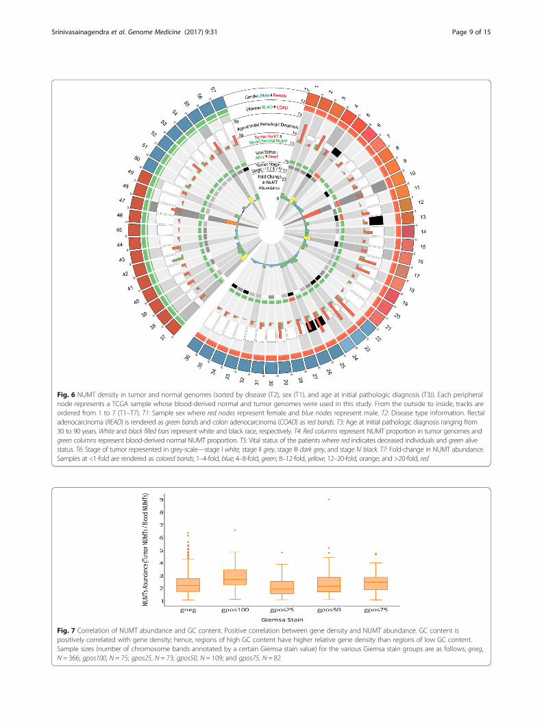
Fig. 6 NUMT density in tumor and normal genomes (sorted by disease (T2), sex (T1), and age at initial pathologic diagnosis (T3)). Each peripheralnode represents a TCGA sample whose blood-derived normal and tumor genomes were used in this study. From the outside to inside, tracks areordered from 1 to 7 (T1–T7). T1: Sample sex where red nodes represent female and blue nodes represent male. T2: Disease type information. Rectaladenocarcinoma (READ) is rendered as green bands and colon adenocarcinoma (COAD) as red bands. T3: Age at initial pathologic diagnosis ranging from30 to 90 years. White and black filled bars represent white and black race, respectively. T4: Red columns represent NUMT proportion in tumor genomes andgreen columns represent blood-derived normal NUMT proportion. T5: Vital status of the patients where red indicates deceased individuals and green alivestatus. T6: Stage of tumor represented in grey-scale—stage I white, stage II grey, stage III dark grey, and stage IV black. T7: Fold-change in NUMT abundance.Samples at <1-fold are rendered as colored bands; 1–4-fold, blue; 4–8-fold, green; 8–12-fold, yellow; 12–20-fold, orange; and >20-fold, red
Fig. 7 Correlation of NUMT abundance and GC content. Positive correlation between gene density and NUMT abundance. GC content ispositively correlated with gene density; hence, regions of high GC content have higher relative gene density than regions of low GC content.Sample sizes (number of chromosome bands annotated by a certain Giemsa stain value) for the various Giemsa stain groups are as follows; gneg,N = 366; gpos100, N = 75; gpos25, N = 73; gpos50, N = 109; and gpos75, N = 82
Srinivasainagendra et al. Genome Medicine (2017) 9:31 Page 9 of 15
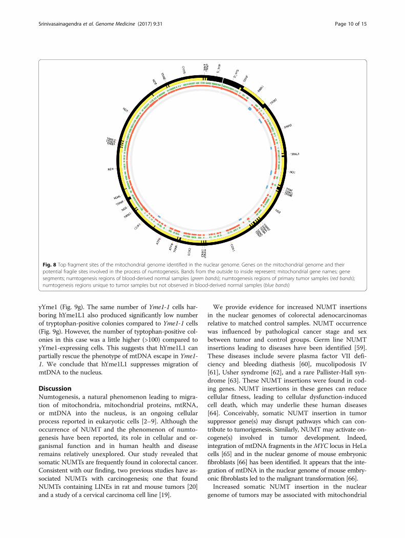
Fig. 8 Top fragment sites of the mitochondrial genome identified in the nuclear genome. Genes on the mitochondrial genome and theirpotential fragile sites involved in the process of numtogenesis. Bands from the outside to inside represent: mitochondrial gene names; genesegments; numtogenesis regions of blood-derived normal samples (green bands); numtogenesis regions of primary tumor samples (red bands);numtogenesis regions unique to tumor samples but not observed in blood-derived normal samples (blue bands)
Srinivasainagendra et al. Genome Medicine (2017) 9:31 Page 10 of 15
yYme1 (Fig. 9g). The same number of Yme1-1 cells har-boring hYme1L1 also produced significantly low numberof tryptophan-positive colonies compared to Yme1-1 cells(Fig. 9g). However, the number of typtophan-positive col-onies in this case was a little higher (>100) compared toyYme1-expressing cells. This suggests that hYme1L1 canpartially rescue the phenotype of mtDNA escape in Yme1-1. We conclude that hYme1L1 suppresses migration ofmtDNA to the nucleus.
DiscussionNumtogenesis, a natural phenomenon leading to migra-tion of mitochondria, mitochondrial proteins, mtRNA,or mtDNA into the nucleus, is an ongoing cellularprocess reported in eukaryotic cells [2–9]. Although theoccurrence of NUMT and the phenomenon of numto-genesis have been reported, its role in cellular and or-ganismal function and in human health and diseaseremains relatively unexplored. Our study revealed thatsomatic NUMTs are frequently found in colorectal cancer.Consistent with our finding, two previous studies have as-sociated NUMTs with carcinogenesis; one that foundNUMTs containing LINEs in rat and mouse tumors [20]and a study of a cervical carcinoma cell line [19].
We provide evidence for increased NUMT insertionsin the nuclear genomes of colorectal adenocarcinomasrelative to matched control samples. NUMT occurrencewas influenced by pathological cancer stage and sexbetween tumor and control groups. Germ line NUMTinsertions leading to diseases have been identified [59].These diseases include severe plasma factor VII defi-ciency and bleeding diathesis [60], mucolipodosis IV[61], Usher syndrome [62], and a rare Pallister-Hall syn-drome [63]. These NUMT insertions were found in cod-ing genes. NUMT insertions in these genes can reducecellular fitness, leading to cellular dysfunction-inducedcell death, which may underlie these human diseases[64]. Conceivably, somatic NUMT insertion in tumorsuppressor gene(s) may disrupt pathways which can con-tribute to tumorigenesis. Similarly, NUMT may activate on-cogene(s) involved in tumor development. Indeed,integration of mtDNA fragments in theMYC locus in HeLacells [65] and in the nuclear genome of mouse embryonicfibroblasts [66] has been identified. It appears that the inte-gration of mtDNA in the nuclear genome of mouse embry-onic fibroblasts led to the malignant transformation [66].Increased somatic NUMT insertion in the nuclear
genome of tumors may be associated with mitochondrial
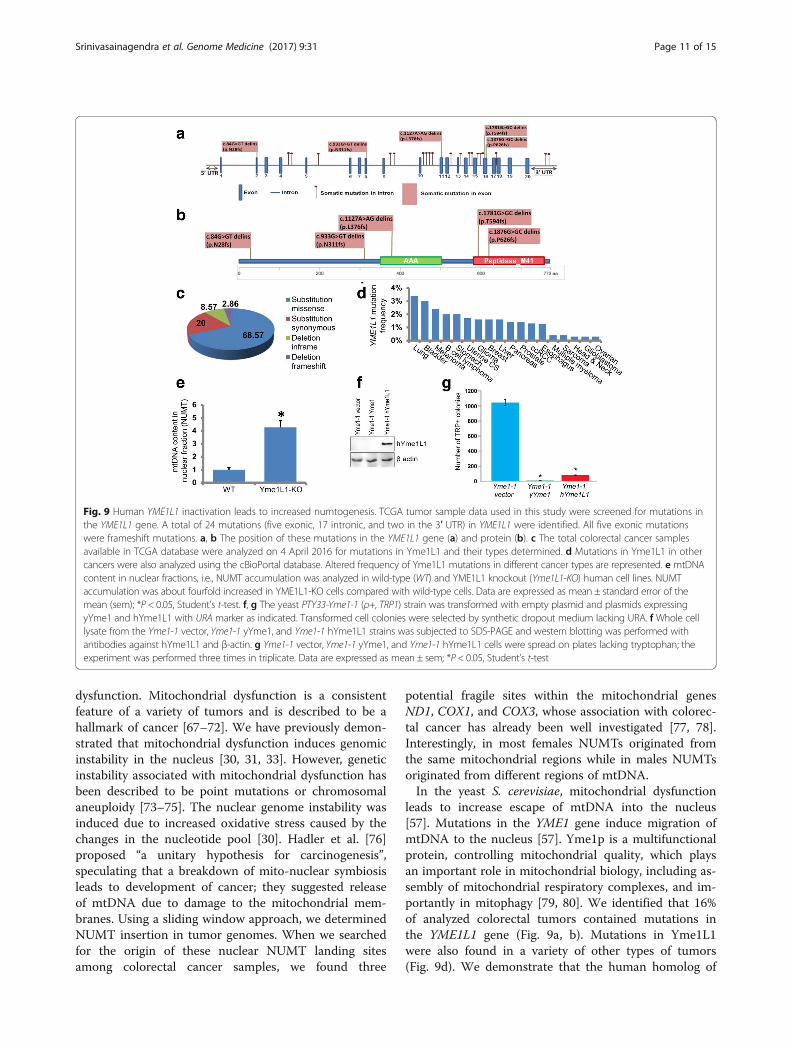
Fig. 9 Human YME1L1 inactivation leads to increased numtogenesis. TCGA tumor sample data used in this study were screened for mutations inthe YME1L1 gene. A total of 24 mutations (five exonic, 17 intronic, and two in the 3′ UTR) in YME1L1 were identified. All five exonic mutationswere frameshift mutations. a, b The position of these mutations in the YME1L1 gene (a) and protein (b). c The total colorectal cancer samplesavailable in TCGA database were analyzed on 4 April 2016 for mutations in Yme1L1 and their types determined. d Mutations in Yme1L1 in othercancers were also analyzed using the cBioPortal database. Altered frequency of Yme1L1 mutations in different cancer types are represented. e mtDNAcontent in nuclear fractions, i.e., NUMT accumulation was analyzed in wild-type (WT) and YME1L1 knockout (Yme1L1-KO) human cell lines. NUMTaccumulation was about fourfold increased in YME1L1-KO cells compared with wild-type cells. Data are expressed as mean ± standard error of themean (sem); *P < 0.05, Student’s t-test. f, g The yeast PTY33-Yme1-1 (ρ+, TRP1) strain was transformed with empty plasmid and plasmids expressingyYme1 and hYme1L1 with URA marker as indicated. Transformed cell colonies were selected by synthetic dropout medium lacking URA. f Whole celllysate from the Yme1-1 vector, Yme1-1 yYme1, and Yme1-1 hYme1L1 strains was subjected to SDS-PAGE and western blotting was performed withantibodies against hYme1L1 and β-actin. g Yme1-1 vector, Yme1-1 yYme1, and Yme1-1 hYme1L1 cells were spread on plates lacking tryptophan; theexperiment was performed three times in triplicate. Data are expressed as mean ± sem; *P < 0.05, Student’s t-test
Srinivasainagendra et al. Genome Medicine (2017) 9:31 Page 11 of 15
dysfunction. Mitochondrial dysfunction is a consistentfeature of a variety of tumors and is described to be ahallmark of cancer [67–72]. We have previously demon-strated that mitochondrial dysfunction induces genomicinstability in the nucleus [30, 31, 33]. However, geneticinstability associated with mitochondrial dysfunction hasbeen described to be point mutations or chromosomalaneuploidy [73–75]. The nuclear genome instability wasinduced due to increased oxidative stress caused by thechanges in the nucleotide pool [30]. Hadler et al. [76]proposed “a unitary hypothesis for carcinogenesis”,speculating that a breakdown of mito-nuclear symbiosisleads to development of cancer; they suggested releaseof mtDNA due to damage to the mitochondrial mem-branes. Using a sliding window approach, we determinedNUMT insertion in tumor genomes. When we searchedfor the origin of these nuclear NUMT landing sitesamong colorectal cancer samples, we found three
potential fragile sites within the mitochondrial genesND1, COX1, and COX3, whose association with colorec-tal cancer has already been well investigated [77, 78].Interestingly, in most females NUMTs originated fromthe same mitochondrial regions while in males NUMTsoriginated from different regions of mtDNA.In the yeast S. cerevisiae, mitochondrial dysfunction
leads to increase escape of mtDNA into the nucleus[57]. Mutations in the YME1 gene induce migration ofmtDNA to the nucleus [57]. Yme1p is a multifunctionalprotein, controlling mitochondrial quality, which playsan important role in mitochondrial biology, including as-sembly of mitochondrial respiratory complexes, and im-portantly in mitophagy [79, 80]. We identified that 16%of analyzed colorectal tumors contained mutations inthe YME1L1 gene (Fig. 9a, b). Mutations in Yme1L1were also found in a variety of other types of tumors(Fig. 9d). We demonstrate that the human homolog of
Srinivasainagendra et al. Genome Medicine (2017) 9:31 Page 12 of 15
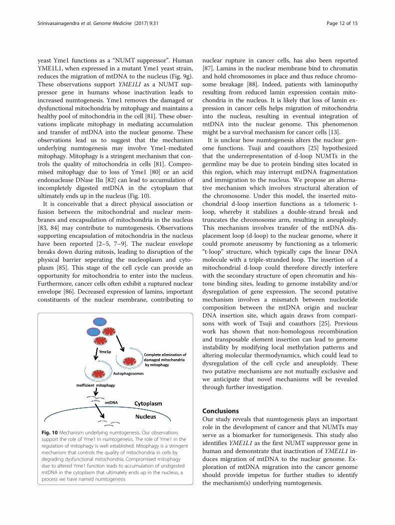
yeast Yme1 functions as a “NUMT suppressor”. HumanYME1L1, when expressed in a mutant Yme1 yeast strain,reduces the migration of mtDNA to the nucleus (Fig. 9g).These observations support YME1LI as a NUMT sup-pressor gene in humans whose inactivation leads toincreased numtogenesis. Yme1 removes the damaged ordysfunctional mitochondria by mitophagy and maintains ahealthy pool of mitochondria in the cell [81]. These obser-vations implicate mitophagy in mediating accumulationand transfer of mtDNA into the nuclear genome. Theseobservations lead us to suggest that the mechanismunderlying numtogenesis may involve Yme1-mediatedmitophagy. Mitophagy is a stringent mechanism that con-trols the quality of mitochondria in cells [81]. Compro-mised mitophagy due to loss of Yme1 [80] or an acidendonuclease DNase IIα [82] can lead to accumulation ofincompletely digested mtDNA in the cytoplasm thatultimately ends up in the nucleus (Fig. 10).It is conceivable that a direct physical association or
fusion between the mitochondrial and nuclear mem-branes and encapsulation of mitochondria in the nucleus[83, 84] may contribute to numtogenesis. Observationssupporting encapsulation of mitochondria in the nucleushave been reported [2–5, 7–9]. The nuclear envelopebreaks down during mitosis, leading to disruption of thephysical barrier seperating the nucleoplasm and cyto-plasm [85]. This stage of the cell cycle can provide anopportunity for mitochondria to enter into the nucleus.Furthermore, cancer cells often exhibit a ruptured nuclearenvelope [86]. Decreased expression of lamins, importantconstituents of the nuclear membrane, contributing to
Fig. 10 Mechanism underlying numtogenesis. Our observationssupport the role of Yme1 in numtogenesis. The role of Yme1 in theregulation of mitophagy is well established. Mitophagy is a stringentmechanism that controls the quality of mitochondria in cells bydegrading dysfunctional mitochondria. Compromised mitophagydue to altered Yme1 function leads to accumulation of undigestedmtDNA in the cytoplasm that ultimately ends up in the nucleus, aprocess we have named numtogenesis
nuclear rupture in cancer cells, has also been reported[87]. Lamins in the nuclear membrane bind to chromatinand hold chromosomes in place and thus reduce chromo-some breakage [88]. Indeed, patients with laminopathyresulting from reduced lamin expression contain mito-chondria in the nucleus. It is likely that loss of lamin ex-pression in cancer cells helps migration of mitochondriainto the nucleus, resulting in eventual integration ofmtDNA into the nuclear genome. This phenomenonmight be a survival mechanism for cancer cells [13].It is unclear how numtogenesis alters the nuclear gen-
ome functions. Tsuji and coauthors [25] hypothesizedthat the underrepresentation of d-loop NUMTs in thegermline may be due to protein binding sites located inthis region, which may interrupt mtDNA fragmentationand immigration to the nucleus. We propose an alterna-tive mechanism which involves structural alteration ofthe chromosome. Under this model, the inserted mito-chondrial d-loop insertion functions as a telomeric t-loop, whereby it stabilizes a double-strand break andtruncates the chromosome arm, resulting in aneuploidy.This mechanism involves transfer of the mtDNA dis-placement loop (d-loop) to the nuclear genome, where itcould promote aneusomy by functioning as a telomeric“t-loop” structure, which typically caps the linear DNAmolecule with a triple-stranded loop. The insertion of amitochondrial d-loop could therefore directly interferewith the secondary structure of open chromatin and his-tone binding sites, leading to genome instability and/ordysregulation of gene expression. The second putativemechanism involves a mismatch between nucleotidecomposition between the mtDNA origin and nuclearDNA insertion site, which again draws from compari-sons with work of Tsuji and coauthors [25]. Previouswork has shown that non-homologous recombinationand transposable element insertion can lead to genomeinstability by modifying local methylation patterns andaltering molecular thermodynamics, which could lead todysregulation of the cell cycle and aneuploidy. Thesetwo putative mechanisms are not mutually exclusive andwe anticipate that novel mechanisms will be revealedthrough further investigation.
ConclusionsOur study reveals that numtogenesis plays an importantrole in the development of cancer and that NUMTs mayserve as a biomarker for tumorigenesis. This study alsoidentifies YME1L1 as the first NUMT suppressor gene inhuman and demonstrate that inactivation of YME1L1 in-duces migration of mtDNA to the nuclear genome. Ex-ploration of mtDNA migration into the cancer genomeshould provide impetus for further studies to identifythe mechanism(s) underlying numtogenesis.
Srinivasainagendra et al. Genome Medicine (2017) 9:31 Page 13 of 15
Additional files
Additional file 1: Table S1. NUMT abundance in all the colorectaltumor samples (n = 57) used in this study. (XLSX 14 kb)
Additional file 2: Figure S1. NUMT density in tumor and normalgenomes (sorted by disease (T2) and fold change in NUMT abundance(T7)). Each peripheral node represents a TCGA sample whose blood-derivednormal and tumor genomes were used in this study. From the outside tothe inside, tracks are ordered from 1 to 7 (T1–T7). T1: Sample gender wherered nodes represent female and blue nodes represent male. T2: Disease typeinformation. Rectal adenocarcinoma (READ) is rendered as green bands andcolon adenocarcinoma (COAD) as red bands. T3: Age at initial pathologicdiagnosis ranging from 30 to 90 years. White and black filled bars representwhite and black race of the individual, respectively. T4: Red columns repre-sent NUMT proportion in tumor genomes and green columns representblood-derived normal NUMT proportion. T5: Vital status of the patients—redfor deceased individuals and green for alive status. T6: Stage of tumorrepresented in grey scale—stage I white, stage II grey, stage III dark grey,stage IV black. T7: Fold change in NUMT abundance. Samples with <1-fold are rendered as colored bands: 1–4-fold, blue; 4–8-fold, green; 8–12-fold,yellow; 12–20-fold, orange; >20-fold, red. (TIF 10991 kb)
Additional file 3: Figure S2. NUMT density in tumor and normalgenomes (sorted by disease (T2) and pathologic tumor stage (T6)). Eachperipheral node represents a TCGA sample whose blood-derived normaland tumor genomes were used in this study. From the outside to theinside, tracks are ordered from 1 to 7 (T1–T7). T1: Sample gender wherered nodes represent female and blue nodes represent male. T2: Diseasetype information. Rectal adenocarcinoma (READ) is rendered as greenbands and colon adenocarcinoma (COAD) as red bands. T3: Age at initialpathologic diagnosis ranging from 30 to 90 years. White and black filledbars represent white and black race of the individual, respectively. T4: Redcolumns represent NUMT proportion in tumor genomes and green columnsrepresent blood-derived normal NUMT proportion. T5: Vital status of thepatients—red for deceased individuals and green for alive status. T6: Stage oftumor represented in grey scale—stage I white, stage II grey, stage III darkgrey, stage IV black. T7: Fold change in NUMT abundance. Samples with <1-fold are rendered as colored bands: 1–4-fold, blue; 4–8-fold, green; 8–12-fold,yellow; 12–20-fold, orange; >20-fold, red. (TIF 10662 kb)
AbbreviationsBAM: Binary alignment; CGHub: Cancer Genomics Hub; COAD: Colonadenocarcinoma; CRC: colorectal cancer; mtDNA: Mitochondrial DNA;mtRNA: Mitochondrial RNA; NUMT: nuclear mtDNA sequence; QC: qualitycontrol; READ: Rectum adenocarcinoma; TCGA: The Cancer Genome Atlas
AcknowledgementsWe thank Dr. Andreas Ivessa for generously providing us with PTY62 (Yme1-1)yeast strains, Dr. Thomas Fox for the yeast expression construct pPT31-yYme1,Dr. Thomas Langer for pYX113-hYme1L1, and Dr. Diana Stojanovski for pRS414-yYme1 constructs.
FundingThis study was supported by grants from the Veterans Administration1I01BX001716 and a NCTN–LAPS Program Translational Research Award toKKS and T32HL072757 (PI: HKT) to MWS.
Availability of data and materialsTCGA data sets are publicly available to researchers upon individual institutionalIRB approval and approval from dbgap.
Authors’ contributionsKKS and HKT conceived the project and designed the experiments. VS, MWS,AS, VPM, and BS performed the experiments and analyzed the data.PB analyzed YME1L1 mutations in tumors. VS, MWS, HKT, BS, and KKS wrotethe manuscript. All authors read and approved the final manuscript.
Authors’ informationMWS and BS contributed equally to this study. MWS’s current affiliation is theDepartment of Biological and Environmental Sciences, School of NaturalSciences and Mathematics, University of West Alabama, Livingston, Alabama.
Competing interestsThe authors declare that they have no competing interests.
Consent for publicationNot applicable.
Ethics approval and consent to participateDatasets utilized in part of this research fall under dbGaP’s “Protected/Controlled Data Access System”, which warrants a multi-step protocol involvingthe IRB team of UAB for gaining complete access to the requested researchdata. The data access process was initiated by Dr. Hemant Tiwari through hisdbGaP eRA Commons account by submitting a research proposal description.Upon successful submission and approval to use identified TCGA Sequencingdata, the Genomic Data Commons (GDC) data portal was used to accessauthorized sequencing datasets. GDC’s data download and storageprotocol was followed carefully to maintain integrity of the downloadeddata and ensure a secure sandbox was staged to store and analyze thedatasets. The research data part of dbGaP’s study ID ‘phs000178.v9.p8’(referenced and approved by TCGA as Project ID 4538) was downloadedand analyzed as part of our research effort. Furthermore, we de-identifiedthe sample IDs by replacing the original Barcode-based TCGA ID conventionwith our internal numeric ID format. No individual consent was acquired as thestudy utilized the TCGA database.
Publisher’s NoteSpringer Nature remains neutral with regard to jurisdictional claims inpublished maps and institutional affiliations.
Author details1Department of Biostatistics, School of Public Health, University of Alabamaat Birmingham, Birmingham, Alabama 35294, USA. 2Department of Genetics,University of Alabama at Birmingham, Birmingham, Alabama 35294, USA.3Departments of Genetics, Environmental Health, Center for Free RadicalBiology, Center for Aging and UAB Comprehensive Cancer Center, Universityof Alabama at Birmingham, Birmingham, Alabama 35294, USA. 4Departmentsof Pathology, Environmental Health, Center for Free Radical Biology, Centerfor Aging and UAB Comprehensive Cancer Center, University of Alabama atBirmingham, Birmingham, Alabama 35294, USA. 5Birmingham Veterans AffairsMedical Center, Birmingham, Alabama 35294, USA. 6Department of Genetics,School of Medicine, University of Alabama at Birmingham, Kaul GeneticsBuilding, Suite 620, 720 20th St. South, Birmingham, AL 35294, USA. 7Presentaddress: Department of Biological and Environmental Sciences, School ofNatural Sciences and Mathematics, University of West Alabama, Livingston,Alabama, USA.
Received: 3 November 2016 Accepted: 9 March 2017
References1. Hazkani-Covo E, Zeller RM, Martin W. Molecular poltergeists: mitochondrial
DNA copies (numts) in sequenced nuclear genomes. PLoS Genet.2010;6:e1000834.
2. Bakeeva LE, Skulachev VP, Sudarikova YV, Tsyplenkova VG. Mitochondriaenter the nucleus (one further problem in chronic alcoholism). Biochemistry(Mosc). 2001;66:1335–41.
3. Bloom GD. A nucleus with cytoplasmic features. J Cell Biol. 1967;35:266–8.4. Brandes D, Schofield BH, Anton E. Nuclear mitochondria? Science. 1965;
149:1373–4.5. De Vos WH, Houben F, Kamps M, Malhas A, Verheyen F, Cox J, et al.
Repetitive disruptions of the nuclear envelope invoke temporary loss ofcellular compartmentalization in laminopathies. Hum Mol Genet. 2011;20:4175–86.
6. Landerer E, Villegas J, Burzio VA, Oliveira L, Villota C, Lopez C, et al. Nuclearlocalization of the mitochondrial ncRNAs in normal and cancer cells. CellOncol (Dordr). 2011;34:297–305.
Srinivasainagendra et al. Genome Medicine (2017) 9:31 Page 14 of 15
7. Matsuyama M, Suzuki H. Seizing mechanism and fate of intranuclearmitochondria. Experientia. 1972;28:1347–8.
8. Sunba MS, Rahi AH, Morgan G. Tumours of the anterior uvea. II. Intranuclearcytoplasmic inclusions in malignant melanoma of the iris. Br J Ophthalmol.1980;64:453–6.
9. Takemura G, Takatsu Y, Sakaguchi H, Fujiwara H. Intranuclear mitochondriain human myocardial cells. Pathol Res Pract. 1997;193:305–11.
10. Farrelly F, Butow RA. Rearranged mitochondrial genes in the yeast nucleargenome. Nature. 1983;301:296–301.
11. Zullo S, Sieu LC, Slightom JL, Hadler HI, Eisenstadt JM. Mitochondrial D-loopsequences are integrated in the rat nuclear genome. J Mol Biol. 1991;221:1223–35.
12. Dayama G, Emery SB, Kidd JM, Mills RE. The genomic landscape ofpolymorphic human nuclear mitochondrial insertions. Nucleic Acids Res.2014;42:12640–9.
13. Hazkani-Covo E, Covo S. Numt-mediated double-strand break repairmitigates deletions during primate genome evolution. PLoS Genet. 2008;4:e1000237.
14. Wang D, Timmis JN. Cytoplasmic organelle DNA preferentially inserts intoopen chromatin. Genome Biol Evol. 2013;5:1060–4.
15. Woischnik M, Moraes CT. Pattern of organization of human mitochondrialpseudogenes in the nuclear genome. Genome Res. 2002;12:885–93.
16. Hallmann A, Milczarek R, Lipinski M, Kossowska E, Spodnik JH, Wozniak M,et al. Fast perinuclear clustering of mitochondria in oxidatively stressedhuman choriocarcinoma cells. Folia Morphol (Warsz). 2004;63:407–12.
17. Kim SJ, Syed GH, Siddiqui A. Hepatitis C virus induces the mitochondrialtranslocation of Parkin and subsequent mitophagy. PLoS Pathog.2013;9:e1003285.
18. Villa AM, Doglia SM. Mitochondria in tumor cells studied by laser scanningconfocal microscopy. J Biomed Opt. 2004;9:385–94.
19. Chen D, Xue W, Xiang J. The intra-nucleus integration of mitochondrialDNA (mtDNA)in cervical mucosa cells and its relation with c-mycexpression. J Exp Clin Cancer Res. 2008;27:36.
20. Hadler HI, Devadas K, Mahalingam R. Selected nuclear LINE elements withmitochondrial-DNA-like inserts are more plentiful and mobile in tumor thanin normal tissue of mouse and rat. J Cell Biochem. 1998;68:100–9.
21. Mourier T. Reverse transcription in genome evolution. Cytogenet GenomeRes. 2005;110:56–62.
22. Mourier T, Hansen AJ, Willerslev E, Arctander P. The Human Genome Projectreveals a continuous transfer of large mitochondrial fragments to thenucleus. Mol Biol Evol. 2001;18:1833–7.
23. Bensasson D, Feldman MW, Petrov DA. Rates of DNA duplication andmitochondrial DNA insertion in the human genome. J Mol Evol. 2003;57:343–54.
24. Lang M, Sazzini M, Calabrese FM, Simone D, Boattini A, Romeo G, et al.Polymorphic NumtS trace human population relationships. Hum Genet.2012;131:757–71.
25. Tsuji J, Frith MC, Tomii K, Horton P. Mammalian NUMT insertion is non-random. Nucleic Acids Res. 2012;40:9073–88.
26. Zhang Z, Harrison PM, Liu Y, Gerstein M. Millions of years of evolutionpreserved: a comprehensive catalog of the processed pseudogenes in thehuman genome. Genome Res. 2003;13:2541–58.
27. Simone D, Calabrese FM, Lang M, Gasparre G, Attimonelli M. The referencehuman nuclear mitochondrial sequences compilation validated andimplemented on the UCSC genome browser. BMC Genomics. 2011;12:517.
28. Amuthan G, Biswas G, Zhang SY, Klein-Szanto A, Vijayasarathy C, Avadhani NG.Mitochondria-to-nucleus stress signaling induces phenotypic changes, tumorprogression and cell invasion. EMBO J. 2001;20:1910–20.
29. Delsite R, Kachhap S, Anbazhagan R, Gabrielson E, Singh KK. Nuclear genesinvolved in mitochondria-to-nucleus communication in breast cancer cells.Mol Cancer. 2002;1:6.
30. Desler C, Munch-Petersen B, Stevnsner T, Matsui S, Kulawiec M, Singh KK,et al. Mitochondria as determinant of nucleotide pools and chromosomalstability. Mutat Res. 2007;625:112–24.
31. Donthamsetty S, Brahmbhatt M, Pannu V, Rida PC, Ramarathinam S, OgdenA, et al. Mitochondrial genome regulates mitotic fidelity by maintainingcentrosomal homeostasis. Cell Cycle. 2014;13:2056–63.
32. Frezza C. The role of mitochondria in the oncogenic signal transduction. IntJ Biochem Cell Biol. 2014;48:11–7.
33. Rasmussen AK, Chatterjee A, Rasmussen LJ, Singh KK. Mitochondria-mediated nuclear mutator phenotype in Saccharomyces cerevisiae. NucleicAcids Res. 2003;31:3909–17.
34. Singh KK, Kulawiec M, Still I, Desouki MM, Geradts J, Matsui S. Inter-genomiccross talk between mitochondria and the nucleus plays an important role intumorigenesis. Gene. 2005;354:140–6.
35. Wallace DC. Mitochondria and cancer. Nat Rev Cancer. 2012;12:685–98.36. Qu F, Liu X, Zhou F, Yang H, Bao G, He X, et al. Association between
mitochondrial DNA content in leukocytes and colorectal cancer risk: a case-control analysis. Cancer. 2011;117:3148–55.
37. Thyagarajan B, Wang R, Barcelo H, Koh WP, Yuan JM. Mitochondrial copynumber is associated with colorectal cancer risk. Cancer EpidemiolBiomarkers Prev. 2012;21:1574–81.
38. Webb E, Broderick P, Chandler I, Lubbe S, Penegar S, Tomlinson IP, et al.Comprehensive analysis of common mitochondrial DNA variants andcolorectal cancer risk. Br J Cancer. 2008;99:2088–93.
39. Skonieczna K, Malyarchuk BA, Grzybowski T. The landscape of mitochondrialDNA variation in human colorectal cancer on the background ofphylogenetic knowledge. Biochim Biophys Acta. 2012;1825:153–9.
40. Theodoratou E, Din FV, Farrington SM, Cetnarskyj R, Barnetson RA, PorteousME, et al. Association between common mtDNA variants and all-cause orcolorectal cancer mortality. Carcinogenesis. 2010;31:296–301.
41. Bensasson D, Zhang D, Hartl DL, Hewitt GM. Mitochondrial pseudogenes:evolution’s misplaced witnesses. Trends Ecol Evol. 2001;16:314–21.
42. Chen T, He J, Shen L, Fang H, Nie H, Jin T, et al. The mitochondrial DNA4,977-bp deletion and its implication in copy number alteration incolorectal cancer. BMC Med Genet. 2011;12:8.
43. Lee HC, Yin PH, Lin JC, Wu CC, Chen CY, Wu CW, et al. Mitochondrialgenome instability and mtDNA depletion in human cancers. Ann N Y AcadSci. 2005;1042:109–22.
44. Polyak K, Li Y, Zhu H, Lengauer C, Willson JK, Markowitz SD, et al. Somaticmutations of the mitochondrial genome in human colorectal tumours. NatGenet. 1998;20:291–3.
45. Wilks C, Cline MS, Weiler E, Diehkans M, Craft B, Martin C, et al. The CancerGenomics Hub (CGHub): overcoming cancer through the power oftorrential data. Database (Oxford). 2014.
46. Chu HW, Rios C, Huang C, Wesolowska-Andersen A, Burchard EG, O’ConnorBP, et al. CRISPR-Cas9-mediated gene knockout in primary human airwayepithelial cells reveals a proinflammatory role for MUC18. Gene Ther. 2015;22:822–9.
47. West AP, Khoury-Hanold W, Staron M, Tal MC, Pineda CM, Lang SM, et al.Mitochondrial DNA stress primes the antiviral innate immune response.Nature. 2015;520:553–7.
48. Gietz RD, Schiestl RH, Willems AR, Woods RA. Studies on the transformation ofintact yeast cells by the LiAc/SS-DNA/PEG procedure. Yeast. 1995;11:355–60.
49. Singh KK, Sigala B, Sikder HA, Schwimmer C. Inactivation of Saccharomycescerevisiae OGG1 DNA repair gene leads to an increased frequency ofmitochondrial mutants. Nucleic Acids Res. 2001;29:1381–8.
50. Soni R, Carmichael JP, Murray JA. Parameters affecting lithium acetate-mediated transformation of Saccharomyces cerevisiae and development ofa rapid and simplified procedure. Curr Genet. 1993;24:455–9.
51. Velarde MC. Mitochondrial and sex steroid hormone crosstalk during aging.Longev Healthspan. 2014;3:2.
52. Bernardi G. Isochores and the evolutionary genomics of vertebrates. Gene.2000;241:3–17.
53. Saccone S, Federico C, Solovei I, Croquette MF, Della Valle G, Bernardi G.Identification of the gene-richest bands in human prometaphasechromosomes. Chromosome Res. 1999;7:379–86.
54. Chen TL, Manuelidis L. SINEs and LINEs cluster in distinct DNA fragments ofGiemsa band size. Chromosoma. 1989;98:309–16.
55. Thorsness PE, Fox TD. Escape of DNA from mitochondria to the nucleus inSaccharomyces cerevisiae. Nature. 1990;346:376–9.
56. Shah ZH, Hakkaart GA, Arku B, de Jong L, van der Spek H, Grivell LA, et al.The human homologue of the yeast mitochondrial AAA metalloproteaseYme1p complements a yeast yme1 disruptant. FEBS Lett. 2000;478:267–70.
57. Thorsness PE, Fox TD. Nuclear mutations in Saccharomyces cerevisiae thataffect the escape of DNA from mitochondria to the nucleus. Genetics. 1993;134:21–8.
58. Cheng X, Ivessa AS. The migration of mitochondrial DNA fragments to thenucleus affects the chronological aging process of Saccharomycescerevisiae. Aging Cell. 2010;9:919–23.
59. Chen JM, Chuzhanova N, Stenson PD, Ferec C, Cooper DN. Meta-analysis ofgross insertions causing human genetic disease: novel mutational mechanismsand the role of replication slippage. Hum Mutat. 2005;25:207–21.
Srinivasainagendra et al. Genome Medicine (2017) 9:31 Page 15 of 15
60. Borensztajn K, Chafa O, Alhenc-Gelas M, Salha S, Reghis A, Fischer AM, et al.Characterization of two novel splice site mutations in human factor VII genecausing severe plasma factor VII deficiency and bleeding diathesis. Br JHaematol. 2002;117:168–71.
61. Goldin E, Stahl S, Cooney AM, Kaneski CR, Gupta S, Brady RO, et al. Transferof a mitochondrial DNA fragment to MCOLN1 causes an inherited case ofmucolipidosis IV. Hum Mutat. 2004;24:460–5.
62. Ahmed ZM, Smith TN, Riazuddin S, Makishima T, Ghosh M, Bokhari S, et al.Nonsyndromic recessive deafness DFNB18 and Usher syndrome type IC areallelic mutations of USHIC. Hum Genet. 2002;110:527–31.
63. Turner C, Killoran C, Thomas NS, Rosenberg M, Chuzhanova NA, Johnston J,et al. Human genetic disease caused by de novo mitochondrial-nuclearDNA transfer. Hum Genet. 2003;112:303–9.
64. Miettinen TP, Bjorklund M. Cellular Allometry of mitochondrial functionalityestablishes the optimal cell size. Dev Cell. 2016;39:370–82.
65. Shay JW, Baba T, Zhan QM, Kamimura N, Cuthbert JA. HeLaTG cells havemitochondrial DNA inserted into the c-myc oncogene. Oncogene. 1991;6:1869–74.
66. Hu Y, Qian G, Mao B, Xiao T, Li Y, Cao S. Malignant transformation of mouseembryonic fibroblast induced by mitochondrial DNA fragments. ZhonghuaBing Li Xue Za Zhi. 2000;29:39–42.
67. de Araujo LF, Fonseca AS, Muys BR, Placa JR, Bueno RB, Lorenzi JC, et al.Mitochondrial genome instability in colorectal adenoma andadenocarcinoma. Tumour Biol. 2015;36:8869–79.
68. Hsu CC, Tseng LM, Lee HC. Role of mitochondrial dysfunction in cancerprogression. Exp Biol Med (Maywood). 2016;241:1281–95.
69. Kulawiec M, Safina A, Desouki MM, Still I, Matsui S, Bakin A, et al.Tumorigenic transformation of human breast epithelial cells induced bymitochondrial DNA depletion. Cancer Biol Ther. 2008;7:1732–43.
70. Modica-Napolitano JS, Kulawiec M, Singh KK. Mitochondria and humancancer. Curr Mol Med. 2007;7:121–31.
71. Owens KM, Kulawiec M, Desouki MM, Vanniarajan A, Singh KK. ImpairedOXPHOS complex III in breast cancer. PLoS ONE. 2011;6:e23846.
72. Singh KK. Mitochondrial dysfunction is a common phenotype in aging andcancer. Ann N Y Acad Sci. 2004;1019:260–4.
73. Abdel-Rahman WM. Genomic instability and carcinogenesis: an update. CurrGenomics. 2008;9:535–41.
74. Giam M, Rancati G. Aneuploidy and chromosomal instability in cancer: ajackpot to chaos. Cell Div. 2015;10:3.
75. Modica-Napolitano JS, Singh KK. Mitochondrial dysfunction in cancer.Mitochondrion. 2004;4:755–62.
76. Hadler HI, Daniel BG, Pratt RD. The induction of ATP energizedmitochondrial volume changes by carcinogenic N-hydroxy-N-acetyl-aminofluorenes when combined with showdomycin. A unitary hypothesisfor carcinogenesis. J Antibiot (Tokyo). 1971;24:405–17.
77. Akouchekian M, Houshmand M, Akbari MH, Kamalidehghan B, Dehghan M.Analysis of mitochondrial ND1 gene in human colorectal cancer. J Res MedSci. 2011;16:50–5.
78. Wang CY, Li H, Hao XD, Liu J, Wang JX, Wang WZ, et al. Uncovering theprofile of somatic mtDNA mutations in Chinese colorectal cancer patients.PLoS ONE. 2011;6:e21613.
79. Francis BR, Thorsness PE. Hsp90 and mitochondrial proteases Yme1 andYta10/12 participate in ATP synthase assembly in Saccharomyces cerevisiae.Mitochondrion. 2011;11:587–600.
80. Wang K, Jin M, Liu X, Klionsky DJ. Proteolytic processing of Atg32 by themitochondrial i-AAA protease Yme1 regulates mitophagy. Autophagy. 2013;9:1828–36.
81. Deffieu M, Bhatia-Kissova I, Salin B, Klionsky DJ, Pinson B, Manon S, et al.Increased levels of reduced cytochrome b and mitophagy components arerequired to trigger nonspecific autophagy following induced mitochondrialdysfunction. J Cell Sci. 2013;126:415–26.
82. Oka T, Hikoso S, Yamaguchi O, Taneike M, Takeda T, Tamai T, et al.Mitochondrial DNA that escapes from autophagy causes inflammation andheart failure. Nature. 2012;485:251–5.
83. Jensen H, Engedal H, Saetersdal TS. Ultrastructure of mitochondria-containiningnuclei in human myocardial cells. Virchows Arch B Cell Pathol. 1976;21:1–12.
84. Thorsness PE, Weber ER. Escape and migration of nucleic acids betweenchloroplasts, mitochondria, and the nucleus. Int Rev Cytol. 1996;165:207–34.
85. Guttinger S, Laurell E, Kutay U. Orchestrating nuclear envelope disassemblyand reassembly during mitosis. Nat Rev Mol Cell Biol. 2009;10:178–91.
86. Zink D, Fischer AH, Nickerson JA. Nuclear structure in cancer cells. Nat RevCancer. 2004;4:677–87.
87. Vargas JD, Hatch EM, Anderson DJ, Hetzer MW. Transient nuclear enveloperupturing during interphase in human cancer cells. Nucleus. 2012;3:88–100.
88. de Las Heras JI, Batrakou DG, Schirmer EC. Cancer biology and the nuclearenvelope: a convoluted relationship. Semin Cancer Biol. 2013;23:125–37.
• We accept pre-submission inquiries
• Our selector tool helps you to find the most relevant journal
• We provide round the clock customer support
• Convenient online submission
• Thorough peer review
• Inclusion in PubMed and all major indexing services
• Maximum visibility for your research
Submit your manuscript atwww.biomedcentral.com/submit
Submit your next manuscript to BioMed Central and we will help you at every step: