The pseudo-mitochondrial genome influences mistakes in heteroplasmy interpretation
13
BioMed Central Page 1 of 13 (page number not for citation purposes) BMC Genomics Open Access Research article The pseudo-mitochondrial genome influences mistakes in heteroplasmy interpretation Ryan L Parr* 1 , Jennifer Maki 1 , Brian Reguly 1 , Gabriel D Dakubo 1 , Andrea Aguirre 1 , Roy Wittock 1 , Kerry Robinson 1 , John P Jakupciak 2 and Robert E Thayer 1 Address: 1 Genesis Genomics Inc, 1294 Balmoral Street, Thunder Bay, Ontario, P7B 5Z5, Canada and 2 Biochemical Science Division, National Institute of Standards and Technology, Gaithersburg, MD 20899, USA Email: Ryan L Parr* - [email protected]; Jennifer Maki - [email protected]; Brian Reguly - [email protected]; Gabriel D Dakubo - [email protected]; Andrea Aguirre - [email protected]; Roy Wittock - [email protected]; Kerry Robinson - [email protected]; John P Jakupciak - [email protected]; Robert E Thayer - [email protected] * Corresponding author Abstract Background: Nuclear mitochondrial pseudogenes (numts) are a potential source of contamination during mitochondrial DNA PCR amplification. This possibility warrants careful experimental design and cautious interpretation of heteroplasmic results. Results: Here we report the cloning and sequencing of numts loci, amplified from human tissue and rho-zero (ρ 0 ) cells (control) with primers known to amplify the mitochondrial genome. This paper is the first to fully sequence 46 paralogous nuclear DNA fragments that represent the entire mitochondrial genome. This is a surprisingly small number due primarily to the primer sets used in this study, because prior to this, BLAST searches have suggested that nuclear DNA harbors between 400 to 1,500 paralogous mitochondrial DNA fragments. Our results indicate that multiple numts were amplified simultaneously with the mitochondrial genome and increased the load of pseudogene signal in PCR reactions. Further, the entire mitochondrial genome was represented by multiple copies of paralogous nuclear sequences. Conclusion: These findings suggest that mitochondrial genome disease-associated biomarkers must be rigorously authenticated to preclude any affiliation with paralogous nuclear pseudogenes. Importantly, the common perception that mitochondrial template "swamps" numts loci precluding detectable amplification, depends on the region of the mitochondrial genome targeted by the PCR reaction and the number of pseudogene loci that may co-amplify. Cloning and relevant sequencing data will facilitate the correct interpretation. This is the first complete, wet-lab characterization of numts that represent the entire mitochondrial genome. Published: 21 July 2006 BMC Genomics 2006, 7:185 doi:10.1186/1471-2164-7-185 Received: 19 April 2006 Accepted: 21 July 2006 This article is available from: http://www.biomedcentral.com/1471-2164/7/185 © 2006 Parr et al; licensee BioMed Central Ltd. This is an Open Access article distributed under the terms of the Creative Commons Attribution License (http://creativecommons.org/licenses/by/2.0 ), which permits unrestricted use, distribution, and reproduction in any medium, provided the original work is properly cited.
-
Upload
independent -
Category
Documents
-
view
0 -
download
0
Transcript of The pseudo-mitochondrial genome influences mistakes in heteroplasmy interpretation

BioMed CentralBMC Genomics
ss
Open AcceResearch articleThe pseudo-mitochondrial genome influences mistakes in heteroplasmy interpretationRyan L Parr*1, Jennifer Maki1, Brian Reguly1, Gabriel D Dakubo1, Andrea Aguirre1, Roy Wittock1, Kerry Robinson1, John P Jakupciak2 and Robert E Thayer1Address: 1Genesis Genomics Inc, 1294 Balmoral Street, Thunder Bay, Ontario, P7B 5Z5, Canada and 2Biochemical Science Division, National Institute of Standards and Technology, Gaithersburg, MD 20899, USA
Email: Ryan L Parr* - [email protected]; Jennifer Maki - [email protected]; Brian Reguly - [email protected]; Gabriel D Dakubo - [email protected]; Andrea Aguirre - [email protected]; Roy Wittock - [email protected]; Kerry Robinson - [email protected]; John P Jakupciak - [email protected]; Robert E Thayer - [email protected]
* Corresponding author
AbstractBackground: Nuclear mitochondrial pseudogenes (numts) are a potential source ofcontamination during mitochondrial DNA PCR amplification. This possibility warrants carefulexperimental design and cautious interpretation of heteroplasmic results.
Results: Here we report the cloning and sequencing of numts loci, amplified from human tissueand rho-zero (ρ0) cells (control) with primers known to amplify the mitochondrial genome. Thispaper is the first to fully sequence 46 paralogous nuclear DNA fragments that represent the entiremitochondrial genome. This is a surprisingly small number due primarily to the primer sets used inthis study, because prior to this, BLAST searches have suggested that nuclear DNA harborsbetween 400 to 1,500 paralogous mitochondrial DNA fragments. Our results indicate that multiplenumts were amplified simultaneously with the mitochondrial genome and increased the load ofpseudogene signal in PCR reactions. Further, the entire mitochondrial genome was represented bymultiple copies of paralogous nuclear sequences.
Conclusion: These findings suggest that mitochondrial genome disease-associated biomarkersmust be rigorously authenticated to preclude any affiliation with paralogous nuclear pseudogenes.Importantly, the common perception that mitochondrial template "swamps" numts loci precludingdetectable amplification, depends on the region of the mitochondrial genome targeted by the PCRreaction and the number of pseudogene loci that may co-amplify. Cloning and relevant sequencingdata will facilitate the correct interpretation. This is the first complete, wet-lab characterization ofnumts that represent the entire mitochondrial genome.
Published: 21 July 2006
BMC Genomics 2006, 7:185 doi:10.1186/1471-2164-7-185
Received: 19 April 2006Accepted: 21 July 2006
This article is available from: http://www.biomedcentral.com/1471-2164/7/185
© 2006 Parr et al; licensee BioMed Central Ltd.This is an Open Access article distributed under the terms of the Creative Commons Attribution License (http://creativecommons.org/licenses/by/2.0), which permits unrestricted use, distribution, and reproduction in any medium, provided the original work is properly cited.
Page 1 of 13(page number not for citation purposes)

BMC Genomics 2006, 7:185 http://www.biomedcentral.com/1471-2164/7/185
BackgroundThe unique maternal inheritance pattern of mitochon-drial DNA (mtDNA), its small genome size, acceleratedmutation rate, lack of recombination, and multiple copynumber per cell, in comparison to nuclear DNA, are idealbiological traits for investigating evolution, populationgenetics and for forensic and medical applications. Thus,the mitochondrial genome has been used as a biosensorfor the timing and movement of human populations inantiquity [1,2]. MtDNA analysis is routinely used in foren-sic biology to type biological material when degradationprevents nuclear STR amplification [3]. In addition, theentire mitochondrial molecule has potential medical util-ity because it can serve as a repository of cancer mutationsand as a biosensor indicative of genetic alterations [4-13].
Frequently, identifying legitimate mtDNA mutations isconfounded by heteroplasmy, a condition in which wild-type and mutant mitochondrial genomes co-exist in a cell.The interpretation of heteroplasmy can further be con-founded by the widespread integration of portions of themitochondrial genome into the nuclear genome [14,15].These homologous, yet divergent nuclear and mtDNAsequences can be co-amplified in PCR reactions intendedto replicate targeted mtDNA sequences only. Althoughthis problem has previously been considered to be mutedbecause of the high copy number of mtDNA over corre-sponding nuclear loci, caution is warranted [16]. Forexample, there are specific regions of the mitochondrialgenome that have corresponding nuclear mitochondrialpseudogenes (numts) distributed across multiple chro-mosomes. Hence, there are regions of the mitochondrialgenome that have a high nuclear copy number, which arenot completely "swamped" during amplification. Wereport that some heteroplasmies detected in prostate can-cer samples are a result of co-amplification of these multi-ple loci.
A large number of manuscripts addressing errors relatedto the interpretation of mtDNA and mtDNA hetero-plasmy has been published [17-25]. Notably not all theseerrors are due to pseudogene co-amplification; however,mistakes from pseudogenes may increase with improvedsequencing methods and highly sensitive re-sequencingmicroarray technologies that have a lower detection limitthan traditional sequencing and which readily detect low-level heteroplasmy [11,26]. In some cases, if the hetero-plasmy is inherited, it substantially increases the power ofmutation detection, which becomes an important aspectsince heteroplasmy has been reported as an early indica-tion of disease [27-31]. In addition, if the disease processinvokes mitochondrial depletion, this could increasenuclear pseudogene signal in reactions as a result ofreduced mitochondrial genome copy number [32]. Lossof mitochondria has been described in several human
cancers [33-36]. As well, the number of mitochondria andmtDNA copy number vary for different cell types [37-39].These important matters relating to sequence interpreta-tion have been generally neglected, in part, due to the lackof numt reference material, which would help investiga-tors determine the relevance of detected mtDNA sequencevariations. Hence, the need to validate somatic mitochon-drial mutations is a pressing one.
Heteroplasmic issues have already complicated dataobtained from other species. For example, in elephanthair, low mtDNA content is the reason why numts wereco-amplified and misinterpreted as authentic mtDNA. Incontrast, numts were not detected in DNA derived fromelephant blood due to the presence of mitochondria-richplatelets [40]. Moreover, the hominid, Gorilla, is wellknown for significant numt interference with mitochon-drial sequences, highlighting the need for diligence wheninterpreting human mtDNA heteroplasmy [41]. Not sur-prisingly, the effort for using mitochondrial cytochrome coxidase as a primate "barcode" is plagued by numt ampli-fication as well [42].
Further, laser capture microscopy has improved the abilityto separate and analyze cancer cells, but because of thedecreased amounts of sample DNA, many primer pairs arerequired to obtain a robust amplification of the entiremitochondrial genome [43]. Moreover, a sufficientnumber of cells must be captured to avoid incorporationerrors associated with low template quantity [44]. Thiswill also be relevant to studies that use formalin-fixed par-affin embedded samples [45]. The use of many primersmeans that smaller amplicons will be synthesized trans-lating into a higher risk of co-amplification of numts andthe potential misinterpretation of heteroplasmic calls.
There is limited in silico and wet-lab evidence indicatingthat fragments of the human mitochondrial genome areembedded in the nuclear DNA archive [46-50]. Thesefindings emphasize the critical need to minimize errone-ous interpretation of heteroplasmy, a vital necessity forprecise forensic discrimination, evolutionary studies, andpotential diagnostics. We provide evidence of numts forthe entire mitochondrial genome by the amplification,cloning, and identification of numts from rho-zero (ρ0)cells and clinical cancer specimens. Here we presentresults from overlapping primers, which co-amplifiednuclear embedded, paralogous mitochondrial sequence.Surprisingly, our data shows a relatively small number(when compared to hypothetical sequence informationobtained from BLAST searches) of multiple nuclear locithat co-amplify with the mitochondrial genome. Thesefindings demonstrate that accurate interpretation of heter-oplasmy not only requires careful primer design and test-ing, but also indicates that a compendium of the sequence
Page 2 of 13(page number not for citation purposes)

BMC Genomics 2006, 7:185 http://www.biomedcentral.com/1471-2164/7/185
information from multiple-copy number numts is animportant reference tool that will facilitate correct mtDNAinterpretation and support reliable mitochondrialgenome sequencing data.
Resultsρ0 cells lack mtDNARho-zero cells were evaluated for the presence of mtDNA.To ensure that total DNA extracted from ρ0 cells wereindeed devoid of mtDNA, we first performed Southernblot analyses on DNA extracts from ρ0 cells. Blood wasused as a positive control. No full length mitochondrialgenome signal was observed in ρ0 lanes when the blotswere probed with mtDNA-specific probes (Figure 1a). Wenext performed PCR on the DNA extracts with primersspecific to the mitochondrial coding regions. Again, therewere no amplifications observed in the ρ0 templates,
while DNA isolated from blood was amplified, asexpected (data not shown). We used RT-PCR to providefurther evidence that ρ0 cells are indeed devoid of mtDNA[51]. RT-PCR analysis was performed on RNA samplesfrom ρ0 cells and normal human skin tissue (epithelialcells) samples with primers to OXPHOS genes and anuclear gene (positive control), 5-aminolevulinate syn-thase (hALAS) (Table 1). Whereas the hALAS primersamplified nuclear targets in template from ρ0 and epithe-lial cells, there was no observable product with mtDNAprimers for the ρ0 cDNA template (Figure 1b), independ-ently confirming the absence of mtDNA in these cells.
Co-amplification of numts and mtDNAAmplification of the complete mitochondrial genome wasperformed on human formalin fixed and paraffin embed-ded (FFPE) prostate cancer samples using a set of 34 prim-ers (Table 2). Due to the amount and quality of DNArecovered, the average amplicon size was limited to 625bp. Surprisingly, 24 (71%) of the primer sets co-amplifiedmitochondrial pseudogenes (Figure 2, and data notshown). A similar ratio was previously reported by anindependent group using 38 primers (26/38 or 68%)[16]. In an effort to fully characterize numts that representthe entire mitochondrial genome, we redesigned theremaining 10 primers to co-amplify nuclear loci. Thus, weamplified template from ρ0 cells and subsequently identi-fied (via sequencing) the cloned fragments from thenucleus. A region of the D-loop (base pairs 16211-420and 15-711) was recalcitrant to co-amplification usingour mitochondrial primers. Therefore, two chromosome17 specific primers were designed to capture this D-loopfragment (Table 2). Hence, a total of 36 primer sets wereused to recover the entire mitochondrial genome from thenucleus. The sequences representative of the entire mito-chondrial genome are provided as an additional material(additional file 1). Figure 3 is an example of an alignmentto rCRS of three numt clones recovered using primer set1488F/2084R (Table 2). These three clones were recov-ered form three separate chromosomes (Chr11 –NT_009237, Chr5 – NT_006713, and Chr3 –NT_005612). Similar alignment of our consensus clonedsequences enabled the assembly of a pseudo-mitochon-drial genome (Figure 4).
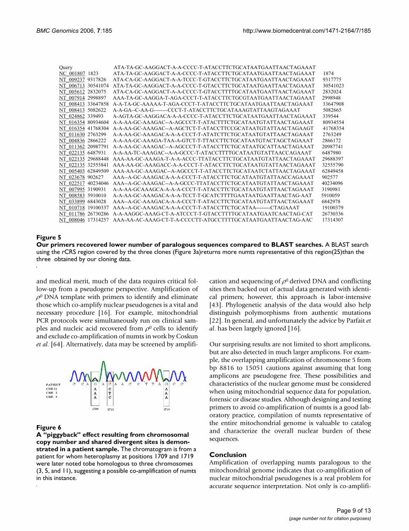
The following chromosomes were represented in the data:1, 2, 3, 4, 5, 7, 8, 9, 11, 16, 17, 20 and X. The number ofparalogous sequences, in some instances, was lower thanthe number predicted from BLAST searches (Figure 3, 5).We demonstrate that there are only a limited number ofmultiple copy numts that potentially contribute to a het-eroplasmic signal. Subsequently, we systematicallyinspected heteroplasmic sites observed in sequences fromthe prostate cancer samples for numt contribution usingour cloned ρ0 data as a reference. We discovered false het-
ρ0 cellscells do not contain mtDNAFigure 1ρ0 cells do not contain mtDNA. a. Southern blot analysis of total DNA extracted from blood (bld) and ρ0 cells and probed with a full length mtDNA probe. Note the absence of hybridization to ρ0 extracts. Lad is a DIG-labeled DNA molecular weight marker III (Roche). b. PCR amplification of cDNA from ρ0 and epithelial cells (EC). Note the amplifica-tion of ρ0 cDNA with primers to the nuclear gene hALAS, whereas primers to ND1, ATPase6, and CYTB failed to amplify ρ0 cDNA, although they all amplified cDNA from EC. Lad is a 100bp DNA size standard (Fermentas life sciences).
Lad bld ρo Lad ρ0 ρ0 EC ρ0 EC ρ0 ECa b
hALAS ND1 ATPase6 CYTB
21226
5148
4973
4268
Table 1: Sequences of four primer sets used in RT-PCR.
Designation Primer sequence (5'-3')
hALASF CCACTGGAAGAGCTGTGTGAhALASR ACCCTCCAACACAACCAAAGND1F GAGCAGTAGCCCAAACAATCND1R GGGTTCGGTTGGTCTCTGCTAGATP6F CCATAAAATTATGAGCGGGCACAGTGATTATP6R GGAAGGTTAATGGTTGATATTGCTAGGCYTBF CTAGCAACACTCCACCTCCTATCYTBR GTAAGCCGAGGGCGTCTTTGCTTG
Page 3 of 13(page number not for citation purposes)

BMC Genomics 2006, 7:185 http://www.biomedcentral.com/1471-2164/7/185
eroplasmic sites occurred when there was co-amplifica-tion of multiple numt loci with the same nucleotide atthat particular site. Base pairs 1709, 1711 and 1719 in onespecific amplicon (1488–2084, 16S rRNA) illustrate thispoint. The amplification of this specific region of themitochondrial genome also co-amplified numts on chro-mosomes 3, 5 and 11 (Figures 3, 6). All three chromo-somes have an A as opposed to a G, which correspond tomitochondrial positions 1709 and 1719. Using auto-mated DNA sequencing, these multi-copy numts weredetected as heteroplasmies at positions 1709 and 1719(Figure 6). At position 1711, chromosomes 5 and 11 havea C as does the tissue; however, chromosome 3 has a T. Aweak heteroplasmic signal is evident by a minute T peak,but because of the poor detection limit of fluorescentsequencing, this peak is virtually equivalent to back-ground (Figure 6). Heteroplasmic signals were detectedfor other sites as well. For instance, the primer pair for theamplicon (3230–3893) co-amplified homologous numtson 5 different chromosomes (2, 4, 16, 17 and X). Thisregion is evident in the pseudo-mitochondrial genomeassembly from our clones (Figure 4).
Multiple numt copies exist in the genomeTo cross-validate our cloned data, we analyzed genomicDNA from ρ0 cells, blood, and human placenta usingmitochondrial primers that co-amplified nuclear loci inthe prostate cancer specimens. In blood and human pla-centa samples, a single mtDNA amplicon was consistentlyobserved (Figure 7, and data not shown). Althoughsequence analysis of the prostate specimens detectednumts, their signals were below the detection limit of aga-rose gel electrophoresis. In contrast, several primers con-
sistently amplified numts from ρ0 cells generating highmolecular weight amplifications in addition to theexpected mtDNA fragments (Figure 7). These findingsconfirm the presence of multiple numts loci in thegenome and translate into real concern that numts arepresent in amplifications that produce more that oneband or different size amplicons.
Survey of mitochondrial genome mutations associated with disease suggests cautionBased on our findings that false heteroplasmic sitesoccurred when there was co-amplification of multiplenumt loci with the same nucleotide at that particular site,we compared our cloning data to possible disease associ-ated mutations listed on MITOMAP [52] and commonsites were noted. In addition, a BLAST search was per-formed for these sites and hits held in common betweenthe marker and cloning information were scored as well.Numerous commonalities were noted, which is cause forconcern (Table 3).
DiscussionIn this study, we recovered and assembled the entire mito-chondrial genome from nuclear loci. Moreover, this"pseudo-mitochondrial genome" involves numts fromover half of the human complement of chromosomes,including the X chromosome. This suggests a widespreadallocation of numts in the human nuclear genome. Sur-prisingly, this distribution was achieved with primers rou-tinely used to amplify mtDNA, yet designed withoutconsideration for numts. Seventy-one percent (24/34) ofthe primers co-amplified numts in prostate cancer tissuesamples. This validates prior suggestions that numts are apotential source of misinformation and serves to illustratethe ease of co-amplification of both mtDNA and nuclearembedded paralogous mitochondrial DNA sequences[16]. Our data demonstrate that contrary to a consensusof opinions that the copy number of the mitochondrialgenome "swamps" the signal from numts loci, there arecircumstances which favor PCR recovery of numts, such asmultiple pseudogene copy number [18]. For instance, het-eroplasmic mutations had been associated with late-onsetAlzheimer's disease [17,53]; however, these false hetero-plasmies resulted from co-amplification of numts[19,54,55]. Indeed, human numts perplexed ancient DNAstudies as well when it was reported that DNA had beenrecovered and amplified from a Cretaceous dinosaur bone[56]. This sequence corresponded to a human numt con-taining cytochrome b sequence [57], probably from rea-gent or sample contamination.
Direct pseudogene contribution is not always obviousand can confound suggested mtDNA biomarkers. Forexample, one set of primers in our data set amplifiestRNAleu and ND1 (3230–3893). Subsequent cloning data
Numts co-amplify from clinical samplesFigure 2Numts co-amplify from clinical samples. A representa-tive gel picture showing amplification of clinical samples with primers that also amplify ρ0 template is shown. Unlabelled lanes are the clinical samples. Subsequent analysis of sequences from this amplification (see Figure 5) revealed the presence of pseudogene contamination.
Page 4 of 13(page number not for citation purposes)

BMC Genomics 2006, 7:185 http://www.biomedcentral.com/1471-2164/7/185
Page 5 of 13(page number not for citation purposes)
Example of an alignment of three clonesFigure 3Example of an alignment of three clones. Example of an alignment of three clones (clones G C11. A1, G C3. A1 and G C5.C1) recovered from three chromosomes (Chr11 - NT_009237, Chr5 - NT_006713, and Chr3 - NT_005612) to the rCRS is shown.
a

BMC Genomics 2006, 7:185 http://www.biomedcentral.com/1471-2164/7/185
Table 2: 5' genome location (according to rCRS) and primer sequences for 36 primers including 34 (#3–36) used to amplify formalin fixed, paraffin embedded tissue. Primers in bold (1, 2) indicate chromosome (Chr) 17 specific primers redesigned to capture rest of D-loop fragment. The region spanned by 15971–16410 was split into two separate amplicons for the ρ 0 amplification (15673–16009 and 15777–16398).
Primer Sequence (5'-3') Mitochondria Position Chr Accession Number Nuclear Position
1 16319F ATC GCT CAT GGT AGA TAG CAC 16319-446 17 NT_024862.13 337395–338087446R TGT TAG TTG AGG GGT GAC TGTT
2 374F AAT CCA ACT CAA CCA GAG CCC A 371–461 17 NT_024862.13 338011–338101781R TGA GCT GCA CTA ATG CGT GC Nuclear Gap 17 NT_024862.13 338102–338279
574–783 17 NT_024862.13 338280–3384893 615F ATG TTT AGA CGG GCT CAC ATC ACC 651–1247 11 NT_009237.17 9318994-9318400
1247R CAA GAG GTG GTG AGG TTG ATC G4 1051F ACA ATA GCT AAG ACC CAA ACT GGG AT 1062–1639 11 NT_009237.17 9318585-9318008
1644R CTC CTA AGT GTA AGT TGG GTG CTT TG 1068–1618 5 NT_006713.14 30541827-305412775 1488F CGT CAC CCT CCT CAA GTA TAC TTC 1497–2049 11 NT_009237.17 9318150-9317598
2084R TAC AAG GGG ATT TAG AGG GTT CTG TG 1497–2049 5 NT_006713.14 30541398-305408461497–2049 3 NT_005612.14 2832399-2832340
6 1938F AGA GCA CAC CCG TCT ATG TAG CAA 1938–2612 11 NT_009237.17 9317709-93170352612R GGA ACA AGT GAT TAT GCT ACC TTT GCA C 1938–2612 3 NT_005612.14 2831958-2831284
1939–2612 17 NT_024862.13 339611-3402811940–2612 8 NT_023678.15 902509-9018441971–2612 5 NT_006713.14 30540924-30540284
7 2417F CAC TGT CAA CCC AAC ACA GGC AT 2402–3078 20 NT_011362.9 20987219-209865643101R TAG AAA CCG ACC TGG ATT ACT CCG 2417–3076 17 NT_024862.13 340090-340749
8 3021F CGC TAT TAA AGG TTC GTT TGT TC 3070–3673 17 NT_024862.13 340743-3413463695R ATC AGG GCG TAG TTT GAG TTT G
9 3230F GTT AAG ATG GCA GAG CCC GGT AA 3253–3859 4 NT_016354.17 80880160-808795593893R GTT CGG TTG GTC TCT GCT AGT GT 3253–3870 2 NT_005058.14 9588111-9587524
3296–3870 16 NT_037887.4 3360917-33603433323–3870 X NT_011630.14 2761822-27612813347–3870 17 NT_024862.13 341020-341543
10 3728F CAT ATG AAG TCA CCC TAG CCA TC 3814–4391 17 NT_010718.15 19103293-191038654417R TTT AGC TGA CCT TAC TTT AGG ATG GG 3816–4391 17 NT_024862.13 341489-342064
11 4337F ATG AGA ATC GAA CCC ATC CCT GAG 4361–5010 1 NT_077913.2 43543-441925035R CAT CCT ATG TGG GTA ATT GAG GAG T
12 4867F GAC AAA AAC TAG CCC CCA TCT CAA 4866–5554 1 NT_077913.2 44048–447365646R GCT TAA TTA AAG TGG CTG ATT TGC GT 5169–5632 2 NT_005403.15 62851661-62852120
13 5468F CAC GCT ACT CCT ACC TAT CTC 5508–6124 17 NT_024862.13 343174–3437946145R CAG TTG CCA AAG CCT CCG ATT ATG 5511–6121 1 NT_077913.2 44693–45303
14 5867F CAA TGC TTC ACT CAG CCA TTT TAC C 5892–6481 1 NT_077913.2 45074–456636482R GAC TGC TGT GAT TAG GAC GG
15 6418F AAC CCC CTG CCA TAA CCC AAT AC 6441–7056 1 NT_077913.2 45623-462397082R GAA GCC TCC TAT GAT GGC AAA TAC AG
16 6911F TGC AGT GCT CTG AGC CCT AGG ATT 6914–7521 1 NT_077913.2 46097-467047554R CTT TGA CAA AGT TAT GAA ATG GTT TTT CTA ATA 6935–7521 5 NT_034772.5 1804907-1804321
17 7400F CCC ACC CTA CCA CAC ATT CGA A 7453–8005 1 NT_077913.2 46636–471888029R GGC TTC AAT CGG GAG TAC TAC TCG
18 7829F CGC ATC CTT TAC ATA ACA GAC GAG G 7853–8472 1 NT_077913.2 47036–476228441R GTT GGG TGA TGA GGA ATA GTG TAA GG
17 8346F CAACACCTCTTTACAGTGAAATGCCC 8471–8941 1 NT_077913.2 47652–481228959R CGATAATAACTAGTATGGGGAT
20 8814F CCA ACT ATC TAT AAA CCT AGC C 8814–9427 1 NT_077913.2 47995–485949413R GCC TTG GTA TGT GCT TTC TCG TGT 8816–9375 5 NT_034772.5 1803025-1802466
21 9247F GCC CAT GAC CCC TAA CAG G 9268–9845 5 NT_034772.5 1802573-18019949868R CGG ATG AAG CAG ATA GTG AGG 9545–9859 7 NT_079592.1 56744603-56744909
22 9711F CTG GGT CTC TAT TTT ACC CTC C 9730–10300 5 NT_034772.5 1802112-180153910285R GGT AGG GGT AAA AGG AGG GCA
23 10198F CCC GCG TCC CTT TCT CCA T 10206–10768 5 NT_034772.5 1801633-180107110766R TTA GCA TTG GAG TAG GTT TAG G
24 10696F CCC TAC TAG TCT CAA TCT CCA A 10694–11509 9 NT_008470.17 2194293–219499911426R CTT CGA CAT GGG CTT TAG GGA G 10751–11426 5 NT_034772.5 36678748-36678073
25 11210F TTC TAC ACC CTA GTA GGC TCC CTT 11234–11786 5 NT_034772.5 36678265-3667771311813R GTA GAG TTT GAA GTC CTT GAG AGA GG
Page 6 of 13(page number not for citation purposes)

BMC Genomics 2006, 7:185 http://www.biomedcentral.com/1471-2164/7/185
identified co-amplification of paralogous numts on fivechromosomes with this amplicon. Of specific interest arebases 3316, 3496, 3697 and 3796, which are reported aspotential disease associated sites [52]. These sites areproblematic since the reported base changes are consist-ent with pseudogene presentation in our data. Further,results from new sequencing technologies have suggestedthat homoplasmic signals may indeed be heteroplasmicin nature [11]. In addition, the heteroplasmic patternsseen at bps 3697 and 3796 are mirrored by the nuclearpseudogene patterns (Table 3).
Re-examination of the raw data from the above studiescould address if the disease mutations are actually due toco-amplified numts. Potential markers must be thor-oughly investigated to preclude the inclusion of falsemutations in the interpretation of mtDNA mutations.BLAST searches of nuclear pseudogenes belie the possibil-ity of widespread integration and/or replication of thesesequences, since primers may amplify homologous numtsembedded elsewhere in the genome. Thus, high copynumbers for these nuclear segments can produce poten-tial misleading heteroplasmic signals.
Comparative marker studies require the same conditionsand primers for meaningful results. For example, pro-posed MELAS sites T3250C and T3291C (tRNAleu) haveparalogous nuclear sites [58-60]. Follow-up work by Aka-numa et al. (2000) [61], demonstrate a corollary numtassociated site for T3250C, but not T3291C. A BLASTsearch shows that these competing sequences are on sep-
arate chromosomes (17 and 20) indicating comparison ofdissimilar data. In addition, the cloning data here identi-fies similar regions on chromosomes 2, 4 and 17 (Table3). Clearly there are numerous paralogous loci for mito-chondrial tRNAleu, the amplification of which depends onlocation and homology of primer sets.
Because of this association in our data, we compared ourcloning results to the suggested mitochondrial genomedisease associated sites listed on MITOMAP [52]. Resultssuggest that many mutations require meticulous scrutinybecause of paralogous nuclear commonalities. Althoughmany of these mutations may well be actual disease mark-ers, the possibility of numt association may confounddetection. For example, proposed prostate cancer muta-tions (G5913A, G5973A and G6081A) are identified, bycloning data, as resident on chromosomes 1 (2 homolo-gous copies) and 17 even though the authors exercisedprecautionary measures by scanning a database of knownnuclear pseudogenes [49,62]. A locus on chromosome 6was identified as a potential co-amplification product, yetchromosomes 1 and 17 were not detected. Co-amplifica-tion of numts is primer dependent, which may explain thedifferences seen here; however, database limitations andthe absence of extensive wet-lab numt data obscure themeaning of the marker. Particularly suspicious are thosesites which demonstrate heteroplasmy in both normaland disease tissue. This may reflect a consistent pattern ofnumt amplification, an unintended characteristic ofprimer design. A subset of the marker work prior to theseminal numt work of Lopez et al. (1994) [14] may need
26 11629F AAT CAG CCA CAT AGC CCT CGT AG 11651–12205 5 NT_034772.5 36677848-3667729412231R GTT AGC AGT TCT TGT GAG CTT TCT CG 11841–12206 5 NT_034772.5 1799998-1799633
27 12096F TCC TAT CCC TCA ACC CCG ACA T 12118–12681 5 NT_034772.5 36677381-3667681812709R GGA AGA TGA GTA GAT ATT TGA AGA ACT G
28 12528F GAA CTG ACA CTG AGC CAC AAC C 12550–13070 5 NT_034772.5 1799289-179876913096R CAA CTA TAG TGC TTG AGT GGA GTA GG
29 12882F CAT CCT CGC CTT AGC ATG ATT TAT CC 12908–13493 5 NT_034772.5 36676591-3667600613516R GGT CTT TGG AGT AGA AAC CTG TG 12908–13493 5 NT_034772.5 1798931-1798346
30 13239F CGT AGC CTT CTC CAC TTC AAG TC 13262–13827 5 NT_023148.12 2217633-221819813851R GTT GAG GTC TAG GGC TGT TAG AAG
31 13354F TTT ATG TGC TCC GGG TCC ATC AT 13377–13935 5 NT_023148.12 2217748-221830613957R CTA GAT AGG GGA TTG TGC GGT G
32 13838F CCC TAG ACC TCA ACT ACC TAA CC 13895–14454 5 NT_023148.12 2218266-221882314458R GAT GGC TAT TGA GGA GTA TCC T 14324–14535 5 NT_034772.5 36675175-36674964
33 14339F ACC CCA TCA TAC TCT TTC ACC C 14550–15042 5 NT_034772.5 1797288-179679615052R TAG GCC TCG CCC GAT GTG TA 14604–15051 5 NT_034772.5 36674895-36674448
34 14971F TGG CTG AAT CAT CCG CTA CCT T 15041–15785 17 NT_024862.13 336116-33685815786R AAA GGG TAG CTT ACT GGT TGT CC 15345–15774 5 NT_023148.12 2219715-2220144
35 15673F ATC CAA ACA ACA AAG CAT AAT ATT TC 15672–15994 5 NT_023148.12 2220042-222036416009R AAT TAG AAT CTT AGC TTT GGG TG
36 15777F GCT ACC CCT TCA TCA CCT TC 15785–16388 17 NT_024862.13 336858-33746316398R CAA CGG ACC ACT ATC TGA GGG
Table 2: 5' genome location (according to rCRS) and primer sequences for 36 primers including 34 (#3–36) used to amplify formalin fixed, paraffin embedded tissue. Primers in bold (1, 2) indicate chromosome (Chr) 17 specific primers redesigned to capture rest of D-loop fragment. The region spanned by 15971–16410 was split into two separate amplicons for the ρ 0 amplification (15673–16009 and 15777–16398). (Continued)
Page 7 of 13(page number not for citation purposes)

BMC Genomics 2006, 7:185 http://www.biomedcentral.com/1471-2164/7/185
clarification as well. For example, an ND2 lesion associ-ated with Alzheimer's and Parkinson's diseases (G5460T/A) has modulated T and A nucleotides. Both mutationsare seen in a BLAST search of numts. Moreover, our clon-ing data also identifies a G at this point, but not a T, againsuggesting the relevance of primer selection; nevertheless,
all possible modulations are described in this early work(see Kosel 1994. [63]), yet it remains a primary referencefor this lesion.
If the use of mitochondrial DNA and in particular somaticmitochondrial genome mutations has important utility
The distribution of numt clones.Figure 4The distribution of numt clones. The distribution of numt clones (based on our primers) across the rCRS reveals regions of the rCRS with multiple numts copies. The pseudo-mitochondrial genome assembled from consensus numt sequences.The distribution of numt clones across the rCRS reveals sites that could be problematic when primers are designed to targets in these regions. Clone name, chromosomal location and rCRS positions are indicated above each clone.
Mitochondria RCRS, 8,437 to 16,569
CLONE S CHR1, 8,437 to 8,941
CLONE T CHR1, 8,814 to 9,427
CLONE T CHR5, 8,816 to 9,375
CLONE U CHR5, 9,268 to 9,845
CLONE U CHR7, 9,546 to 9,859
CLONE V CHR5, 9,730 to 10,300
CLONE W CHR5, 10,206 to 10,768
CLONE X CHR9, 10,694 to 11,509
CLONE X CHR5-B, 10,751 to 11,426
CLONE Y CHR5-B, 11,234 to 11,786
CLONE Z CHR5-B, 11,651 to 12,206
CLONE Z CHR5, 11,841 to 12,206
CLONE AA CHR5-B, 12,118 to 12,681
CLONE BB CHR5, 12,550 to 13,070
CLONE CC CHR5, 12,908 to 13,493
CLONE CC CHR5-B, 12,908 to 13,493
CLONE DD CHR5-C, 13,262 to 13,827
CLONE EE CHR5-C, 13,377 to 13,935
CLONE FF CHR5-C, 13,895 to 14,454
CLONE FF CHR5-B, 14,324 to 14,454
CLONE GG CHR5, 14,450 to 15,042
CLONE GG CHR5-B , 14,604 to 15,051
CLONE HH CHR17, 15,050 to 15,760
CLONE HH CHR5-C, 15,345 to 15,774
CLONE II CHR5-C, 15,672 to 15,994
CLONE JJ CHR17, 15,785 to 16,388
CLONE A CHR17, 16,319 to 16,569
8,437 16,569
Mitochondria RCRS, 1 to 8,441
CLONE A CHR17 , 1 to 446
CLONE B CHR17 , 371 to 461
CLONE B CHR17, 568 to 780
CLONE C CHR11, 651 to 1,247
CLONE D CHR11, 1,062 to 1,639
CLONE D CHR5-D, 1,068 to 1,618
CLONE E CHR3, 1,497 to 2,049
CLONE E CHR11, 1,497 to 2,049
CLONE E CHR5-D, 1,497 to 2,049
CLONE F CHR3, 1,938 to 2,612
CLONE F CHR11, 1,938 to 2,612
CLONE F CHR17, 1,939 to 2,608
CLONE F CHR8, 1,940 to 2,612
CLONE F CHR5-D, 1,971 to 2,612
CLONE G CHR17, 2,417 to 3,076
CLONE G CHR20, 2,417 to 3,078
CLONE H CHR17, 3,070 to 3,673
CLONE I CHR4, 3,253 to 3,859
CLONE I CHR2, 3,253 to 3,870
CLONE I CHR16, 3,296 to 3,870
CLONE I CHRX, 3,323 to 3,870
CLONE I CHR17, 3,347 to 3,870
CLONE J CHR17, 3,816 to 4,391
CLONE J CHR17-B, 3,814 to 4,391
CLONE K CHR1, 4,361 to 5,010
CLONE L CHR1, 4,866 to 5,554
CLONE L CHR2, 5,169 to 5,632
CLONE L CHR1, 5,361 to 5,629
CLONE M CHR17, 5,508 to 6,124
CLONE M CHR1, 5,511 to 6,121
CLONE N CHR1, 5,892 to 6,481
CLONE O CHR1, 6,441 to 7,056
CLONE P CHR1, 6,914 to 7,521
CLONE P CHR5, 6,935 to 7,521
CLONE Q CHR1, 7,453 to 8,005
CLONE R CHR1, 7,853 to 8,442
1 8,442
Page 8 of 13(page number not for citation purposes)
BMC Genomics 2006, 7:185 http://www.biomedcentral.com/1471-2164/7/185
and medical merit, much of the data requires critical fol-low-up from a pseudogene perspective. Amplification ofρ0 DNA template with primers to identify and eliminatethose which co-amplify nuclear pseudogenes is a vital andnecessary procedure [16]. For example, mitochondrialPCR protocols were simultaneously run on clinical sam-ples and nucleic acid recovered from ρ0 cells to identifyand exclude co-amplification of numts in work by Coskunet al. [64]. Alternatively, data may be screened by amplifi-
cation and sequencing of ρ0 derived DNA and conflictingsites then backed out of actual data generated with identi-cal primers; however, this approach is labor-intensive[43]. Phylogenetic analysis of the data would also helpdistinguish polymorphisms from authentic mutations[22]. In general, and unfortunately the advice by Parfait etal. has been largely ignored [16].
Our surprising results are not limited to short amplicons,but are also detected in much larger amplicons. For exam-ple, the overlapping amplification of chromosome 5 frombp 8816 to 15051 cautions against assuming that longamplicons are pseudogene free. These possibilities andcharacteristics of the nuclear genome must be consideredwhen using mitochondrial sequence data for population,forensic or disease studies. Although designing and testingprimers to avoid co-amplification of numts is a good lab-oratory practice, compilation of numts representative ofthe entire mitochondrial genome is valuable to catalogand characterize the overall nuclear burden of thesesequences.
ConclusionAmplification of overlapping numts paralogous to themitochondrial genome indicates that co-amplification ofnuclear mitochondrial pseudogenes is a real problem foraccurate sequence interpretation. Not only is co-amplifi-
A “piggyback” effect resulting from chromosomal copy number and shared divergent sites is demon-strated in a patient sample.Figure 6A “piggyback” effect resulting from chromosomal copy number and shared divergent sites is demon-strated in a patient sample. The chromatogram is from a patient for whom heteroplasmy at positions 1709 and 1719 were later noted tobe homologous to three chromosomes (3, 5, and 11), suggesting a possible co-amplification of numts in this instance.
Our primers recovered lower number of paralogous sequences compared to BLAST searches.Figure 5Our primers recovered lower number of paralogous sequences compared to BLAST searches. A BLAST search using the rCRS region covered by the three clones (Figure 3a)returns more numts representative of this region(25)than the three obtained by our cloning data.
Query ATA-TA-GC-AAGGACT-A-A-CCCC-T-ATACCTTCTGCATAATGAATTAACTAGAAAT
NC_001807 1823 ATA-TA-GC-AAGGACT-A-A-CCCC-T-ATACCTTCTGCATAATGAATTAACTAGAAAT 1874
NT_009237 9317826 ATA-CA-GC-AAGGACT-A-A-TCCC-T-GTACCTTCTGCATAATGAATTAACTAGAAAT 9317775
NT_006713 30541074 ATA-TA-GC-AAGGACT-A-A-CCCC-T-GTACCTTCTGCATAATGAATTAACTAGAAAT 30541023
NT_005612 2832075 ATA-CA-GC-AAGGACT-A-A-CCCC-T-GTACCTTTTGCATAATGAATTAACTAGAAAT 2832024
NT_007914 2998897 AAA-TA-GC-AAGGA-T-AGA-CCCT-T-ATACCTTCTGCGTAATGAATTAACTAGAAAT 2998948
NT_008413 33647858 A-A-TA-GC-AAAAA-T-AGA-CCCT-T-ATACCTTCTGCATAATGAATTAACTAGAAAT 33647908
NT_008413 5082622 A-A-GA--C-AA-G--------CCCT-T-ATACCTTCTGCATAAAGTATTAAGTAGAAAT 5082665
NT_024862 339493 A-AGTA-GC-AAGGACA-A-A-CCCC-T-ATACCTTCTGCATAATGAATTAACTAGAAAT 339544
NT_016354 80934604 A-A-AA-GC-AAAGAC--A-AGCCCT-T-ATACTTTCTGCATAATGTATTAACTAGAAAT 80934554
NT_016354 41768304 A-A-AA-GC-AAAGAC--A-AGCTCT-T-ATACCTTCCGCATAATGTATTAACTAGAAGT 41768354
NT_011630 2763299 A-A-AA-GC-AAAGACA-A-A-CCCT-T-ATATCTTCTGCATAATGTATTAACTAGAAAT 2763249
NT_004836 2866222 A-A-AA-GC-AAAGA-TAA-A-GTCT-T-TTACCTTCTGCATAATGTACTAGCTAGAAAT 2866172
NT_011362 20987791 A-A-AA-GC-AAAGAC--A-AGCCCT-T-ATACCTTCTGCATAATGCATTAACTAGAAAT 20987741
NT_022135 6487931 A-A-AA-TC-AAAGAC--A-A-GCCC-T-ATACCTTTTGCATAATGTATTAACCAGAAAT 6487980
NT_022135 29688448 AAA-AA-GC-AAAGA-T-A-A-ACCC-TTATACCTTCTGCATAATGTATTAACTAGAAAT 29688397
NT_022135 32555841 AAA-AA-GC-AAAGACC-A-A-CCCT-T-ATACCTTCTGCATAATGTATTAACTAGAAAT 32555790
NT_005403 62849509 AAA-AA-GC-AAAGAC--A-AGCCCT-T-ATACCTTCTGCATAATCTATTAACTAGAAAT 62849458
NT_023678 902627 AAA--A-GC-AAAGACA-A-A-CCCT-T-ATACCTTCTGCATAATGTATTAACCAGAAAT 902577
NT_022517 40234046 AAA--A-GC-AAAGAC--A-A-GCCC-TTATACCTTCTGCATAATGTATTAACTAGAAAT 40234096
NT_007995 3190931 A-A-AA-GCAAAGCA-A-A-A-CCCT-T-ATACCTTCTGCATAATGTATTAACTAGAAAT 3190981
NT_008583 5910010 A-A-AA-GC-AAAGACA-A-A-TCCT-T-GCATCTTTTGAATAATGAATTAACTAG-AAT 5910059
NT_033899 6843028 AAA--A-GC-AAAGACA-A-A-CCCT-T-ATACCTTCTGCATAATGTATTAACTAGAAAT 6842978
NT_010718 19100337 AAA--A-GC-AAAGACA-A-A-CCCT-T-ATACCTTCTGCATAA--------CTAGAAAT 19100379
NT_011786 26730286 A-A-AAGGC-AAAG-CT-A-ATCCCT-T-GTACCTTTTGCATAATGAATCAACTAG-CAT 26730336
NT_008046 17314257 AAA-AA-AC-AAAG-CT-T-A-CCCCTT-ATGCCTTTTGCATAATGAATTAACTAG-AAC 17314307
Page 9 of 13(page number not for citation purposes)

BMC Genomics 2006, 7:185 http://www.biomedcentral.com/1471-2164/7/185
cation dependent on the particular amplicon used, butthe copy number of these loci is also important. Only cer-tain positions across the mitochondrial genome are asso-ciated with multiple copies of numts. Mitochondrial DNAheteroplasmy should be interpreted with caution sincethey can be the result of nuclear/cytoplasmic co-amplifi-cation. Herein, we have demonstrated the robust amplifi-cation of numts. This paper is the first to fully sequencethe 46 paralogous DNA fragments that represent theentire mitochondrial genome using 36 primer pairs. Thisis a surprisingly low number, but reveals that only a lim-ited number of paralogous numts are relevant when con-sidering if heteroplasmic call are authentic mutations.Compilation of a complete data set of numt sequenceswill help others distinguish paralogous nuclear based het-eroplasmy in forensic, population and medical applica-tions.
MethodsNucleic acid extractionAll research involving human tissue was approved by theThunder Bay Regional Health Sciences Centre EthicsCommittee in accordance with the Tri-Council PolicyStatement for Research Involving Humans http://www.nserc.ca/programs/ethics.htm. Archived formalinfixed and paraffin embedded (FFPE) prostatectomy sam-ples were laser capture microdissected (LCM) and DNAisolated by proteinase K digestion. DNA was isolated from
blood using the UltraClean™ DNA BloodSpin kit (MOBIO laboratories, Inc). Human placenta DNA was pur-chased from Sigma-Aldrich (D4642). ρ0cells were pre-pared from a human osteocarcoma cell line 143B (ATCCCRL-8303) treated with ethidium bromide to depletecytoplasmic mitochondrial DNA(kindly provided by EricShoubridge) [65]. Cells were grown to confluence in highglucose DMEM with pyruvate, L-glutamine, uridine (50µg/ml) and 5% FBS. At confluence cultures were harvestedand DNA was extracted using QIAmp DNA Mini Kit.
Amplification, cloning and sequencingTemplate from FFPE tissue samples and ρ0 cells wereamplified using 34 mitochondrial and 2 chromosome 17primers. Using TaKaRa LA Taq DNA polymerase (TakaraBio Inc.), PCR reactions were performed using the follow-ing conditions: 1X LA PCR Buffer II (Mg2+plus), 0.4 mMeach dNTP mixture, 1X BSA (New England Biolabs Inc.),0.6 µM each primer, 1.25 Units LA Taq, 0.5% Ficoll 400and 1 mM tartrazine (20,195-2, Aldrich). Total reactionvolume was 25 µl. Cycling parameters were 94°C for 2minutes, followed by 40 cycles of 94°C for 20 seconds, 30seconds annealing at primer-specific optimized tempera-tures, and 72°C for 90 seconds. Cycling was performed ona DNA Engine Tetrad 2 (Bio-Rad, Hercules, CA). PCRproducts were purified, cloned and sequenced at Lark™Technologies using in-house standard operating proce-dures (Houston, Texas). In general, 40 clones from eachρ0 amplicon were selected and sequenced in both forwardand reverse directions.
AnalysisSequences were analyzed using the Phred-Phrap-Consedsoftware package [66]. The sequences were then groupedbased on similarity and a megaBLAST search of NCBIdatabase was performed (using default parameters) toidentify all the nuclear co-ordinates of the fragments. Thisenabled the chromosomal location and nuclear copynumber of each amplicon to be determined. Pairwisesequence alignment was performed between the revisedCambridge Reference Sequence (rCRS)[67] and the ρ0
clones from the suite of amplicons covering the entiremitochondrial genome using the Sequencher™ soft-ware(Gene Codes Corporation).
Southern BlottingMitochondrial genomes were cut with PvuII from 2 ug oftotal DNA extracted from normal blood and ρ0 cells.Digested product was electrophoresed through a 0.4%agarose gel and blotted onto a membrane (Hybond-N+,Roche Applied Sciences). Probes were generated from fulllength mtDNA (16.5 kb) by random primer labelingusing the DIG System (Roche Diagnostics). Blots wereincubated with probe, washed, blocked, incubated withanti-digoxigenin-AP fragments (Roche Applied Science)
Multiple numt copies are present in the nucleusFigure 7Multiple numt copies are present in the nucleus. PCR amplification of total DNA extracted from ?0 and blood (bld) cells with primers targeting ND1, ATPase6 and CYTB genes. In contrast to the single amplicons obtained from blood, tem-plate from ?0 contains additional high molecular weight ampli-cons. Lad is a 100bp DNA size standard (Fermentas life sciences).
Lad ρ0 bld ρ0 bld ρ0 bld
ND1 ATPase6 CYTB
Page 10 of 13(page number not for citation purposes)

BMC Genomics 2006, 7:185 http://www.biomedcentral.com/1471-2164/7/185
and reacted with a chemiluminescent substrate (CDP-Star®) and exposed to X-ray film (Kodak) as recom-mended by the DIG Application Manual for Filter Hybrid-ization (Roche Diagnostics, 2000).
PCRFor reverse transcriptase PCR analysis, total RNA wasextracted from ρ0 cells and a snap frozen skin sampleusing standard protocols outlined in the RNeasy Micro Kitmanual (Qiagen). A DNase1 treatment step was includedin the RNA extraction process to ensure the completeremoval of all genomic DNA. We assessed RNA quantityand quality with the ND-1000 spectrophotometer (Nano-Drop® technologies) and by gel electrophoresis. Firststrand DNA was synthesized with the Omniscript® RT(Qiagen) kit. 2 ul of the cDNA was amplified with primersets to coding mitochondrial genes and a nuclear gene, 5-aminolaevulinate synthase (hALAS) (Table 3), using thePCR conditions described above except the annealingtemperature for these primers was 54°C.
To examine for multiple copy numts, 50 ng of genomicDNA from ρ0 cells, blood and human placenta wereamplified as described above, using primer sets toOXPHOS genes,.
Authors' contributionsRLP conceived of and supervised the study, and draftedthe manuscript, JM, BR AA, and KR conducted experi-ments and participated in sequence alignment and dataanalysis, RW co-coordinated sample collection, GDD con-ducted experiments and helped draft the manuscript, JPJprovided intellectual insight and helped draft the manu-script, RET designed experiment and helped draft manu-script. All authors read and approved the finalmanuscript.
Table 3:
Locus Disease Allele Hom/Het BLAST hits Cloning data
Leu MM/CPEO T3250C -/+ C = 16; A = 1 C = 17Leu CPEO C3254T +/- T = 18; A = 2 T = 2, 4 and 17Leu MELAS T3291C -/+ C = 17 C = 2, 4 and 17ND1 NIDDM; LHON; PEO G3316A +/- A = 17 A = 2, 4, 16, 17ND1 LHON G3496T +/- T = 14; A = 2 T = X, 2, 4, 16, & 17ND1 MELAS G3697A -/+ A = 3; C = 1 A = 2 & 4; G = X, 16, 17ND1 Adult-Onset Dystonia A3796G -/+ G = 6 A = 2,4 & 17; G = X & 16
ADPD/Hearing Loss &Gln Migrane T4336C +/+ C = 1 C = 17ND2 AD;PD G5460T/A +/+ T = 5; A = 9 G = 1 (2); T = 2Asn CPEO/MM G5703A -/+ A = 24 A = 17COI PCA G5913A +/- A = 18; T = 1 G = 1(2); A = 17
Myogloinuria; ExerciseCO1 Intolerance G5920A -/+ A = 0 G = 1 (2); A = 17CO1 PCA G5973A +/- A = 3 G = 1(2); A = 17COI PCA G6081A +/- A = 1; T = 1 G = 1 (2); A = 17Ser MM/Exercise Intolerance G7497A +/+ A = 14 A = 5ATP6 LDYT G8950A +/- A = 2 A = 5ATP6 Leigh Disease T9185C -/+ C = 1 C = 5ND4 MELAS A11084G +/+ G = 9 G = 9His MICM G12192A +/- A = 9 G = 5 (3); A = 2(5)ND5 LHON G13708A +/- A = 15 A = 5 & 9
Paracrystalline InclusionsCYTB with Exercise Intolerance G15497A +/- A = 2 A = 17Thr Multiple Sclerosis G15927A +/- A = 5 G = 5; A = 17
Type 2 Diabetes;D-Loop Cardiomyopathy T16189C +/+ C = 0 C = 17
Hom: homoplasmy; Het: heteroplasmy;BLAST hits: number of times a unique numt nucleotide location was returned with a BLAST search; Cloning data: designates chromosome(s) location of nucleotide from cloning data; AD: Alzeimer's Disease; ADPD: Alzeimer's and Parkinson's Disease; PD: Parkinson's Disease; CPEO: Chronic Progressive External Opthalmoplegia; PEO: Progressive External Opthalmoplegia; LDYT: Leber's hereditary optic neuropathy and Dystonia; MELAS: Mitochondrial Encephalomyopathy, Lactic Acidosis and Stroke-like episodes; LHON: Leber's Hereditary Optic Neuropathy; MICM: Maternally Inherited Cardiomyopathy; MM: Mitochondrial Myopathy;NIDDM: Non-Insulin Dependent Diabetes; PCA: Prostate Cancer.
Page 11 of 13(page number not for citation purposes)

BMC Genomics 2006, 7:185 http://www.biomedcentral.com/1471-2164/7/185
Additional material
AcknowledgementsWe thank Eric Shoubridge for ρ0 cells and Lark Technologies for cloning and sequencing. Financial support for this study was provided to Genesis Genomics Inc. by Industry Canada (FedNor), National Research Council-Industrial Research Assistance Program (NRC-IRAP), and Northern Ontario Heritage Fund Corporation (NOHFC).
References1. Cavalli-Sforza LL: The DNA revolution in population genetics.
Trends Genet 1998, 14(2):60-65.2. Pakendorf B, Stoneking M: Mitochondrial DNA and Human Evo-
lution. Annu Rev Genomics Hum Genet 2005.3. Budowle B, Allard MW, Wilson MR, Chakraborty R: Forensics and
mitochondrial DNA: applications, debates, and foundations.Annu Rev Genomics Hum Genet 2003, 4:119-141.
4. Parr RL, Dakubo GD, Thayer RE, McKenney K, Birch-Machin MA:Mitochondrial DNA as a potential tool for early cancerdetection. Hum Genomics 2006, 2(4):252-257.
5. Fliss MS, Usadel H, Caballero OL, Wu L, Buta MR, Eleff SM, Jen J, Sid-ransky D: Facile detection of mitochondrial DNA mutationsin tumors and bodily fluids. Science 2000, 287(5460):2017-2019.
6. Warburg O: On respiratory impairment in cancer cells. Science1956, 124(3215):269-270.
7. Carew JS, Huang P: Mitochondrial defects in cancer. Mol Cancer2002, 1(1):9.
8. Copeland WC, Wachsman JT, Johnson FM, Penta JS: MitochondrialDNA alterations in cancer. Cancer Invest 2002, 20(4):557-569.
9. Penta JS, Johnson FM, Wachsman JT, Copeland WC: MitochondrialDNA in human malignancy. Mutat Res 2001, 488(2):119-133.
10. Taylor RW, Turnbull DM: Mitochondrial DNA mutations inhuman disease. Nat Rev Genet 2005, 6(5):389-402.
11. Jakupciak JP, Wang W, Markowitz ME, Ally D, Coble M, Srivastava S,Maitra A, Barker PE, Sidransky D, O'Connell CD: MitochondrialDNA as a cancer biomarker. J Mol Diagn 2005, 7(2):258-267.
12. O'Connell CD, Atha DH, Jakupciak JP: Standards for validation ofcancer biomarkers. Cancer Biomarkers 2005, 1:233-239.
13. Czarnecka AM, Golik P, Bartnik E: Mitochondrial DNA muta-tions in human neoplasia. J Appl Genet 2006, 47(1):67-78.
14. Lopez JV, Yuhki N, Masuda R, Modi W, O'Brien SJ: Numt, a recenttransfer and tandem amplification of mitochondrial DNA tothe nuclear genome of the domestic cat. J Mol Evol 1994,39(2):174-190.
15. Bensasson D, Feldman MW, Petrov DA: Rates of DNA duplicationand mitochondrial DNA insertion in the human genome. JMol Evol 2003, 57(3):343-354.
16. Parfait B, Rustin P, Munnich A, Rotig A: Co-amplification ofnuclear pseudogenes and assessment of heteroplasmy ofmitochondrial DNA mutations. Biochem Biophys Res Commun1998, 247(1):57-59.
17. Davis RE, Miller S, Herrnstadt C, Ghosh SS, Fahy E, Shinobu LA,Galasko D, Thal LJ, Beal MF, Howell N, et al.: Mutations in mito-chondrial cytochrome c oxidase genes segregate with late-onset Alzheimer disease. Proc Natl Acad Sci USA 1997,94(9):4526-4531.
18. Hirano M, Shtilbans A, Mayeux R, Davidson MM, DiMauro S, KnowlesJA, Schon EA: Apparent mtDNA heteroplasmy in Alzheimer'sdisease patients and in normals due to PCR amplification ofnucleus-embedded mtDNA pseudogenes. Proc Natl Acad SciUSA 1997, 94(26):14894-14899.
19. Davis JN 2nd, Parker WD Jr: Evidence that two reports ofmtDNA cytochrome c oxidase "mutations" in Alzheimer'sdisease are based on nDNA pseudogenes of recent evolu-tionary origin. Biochem Biophys Res Commun 1998, 244(3):877-883.
20. Bandelt HJ, Salas A, Bravi C: Problems in FBI mtDNA database.Science 2004, 305(5689):1402-1404.
21. Yao YG, Bravi CM, Bandelt HJ: A call for mtDNA data qualitycontrol in forensic science. Forensic Sci Int 2004, 141(1):1-6.
22. Salas A, Yao YG, Macaulay V, Vega A, Carracedo A, Bandelt HJ: Acritical reassessment of the role of mitochondria in tumori-genesis. PLoS Med 2005, 2(11):e296.
23. Salas A, Carracedo A, Macaulay V, Richards M, Bandelt HJ: A practi-cal guide to mitochondrial DNA error prevention in clinical,forensic, and population genetics. Biochem Biophys Res Commun2005, 335(3):891-899.
24. Brandstatter A, Sanger T, Lutz-Bonengel S, Parson W, Beraud-Colomb E, Wen B, Kong QP, Bravi CM, Bandelt HJ: Phantommutation hotspots in human mitochondrial DNA. Electro-phoresis 2005, 26(18):3414-3429.
25. Yao YG, Salas A, Bravi CM, Bandelt HJ: A reappraisal of completemtDNA variation in East Asian families with hearing impair-ment. Hum Genet 2006.
26. Maitra A, Cohen Y, Gillespie SE, Mambo E, Fukushima N, Hoque MO,Shah N, Goggins M, Califano J, Sidransky D, et al.: The Human Mito-Chip: a high-throughput sequencing microarray for mito-chondrial mutation detection. Genome Res 2004, 14(5):812-819.
27. Lynn S, Wardell T, Johnson MA, Chinnery PF, Daly ME, Walker M,Turnbull DM: Mitochondrial diabetes: investigation and identi-fication of a novel mutation. Diabetes 1998, 47(11):1800-1802.
28. Dimauro S, Davidzon G: Mitochondrial DNA and disease. AnnMed 2005, 37(3):222-232.
29. White HE, Durston VJ, Seller A, Fratter C, Harvey JF, Cross NC:Accurate detection and quantitation of heteroplasmic mito-chondrial point mutations by pyrosequencing. Genet Test2005, 9(3):190-199.
30. Wong LJ, Boles RG: Mitochondrial DNA analysis in clinical lab-oratory diagnostics. Clin Chim Acta 2005, 354(1–2):1-20.
31. Grzybowski T: Extremely high levels of human mitochondrialDNA heteroplasmy in single hair roots. Electrophoresis 2000,21(3):548-553.
32. Swerdlow RH, Redpath GT, Binder DR, Davis JN 2nd, VandenBergSR: Mitochondrial DNA depletion analysis by pseudogeneratioing. J Neurosci Methods 2006, 150(2):265-271.
33. Wu CW, Yin PH, Hung WY, Li AF, Li SH, Chi CW, Wei YH, Lee HC:Mitochondrial DNA mutations and mitochondrial DNAdepletion in gastric cancer. Genes Chromosomes Cancer 2005,44(1):19-28.
34. Tseng LM, Yin PH, Chi CW, Hsu CY, Wu CW, Lee LM, Wei YH, LeeHC: Mitochondrial DNA mutations and mitochondrial DNAdepletion in breast cancer. Genes Chromosomes Cancer 2006.
35. Higuchi M, Kudo T, Suzuki S, Evans TT, Sasaki R, Wada Y, ShirakawaT, Sawyer JR, Gotoh A: Mitochondrial DNA determines andro-gen dependence in prostate cancer cell lines. Oncogene 2006,25(10):1437-1445.
36. Lee HC, Yin PH, Lin JC, Wu CC, Chen CY, Wu CW, Chi CW, TamTN, Wei YH: Mitochondrial Genome Instability and mtDNADepletion in Human Cancers. Ann N Y Acad Sci 2005,1042:109-122.
37. Satoh M, Kuroiwa T: Organization of multiple nucleoids andDNA molecules in mitochondria of a human cell. Exp Cell Res1991, 196(1):137-140.
38. Cavelier L, Johannisson A, Gyllensten U: Analysis of mtDNA copynumber and composition of single mitochondrial particlesusing flow cytometry and PCR. Exp Cell Res 2000, 259(1):79-85.
39. Robin ED, Wong R: Mitochondrial DNA molecules and virtualnumber of mitochondria per cell in mammalian cells. J CellPhysiol 1988, 136(3):507-513.
40. Greenwood AD, Paabo S: Nuclear insertion sequences of mito-chondrial DNA predominate in hair but not in blood of ele-phants. Mol Ecol 1999, 8(1):133-137.
41. Thalmann O, Hebler J, Poinar HN, Paabo S, Vigilant L: UnreliablemtDNA data due to nuclear insertions: a cautionary talefrom analysis of humans and other great apes. Mol Ecol 2004,13(2):321-335.
42. Lorenz JG, Jackson WE, Beck JC, Hanner R: The problems andpromise of DNA barcodes for species diagnosis of primate
Additional File 1Sequences of numt clones. The chromosomal locations and sequences of numts amplified using our primersClick here for file[http://www.biomedcentral.com/content/supplementary/1471-2164-7-185-S1.pdf]
Page 12 of 13(page number not for citation purposes)
http://www.ncbi.nlm.nih.gov/entrez/query.fcgi?cmd=Retrieve&db=PubMed&dopt=Abstract&list_uids=9520599
http://www.ncbi.nlm.nih.gov/entrez/query.fcgi?cmd=Retrieve&db=PubMed&dopt=Abstract&list_uids=7932781
http://www.ncbi.nlm.nih.gov/entrez/query.fcgi?cmd=Retrieve&db=PubMed&dopt=Abstract&list_uids=7932781
http://www.ncbi.nlm.nih.gov/entrez/query.fcgi?cmd=Retrieve&db=PubMed&dopt=Abstract&list_uids=7932781
http://www.ncbi.nlm.nih.gov/entrez/query.fcgi?cmd=Retrieve&db=PubMed&dopt=Abstract&list_uids=9636653
http://www.ncbi.nlm.nih.gov/entrez/query.fcgi?cmd=Retrieve&db=PubMed&dopt=Abstract&list_uids=9636653
http://www.ncbi.nlm.nih.gov/entrez/query.fcgi?cmd=Retrieve&db=PubMed&dopt=Abstract&list_uids=9636653
http://www.ncbi.nlm.nih.gov/entrez/query.fcgi?cmd=Retrieve&db=PubMed&dopt=Abstract&list_uids=9114023
http://www.ncbi.nlm.nih.gov/entrez/query.fcgi?cmd=Retrieve&db=PubMed&dopt=Abstract&list_uids=9114023
http://www.ncbi.nlm.nih.gov/entrez/query.fcgi?cmd=Retrieve&db=PubMed&dopt=Abstract&list_uids=9114023
http://www.ncbi.nlm.nih.gov/entrez/query.fcgi?cmd=Retrieve&db=PubMed&dopt=Abstract&list_uids=9405710
http://www.ncbi.nlm.nih.gov/entrez/query.fcgi?cmd=Retrieve&db=PubMed&dopt=Abstract&list_uids=9405710
http://www.ncbi.nlm.nih.gov/entrez/query.fcgi?cmd=Retrieve&db=PubMed&dopt=Abstract&list_uids=9405710
http://www.ncbi.nlm.nih.gov/entrez/query.fcgi?cmd=Retrieve&db=PubMed&dopt=Abstract&list_uids=9535760
http://www.ncbi.nlm.nih.gov/entrez/query.fcgi?cmd=Retrieve&db=PubMed&dopt=Abstract&list_uids=9535760
http://www.ncbi.nlm.nih.gov/entrez/query.fcgi?cmd=Retrieve&db=PubMed&dopt=Abstract&list_uids=9535760
http://www.ncbi.nlm.nih.gov/entrez/query.fcgi?cmd=Retrieve&db=PubMed&dopt=Abstract&list_uids=9792552
http://www.ncbi.nlm.nih.gov/entrez/query.fcgi?cmd=Retrieve&db=PubMed&dopt=Abstract&list_uids=9792552
http://www.ncbi.nlm.nih.gov/entrez/query.fcgi?cmd=Retrieve&db=PubMed&dopt=Abstract&list_uids=1715276
http://www.ncbi.nlm.nih.gov/entrez/query.fcgi?cmd=Retrieve&db=PubMed&dopt=Abstract&list_uids=1715276
http://www.ncbi.nlm.nih.gov/entrez/query.fcgi?cmd=Retrieve&db=PubMed&dopt=Abstract&list_uids=3170646
http://www.ncbi.nlm.nih.gov/entrez/query.fcgi?cmd=Retrieve&db=PubMed&dopt=Abstract&list_uids=3170646
http://www.ncbi.nlm.nih.gov/entrez/query.fcgi?cmd=Retrieve&db=PubMed&dopt=Abstract&list_uids=9919702
http://www.ncbi.nlm.nih.gov/entrez/query.fcgi?cmd=Retrieve&db=PubMed&dopt=Abstract&list_uids=9919702

BMC Genomics 2006, 7:185 http://www.biomedcentral.com/1471-2164/7/185
Publish with BioMed Central and every scientist can read your work free of charge
"BioMed Central will be the most significant development for disseminating the results of biomedical research in our lifetime."
Sir Paul Nurse, Cancer Research UK
Your research papers will be:
available free of charge to the entire biomedical community
peer reviewed and published immediately upon acceptance
cited in PubMed and archived on PubMed Central
yours — you keep the copyright
Submit your manuscript here:http://www.biomedcentral.com/info/publishing_adv.asp
BioMedcentral
biomaterials. Philos Trans R Soc Lond B Biol Sci 2005,360(1462):1869-1877.
43. Parr RL, Dakubo GD, Crandall KA, Maki J, Reguly B, Aguirre A, Wit-tock R, Robinson K, Alexander JS, Birch-Machin MA, et al.: SomaticMitochondrial DNA Mutations in Prostate Cancer and Nor-mal Appearing Adjacent Glands in Comparison to Age-Matched Prostate Samples without Malignant Histology. JMol Diagn 2006, 8(3):312-319.
44. Williams C, Ponten F, Moberg C, Soderkvist P, Uhlen M, Ponten J, Sit-bon G, Lundeberg J: A high frequency of sequence alterations isdue to formalin fixation of archival specimens. Am J Pathol1999, 155(5):1467-1471.
45. Srinivasan M, Sedmak D, Jewell S: Effect of fixatives and tissueprocessing on the content and integrity of nucleic acids. AmJ Pathol 2002, 161(6):1961-1971.
46. Tourmen Y, Baris O, Dessen P, Jacques C, Malthiery Y, Reynier P:Structure and chromosomal distribution of human mito-chondrial pseudogenes. Genomics 2002, 80(1):71-77.
47. Ricchetti M, Tekaia F, Dujon B: Continued colonization of thehuman genome by mitochondrial DNA. PLoS Biol 2004,2(9):E273.
48. Richly E, Leister D: NUMTs in sequenced eukaryotic genomes.Mol Biol Evol 2004, 21(6):1081-1084.
49. Mishmar D, Ruiz-Pesini E, Brandon M, Wallace DC: MitochondrialDNA-like sequences in the nucleus (NUMTs): insights intoour African origins and the mechanism of foreign DNA inte-gration. Hum Mutat 2004, 23(2):125-133.
50. Woischnik M, Moraes CT: Pattern of organization of humanmitochondrial pseudogenes in the nuclear genome. GenomeRes 2002, 12(6):885-893.
51. Collura RV, Auerbach MR, Stewart CB: A quick, direct methodthat can differentiate expressed mitochondrial genes fromtheir nuclear pseudogenes. Curr Biol 1996, 6(10):1337-1339.
52. Brandon MC, Lott MT, Nguyen KC, Spolim S, Navathe SB, Baldi P,Wallace DC: MITOMAP: a human mitochondrial genomedatabase – 2004 update. Nucleic Acids Res 2005, 33(Data-base):D611-613.
53. Fahy E, Nazarbaghi R, Zomorrodi M, Herrnstadt C, Parker WD,Davis RE, Ghosh SS: Multiplex fluorescence-based primerextension method for quantitative mutation analysis ofmitochondrial DNA and its diagnostic application for Alzhe-imer's disease. Nucleic Acids Res 1997, 25(15):3102-3109.
54. Wallace DC, Stugard C, Murdock D, Schurr T, Brown MD: AncientmtDNA sequences in the human nuclear genome: a poten-tial source of errors in identifying pathogenic mutations. ProcNatl Acad Sci USA 1997, 94(26):14900-14905.
55. Herrnstadt C, Clevenger W, Ghosh SS, Anderson C, Fahy E, Miller S,Howell N, Davis RE: A novel mitochondrial DNA-like sequencein the human nuclear genome. Genomics 1999, 60(1):67-77.
56. Woodward SR, Weyand NJ, Bunnell M: DNA sequence from Cre-taceous period bone fragments. Science 1994,266(5188):1229-1232.
57. Collura RV, Stewart CB: Insertions and duplications of mtDNAin the nuclear genomes of Old World monkeys and homi-noids. Nature 1995, 378(6556):485-489.
58. Taylor RW, Taylor GA, Morris CM, Edwardson JM, Turnbull DM:Diagnosis of mitochondrial disease: assessment of mitochon-drial DNA heteroplasmy in blood. Biochem Biophys Res Commun1998, 251(3):883-887.
59. Goto Y, Tojo M, Tohyama J, Horai S, Nonaka I: A novel pointmutation in the mitochondrial tRNA(Leu)(UUR) gene in afamily with mitochondrial myopathy. Ann Neurol 1992,31(6):672-675.
60. Goto Y, Tsugane K, Tanabe Y, Nonaka I, Horai S: A new pointmutation at nucleotide pair 3291 of the mitochondrialtRNA(Leu(UUR)) gene in a patient with mitochondrialmyopathy, encephalopathy, lactic acidosis, and stroke-likeepisodes (MELAS). Biochem Biophys Res Commun 1994,202(3):1624-1630.
61. Akanuma J, Muraki K, Komaki H, Nonaka I, Goto Y: Two patho-genic point mutations exist in the authentic mitochondrialgenome, not in the nuclear pseudogene. J Hum Genet 2000,45(6):337-341.
62. Petros JA, Baumann AK, Ruiz-Pesini E, Amin MB, Sun CQ, Hall J, LimS, Issa MM, Flanders WD, Hosseini SH, et al.: mtDNA mutations
increase tumorigenicity in prostate cancer. Proc Natl Acad SciUSA 2005, 102(3):719-724.
63. Kosel S, Egensperger R, Mehraein P, Graeber MB: No associationof mutations at nucleotide 5460 of mitochondrial NADHdehydrogenase with Alzheimer's disease. Biochem Biophys ResCommun 1994, 203(2):745-749.
64. Coskun PE, Beal MF, Wallace DC: Alzheimer's brains harborsomatic mtDNA control-region mutations that suppressmitochondrial transcription and replication. Proc Natl Acad SciUSA 2004, 101(29):10726-10731.
65. King MP, Attardi G: Human cells lacking mtDNA: repopulationwith exogenous mitochondria by complementation. Science1989, 246(4929):500-503.
66. Nickerson DA, Tobe VO, Taylor SL: PolyPhred: automating thedetection and genotyping of single nucleotide substitutionsusing fluorescence-based resequencing. Nucleic Acids Res 1997,25(14):2745-2751.
67. Andrews RM, Kubacka I, Chinnery PF, Lightowlers RN, Turnbull DM,Howell N: Reanalysis and revision of the Cambridge referencesequence for human mitochondrial DNA. Nat Genet 1999,23(2):147.
Page 13 of 13(page number not for citation purposes)
http://www.ncbi.nlm.nih.gov/entrez/query.fcgi?cmd=Retrieve&db=PubMed&dopt=Abstract&list_uids=8939570
http://www.ncbi.nlm.nih.gov/entrez/query.fcgi?cmd=Retrieve&db=PubMed&dopt=Abstract&list_uids=8939570
http://www.ncbi.nlm.nih.gov/entrez/query.fcgi?cmd=Retrieve&db=PubMed&dopt=Abstract&list_uids=8939570
http://www.ncbi.nlm.nih.gov/entrez/query.fcgi?cmd=Retrieve&db=PubMed&dopt=Abstract&list_uids=9224611
http://www.ncbi.nlm.nih.gov/entrez/query.fcgi?cmd=Retrieve&db=PubMed&dopt=Abstract&list_uids=9224611
http://www.ncbi.nlm.nih.gov/entrez/query.fcgi?cmd=Retrieve&db=PubMed&dopt=Abstract&list_uids=9224611
http://www.ncbi.nlm.nih.gov/entrez/query.fcgi?cmd=Retrieve&db=PubMed&dopt=Abstract&list_uids=9405711
http://www.ncbi.nlm.nih.gov/entrez/query.fcgi?cmd=Retrieve&db=PubMed&dopt=Abstract&list_uids=9405711
http://www.ncbi.nlm.nih.gov/entrez/query.fcgi?cmd=Retrieve&db=PubMed&dopt=Abstract&list_uids=9405711
http://www.ncbi.nlm.nih.gov/entrez/query.fcgi?cmd=Retrieve&db=PubMed&dopt=Abstract&list_uids=7973705
http://www.ncbi.nlm.nih.gov/entrez/query.fcgi?cmd=Retrieve&db=PubMed&dopt=Abstract&list_uids=7973705
http://www.ncbi.nlm.nih.gov/entrez/query.fcgi?cmd=Retrieve&db=PubMed&dopt=Abstract&list_uids=7477403
http://www.ncbi.nlm.nih.gov/entrez/query.fcgi?cmd=Retrieve&db=PubMed&dopt=Abstract&list_uids=7477403
http://www.ncbi.nlm.nih.gov/entrez/query.fcgi?cmd=Retrieve&db=PubMed&dopt=Abstract&list_uids=7477403
http://www.ncbi.nlm.nih.gov/entrez/query.fcgi?cmd=Retrieve&db=PubMed&dopt=Abstract&list_uids=9791004
http://www.ncbi.nlm.nih.gov/entrez/query.fcgi?cmd=Retrieve&db=PubMed&dopt=Abstract&list_uids=9791004
http://www.ncbi.nlm.nih.gov/entrez/query.fcgi?cmd=Retrieve&db=PubMed&dopt=Abstract&list_uids=9791004
http://www.ncbi.nlm.nih.gov/entrez/query.fcgi?cmd=Retrieve&db=PubMed&dopt=Abstract&list_uids=1514779
http://www.ncbi.nlm.nih.gov/entrez/query.fcgi?cmd=Retrieve&db=PubMed&dopt=Abstract&list_uids=1514779
http://www.ncbi.nlm.nih.gov/entrez/query.fcgi?cmd=Retrieve&db=PubMed&dopt=Abstract&list_uids=1514779
http://www.ncbi.nlm.nih.gov/entrez/query.fcgi?cmd=Retrieve&db=PubMed&dopt=Abstract&list_uids=7520241
http://www.ncbi.nlm.nih.gov/entrez/query.fcgi?cmd=Retrieve&db=PubMed&dopt=Abstract&list_uids=7520241
http://www.ncbi.nlm.nih.gov/entrez/query.fcgi?cmd=Retrieve&db=PubMed&dopt=Abstract&list_uids=7520241
http://www.ncbi.nlm.nih.gov/entrez/query.fcgi?cmd=Retrieve&db=PubMed&dopt=Abstract&list_uids=8093052
http://www.ncbi.nlm.nih.gov/entrez/query.fcgi?cmd=Retrieve&db=PubMed&dopt=Abstract&list_uids=8093052
http://www.ncbi.nlm.nih.gov/entrez/query.fcgi?cmd=Retrieve&db=PubMed&dopt=Abstract&list_uids=8093052
http://www.ncbi.nlm.nih.gov/entrez/query.fcgi?cmd=Retrieve&db=PubMed&dopt=Abstract&list_uids=2814477
http://www.ncbi.nlm.nih.gov/entrez/query.fcgi?cmd=Retrieve&db=PubMed&dopt=Abstract&list_uids=2814477
http://www.ncbi.nlm.nih.gov/entrez/query.fcgi?cmd=Retrieve&db=PubMed&dopt=Abstract&list_uids=9207020
http://www.ncbi.nlm.nih.gov/entrez/query.fcgi?cmd=Retrieve&db=PubMed&dopt=Abstract&list_uids=9207020