Microbiota of Cow's Milk with Udder Pathologies - MDPI
11
microorganisms Article Microbiota of Cow’s Milk with Udder Pathologies Mariya V. Gryaznova 1,2 , Mikhail Y. Syromyatnikov 1,2, *, Yulia D. Dvoretskaya 1,2 , Sergey A. Solodskikh 1 , Nikolay T. Klimov 3 , Vitaliy I. Mikhalev 3 , Vitaliy I. Zimnikov 3 , Evgeniy V. Mikhaylov 3 and Vasily N. Popov 1,2 Citation: Gryaznova, M.V.; Syromyatnikov, M.Y.; Dvoretskaya, Y.D.; Solodskikh, S.A.; Klimov, N.T.; Mikhalev, V.I.; Zimnikov, V.I.; Mikhaylov, E.V.; Popov, V.N. Microbiota of Cow’s Milk with Udder Pathologies. Microorganisms 2021, 9, 1974. https://doi.org/10.3390/ microorganisms9091974 Academic Editor: Christian Ahrens Received: 9 August 2021 Accepted: 14 September 2021 Published: 17 September 2021 Publisher’s Note: MDPI stays neutral with regard to jurisdictional claims in published maps and institutional affil- iations. Copyright: © 2021 by the authors. Licensee MDPI, Basel, Switzerland. This article is an open access article distributed under the terms and conditions of the Creative Commons Attribution (CC BY) license (https:// creativecommons.org/licenses/by/ 4.0/). 1 Laboratory of Metagenomics and Food Biotechnology, Voronezh State University of Engineering Technologies, 394036 Voronezh, Russia; [email protected] (M.V.G.); [email protected] (Y.D.D.); [email protected] (S.A.S.); [email protected] (V.N.P.) 2 Department of Genetics, Cytology and Bioengineering, Voronezh State University, 394018 Voronezh, Russia 3 FSBSI All-Russian VeterinaryResearch Institute of Pathology, Pharmacologyand Therapy, 394061 Voronezh, Russia; [email protected] (N.T.K.); [email protected] (V.I.M.); [email protected] (V.I.Z.); [email protected] (E.V.M.) * Correspondence: [email protected]; Tel.: +7-473-220-0876 Abstract: Mastitis is the most common disease for cattle, causing great economic losses for the global dairy industry. Recent studies indicate the multi-agent and microbiome diversity of this disease. To understand the nature of mastitis and investigate the role of the microbiome in the development of pathologies in the udder of bovines, we performed NGS sequencing of the 16S rRNA gene of cow’s milk with pathologies of the udder. The obtained data show a significant increase in the Cutibacterium, Blautia, Clostridium sensu stricto 2, Staphylococcus, Streptococcus and Microbacterium genera for groups of cows with udder pathologies. Increasing relative abundance of the Staphylococcus and Streptococcus genera was associated with subclinical mastitis. Our data show that a relative increase in abundance of the Staphylococcus and Microbacterium genera may be an early sign of infection. We have shown, for the first time, an increase in the Colidextribacter, Paeniclostridium and Turicibacter genera in groups of cows with mastitis. These results expand our understanding of the role of the microbiome in the development of bovine mastitis. Keywords: microbiome; udder pathologies; sequencing; mastitis; 16S rRNA 1. Introduction Mastitis is the most common inflammatory disease of the mammary gland of cattle, detected by an increase in the number of somatic cells (SCC) or visible abnormalities in milk [1]. The clinical manifestation is characterized by physical changes in the gland, which lead to a deterioration in the quality of milk and a decrease in its volume [2]. In contrast, in subclinical mastitis, visual symptoms and signs are absent, but there is an increase in milk SCC, which reduces the value of milk [3,4]. Mastitis is a major production and economic burden in the global dairy industry [5,6]. The main consequences of mastitis include reduced milk yield, milk illiquidity due to an- tibiotic residues, veterinary costs, culling of chronically infected cows, and accidental death of cows [7]. Moreover, mastitis has serious zoonotic potential associated with the release of bacteria and their toxins into milk [8]. Although many bacteria can cause mastitis, until recently the disease was mainly associated with Streptococcus spp. or Staphylococcus spp. However, recent research suggests that mastitis can be a multi-agent disease, in contrast to the generally accepted concept that mastitis is usually caused by a single pathogen [9,10]. This is indicated by the fact that the milk of cattle with clinical mastitis (CM) is a source of complex microbial communities with a wide diversity [11,12]. The most frequently isolated pathogens are Staphylococcus aureus, Escherichia coli, Klebsiella spp., Streptococcus spp., Mycoplasma spp., Enterobacter spp., Bacillus spp. and Corynebacterium [13–15]. Rapid progress in high-throughput NGS technology and bioinformatics tools over the past decade has prompted a transition from clinical microbiology to the genomic character- Microorganisms 2021, 9, 1974. https://doi.org/10.3390/microorganisms9091974 https://www.mdpi.com/journal/microorganisms
-
Upload
khangminh22 -
Category
Documents
-
view
0 -
download
0
Transcript of Microbiota of Cow's Milk with Udder Pathologies - MDPI
microorganisms
Article
Microbiota of Cow’s Milk with Udder Pathologies
Mariya V. Gryaznova 1,2 , Mikhail Y. Syromyatnikov 1,2,*, Yulia D. Dvoretskaya 1,2 , Sergey A. Solodskikh 1 ,Nikolay T. Klimov 3, Vitaliy I. Mikhalev 3, Vitaliy I. Zimnikov 3, Evgeniy V. Mikhaylov 3 and Vasily N. Popov 1,2
�����������������
Citation: Gryaznova, M.V.;
Syromyatnikov, M.Y.; Dvoretskaya,
Y.D.; Solodskikh, S.A.; Klimov, N.T.;
Mikhalev, V.I.; Zimnikov, V.I.;
Mikhaylov, E.V.; Popov, V.N.
Microbiota of Cow’s Milk with Udder
Pathologies. Microorganisms 2021, 9,
1974. https://doi.org/10.3390/
microorganisms9091974
Academic Editor: Christian Ahrens
Received: 9 August 2021
Accepted: 14 September 2021
Published: 17 September 2021
Publisher’s Note: MDPI stays neutral
with regard to jurisdictional claims in
published maps and institutional affil-
iations.
Copyright: © 2021 by the authors.
Licensee MDPI, Basel, Switzerland.
This article is an open access article
distributed under the terms and
conditions of the Creative Commons
Attribution (CC BY) license (https://
creativecommons.org/licenses/by/
4.0/).
1 Laboratory of Metagenomics and Food Biotechnology, Voronezh State University of EngineeringTechnologies, 394036 Voronezh, Russia; [email protected] (M.V.G.); [email protected] (Y.D.D.);[email protected] (S.A.S.); [email protected] (V.N.P.)
2 Department of Genetics, Cytology and Bioengineering, Voronezh State University, 394018 Voronezh, Russia3 FSBSI All-Russian Veterinary Research Institute of Pathology, Pharmacology and Therapy, 394061 Voronezh,
Russia; [email protected] (N.T.K.); [email protected] (V.I.M.);[email protected] (V.I.Z.); [email protected] (E.V.M.)
* Correspondence: [email protected]; Tel.: +7-473-220-0876
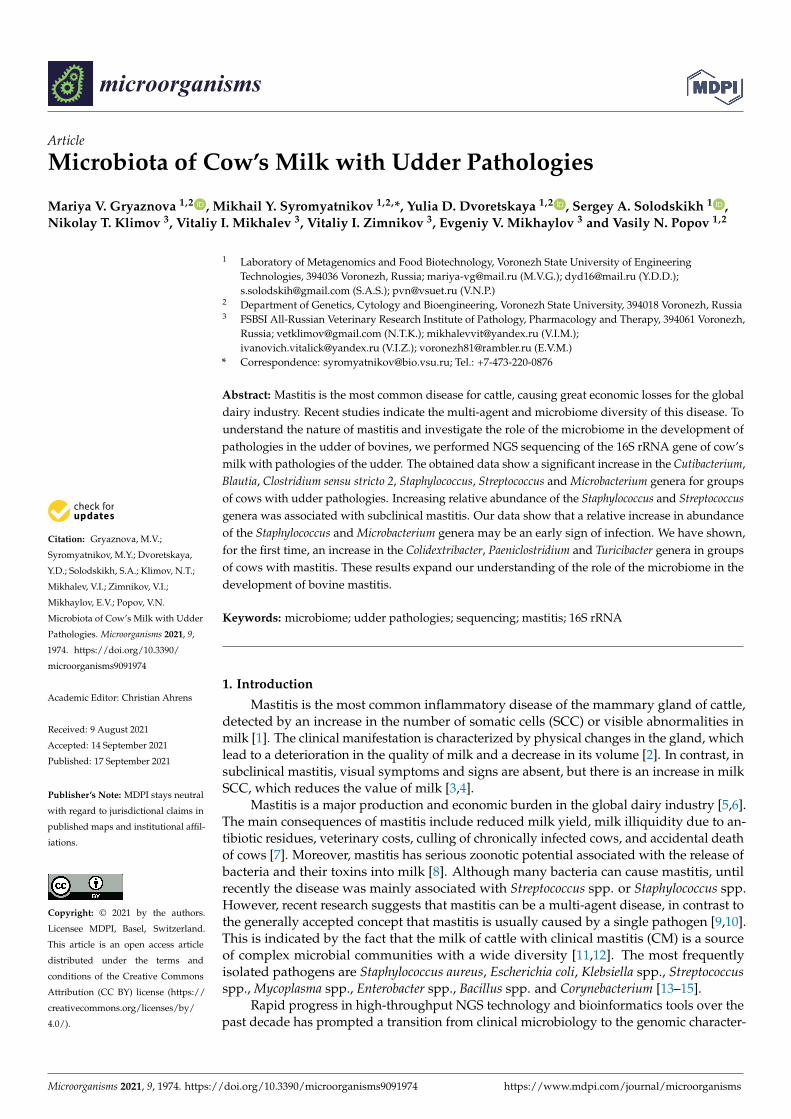
Abstract: Mastitis is the most common disease for cattle, causing great economic losses for the globaldairy industry. Recent studies indicate the multi-agent and microbiome diversity of this disease. Tounderstand the nature of mastitis and investigate the role of the microbiome in the development ofpathologies in the udder of bovines, we performed NGS sequencing of the 16S rRNA gene of cow’smilk with pathologies of the udder. The obtained data show a significant increase in the Cutibacterium,Blautia, Clostridium sensu stricto 2, Staphylococcus, Streptococcus and Microbacterium genera for groupsof cows with udder pathologies. Increasing relative abundance of the Staphylococcus and Streptococcusgenera was associated with subclinical mastitis. Our data show that a relative increase in abundanceof the Staphylococcus and Microbacterium genera may be an early sign of infection. We have shown,for the first time, an increase in the Colidextribacter, Paeniclostridium and Turicibacter genera in groupsof cows with mastitis. These results expand our understanding of the role of the microbiome in thedevelopment of bovine mastitis.
Keywords: microbiome; udder pathologies; sequencing; mastitis; 16S rRNA
1. Introduction
Mastitis is the most common inflammatory disease of the mammary gland of cattle,detected by an increase in the number of somatic cells (SCC) or visible abnormalities inmilk [1]. The clinical manifestation is characterized by physical changes in the gland, whichlead to a deterioration in the quality of milk and a decrease in its volume [2]. In contrast, insubclinical mastitis, visual symptoms and signs are absent, but there is an increase in milkSCC, which reduces the value of milk [3,4].
Mastitis is a major production and economic burden in the global dairy industry [5,6].The main consequences of mastitis include reduced milk yield, milk illiquidity due to an-tibiotic residues, veterinary costs, culling of chronically infected cows, and accidental deathof cows [7]. Moreover, mastitis has serious zoonotic potential associated with the release ofbacteria and their toxins into milk [8]. Although many bacteria can cause mastitis, untilrecently the disease was mainly associated with Streptococcus spp. or Staphylococcus spp.However, recent research suggests that mastitis can be a multi-agent disease, in contrast tothe generally accepted concept that mastitis is usually caused by a single pathogen [9,10].This is indicated by the fact that the milk of cattle with clinical mastitis (CM) is a sourceof complex microbial communities with a wide diversity [11,12]. The most frequentlyisolated pathogens are Staphylococcus aureus, Escherichia coli, Klebsiella spp., Streptococcusspp., Mycoplasma spp., Enterobacter spp., Bacillus spp. and Corynebacterium [13–15].
Rapid progress in high-throughput NGS technology and bioinformatics tools over thepast decade has prompted a transition from clinical microbiology to the genomic character-
Microorganisms 2021, 9, 1974. https://doi.org/10.3390/microorganisms9091974 https://www.mdpi.com/journal/microorganisms
Microorganisms 2021, 9, 1974 2 of 11
ization of the microbiome associated with infectious diseases, including mastitis [16,17].The method of selective sequencing of the 16S rRNA gene is the most commonly usedgenomic research tool in studying the microbiome of bovine mastitis, and it allows theseparation of more than 90% of isolates at the genus level [12].
The purpose of this study is to investigate the microbiome in milk samples, obtainedfrom bovines with subclinical and clinical mastitis, as well as bovines with udder irritation,by sequencing the 16S rRNA gene (hypervariable region V3) on the Ion Torrent PGMplatform. We also performed a comparative assessment of the microbial community amongmilk samples from different groups. This study can help to better understand the natureof mastitis and the role of the microbiome in this pathology, as well as contribute to thedevelopment of more effective prevention and treatment of bovine mastitis.
2. Materials and Methods2.1. Samples
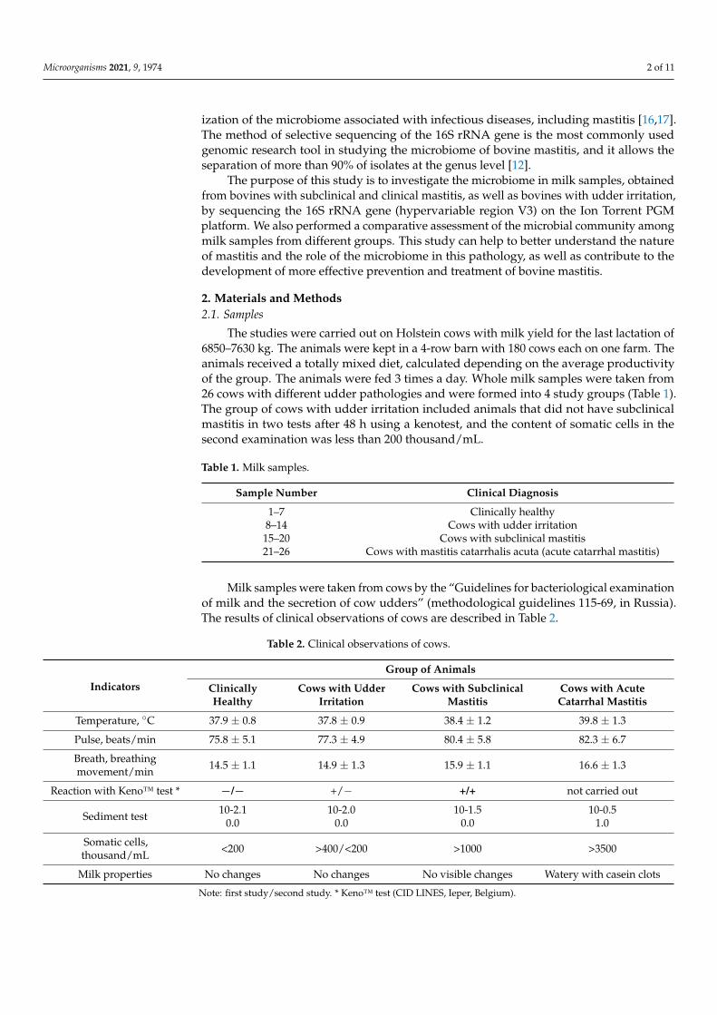
The studies were carried out on Holstein cows with milk yield for the last lactation of6850–7630 kg. The animals were kept in a 4-row barn with 180 cows each on one farm. Theanimals received a totally mixed diet, calculated depending on the average productivityof the group. The animals were fed 3 times a day. Whole milk samples were taken from26 cows with different udder pathologies and were formed into 4 study groups (Table 1).The group of cows with udder irritation included animals that did not have subclinicalmastitis in two tests after 48 h using a kenotest, and the content of somatic cells in thesecond examination was less than 200 thousand/mL.
Table 1. Milk samples.
Sample Number Clinical Diagnosis
1–7 Clinically healthy8–14 Cows with udder irritation
15–20 Cows with subclinical mastitis21–26 Cows with mastitis catarrhalis acuta (acute catarrhal mastitis)
Milk samples were taken from cows by the “Guidelines for bacteriological examinationof milk and the secretion of cow udders” (methodological guidelines 115-69, in Russia).The results of clinical observations of cows are described in Table 2.
Table 2. Clinical observations of cows.
Indicators
Group of Animals
ClinicallyHealthy
Cows with UdderIrritation
Cows with SubclinicalMastitis
Cows with AcuteCatarrhal Mastitis
Temperature, ◦C 37.9 ± 0.8 37.8 ± 0.9 38.4 ± 1.2 39.8 ± 1.3
Pulse, beats/min 75.8 ± 5.1 77.3 ± 4.9 80.4 ± 5.8 82.3 ± 6.7
Breath, breathingmovement/min 14.5 ± 1.1 14.9 ± 1.3 15.9 ± 1.1 16.6 ± 1.3
Reaction with Keno™ test * −/− +/− +/+ not carried out
Sediment test 10-2.10.0
10-2.00.0
10-1.50.0
10-0.51.0
Somatic cells,thousand/mL <200 >400/<200 >1000 >3500
Milk properties No changes No changes No visible changes Watery with casein clots
Note: first study/second study. * Keno™ test (CID LINES, Ieper, Belgium).

Microorganisms 2021, 9, 1974 3 of 11
Milk was taken from the udder quarters in compliance with the rules of asepsis; thus,the teats of the udder and the hands of the personnel were treated with a 70% alcoholsolution before taking a milk sample. Samples of 5–10 mL of alveolar milk were takenat the end of milking in sterile plastic tubes. During the sampling process, under nocircumstances did the udder nipple touch the edge of the tube, which was tightly closedwith sterile caps afterwards. The milk samples were immediately placed in liquid nitrogen.The samples were stored at a temperature of −80 ◦C.
After sampling, all cows with acute catarrhal mastitis were treated with antibioticswith a wide spectrum of action-injected intracisternal. Cows with subclinical mastitis weretreated by antibiotics if Staphylococcus aureus and/or Staphylococcus agalactiae were isolatedfrom the affected quarters of the udder. All animals after the last milking were injected withorbenin DC (Zoetis, Parsippany-Troy Hills, NJ, USA) in all lobes, regardless of whether theanimals had mastitis or not.
2.2. Isolation of DNA
DNA was isolated from milk samples using a commercial ZymoBIOMICS DNAMicroprep Kit (Zymo research, Irvine, CA, USA) according to the manufacturer’s protocol.
During the DNA extraction step, an extra sample containing Milli-Q water was addedas a negative control. This sample was subjected to all the same sample preparationsteps as the test samples from DNA extraction to library preparation and sequencing.Negative control was added to exclude the contamination of the study samples in thelaboratory. At the stage of bioinformatics analysis, we used the decontam R package toidentify and visualize contaminating DNA features, allowing them to be removed and amore accurate picture of observed communities to be constructed from marker-gene andmetagenomics data.
2.3. Amplification of the 16S rRNA Gene
We selected the variable region V3 of the 16S rRNA gene to study the microbiomeusing sequencing on the Ion Torrent PGM. Bacterial DNA was amplified with the universalforward 337F and reverse 518R primers [18,19].
The primer sequences were as follows:337F: 5′-GACTCCTACGGGAGGCWGCAG-3′;518R: 5′-GTATTACCGCGGCTGCTGG-3′.PCR was performed using a 5X ScreenMix-HS Master Mix (Evrogen, Moscow, Russia)
in the following mode: 94 ◦C for 4 min followed by 37 cycles of 94 ◦C for 30 s, 53 ◦C for30 s, and 72 ◦C for 30 s with the final elongation at 72 ◦C for 5 min.
2.4. High-Throughput Sequencing
PCR products were purified with AMPure XP magnetic beads (Beckman Coulter, Miami,FL, USA). Sequencing libraries were prepared using the NEBNext Fast DNA Library Prep kit(New England Biolabs, Ipswich, MA, USA) by following the manufacturer’s protocol.
The quality of the sequencing libraries was evaluated using qPCR and the LibraryQuantification Kit Ion Torrent Platforms (Kapa Biosystems, Wilmington, MA, USA). Atthis stage of the study, the library corresponding to sample number 2 was excluded due toits low concentration.
After that, the libraries were mixed in equimolar volumes for emulsion PCR usingthe OneTouch 2 system (Thermo Fisher Scientific, Waltham, MA, USA). Sequencing wasperformed on the IonTorrent PGM system using the Ion PGM Hi-Q View SequencingKit, Ion OneTouch 2 System, and Ion PGM Hi-Q View OT2 Kit (Thermo Fisher Scientific,Waltham, MA, USA).
2.5. Bioinformatic and Statistical Analysis
Sequencing results were obtained in BAM format and converted to FASTQ formatusing SAMtools v.1.2 software [20]. Demultiplexing was done with the fastq-multx applica-
Microorganisms 2021, 9, 1974 4 of 11
tion of the ea-utils v.1.3 program package. The reads were filtered according to the readingquality based on the expected number of errors using the maximum expected error cutoff of1.0 [21]. The samples were pooled, and unique sequences were identified before searchingfor the operational taxonomic units (OTUs). We searched for the OTUs using the UNOISE2algorithm, which reduces the noise through error correction [22]. We combined all reads ofthe samples for generating OTUs and making an OTU table. The most important reasonfor pooling is the fact that it enhances the signal of abundance for correct sequences. Whenthe samples are pooled, a sequence that appears as a singleton in one sample may alsoappear in another sample. Thus, it is retained and included in the OTU table. If singletonsare discarded after pooling (as usually recommended to reduce spurious OTUs), then morelow-abundance species will be retained compared with discarding singletons for eachsample separately.
Filtration of reads, identification of unique sequences, and clusterization of searchingfor the OTUs were performed using the VSEARCH v.2.8.2 software. Microbial species inthe samples were identified using the SILVA database v.123 (https://www.arb-silva.de,(accessed on 30 July 2021)).
To compare relative abundances between different experimental groups, we used thegeneralized linear modelling (GLM) method implemented in the DeSEQ2 R package [23].In brief, the final estimation of logarithmic fold changes for each OTU performed byDeSEQ2, which is based on the gene-wise dispersion of estimates comparison. The startingpoint of a DESeq2 analysis is a count matrix K with one row for each taxa i and one columnfor each sample j. The matrix entries Kij indicate the size of the OTU.
In our case, a comparison between four groups was performed. It produces the designmatrix where elements indicate whether a sample j belongs to the experimental group(clinically healthy cows or the group with subclinical mastitis) or not. p values for eachOTU were obtained using the Wald test [24].
3. Results
We estimated the total abundance of all identified taxa based on the number ofreads. Raw sequencing data are available in the NCBI BioProject database (BioProject ID:PRJNA736244). The most common phyla are Actinobacteriota, Firmicutes, Proteobacteria andBacteroidota. The values of the mean abundance of phyla for each group are presented inTable 3.
Table 3. Relative abundance of bacterial phyla in the studied groups.
Phyla Mean Abundance SD Group
Actinobacteriota 0.2852 ±0.1655
Clinically healthyBacteroidota 0.0482 ±0.0273Firmicutes 0.5538 ±0.1603
Proteobacteria 0.1115 ±0.0452Other 0.0002 ±0.0008
Actinobacteriota 0.1712 ±0.2073
Udder irritationBacteroidota 0.0446 ±0.0318Firmicutes 0.6790 ±0.3069
Proteobacteria 0.1497 ±0.1079Other 0.0006 ±0.0019
Actinobacteriota 0.5419 ±0.2424
Subclinical mastitisBacteroidota 0.0969 ±0.0755Firmicutes 0.6202 ±0.4266
Proteobacteria 0.1032 ±0.0427Other 0.0010 ±0.0022
Actinobacteriota 0.5144 ±0.1782
Acute catarrhal mastitisBacteroidota 0.0333 ±0.0125Firmicutes 0.3393 ±0.2169
Proteobacteria 0.1201 ±0.0541Other 0.0014 ±0.0028
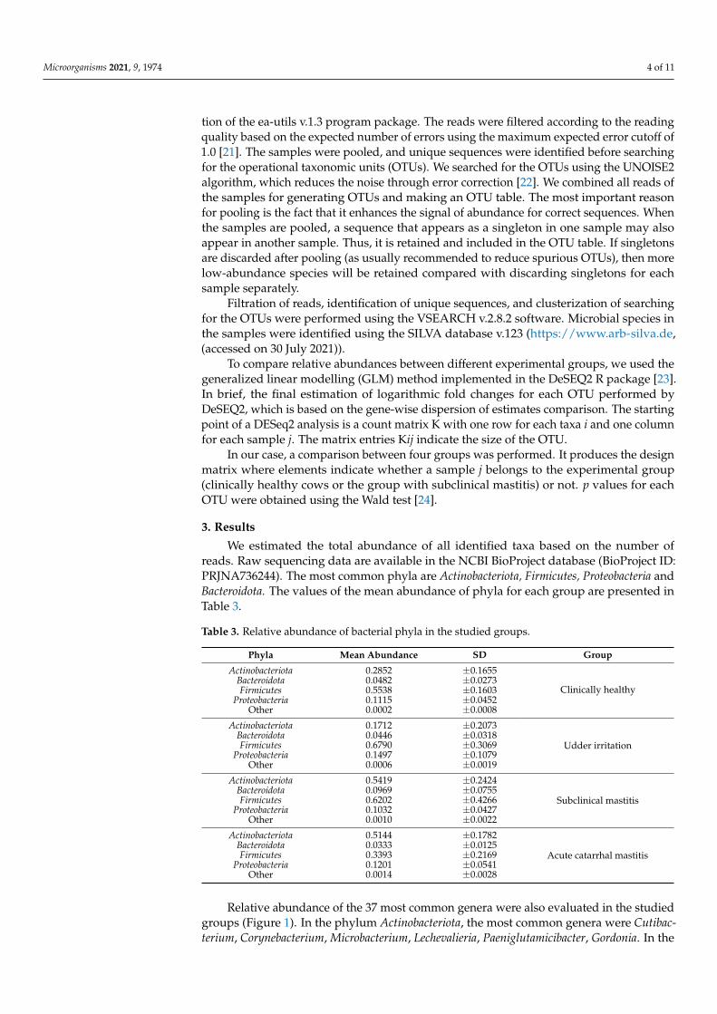
Relative abundance of the 37 most common genera were also evaluated in the studiedgroups (Figure 1). In the phylum Actinobacteriota, the most common genera were Cutibac-terium, Corynebacterium, Microbacterium, Lechevalieria, Paeniglutamicibacter, Gordonia. In the
Microorganisms 2021, 9, 1974 5 of 11
phylum, Firmicutes, the most common genera were Streptococcus, Staphylococcus, Lactococcus,Clostridium sensu stricto 2, Paeniclostridium, Blautia, Turicibacter. In the phylum Proteobacteriaand Bacteroidota, the most common genera were Methylobacterium, Afipia, Acinetobacter,Microvirga and Prevotella, and Prevotellaceae (Figure 1). Data showing the abundance ofmicroorganisms for each sample are presented in the Supplementary Materials (Table S1).
Microorganisms 2021, 9, x FOR PEER REVIEW 5 of 12
Firmicutes 0.6202 ±0.4266 Proteobacteria 0.1032 ±0.0427
Other 0.0010 ±0.0022 Actinobacteriota 0.5144 ±0.1782
Acute catarrhal mastitis Bacteroidota 0.0333 ±0.0125 Firmicutes 0.3393 ±0.2169
Proteobacteria 0.1201 ±0.0541 Other 0.0014 ±0.0028
Relative abundance of the 37 most common genera were also evaluated in the studied groups (Figure 1). In the phylum Actinobacteriota, the most common genera were Cutibacterium, Corynebacterium, Microbacterium, Lechevalieria, Paeniglutamicibacter, Gordo-nia. In the phylum, Firmicutes, the most common genera were Streptococcus, Staphylococ-cus, Lactococcus, Clostridium sensu stricto 2, Paeniclostridium, Blautia, Turicibacter. In the phylum Proteobacteria and Bacteroidota, the most common genera were Methylobacterium, Afipia, Acinetobacter, Microvirga and Prevotella, and Prevotellaceae (Figure 1). Data showing the abundance of microorganisms for each sample are presented in the Supplementary Materials (Table S1).
Figure 1. Taxonomic profile of the 37 most common bacterial genera in cow milk samples.
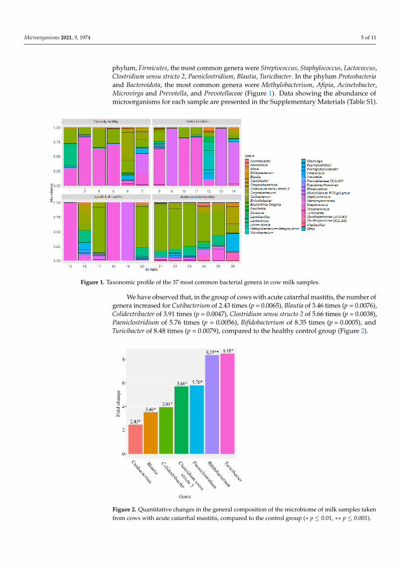
We have observed that, in the group of cows with acute catarrhal mastitis, the number of genera increased for Cutibacterium of 2.43 times (p = 0.0065), Blautia of 3.46 times (p = 0.0076), Colidextribacter of 3.91 times (p = 0.0047), Clostridium sensu structo 2 of 5.66 times (p = 0.0038), Paeniclostridium of 5.76 times (p = 0.0056), Bifidobacterium of 8.35 times (p = 0.0005), and Turicibacter of 8.48 times (p = 0.0079), compared to the healthy control group (Figure 2).
Figure 1. Taxonomic profile of the 37 most common bacterial genera in cow milk samples.
We have observed that, in the group of cows with acute catarrhal mastitis, the number ofgenera increased for Cutibacterium of 2.43 times (p = 0.0065), Blautia of 3.46 times (p = 0.0076),Colidextribacter of 3.91 times (p = 0.0047), Clostridium sensu structo 2 of 5.66 times (p = 0.0038),Paeniclostridium of 5.76 times (p = 0.0056), Bifidobacterium of 8.35 times (p = 0.0005), andTuricibacter of 8.48 times (p = 0.0079), compared to the healthy control group (Figure 2).
Microorganisms 2021, 9, x FOR PEER REVIEW 6 of 12
Figure 2. Quantitative changes in the general composition of the microbiome of milk samples taken from cows with acute catarrhal mastitis, compared to the control group (∗ p ≤ 0.01, ∗∗ p ≤ 0.001).
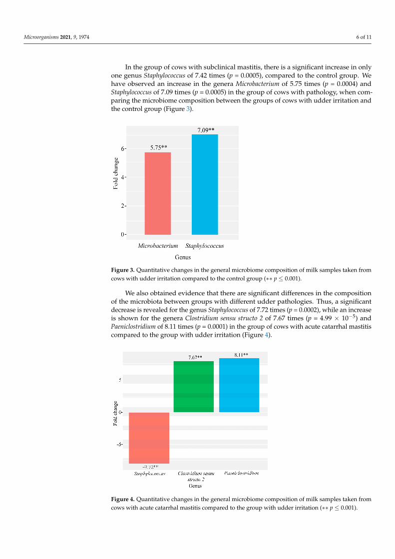
In the group of cows with subclinical mastitis, there is a significant increase in only one genus Staphylococcus of 7.42 times (p = 0.0005), compared to the control group. We have observed an increase in the genera Microbacterium of 5.75 times (p = 0.0004) and Staphylococcus of 7.09 times (p = 0.0005) in the group of cows with pathology, when comparing the microbiome composition between the groups of cows with udder irrita-tion and the control group (Figure 3).
Figure 3. Quantitative changes in the general microbiome composition of milk samples taken from cows with udder irritation compared to the control group (∗∗ p ≤ 0.001).
Figure 2. Quantitative changes in the general composition of the microbiome of milk samples takenfrom cows with acute catarrhal mastitis, compared to the control group (∗ p ≤ 0.01, ∗∗ p ≤ 0.001).
Microorganisms 2021, 9, 1974 6 of 11
In the group of cows with subclinical mastitis, there is a significant increase in onlyone genus Staphylococcus of 7.42 times (p = 0.0005), compared to the control group. Wehave observed an increase in the genera Microbacterium of 5.75 times (p = 0.0004) andStaphylococcus of 7.09 times (p = 0.0005) in the group of cows with pathology, when com-paring the microbiome composition between the groups of cows with udder irritation andthe control group (Figure 3).
Microorganisms 2021, 9, x FOR PEER REVIEW 6 of 12
Figure 2. Quantitative changes in the general composition of the microbiome of milk samples taken from cows with acute catarrhal mastitis, compared to the control group (∗ p ≤ 0.01, ∗∗ p ≤ 0.001).
In the group of cows with subclinical mastitis, there is a significant increase in only one genus Staphylococcus of 7.42 times (p = 0.0005), compared to the control group. We have observed an increase in the genera Microbacterium of 5.75 times (p = 0.0004) and Staphylococcus of 7.09 times (p = 0.0005) in the group of cows with pathology, when comparing the microbiome composition between the groups of cows with udder irrita-tion and the control group (Figure 3).
Figure 3. Quantitative changes in the general microbiome composition of milk samples taken from cows with udder irritation compared to the control group (∗∗ p ≤ 0.001). Figure 3. Quantitative changes in the general microbiome composition of milk samples taken fromcows with udder irritation compared to the control group (∗∗ p ≤ 0.001).
We also obtained evidence that there are significant differences in the compositionof the microbiota between groups with different udder pathologies. Thus, a significantdecrease is revealed for the genus Staphylococcus of 7.72 times (p = 0.0002), while an increaseis shown for the genera Clostridium sensu structo 2 of 7.67 times (p = 4.99 × 10⁻⁵) andPaeniclostridium of 8.11 times (p = 0.0001) in the group of cows with acute catarrhal mastitiscompared to the group with udder irritation (Figure 4).
Microorganisms 2021, 9, x FOR PEER REVIEW 7 of 12
We also obtained evidence that there are significant differences in the composition of the microbiota between groups with different udder pathologies. Thus, a significant decrease is revealed for the genus Staphylococcus of 7.72 times (p = 0.0002), while an in-crease is shown for the genera Clostridium sensu structo 2 of 7.67 times (p = 4.99×10⁻⁵) and Paeniclostridium of 8.11 times (p = 0.0001) in the group of cows with acute catarrhal mas-titis compared to the group with udder irritation (Figure 4).
Figure 4. Quantitative changes in the general microbiome composition of milk samples taken from cows with acute catarrhal mastitis compared to the group with udder irritation (∗∗ p ≤ 0.001).
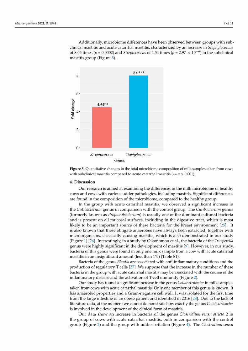
Additionally, microbiome differences have been observed between groups with subclinical mastitis and acute catarrhal mastitis, characterized by an increase in Staphy-lococcus of 8.05 times (p = 0.0002) and Streptococcus of 4.54 times (p = 2.97×10⁻⁶) in the sub-clinical mastitis group (Figure 5).
Figure 4. Quantitative changes in the general microbiome composition of milk samples taken fromcows with acute catarrhal mastitis compared to the group with udder irritation (∗∗ p ≤ 0.001).
Microorganisms 2021, 9, 1974 7 of 11
Additionally, microbiome differences have been observed between groups with sub-clinical mastitis and acute catarrhal mastitis, characterized by an increase in Staphylococcusof 8.05 times (p = 0.0002) and Streptococcus of 4.54 times (p = 2.97 × 10⁻⁶) in the subclinicalmastitis group (Figure 5).
Microorganisms 2021, 9, x FOR PEER REVIEW 7 of 12
We also obtained evidence that there are significant differences in the composition of the microbiota between groups with different udder pathologies. Thus, a significant decrease is revealed for the genus Staphylococcus of 7.72 times (p = 0.0002), while an in-crease is shown for the genera Clostridium sensu structo 2 of 7.67 times (p = 4.99×10⁻⁵) and Paeniclostridium of 8.11 times (p = 0.0001) in the group of cows with acute catarrhal mas-titis compared to the group with udder irritation (Figure 4).
Figure 4. Quantitative changes in the general microbiome composition of milk samples taken from cows with acute catarrhal mastitis compared to the group with udder irritation (∗∗ p ≤ 0.001).
Additionally, microbiome differences have been observed between groups with subclinical mastitis and acute catarrhal mastitis, characterized by an increase in Staphy-lococcus of 8.05 times (p = 0.0002) and Streptococcus of 4.54 times (p = 2.97×10⁻⁶) in the sub-clinical mastitis group (Figure 5).
Figure 5. Quantitative changes in the total microbiome composition of milk samples taken from cowswith subclinical mastitis compared to acute catarrhal mastitis (∗∗ p ≤ 0.001).
4. Discussion
Our research is aimed at examining the differences in the milk microbiome of healthycows and cows with various udder pathologies, including mastitis. Significant differencesare found in the composition of the microbiome, compared to the healthy group.
In the group with acute catarrhal mastitis, we observed a significant increase inthe Cutibacterium genus in comparison with the control group. The Cutibacterium genus(formerly known as Propionibacterium) is usually one of the dominant cultured bacteriaand is present on all mucosal surfaces, including in the digestive tract, which is mostlikely to be an important source of these bacteria for the breast environment [25]. Itis also known that these obligate anaerobes have always been extracted, together withmicroorganisms, classically causing mastitis, which is also demonstrated in our study(Figure 1) [26]. Interestingly, in a study by Oikonomou et al., the bacteria of the Trueperellagenus were highly significant in the development of mastitis [9]. However, in our study,bacteria of this genus were found in only one milk sample from a cow with acute catarrhalmastitis in an insignificant amount (less than 1%) (Table S1).
Bacteria of the genus Blautia are associated with anti-inflammatory conditions and theproduction of regulatory T cells [27]. We suppose that the increase in the number of thesebacteria in the group with acute catarrhal mastitis may be associated with the course of theinflammatory disease and the activation of T-cell immunity (Figure 2).
Our study has found a significant increase in the genus Colidextribacter in milk samplestaken from cows with acute catarrhal mastitis. Only one member of this genus is known. Ithas anaerobic properties and a Gram-negative cell wall. It was isolated for the first timefrom the large intestine of an obese patient and identified in 2016 [28]. Due to the lack ofliterature data, at the moment we cannot demonstrate how exactly the genus Colidextribacteris involved in the development of the clinical form of mastitis.
Our data show an increase in bacteria of the genus Clostridium sensu stricto 2 inthe group of cows with acute catarrhal mastitis, both in comparison with the controlgroup (Figure 2) and the group with udder irritation (Figure 4). The Clostridium sensu
Microorganisms 2021, 9, 1974 8 of 11
stricto 2 genus (also known as Hathewaya) is known to include proteolytic and pathogenicanaerobic organisms. Previously, bacteria of this genus have been isolated from soil, thelower digestive tract of some animals, infected cattle, water buffaloes, human faeces,human clinical specimens, including blood, peritoneal fluid, pleural fluid, and lung biopsyfor pulmonary infections [29]. In addition, it is known that members of this genus areresponsible for causing gas gangrene. They are also found as a part of mixed infections,moreover they are associated with the development of metritis [30]. Thus, the data obtainedmay indicate that this bacterial genus has high pathogenicity and plays a significant role inthe development of acute catarrhal mastitis in cattle.
In our study we have identified, an increase in the genus Paeniclostridium in thegroup of cows with acute catarrhal mastitis compared to the group with udder irritation(Figure 4) and the healthy group (Figure 2). It is known for certain that representatives ofthe genus Paeniclostridium are widespread in nature and have a variety of physiologicalcharacteristics. Some species of this genus of bacteria are often found in wounds, moreoften in association with other anaerobic and aerobic microorganisms. The virulenceof these bacteria for animals is due to β-toxin, which has high biological activity andspecificity. These microorganisms are often isolated in diseases, the symptoms of which arecharacteristic of other clostridiosis, for example, sudden death in sheep, acute abomazitisin cows and lambs, hemorrhagic enteritis, gangrenous lesions of the reproductive tractof newborn cows [31]. Thus, based on previous studies, as well as on our data, wesuppose that representatives of the genus Paeniclostridium can be related to the clinical formof mastitis.
Members of the genus Bifidobacterium are probiotic microorganisms that have a ben-eficial effect on the host organism. Taking into consideration this fact, we have assumedthat this genus would be reduced in groups of cows with udder pathologies. The Bifidobac-terium genus is one of the markers of “healthy microbiota” and is usually reduced in cowswith udder pathology [12,32]. However, we have obtained the opposite data, accordingto which the genus Bifidobacterium has significantly increased in the group of cows withacute catarrhal mastitis, compared to the healthy group (Figure 2). We found a study inwhich the microbiota of milk and faecal samples from cows with mastitis was compared.The researchers have found out that the nature of changes in the microbial community offaeces in cows with mastitis was similar to that in milk, characterized by a general increasein the number of mastitis pathogens and a decrease in Lactobacillus and their members(L. salivarius, L. sakei, L. ruminis, L. delbrueckii, L. buchneri and L. acidophilus). At the sametime, the genus Bifidobacterium did not correlate with the microbial community of milkand SCC in cows with mastitis [33]. Thus, literature data and our findings suggest thatthe biological significance of the genus Bifidobacterium in connection with bovine mastitisrequires a thorough analysis.
According to our data, the Turicibacter genus increased 8.48 times in samples of cowswith acute catarrhal mastitis compared to the healthy group. Recent studies based on theanalysis of the 16S rRNA gene and ribosomal intergenic spacers indicate the presence ofTuricibacter bacteria in the rumen and faeces of cattle [34]. It is also reported that Turicibacteris present in the intestines of pigs, rats and insects, as well as dairy wastewater and wholemilk [35]. Since only one species has been isolated (Turicibacter sanguinis), the physiologicaldiversity of this genus is unknown [36]. However, since the isolated strain is a suspectedpathogen, there is a possibility that Turicibacter bacteria, which is present in farm animals,can cause infections or some other deleterious effect on the gastrointestinal tract, which isconsistent with our study.
The Staphylococcus and Streptococcus genera are major contributors to the developmentof mastitis in cattle [37,38]. One of the most important etiological agents of bovine mastitis isStaphylococcus aureus, which is mainly associated with subclinical infection that is persistentand can easily recur [39,40]. Our data also demonstrate an increase in bacteria of the genusStaphylococcus in the group with subclinical mastitis, both in comparison with the healthygroup and the group with acute catarrhal mastitis (Figure 4). Our data confirm the leading
Microorganisms 2021, 9, 1974 9 of 11
role of this genus in the development of the subclinical form of the disease. In addition, wehave also found an increase in Staphylococcus in the group of cows with udder irritation(Figure 3). The data obtained may indicate that the colonization of Staphylococcus occursin the outer region of the teat duct and firstly leads to irritation of the udder, which laterturns into an inflammatory process, the result of which is serious damage to the epithelialcell of the mammary gland and a subclinical form of mastitis developing [41].
In addition to the increase in bacteria of the genus Staphylococcus, which are the triggermechanism for the development of udder irritation, which subsequently turns into asubclinical form of mastitis, the significant increase in bacteria of the Microbacterium genusis also observed in the group of cows with udder irritation (Figure 3). Bacteria of the genusMicrobacterium are members of the Corynebacterium, which are often isolated from milktaken from infected mammary glands of dairy cows, in addition, their increased numberis associated with a decrease in milk yield [42]. However, little is known so far about theepidemiology of the genus Microbacterium. We consider that a significant increase in thegenus Microbacterium in the group of cows with udder irritation may be caused by infectionof the cow’s mammary gland.
5. Conclusions
Our data show that the milk microbiome of cows with udder pathologies differssignificantly from the milk microbiome of healthy cows. The role of bacteria, such asCutibacterium, Blautia, Clostridium sensu stricto 2, Staphylococcus, Streptococcus and Microbac-terium in groups with various udder pathologies and their association with the developmentof inflammation and mastitis has been identified. In addition, our study has revealed thatthe Staphylococcus and Streptococcus genera are associated with subclinical mastitis. Wealso suggest that an increase in the genera of Staphylococcus and Microbacterium may beearly indicators of infection, which firstly leads to irritation of the udder, and then to aninflammatory process that turns into subclinical mastitis.
Moreover, we would like to note that for the genus Colidextribacter, Paeniclostridiumand Turicibacter our study has shown the association with cattle udder diseases for the firsttime. Additionally, we attained results for the Bifidobacterium genus. Our data show anincrease in the Bifidobacterium genus in the group of cows with acute catarrhal mastitis.
These results expand our understanding of the role of the microbiome in the develop-ment of pathologies of the udder of cattle and in the future may help to create methods formastitis prevention and treatment. Molecular methods may be developed for the rapid andearly identification of the bacteria, which are associated with udder irritation and mastitis,according to our research.
Supplementary Materials: The following are available online at https://www.mdpi.com/article/10.3390/microorganisms9091974/s1, Table S1: The abundance of microorganisms for the samples.
Author Contributions: Conceptualization, M.Y.S., N.T.K. and V.N.P.; methodology, M.Y.S., E.V.M.and V.I.Z.; software, S.A.S., Y.D.D. and M.V.G.; validation, M.G, M.Y.S. and Y.D.D.; formal anal-ysis, S.A.S.; investigation, M.V.G., V.I.M. and Y.D.D.; resources, N.T.K. and V.N.P.; data curation,S.A.S.; writing—original draft preparation, M.V.G. and M.Y.S.; writing—review and editing, M.Y.S.and V.N.P.; visualization, M.V.G., Y.D.D. and S.A.S.; supervision, M.Y.S.; project administration,V.N.P.; funding acquisition, V.N.P. All authors have read and agreed to the published version ofthe manuscript.
Funding: This work was supported by Ministry of Science and Higher Education of the RussianFederation in the framework of the national project “Science” (project FZGW-2020-0001, uniquenumber of the register of State tasks 075001X39782002).
Institutional Review Board Statement: Not applicable.
Informed Consent Statement: Not applicable.
Data Availability Statement: Sequencing data are available in NCBI BioProject database (BioProjectID: PRJNA736244).

Microorganisms 2021, 9, 1974 10 of 11
Conflicts of Interest: The authors declare no conflict of interest.
References1. Metzger, S.A.; Hernandez, L.L.; Suen, G.; Ruegg, P.L. Understanding the Milk Microbiota. Vet. Clin. N. Am.-Food Anim. Pract.
2018, 34, 427–438. [CrossRef]2. Dubuc, J.; Duffield, T.F.; Leslie, K.E.; Walton, J.S.; LeBlanc, S.J. Risk factors for postpartum uterine diseases in dairy cows. J. Dairy
Sci. 2010, 93, 5764–5771. [CrossRef]3. Duffield, T.F.; Lissemore, K.D.; McBride, B.W.; Leslie, K.E. Impact of hyperketonemia in early lactation dairy cows on health and
production. J. Dairy Sci. 2009, 92, 571–580. [CrossRef]4. De Vliegher, S.; Fox, L.K.; Piepers, S.; McDougall, S.; Barkema, H.W. Invited review: Mastitis in dairy heifers: Nature of the
disease, potential impact, prevention, and control. J. Dairy Sci. 2012, 95, 1025–1040. [CrossRef]5. Reyes-Jara, A.; Cordero, N.; Aguirre, J.; Troncoso, M.; Figueroa, G. Antibacterial effect of copper on microorganisms isolated from
bovine mastitis. Front. Microbiol. 2016, 7, 626. [CrossRef]6. Hoque, M.N.; Istiaq, A.; Clement, R.A.; Sultana, M.; Crandall, K.A.; Siddiki, A.Z.; Hossain, M.A. Metagenomic deep sequencing
reveals association of microbiome signature with functional biases in bovine mastitis. Sci. Rep. 2019, 9, 13536. [CrossRef][PubMed]
7. Seegers, H.; Fourichon, C.; Beaudeau, F. Production effects related to mastitis and mastitis economics in dairy cattle herds. Vet.Res. 2003, 34, 475–491. [CrossRef]
8. González, R.N.; Wilson, D.J. Mycoplasmal mastitis in dairy herds. Vet. Clin. N. Am.-Food Anim. Pract. 2003, 19, 199–221. [CrossRef]9. Oikonomou, G.; Machado, V.S.; Santisteban, C.; Schukken, Y.H.; Bicalho, R.C. Microbial Diversity of Bovine Mastitic Milk as
Described by Pyrosequencing of Metagenomic 16s rDNA. PLoS ONE 2012, 7, e47671. [CrossRef]10. Bhatt, V.D.; Ahir, V.B.; Koringa, P.G.; Jakhesara, S.J.; Rank, D.N.; Nauriyal, D.S.; Kunjadia, A.P.; Joshi, C.G. Milk microbiome
signatures of subclinical mastitis-affected cattle analysed by shotgun sequencing. J. Appl. Microbiol. 2012, 112, 639–650. [CrossRef][PubMed]
11. Oikonomou, G.; Bicalho, M.L.; Meira, E.; Rossi, R.E.; Foditsch, C.; Machado, V.S.; Teixeira, A.G.V.; Santisteban, C.; Schukken, Y.H.;Bicalho, R.C. Microbiota of cow’s milk; distinguishing healthy, sub-clinically and clinically diseased quarters. PLoS ONE 2014, 9,e85904. [CrossRef]
12. Falentin, H.; Rault, L.; Nicolas, A.; Bouchard, D.S.; Lassalas, J.; Lamberton, P.; Aubry, J.M.; Marnet, P.G.; Le Loir, Y.; Even, S.Bovine teat microbiome analysis revealed reduced alpha diversity and significant changes in taxonomic profiles in quarters witha history of mastitis. Front. Microbiol. 2016, 7, 480. [CrossRef]
13. Abebe, R.; Hatiya, H.; Abera, M.; Megersa, B.; Asmare, K. Bovine mastitis: Prevalence, risk factors and isolation of Staphylococcusaureus in dairy herds at Hawassa milk shed, South Ethiopia. BMC Vet. Res. 2016, 12, 1–11. [CrossRef] [PubMed]
14. Gao, J.; Barkema, H.W.; Zhang, L.; Liu, G.; Deng, Z.; Cai, L.; Shan, R.; Zhang, S.; Zou, J.; Kastelic, J.P.; et al. Incidence of clinicalmastitis and distribution of pathogens on large Chinese dairy farms. J. Dairy Sci. 2017, 100, 4797–4806. [CrossRef] [PubMed]
15. Hoque, M.N.; Das, Z.C.; Rahman, A.N.M.A.; Haider, M.G.; Islam, M.A. Molecular characterization of Staphylococcus aureusstrains in bovine mastitis milk in Bangladesh. Int. J. Vet. Sci. Med. 2018, 6, 53–60. [CrossRef] [PubMed]
16. D’Argenio, V.; Casaburi, G.; Precone, V.; Salvatore, F. Comparative metagenomic analysis of human gut microbiome compositionusing two different bioinformatic pipelines. Biomed Res. Int. 2014, 325340. [CrossRef] [PubMed]
17. Cremonesi, P.; Ceccarani, C.; Curone, G.; Severgnini, M.; Pollera, C.; Bronzo, V.; Riva, F.; Addis, M.F.; Filipe, J.; Amadori, M.; et al.Milk microbiome diversity and bacterial group prevalence in a comparison between healthy holstein friesian and rendena cows.PLoS ONE 2018, 13, e0205054. [CrossRef]
18. Muyzer, G.; De Waal, E.C.; Uitterlinden, A.G. Profiling of complex microbial populations by denaturing gradient gel elec-trophoresis analysis of polymerase chain reaction-amplified genes coding for 16S rRNA. Appl. Environ. Microbiol. 1993, 59, 695.[CrossRef]
19. 16S rRNA and 16S rRNA Gene–EzBioCloud Help Center. Available online: https://help.ezbiocloud.net/16s-rrna-and-16s-rrna-gene/ (accessed on 12 July 2021).
20. Li, H.; Handsaker, B.; Wysoker, A.; Fennell, T.; Ruan, J.; Homer, N.; Marth, G.; Abecasis, G.; Durbin, R. The SequenceAlignment/Map format and SAMtools. Bioinformatics 2009, 25, 2078–2079. [CrossRef]
21. Edgar, R.C.; Flyvbjerg, H. Error filtering, pair assembly and error correction for next-generation sequencing reads. Bioinformatics2015, 31, 3476–3482. [CrossRef]
22. Edgar, R.C. Search and clustering orders of magnitude faster than BLAST. Bioinformatics 2010, 26, 2460–2461. [CrossRef]23. Love, M.I.; Huber, W.; Anders, S. Moderated estimation of fold change and dispersion for RNA-seq data with DESeq2. Genome
Biol. 2014, 15, 1–21. [CrossRef]24. Gryaznova, M.V.; Solodskikh, S.A.; Panevina, A.V.; Syromyatnikov, M.Y.; Dvoretskaya, Y.D.; Sviridova, T.N.; Popov, E.S.; Popov,
V.N. Study of microbiome changes in patients with ulcerative colitis in the Central European part of Russia. Heliyon 2021, 7,e06432. [CrossRef]
25. Fernández, L.; Pannaraj, P.S.; Rautava, S.; Rodríguez, J.M. The Microbiota of the Human Mammary Ecosystem. Front. Cell. Infect.Microbiol. 2020, 10, 689. [CrossRef]
Microorganisms 2021, 9, 1974 11 of 11
26. Du Preez, J.H.; Greeff, A.S.; Eksteen, N. Isolation and significance of anaerobic bacteria isolated from cases of bovine mastitis.Onderstepoort J. Vet. Res. 1981, 48, 123–126. [PubMed]
27. Saxena, R.; Sharma, V.K. Chapter 9—A Metagenomic Insight Into the Human Microbiome: Its Implications in Health and Disease.In Medical and Health Genomics; Academic Press: Amsterdam, The Netherlands, 2016; pp. 107–119. [CrossRef]
28. Ricaboni, D.; Mailhe, M.; Cadoret, F.; Vitton, V.; Fournier, P.E.; Raoult, D. ‘Colidextribacter massiliensis’ gen. nov., sp. nov.,isolated from human right colon. New Microbes New Infect. 2017, 17, 27–29. [CrossRef] [PubMed]
29. Lawson, P.A.; Rainey, F.A. Proposal to restrict the genus Clostridium prazmowski to Clostridium butyricum and related species.Int. J. Syst. Evol. Microbiol. 2016, 66, 1009–1016. [CrossRef]
30. Biström, M.; Moisander-Jylhä, A.M.; Heinikainen, S.; Pelkola, K.; Raunio-Saarnisto, M. Isolation of Clostridium limosum from anoutbreak of metritis in farmed mink. Acta Vet. Scand. 2016, 58, 1–4. [CrossRef]
31. Vidor, C.; Awad, M.; Lyras, D. Antibiotic resistance, virulence factors and genetics of Clostridium sordellii. Res. Microbiol. 2015,166, 368–374. [CrossRef]
32. Rudenko, P.; Sachivkina, N.; Vatnikov, Y.; Shabunin, S.; Engashev, S.; Kontsevaya, S.; Karamyan, A.; Bokov, D.; Kuznetsova, O.;Vasilieva, E. Role of microorganisms isolated from cows with mastitis in Moscow region in biofilm formation. Int. J. One Health2021, 14, 40–48. [CrossRef]
33. Ma, C.; Zhao, J.; Xi, X.; Ding, J.; Wang, H.; Zhang, H.; Kwok, L.Y. Bovine mastitis may be associated with the deprivation of gutLactobacillus. Benef. Microbes 2016, 7, 95–102. [CrossRef]
34. Mao, S.Y.; Zhang, R.Y.; Wang, D.S.; Zhu, W.Y. Impact of subacute ruminal acidosis (SARA) adaptation on rumen microbiota indairy cattle using pyrosequencing. Anaerobe 2013, 24, 12–19. [CrossRef]
35. Gagnon, N.; Talbot, G.; Ward, P.; Roy, D.; Dupuis, M.; Farnworth, E.; Tompkins, T.A.; Lessard, M. Evaluation of bacterial diversityin the gut of piglets supplemented with probiotics using ribosomal intergenic spacer analysis. Can. J. Anim. Sci. 2007, 87, 207–219.[CrossRef]
36. Bosshard, P.P.; Zbinden, R.; Altwegg, M. Turicibacter sanguinis gen. nov., sp. nov., a novel anaerobic, Gram-positive bacterium.Int. J. Syst. Evol. Microbiol. 2002, 52, 1263–1266. [CrossRef]
37. McDonald, J.S. Streptococcal and staphylococcal mastitis. Vet. Clin. N. Am. Large Anim. Pract. 1984, 6, 269–285. [CrossRef]38. Pang, M.; Xie, X.; Bao, H.; Sun, L.; He, T.; Zhao, H.; Zhou, Y.; Zhang, L.; Zhang, H.; Wei, R.; et al. Insights into the bovine milk
microbiota in dairy farms with different incidence rates of subclinical mastitis. Front. Microbiol. 2018, 9, 2379. [CrossRef]39. Rossi, B.F.; Bonsaglia, E.C.R.; Castilho, I.G.; Dantas, S.T.A.; Salina, A.; Langoni, H.; Pantoja, J.C.F.; Budri, P.E.; Fitzgerald-Hughes,
D.; Júnior, A.F.; et al. Genotyping of long term persistent Staphylococcus aureus in bovine subclinical mastitis. Microb. Pathog.2019, 132, 45–50. [CrossRef] [PubMed]
40. Hoekstra, J.; Zomer, A.L.; Rutten, V.P.; Benedictus, L.; Stegeman, A.; Spaninks, M.P.; Bennedsgaard, T.W.; Biggs, A.; De Vliegher,S.; Mateo, D.H.; et al. Genomic analysis of European bovine Staphylococcus aureus from clinical versus subclinical mastitis. Sci.Rep. 2020, 10, 18172. [CrossRef] [PubMed]
41. Younis, A.; Krifucks, O.; Heller, E.D.; Samra, Z.; Glickman, A.; Saran, A.; Leitner, G. Staphylococcus aureus exosecretions andbovine mastitis. J. Vet. Med. Ser. B 2003, 50, 1–7. [CrossRef]
42. Watts, J.L.; Lowery, D.E.; Teel, J.F.; Rossbach, S. Identification of Corynebacterium bovis and other Coryneforms isolated frombovine mammary glands. J. Dairy Sci. 2000, 83, 2373–2379. [CrossRef]