Metabolomic Profiling of Brain Protective Effect of Edaravone ...
14
Metabolomic Profiling of Brain Protective Effect of Edaravone on Cerebral Ischemia-Reperfusion Injury in Mice Hui-fen Ma † , Fan Zheng † , Lin-jie Su, Da-wei Zhang, Yi-ning Liu, Fang Li, Yuan-yuan Zhang, Shuai-shuai Gong * and Jun-ping Kou * State Key Laboratory of Natural Medicines, Jiangsu Key Laboratory of TCM Evaluation and Translational Research, Department of Pharmacology of Chinese Materia Medica, School of Traditional Pharmacy, China Pharmaceutical University, Nanjing, China Edaravone (EDA) injection has been extensively applied in clinics for treating stroke. Nevertheless, the metabolite signatures and underlying mechanisms associated with EDA remain unclear, which deserve further elucidation for improving the accurate usage of EDA. Ischemia stroke was simulated by intraluminal occlusion of the right middle cerebral artery for 1 h, followed by reperfusion for 24 h in mice. Brain infarct size, neurological deficits, and lactate dehydrogenase (LDH) levels were improved by EDA. Significantly differential metabolites were screened with untargeted metabolomics by cross-comparisons with pre- and posttreatment of EDA under cerebral ischemia/ reperfusion (I/R) injury. The possibly involved pathways, such as valine, leucine, and isoleucine biosynthesis, and phenylalanine, taurine, and hypotaurine metabolisms, were enriched with differential metabolites and relevant regulatory enzymes, respectively. The network of differential metabolites was constructed for the integral exhibition of metabolic characteristics. Targeted analysis of taurine, an important metabolic marker, was performed for further validation. The level of taurine decreased in the MCAO/R group and increased in the EDA group. The inhibition of EDA on cerebral endothelial cell apoptosis was confirmed by TdT-mediated dUTP nick-end labeling (TUNEL) stain. Cysteine sulfinic acid decarboxylase (CSAD), the rate-limiting enzyme of taurine generation, significantly increased along with inhibiting endothelial cell apoptosis after treatment of EDA. Thus, CSAD, as the possible new therapeutic target of EDA, was selected and validated by Western blot and immunofluorescence. Together, this study provided the metabolite signatures and identified CSAD as an unrecognized therapeutic intervention for EDA in the treatment of ischemic stroke via inhibiting brain endothelial cell apoptosis. Keywords: metabolomic, edaravone, ischemia stroke, taurine, endothelial cells Edited by: Li-Nan Zhang, Hebei Medical University, China Reviewed by: Achuthan Raghavamenon, Amala Cancer Research Centre, India Guoxiang Xie, University of Hawaii Cancer Center, United States *Correspondence: Shuai-shuai Gong [email protected] Jun-ping Kou [email protected] † These authors have contributed equally to this work Specialty section: This article was submitted to Neuropharmacology, a section of the journal Frontiers in Pharmacology Received: 14 November 2021 Accepted: 14 January 2022 Published: 14 February 2022 Citation: Ma H-f, Zheng F, Su L-j, Zhang D-w, Liu Y-n, Li F, Zhang Y-y, Gong S-s and Kou J-p (2022) Metabolomic Profiling of Brain Protective Effect of Edaravone on Cerebral Ischemia-Reperfusion Injury in Mice. Front. Pharmacol. 13:814942. doi: 10.3389/fphar.2022.814942 Abbreviations: CSAD, cysteine sulfinic acid decarboxylase; EDA, edaravone; LDH, lactate dehydrogenase; MCAO/R, middle cerebral artery occlusion/reperfusion; TUNEL, TdT-mediated dUTP nick-end labeling; H&E, hematoxylin and eosin; EC, endothelial cell; I/R, ischemia/reperfusion. Frontiers in Pharmacology | www.frontiersin.org February 2022 | Volume 13 | Article 814942 1 ORIGINAL RESEARCH published: 14 February 2022 doi: 10.3389/fphar.2022.814942
-
Upload
khangminh22 -
Category
Documents
-
view
3 -
download
0
Transcript of Metabolomic Profiling of Brain Protective Effect of Edaravone ...

Metabolomic Profiling of BrainProtective Effect of Edaravone onCerebral Ischemia-Reperfusion Injuryin MiceHui-fen Ma†, Fan Zheng†, Lin-jie Su, Da-wei Zhang, Yi-ning Liu, Fang Li, Yuan-yuan Zhang,Shuai-shuai Gong* and Jun-ping Kou*
State Key Laboratory of Natural Medicines, Jiangsu Key Laboratory of TCMEvaluation and Translational Research, Department ofPharmacology of Chinese Materia Medica, School of Traditional Pharmacy, China Pharmaceutical University, Nanjing, China
Edaravone (EDA) injection has been extensively applied in clinics for treating stroke.Nevertheless, the metabolite signatures and underlying mechanisms associated withEDA remain unclear, which deserve further elucidation for improving the accurateusage of EDA. Ischemia stroke was simulated by intraluminal occlusion of the rightmiddle cerebral artery for 1 h, followed by reperfusion for 24 h in mice. Brain infarctsize, neurological deficits, and lactate dehydrogenase (LDH) levels were improved by EDA.Significantly differential metabolites were screened with untargeted metabolomics bycross-comparisons with pre- and posttreatment of EDA under cerebral ischemia/reperfusion (I/R) injury. The possibly involved pathways, such as valine, leucine, andisoleucine biosynthesis, and phenylalanine, taurine, and hypotaurine metabolisms, wereenriched with differential metabolites and relevant regulatory enzymes, respectively. Thenetwork of differential metabolites was constructed for the integral exhibition of metaboliccharacteristics. Targeted analysis of taurine, an important metabolic marker, wasperformed for further validation. The level of taurine decreased in the MCAO/R groupand increased in the EDA group. The inhibition of EDA on cerebral endothelial cellapoptosis was confirmed by TdT-mediated dUTP nick-end labeling (TUNEL) stain.Cysteine sulfinic acid decarboxylase (CSAD), the rate-limiting enzyme of taurinegeneration, significantly increased along with inhibiting endothelial cell apoptosis aftertreatment of EDA. Thus, CSAD, as the possible new therapeutic target of EDA, wasselected and validated by Western blot and immunofluorescence. Together, this studyprovided the metabolite signatures and identified CSAD as an unrecognized therapeuticintervention for EDA in the treatment of ischemic stroke via inhibiting brain endothelial cellapoptosis.
Keywords: metabolomic, edaravone, ischemia stroke, taurine, endothelial cells
Edited by:Li-Nan Zhang,
Hebei Medical University, China
Reviewed by:Achuthan Raghavamenon,
Amala Cancer Research Centre, IndiaGuoxiang Xie,
University of Hawaii Cancer Center,United States
*Correspondence:Shuai-shuai Gong
†These authors have contributedequally to this work
Specialty section:This article was submitted to
Neuropharmacology,a section of the journal
Frontiers in Pharmacology
Received: 14 November 2021Accepted: 14 January 2022
Published: 14 February 2022
Citation:Ma H-f, Zheng F, Su L-j, Zhang D-w,Liu Y-n, Li F, Zhang Y-y, Gong S-s andKou J-p (2022) Metabolomic Profilingof Brain Protective Effect of Edaravone
on Cerebral Ischemia-ReperfusionInjury in Mice.
Front. Pharmacol. 13:814942.doi: 10.3389/fphar.2022.814942
Abbreviations: CSAD, cysteine sulfinic acid decarboxylase; EDA, edaravone; LDH, lactate dehydrogenase; MCAO/R, middlecerebral artery occlusion/reperfusion; TUNEL, TdT-mediated dUTP nick-end labeling; H&E, hematoxylin and eosin; EC,endothelial cell; I/R, ischemia/reperfusion.
Frontiers in Pharmacology | www.frontiersin.org February 2022 | Volume 13 | Article 8149421
ORIGINAL RESEARCHpublished: 14 February 2022
doi: 10.3389/fphar.2022.814942

INTRODUCTION
Stroke, one of the neurovascular diseases, is the leading cause ofdisability and death globally, resulting from an increasing burdenof vascular risk factors. Ischemic stroke is the most common type,accounting for 70% of all strokes (Benjamin et al., 2019;Montaner et al., 2020). Although the precise mechanismunderlying ischemic injury has not been fully elucidated,vascular pathology has been reported the most common cause.As a part of the vascular pathologies, endothelial cell (EC) deathcould affect the surrounding cellular environment, which made ita potential target mechanism for the treatment and prevention ofstroke, and ECs line the entire microvasculature and are alsoimportant for maintaining normal brain function. Therefore, it isnecessary to choose the appropriate drugs for ischemic stroke.
Edaravone injection (EDA), as a commonly neurovascularprotective agent, has been widely used in patients with acuteischemic stroke owing to its scavenging effect on oxygen-freeradical and neurovascular protective effects (Kikuchi et al., 2013).It has been proven that EDA attenuates the Ca2+-inducedswelling of mitochondria and inhibits neuron apoptosis bydecreasing the expression of Fas-associated death domainprotein, death-associated protein, and caspase-8immunoreactivity in the middle cerebral artery occlusion(MCAO) model (Zhang et al., 2005). EDA could suppress theresponse to endoplasmic reticulum stress and subsequentapoptotic signaling in hypoxic/ischemic injury and exhibitneuroprotective effects via its antioxidant actions, such assuppression of lipid peroxidation and oxidant-induced DNAdamage (Amemiya et al., 2005; Yung et al., 2007). In addition,EDA also could inhibit vascular endothelial growth factor(VEGF) expression, aquaporin-4 expression, nuclear factor-κB(NF-κB), inducible nitric oxide synthase (iNOS), cytokines,cyclooxygenase-2, reactive oxygen species (ROS) generation,and ROS-induced inflammatory reactions in stroke mice andpatients (Kikuchi et al., 2013). However, few of the literaturecomprehensively elucidate action characteristics of EDA; thus,further studies are still needed.
Metabolites are small molecules (typically <1.5 kDa),including lipids, amino acids, carbohydrates, and nucleotides,that could reflect the downstream function of the gene, proteinexpression, and environmental changes, such as drug intake; as aresult, metabolome could provide information about relatedmechanisms (Shah et al., 2012). What is more, disease-specificmetabolites can be biomarkers for the diagnosis of diseases andprovide reference for the precise use of drugs in the clinic. Thefunctional characteristics of Huang-Lian-Jie-Du decoction andgross saponins of Tribulus terrestris fruit were elucidated forischemic stroke with metabolomics (Fu et al., 2019; Wang et al.,2019). By contrast, the value of metabolites of EDA for stroke hasnot been systematically studied. Therefore, the metabolomics wasselected to investigate the potential mechanism of EDA.
Herein, we intended to discover the therapeutic mechanism ofEDA for stroke as comprehensively as possible according to themetabolite variation characteristics. In this study, untargetedmetabolic profiling was applied to examine the serum andurine metabolic signature of EDA for improving stroke. The
metabolic network was constructed with differential metabolites.Finally, we performed targeted metabolic profiling and verifiedthe potential therapeutic targets.
MATERIALS AND METHODS
Chemicals and ReagentsThe standard compounds of taurine and caffeic acid wereobtained from Shanghai Yuanye Bio-technology Co., Ltd.(Shanghai, China). Deionized water used in the experimentwas supplied by a Milli-Q Academic ultrapure water system(Milford, Millipore, United States). Acetonitrile and methanolwere obtained from Merck (Chromatographic, Germany);formic acid was obtained from Tedia (Chromatographic,United States). Edaravone injection was obtained fromChina National Medicines Guorui pharmaceutical Co., Ltd.(Anhui, China; lot number: 2005018). The lactatedehydrogenase (LDH) assay kit was purchased fromNanjing Jiancheng Bioengineering Institute (Nanjing,China), and cysteine sulfinic acid decarboxylase (CSAD)was obtained from Abcam (Cambridge, England).
Animals and Middle Cerebral ArteryOcclusion/Reperfusion (MCAO/R) ModelAdult male specified-pathogen-free (SPF) C57BL/6J miceweighing 18–22 g were obtained from the ExperimentalAnimal Research Centre of Yangzhou University(Yangzhou, China; certificate no SCXK 2017–0007). Allexperimental protocols were performed according to theNational Institutes of Health (NIH) guidelines and theresearch was approved by the Institutional Animal Careand Use Committee of the Animal Ethics Committee of theSchool of Chinese Materia Medica, China PharmaceuticalUniversity. All mice were housed with a 12:12 h light–darkcycle at 23 ± 1°C. Prior to experiments, mice were splitrandomly into three groups: sham, MCAO/R, and MCAOR+ EDA. Stroke was induced by the MCAO/R model in mice asreported previously (Cao et al., 2016). In addition, the rightmiddle cerebral artery was occluded with a blunt-tip 6-0 nylonmonofilament for 1 h. Then the animals were reperfused bythe careful withdrawal of the filament. Sham-operated controlmice underwent the same surgical procedures except for theocclusion by nylon monofilament. EDA was administratedintraperitoneally to mice with 3 mg/kg (refer to the clinicaldose) after 1 h of ischemia, the remaining model mice weregiven an equal volume of normal saline. Neurological functionwas evaluated at 24 h after reperfusion. Neurological deficitwas graded on a score of 0–4 as previously reported (Cao et al.,2016) with slight modifications, as follows: 0, no observabledeficit; 1, forelimb flexion and preference to walk in onedirection; 2, unable to walk straight or to turn in bothdirections, circling to the affected side when held by thetail on the bench; 3, circling on the spot and walkingcircling; and 4, no spontaneous locomotor activity or barrelrolling, upon stimulation circling.
Frontiers in Pharmacology | www.frontiersin.org February 2022 | Volume 13 | Article 8149422
Ma et al. Metabolomic of Edaravone on Stroke

Hematoxylin and Eosin (H&E) StainingH&E staining was used for histomorphological analysis. In short,brain slices were put into hematoxylin and eosin solution,redehydrated in gradient ethanol solution again, treated withdimethylbenzene, and covered with coverslips. The pathologicalimages were scanned with a digital pathological section scanner(Hamamatsu, Japan) and analyzed with NDPView2 software.
TTC StainingAfter ischemia/reperfusion (I/R), mice were euthanized andperfused by normal saline. Then, the whole brains were takenout, frozen at −20° followed by cutting into 1 mm thick slicesrapidly. These brain slices were incubated in 1% TTC for 10 minat 37°C. The infarcted areas were analyzed with ImageJ software(NIH, Bethesda, MD).
Transmission Electron MicroscopyAfter I/R, mice were euthanized and perfused by normal salinefollowed by perfusion with the fixative (2% glutaraldehyde and2% lanthanum nitrate in 0.1M sodium cacodylate pH 7.4–7.5) atroom temperature, as previously described (Wang Q. et al., 2007).1 mm3 sample obtained from the region encompassing ischemicinfarction of removed brains was kept in the same fixativeovernight at 4°C. The samples were postfixed in 1% osmiumtetroxide for 1 h followed by embedding in Epon 812. Afterpolymerization, three blocks were randomly selected fromeach brain sample. An Ultratome (Nova, LKB, Bromma,Sweden) was used for cutting ultrathin sections. Then, ultrathinsections were mounted on mesh grids (6–8 sections/grid) andstained with uranyl acetate and lead citrate. Finally, the preparedsamples were examined under a transmission electronmicroscope (JEOL Ltd., Tokyo, Japan).
Untargeted Metabolomics AnalysisSample PretreatmentSerum and urine of mice were collected after 24 h reperfusion.After standing for about 60 min, the blood was centrifuged with3,500 r/min for 10 min at 15°C. The obtained serum samples weresub-packed and stored at −80°C until the analysis. Urine sampleswere collected at 4°C and kept at −80 °C until the analysis. 200 μlof serum and urine were used for untargeted metabolomicsanalysis and 600 μl of methanol was added into samples forprecipitating protein. Samples were subsequently centrifuged(13,000 rpm, 15 min) at 4°C followed by swirling 60 s. Thesupernatant was transferred to a tube and dried under a gentlestream of nitrogen at room temperature. Then, the residue wasdissolved with 200 μl methanol and centrifuged (13,000 rpm,15 min) at 4°C for further analysis.
HPLC-Q-TOF/MS AnalysisThe detection of metabolites in urine and serum samples wasperformed on an Agilent Technologies 6540 Accurate-MassQ-TOF LC/MS (United States) with electrospray ionization(ESI) source and the data were collected by a mass hunterworkstation. The eluant A and B were deionized water (0.1%formic acid) and acetonitrile (0.1% formic acid), respectively.Serum analyses were achieved on a SynergiTM Fusion-RP C18
column (50 × 2 mm i.d., 2.5 μm) with a gradient elution program:0–5 min, 5–5% B; 5–10 min, 5–30% B; 10–15 min, 30–60% B;15–20 min, 60–70% B; 20–22 min, 70–80% B; 22–25 min,80–95% B; 25–30 min, 95–95% B. Urine analyses wereachieved on a TSK-GEL Amide-80 column (150 × 2.0 mm i.d.,5 μm) with a gradient elution program: 0–7 min, 90–90% B;7–9 min, 90–75% B; 9–11 min, 75–75% B; 11–13 min, 75–50%B; 13–20 min, 50–50% B. Both of the flow rates were set at 0.2 ml/min with the injection volume of 10 μl. The Q-TOF/MS operatingparameters were set as follows: fragment voltage, 120 V; nebulizergas, 35 psig; capillary voltage, 4000 V; drying gas flow rate, 9 L/min; temperature, 325°C; detection range, m/z 50–1,500 in fullscan mass spectra. The MS data acquisition was carried out inpositive and negative ionization modes.
Validation of System StabilityThe repeatability and robustness of the experiment were validatedwith the pooled quality control sample (QC) (Peron et al., 2020).The QC sample was prepared to mix equal volumes (30 μl) ofeach test sample, and treated with the same method as the testsamples. QC samples were randomly injected throughout thesequence list.
Data Analysis of Metabolomics StrategiesBefore multivariate analysis, the data format (.mzdata) filesobtained by MassHunter Workstation Software (versionB.06.00, Agilent Technologies) were processed by XCMSsoftware performing on the R+ package (R Foundation forStatistical Computing, Vienna, Austria), and the datapretreatment procedures include non-linear retention timealignment, peak discrimination, filtering, alignment, andmatching. All detected peaks were tabulated with tR-m/z pairsand outputted for statistical analyses. In order to screen thesignificant compounds that were responsible for the differencebetween model and model + EDA, metabolomic strategies weresubsequently used to dispose the data. Principal componentanalysis (PCA), orthogonal partial least square discriminantanalysis (OPLS-DA), volcano Plot, and heatmap developed byMetaboanalyst (https://www.metaboanalyst.ca/) were adopted todo the preliminary screening. PCA is a multivariate techniquewhich can select the typical variables from a data table by severallinear transformations, and OPLS-DA is a supervised machinelearning model. The online database including HMDB (http://www.hmdb.ca/), METLIN (http://metlin.scripps.edu/), andMassBank (http://www.massbank.jp/) was performed toidentify the potential metabolites by matching with themessage of ion fragments.
Targeted Analysis for Taurine byHPLC-QQQ-MS/MSTargeted analysis was performed on a triple quadrupole tandemhigh-performance liquid chromatography-mass spectrometry(HPLC-QQQ-MS/MS) system (Agilent, 6465) with caffeic acidas the internal standard. Chromatographic separation wasperformed on a TSK-GEL Amide-80 column (150 × 2.0 mmi.d., 5 μm) with a gradient elution program: 0–1 min, 75–75% B;
Frontiers in Pharmacology | www.frontiersin.org February 2022 | Volume 13 | Article 8149423
Ma et al. Metabolomic of Edaravone on Stroke

1–2 min, 75–60% B; 2–3 min, 60–60% B; 3–5 min, 60–50% B. Themobile phase system consists of deionized water containing 0.1%formic acid (A) and acetonitrile containing 0.1% formic acid (B)at a flow rate of 0.2 ml/min. Multiple reaction monitoringtransitions in the negative mode were performed atm/z124→79.9 for the target analyte taurine and m/z 179→135for the internal standard compound. MS parameters for the LC-MS/MS system, including the fragment and voltage collisionenergy of taurine and internal standard were 110, 21, and90 V, 17 V, respectively.
Western Blot AnalysisThe RIPA buffer supplemented with protease inhibitor cocktailwas adopted for lysing ischemic penumbra of the brain tissues,and obtained samples were used forWestern blotting as describedpreviously (Zhai et al., 2017). Protein concentration of tissues wasdetermined by Bicinchoninic Acid (BCA) Protein Assay Kit(Biyuntian Biotech. Co., Ltd., China) after centrifuging(12,000 rpm, 10 min, 4°C). The supernatant was diluted byloading buffer to 1 μg/μl followed by heating at 100°C for5 min. Equal protein amounts of different groups wereelectrophoresed on SDS-PAGE gels and transferred to apolyvinylidene fluoride (PVDF) membrane. Then, the obtainedPVDF membrane was blocked with 5% BSA solution for 2 h andincubated with specific primary antibodies overnight at 4°Cfollowed by suitable secondary antibodies at room temperaturefor 2 h. Protein signals were detected with the ECL plus systemand imaged by the gel imaging system (BioRad, Hercules, CA,United States). The protein levels were calculated by proteinsignals to correlative GAPDH or β-actin.
Immunofluorescence StainingAfter perfusion with PBS and 4% paraformaldehyde, braintissues were picked up and put into 4% paraformaldehyde.After 24 h, brain tissues were dehydrated with 40% sucrose for5 days, embedded in OTC, and frozen at −80°C. Brain tissueswere sectioned into slices of 10 μm thickness with a cryotome(Leica, Mannheim, Germany). Brain sections were fixed in 4%paraformaldehyde, permeabilized with 0.3% Triton X-100 inPBS, blocked with 5% bovine serum albumin, and incubatedwith specific primary antibodies overnight at 4°C. The nextday, tissue sections were incubated with appropriatefluorescence-conjugated secondary antibodies at roomtemperature, and the cell nucleus was stained with DAPI.The immunofluorescence TUNEL assay was performedaccording to the instructions of the manufacturer.Fluorescent images were observed by confocal laserscanning microscopy (CLSM, LSM700, Zeiss, Germany).
Statistical AnalysisStudent’s t-test and one-way analysis of variance (ANOVA)followed by Dunnett’s post hoc test operating on theGraphPad Prism 8.0 (Graph Pad Software, La Jolla, CA,United States) were used for analyzing two group comparisonsand multiple comparisons, respectively. Differences wereconsidered significant at p < .05.
RESULTS
EDA Effectively Ameliorated Brain IschemiaReperfusion Injury in MiceThe results of TTC staining demonstrated the marked infarct areaof the brain appeared after cerebral I/R and could be reduced byEDA (Figures 1A,B). H&E staining of brain sections showed thatcerebral I/R induced cell loss and numerous vacuolated spaces,whereas EDA ameliorated such histopathological damage, asshown in Figure 1C. Additionally, the neurobehavioral deficitscould be improved by EDA administration compared with themodel group (Figure 1D). The electron microscope was appliedfor observing the morphology of endothelium, the key elementsof the blood–brain barrier. Obviously, the endothelial cells weredestroyed after cerebral I/R and improved by EDA (Figure 1E).The morphology of cerebral microvascular endothelial cells inMCAO/R mice changed. Additionally, the cell membraneintegrity was also destroyed in MCAO/R mice. These injuriesof microvascular endothelial cells could be improved by EDA.Besides, the level of LDH in serum increased in MCAO/R miceand could be significantly inhibited by EDA (Figure 1F). Takentogether, EDA effectively alleviated the brain injury andinflammation in MCAO/R mice.
Multivariate Statistical Analysis ofMetabolites in Urine and Serum SamplesAnalytical stability was validated by contrasting the difference inretention time of the QC samples. The overlapped total ionchromatograms of QC samples showed that retention timedeviation was acceptable (Supplementary Figures 1A–D).Three ions were randomly chosen from QC samples includingserum-positive, serum-negative, urine-positive, and urine-negative to evaluate the system reproducibility in themetabolomic raw data acquisition throughout the wholeexperiment. The relative standard deviations (RSD) of theretention times and corresponding peak areas of the 3 selectedions in the QC samples were 0.59–2.54 and 1.14%–3.78%, asshown in Table 1. The results proved that the repeatability andstability of the HPLC-Q-TOF/MS system were reliable.
PCA was applied to perform unsupervised data analysis onSham, MCAO/R, and EDA groups, and these groups could beeasily distinguished from each other (Figures 2A–D). Thephenomenon of the EDA group closing to the sham groupcompared with the MCAO/R group showed the improvementof EDA on brain injury. To screen the influential compounds thatcaused the difference between EDA and the model group, OPLS-DA was applied to classify the different samples and select thedifferential compounds from obtained data. Figures 2E–Hsuggested that the metabolic profiles in the EDA group weresignificantly different from those in the MCAO/R group in bothurine and serum samples, and the ions of variable importanceparameters (VIP > 1) were obtained. S-plot was applied to showthose changed ions which significantly contributed to theclassification between EDA and MCAO/R group (Figures2I–L). Depending on VIP > 1 and p-value (p < .05) acquired
Frontiers in Pharmacology | www.frontiersin.org February 2022 | Volume 13 | Article 8149424
Ma et al. Metabolomic of Edaravone on Stroke

FIGURE 1 | EDA protects against cerebral I/R injury and endothelial injury. Mice were subjected to 1 h of ischemia, followed by 24 h of reperfusion. EDA (3 mg/kg)was administered intraperitoneally after ischemia. (A) Representative TTC-stained brain sections. (B)Quantitative analysis of infarct volume. (C) Stained H&E sections ofmice brains. Shrunken cells with pyknotic nuclei are indicated with yellow arrows, while intact cells are indicated with green arrows. (D) Neurological deficit scores indifferent groups. (E) The structure and morphology of cerebral microvascular endothelial cells in different groups were examined by electron microscopy. Redarrow: brain microvascular endothelial cell membrane. Blue arrow: the degree of edema around brain microvascular endothelial cells. (F) LDH activity. All data arepresented as the means ± SEM, n = 6. Scale bar = 50 μm. #p < .05, ##p < .01, ###p < .001, vs. Sham group, *p < .05, **p < .01, ***p < .001, vs. MCAO/R group.
TABLE 1 | Relative standard deviation (RSD%) of retention time and peak area in QC samples.
Sample Model m/z Retention time (RSD%) Peak area (RSD%)
Serum Positive 203.0541 1.34 2.01274.2751 0.95 1.93675.6783 1.12 3.56
Negative 215.0316 1.27 3.78809.2477 2.08 2.69279.2312 2.54 2.17
Urine Positive 174.1122 1.83 2.98114.0654 0.81 1.74263.1456 1.96 3.14
Negative 172.9869 1.53 1.14208.0667 0.59 1.22195.0460 1.21 1.65
Frontiers in Pharmacology | www.frontiersin.org February 2022 | Volume 13 | Article 8149425
Ma et al. Metabolomic of Edaravone on Stroke

through two-tailed Student’s t-test and showed in volcano plot(Figures 3A–D), the variables can be selected for furtherscreening. According to the above screening procedures, theions were screened and the metabolites were identified, whichwere considered as potential biomarkers listed in SupplementaryTable S1. Comparing EDA and MCAO/R groups, 51 and 56differential metabolites were identified in serum and urine,respectively. The hierarchical clustering heatmap exhibited thechange of metabolites more intuitively (Figures 3E,F). Theheatmaps showed that EDA and MCAO/R could be grouped
into two parts according to the identified metabolites. The abovedata exposed that numerous metabolites changed by EDA.Among these metabolites, taurine with 30.795 of fold changeshowed the greatest change.
Enrichment Analysis of Metabolic Pathwayand Regulatory Enzymes Changed by EDAIn order to comprehensively observe the changes in metabolicpathways, Metaboanalyst 4.0 (https://www.metaboanalyst.ca/)
FIGURE 2 | PCA, OPLS-DA score plot, and S-plot of sham, model, and EDA group based on HPLC-Q-TOF/MS system for serum and urine analysis. (A–D) PCAscore plot of model and EDA group. (E–H) OPLS-DA score plot of model and EDA group. (I–L) S-plot of model and EDA group.
Frontiers in Pharmacology | www.frontiersin.org February 2022 | Volume 13 | Article 8149426
Ma et al. Metabolomic of Edaravone on Stroke

was applied for pathway and biological function enrichment byintroducing all significant metabolites of serum and urine. Theperturbed pathways including valine, leucine, and isoleucine
biosynthesis, and phenylalanine, taurine, and hypotaurinemetabolism were screened out (Figures 4A,B). And thecorrelations between biological functions are also shown in
FIGURE 3 | Volcano plot and heatmap of the differential endogenous metabolites between the model and EDA group in serum and urine. (A–D) Volcano plot ofmodel and EDA group. (E) Heatmap of the differential endogenous metabolites between the model and EDA group in serum. (F)Heatmap of the differential endogenousmetabolites between the model and EDA group in urine. Red represented the metabolites in high abundance; green represented the metabolites in low abundance.
Frontiers in Pharmacology | www.frontiersin.org February 2022 | Volume 13 | Article 8149427
Ma et al. Metabolomic of Edaravone on Stroke

Figure 4C. The results suggested that EDA could improve manypathways under MCAO/R. Moreover, the interaction network ofrelated regulatory enzymes built up with STRING (https://string-db.org/) is exhibited in Supplementary Figure S2A. And the GOenrichment analysis of related regulatory enzymes performed byMetascape (https://www.metascape.org/) showed that cellularamino acid metabolic process, monocarboxylic acid metabolicprocess, metabolism of lipids, and so on were regulated by EDA(Figure 4D), and the relations of them are exhibited inSupplementary Figure S2B. According to the resultsdescribed previously, a schematic diagram of the changedmetabolic pathways in serum and urine is exhibited in Figure 5.
Semiquantitative Analysis of Taurine andValidation of CSAD Expression in Pre- andPosttreatment by EDAIdentification of taurine was characterized by MS profile andconfirmed with a standard compound, as shown inSupplementary Figure S3. Analyses of all samples showed
that taurine decreased in MCAO/R mice compared with shamgroups and could be improved by EDA (Figure 6A). To explorethe possible reasons for the change of taurine, the level of CSAD,which is the predominant enzyme that regulates taurinebiosynthesis in the brain, was determined. The expression ofCSAD in the brain decreased in MCAO/R mice and dramaticallyincreased in mice with the treatment of EDA (Figure 6B). Theresults of immunofluorescent staining proved the same tendencyof CSAD expression in brain ECs (Figures 6C,D). These resultsdemonstrate that the level of taurine was increased by EDAthrough promoting the expression of CSAD.
EDA Alleviates MCAO/R Induced Brain ECApoptosis In VivoAs shown in Figures 7A,B, TUNEL assays of brain sectionscounterstained with CD31 to mark endothelium proved thatTUNEL-positive brain ECs increased significantly in theMCAO/R mice, while the number of TUNEL-positive brainECs was decreased after treatment with EDA. The levels of
FIGURE 4 | Enrichment analysis of metabolic pathway and related regulatory enzymes. (A) Bubble plots of altered metabolic pathways. (B) Overview of biologicalfunction related to the differential endogenous metabolites. (C) Network map of pathways. (D) Regulatory enzyme GO enrichment analysis results.
Frontiers in Pharmacology | www.frontiersin.org February 2022 | Volume 13 | Article 8149428
Ma et al. Metabolomic of Edaravone on Stroke

apoptosis proteins were measured with Western blot. The resultsshowed that EDA significantly inhibited the expression of Baxand cleaved caspase-3, and upregulated the expression of Bcl-2compared with the MCAO/R group (Figures 7C,D). Theseresults suggested that EDA had a protective effect on MCAO/R-induced brain EC apoptosis.
DISCUSSION
In this study, through analysis of high-through metabolomicsdata andmultistep validations, we attempted to find the untappedtherapeutic targets of EDA, a first-line drug for the clinicaltreatment of stroke, toward elucidating the therapeuticmechanisms. Initially, we verified that EDA could significantlydecrease cerebral infarction, inflammatory infiltration,neurological deficits, endothelium injury, and apoptosis inMCAO/R mice (Figure 1). The above investigations showedthat EDA could effectively alleviate the cerebral ischemia-reperfusion injury in MCAO/R mice, thereby providingreliable samples for subsequent metabolomic analysis.
The results of metabolomic analyses presented the metabolicsignature of EDA improvement of cerebral I/R injury, offeringinsights into the therapeutic mechanisms (Figure 5). Themetabolites influenced by EDA were mainly lipids, fatty acids inserum, and were mainly amino acids in urine. Notably, oleic acid,
linoleic acid, triacylglycerol (TG), palmitic acid, prostaglandin I2,urea, and leucine were reduced by EDA, while sphingosine-1-phosphate, taurine, valine, glutamine, and creatine were improvedby EDA, especially taurine (Supplementary Table S1). The increasedlevel of oleic acid leads to mitochondrial-derived reactive oxygenspecies production, resulting in endothelial dysfunction andblood–brain barrier disruption (Han et al., 2013; Gremmels et al.,2015). Linoleic acid associated with cardiovascular andcerebrovascular diseases significantly activates pro-inflammatorysignaling in ECs, such as PI3K/Akt and ERK1/2, thus causingvessel inflammation, endothelial dysfunction, and death (Henniget al., 2006; Bin et al., 2013; Satoh, 2013; Marchix et al., 2015). Adultswith high triacylglycerol have increased risks of incident coronaryheart disease and stroke, while lowering triglyceride levels of serumimproves endothelial function, leading to a decrease in cardiovasculardiseases (Hirano et al., 2008; Kajikawa et al., 2016; Lee et al., 2017).Similarly, the elevated palmitic acid level is related to the developmentof inflammation and endothelial dysfunction (Yang et al., 2019).Palmitic acid also induces energymetabolism disorders and apoptosisvia activation of the apoptotic mitochondrial pathway (Adrian et al.,2017; Wen et al., 2017). Additionally, excess prostaglandin I2, urea,and leucine could similarly result in vascular endothelial injury andeven lead to the disruption of barrier (Dorovini-Zis et al., 1987; DeBock et al., 2013; Lau and Vaziri, 2017; d’Apolito et al., 2018;Zhenyukh et al., 2018). Vessel inflammation, endothelialdysfunction, and death were the main factors causing
FIGURE 5 | Metabolic network of the significantly changed endogenous metabolites in both serum and urine. Compared with the model group, elevatedmetabolites in the EDA group were represented by red, and the reduced metabolites were represented by green.
Frontiers in Pharmacology | www.frontiersin.org February 2022 | Volume 13 | Article 8149429
Ma et al. Metabolomic of Edaravone on Stroke

cardiovascular and cerebrovascular diseases including stroke. Thus,reducing levels of differential metabolites damaging ECs is thepotential mechanism of EDA for improving I/R injury.Sphingosine-1-phosphate, a bioactive intermediate of thesphingolipid metabolism, serves important physiological functions,such as proliferation, differentiation, survival, and migration, and is akey regulator of lymphocyte trafficking, endothelial barrier function,and vascular tone (Książek et al., 2015). Taurine, a semi-essentialsulfur-containing amino acid, is present in several organs includingthe brain and has extensive physiological activities such as anti-inflammation and anti-oxidative stress, as well as regulation of energymetabolism, gene expression, osmosis, and quality control of protein.Thus, taurine protects against injuries of ECs and has potentialameliorating effects against cardiovascular diseases andneurological disorder events such as neurodegenerative diseases,stroke, and diabetic neuropathy (Ulrich-Merzenich et al., 2007;Murakami, 2014; Jakaria et al., 2019). Additionally, taurine hasbeen reported to have a protective effect on the brain in stroke bydown-regulating PARP and NF-κB, and activating GABAA andglycine receptors, as well as attenuating cell death (Wang GH. et al.,2007; Sun et al., 2012). Valine, one of the eight essential amino acids
and sugar-producing amino acids for the human body, couldpromote the normal growth of the body, regulate protein andenergy metabolism, and neurological functions (Shimomura andKitaura, 2018). Glutamine metabolism is important for ECs inhealth and disease conditions, especially in cardiovascular diseases.Glutamine not only possesses potent antioxidant and anti-inflammatory effects in the circulation but also drives keyprocesses in vascular cells, including proliferation, migration,apoptosis, senescence, and extracellular matrix deposition byserving as a substrate for the synthesis of DNA, ATP, proteins,and lipids (Rohlenova et al., 2018; Durante, 2019). Creatine exhibitsergogenic effects under a number of conditions includingneurodegenerative diseases by maintaining cellular ATP stores.Moreover, creatine could improve ischemic stroke and othercerebrovascular diseases due to antioxidant activity,neurotransmitter-like behavior, and prevention of the opening ofthe mitochondrial permeability pore (Balestrino et al., 2016). Themetabolites described above could replicate some of the previousresearch findings of the treatment of stroke. In this study, the level ofoleic acid and palmitic acid decreased after treating with EDA, whichappears to be in line with the treatment of gross saponins of Tribulus
FIGURE 6 | Effect of EDA on taurine and CSAD in MCAO/R mice. (A) Level of taurine in the different groups. (B) Representative Western blots and quantitativeanalyses of CSAD expression. (C) Immunofluorescence staining for CSAD (green) and CD31 (red) were performed on frozen brain sections, and the nuclei werecounterstained with DAPI (blue) (scale bar = 20 μm). (D)Quantitative analyses of CSAD expression in endothelial cells. All data are presented as the means ± SEM, n = 6.#p < .05, ##p < .01, ###p < .001, vs. Sham group, *p < .05, **p < .01, ***p < .001, vs. MCAO/R group.
Frontiers in Pharmacology | www.frontiersin.org February 2022 | Volume 13 | Article 81494210
Ma et al. Metabolomic of Edaravone on Stroke

terrestris fruit (Wang et al., 2019). Similarly, the decrease ofphenylalanine was in accordance with the previous report (Fuet al., 2019). Therefore, the possible mechanisms of EDAimprovement of stroke were anti-inflammation and anti-oxidativestress, as well as a decrease of endothelial dysfunction andblood–brain barrier disruption by regulating the metabolitesdescribed above.
Subsequently, the pathways mediated by EDA were enrichedwith the differential metabolites. The results highlighted aminoacid metabolisms, fatty acid metabolisms, and lipid metabolisms,such as valine, leucine, and isoleucine biosynthesis, biosynthesisof unsaturated fatty acids, sphingolipid metabolism as well astaurine and hypotaurine metabolism pathways (Figures 4A–C).As described above, valine, leucine, and isoleucine metabolismand most fatty acid metabolisms were directly associated withendothelial dysfunction through increasing reactive oxygenspecies generation and inflammation, and the change of these
pathways, as well as taurine and hypotaurine metabolism, werethe important pathological factors in stroke (Hennig et al., 2006;Gremmels et al., 2015; Zhenyukh et al., 2018). Consequently,EDA mainly improves endothelial dysfunction and blood–brainbarrier function by interfering with these metabolic pathways,which might be the metabolism mechanism of EDA alleviatingcerebral impairment induced by ischemia-reperfusion.
ECs are the key part of the blood–brain barrier whichmaintains the normal function of the central nervous systemand metabolism activity of brain tissue. The death of ECs occursat the primary stage of stroke which plays a vital role in the earlyimpairment of neurological functions andmay interfere with laterrecovery (Zille et al., 2019). Thus, EC is the potential targetmechanism for the treatment of stroke. Interestingly, taurine, oneof the mainly increased metabolites by EDA, possesses the effectof endothelium protection. EDA might inhibit the death of ECsby increasing the level of taurine. Hence, taurine was selected for
FIGURE 7 | EDA mitigates cerebral endothelial apoptosis stimulated by MCAO/R in mice. (A) Brain-frozen sections were stained with TUNEL (green) and CD31was used as amarker for endothelial cells; the nuclei were stained with DAPI (blue) (scale bar = 20 μm). (B)Quantitative analyses of apoptotic cells in endothelial cells. (C)Western blot analysis for the expression of Bax and Bcl-2 in brain tissues. (D)Western blot analysis for the expression of cleaved caspase-3 in brain tissues. All data arepresented as the means ± SEM, n = 6. #p < .05, ##p < .01, ###p < .001, vs. Sham group, *p < .05, **p < .01, ***p < .001, vs. MCAO/R group.
Frontiers in Pharmacology | www.frontiersin.org February 2022 | Volume 13 | Article 81494211
Ma et al. Metabolomic of Edaravone on Stroke

the follow-up validation to explore the potential targets of EDA.Our research demonstrated that EDA could effectively elevate thelevel of taurine. In order to further confirm the mechanism ofEDA increase of taurine, the key regulatory enzymes of taurinewere verified. CSAD is the key synthetase of taurine and expressesin the brain, while its biofunction in MCAO/R has not beenclarified yet (Park et al., 2014). We found that EDA couldsignificantly increase the expression of CSAD, which indicatedthat EDA might elevate the level of taurine in MCAO/R mice byincreasing CSAD. Therefore, CSAD is a potential target of EDAtherapy for stroke.
Apoptosis is a common way of cell death, and the apoptosis ofECs is an important pathological process in stroke (Yang et al.,2018). Bax, Bcl-2, and cleaved caspase-3 are the characteristicproteins of apoptosis. We found that EDA could effectivelyinhibit Bax, Bcl-2, and cleaved caspase-3 by increasing taurine(Figure 7). Thus, EDA inhibited apoptosis of ECs andameliorated cerebral microvascular endothelial dysfunction,thereby alleviating brain injury induced by I/R. Meanwhile, theEC apoptosis was inhibited along with the expression of CSADincreasing. In summary, taurine and CSAD have a critical role ininhibiting ECs apoptosis, which might be an importantmetabolism mechanism of EDA treatment stroke.
Our current study still has several limitations. EDA treatsstroke with many complex mechanisms. Numerous differentialmetabolites and pathways were found to be associated withtherapeutic stroke of EDA. Thus, more differential metabolitesneed to be further investigated.
CONCLUSION
In the present study, a functional metabolomics strategy was usedto characterize metabolite signatures and their underlyingmechanisms associated with the therapeutic stroke of EDA.We not only constructed the differential metabolic networkmap providing clues for investigating mechanisms but alsoidentified the biological function of taurine in the process ofEDA improving stroke. It is interesting to note that taurine and itsregulatory enzyme CSAD seem to play a key role in inhibiting ECapoptosis induced by I/R. Therefore, this study elucidated thatEDA improves stroke via the influence of metabolites andprovided a potential therapeutic target for stroke.
DATA AVAILABILITY STATEMENT
The original contributions presented in the study are included inthe article/Supplementary Material, further inquiries can bedirected to the corresponding authors.
ETHICS STATEMENT
The animal study was reviewed and approved by the InstitutionalAnimal Care and Use Committee of the Animal EthicsCommittee of the School of Chinese Materia Medica, ChinaPharmaceutical University.
AUTHOR CONTRIBUTIONS
H-fM, S-sG and J-pK conceived this project and designed theexperiments. H-fM, FZ and L-jS performed most experimentsand interpreted the data. D-wZ and Y-nL collected samples. FZand Y-yZ aided in the data analysis. H-fM wrote the manuscript.J-pK, S-sG and FL revised the manuscript. All authors read andapproved the final article.
FUNDING
This research was supported by the National Natural ScienceFoundation of China (Nos. 82074058, 82104438, and 82104437),Project funded by the China Postdoctoral Science Foundation (Nos.2021M693518 and 2021M693519), theNatural Science Foundation ofJiangsu Province (Nos. SBK20210432 and SBK20210431), and theNational Science Foundation for the third batch of special funding forpostdoctoral fellows (No. 2021TQ0367), and supported by “DoubleFirst-Class”University project (CPU2018GF07). This research projectwas supported by the grant from China Pharmaceutical University.
SUPPLEMENTARY MATERIAL
The SupplementaryMaterial for this article can be found online at:https://www.frontiersin.org/articles/10.3389/fphar.2022.814942/full#supplementary-material
REFERENCES
Adrian, L., Lenski, M., Tödter, K., Heeren, J., Böhm, M., and Laufs, U. (2017).AMPK Prevents Palmitic Acid-Induced Apoptosis and LipidAccumulation in Cardiomyocytes. Lipids 52, 737–750. doi:10.1007/s11745-017-4285-7
Amemiya, S., Kamiya, T., Nito, C., Inaba, T., Kato, K., Ueda, M., et al. (2005). Anti-apoptotic and Neuroprotective Effects of Edaravone Following Transient FocalIschemia in Rats. Eur. J. Pharmacol. 516, 125–130. doi:10.1016/j.ejphar.2005.04.036
Balestrino, M., Sarocchi, M., Adriano, E., and Spallarossa, P. (2016). Potential ofCreatine or Phosphocreatine Supplementation in Cerebrovascular Disease and
in Ischemic Heart Disease. Amino Acids 48, 1955–1967. doi:10.1007/s00726-016-2173-8
Benjamin, E. J., Muntner, P., Alonso, A., Bittencourt, M. S., Callaway, C. W.,Carson, A. P., et al. (2019). Heart Disease and Stroke Statistics-2019 Update: aReport from the American Heart Association. Circulation 139, e56–e528.doi:10.1161/CIR.0000000000000659
Bin, Q., Rao, H., Hu, J. N., Liu, R., Fan, Y. W., Li, J., et al. (2013). The CaspasePathway of Linoelaidic Acid (9t, 12t-C18:2)-Induced Apoptosis in HumanUmbilical Vein Endothelial Cells. Lipids 48, 115–126. doi:10.1007/s11745-012-3728-4
Cao, G., Jiang, N., Hu, Y., Zhang, Y., Wang, G., Yin, M., et al. (2016).Ruscogenin Attenuates Cerebral Ischemia-Induced Blood-Brain BarrierDysfunction by Suppressing TXNIP/NLRP3 Inflammasome Activation
Frontiers in Pharmacology | www.frontiersin.org February 2022 | Volume 13 | Article 81494212
Ma et al. Metabolomic of Edaravone on Stroke
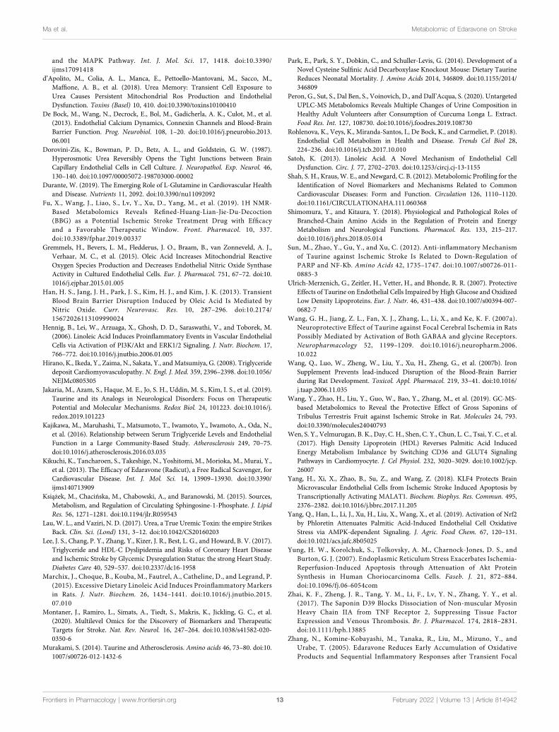
and the MAPK Pathway. Int. J. Mol. Sci. 17, 1418. doi:10.3390/ijms17091418
d’Apolito, M., Colia, A. L., Manca, E., Pettoello-Mantovani, M., Sacco, M.,Maffione, A. B., et al. (2018). Urea Memory: Transient Cell Exposure toUrea Causes Persistent Mitochondrial Ros Production and EndothelialDysfunction. Toxins (Basel) 10, 410. doi:10.3390/toxins10100410
De Bock, M., Wang, N., Decrock, E., Bol, M., Gadicherla, A. K., Culot, M., et al.(2013). Endothelial Calcium Dynamics, Connexin Channels and Blood-BrainBarrier Function. Prog. Neurobiol. 108, 1–20. doi:10.1016/j.pneurobio.2013.06.001
Dorovini-Zis, K., Bowman, P. D., Betz, A. L., and Goldstein, G. W. (1987).Hyperosmotic Urea Reversibly Opens the Tight Junctions between BrainCapillary Endothelial Cells in Cell Culture. J. Neuropathol. Exp. Neurol. 46,130–140. doi:10.1097/00005072-198703000-00002
Durante, W. (2019). The Emerging Role of L-Glutamine in Cardiovascular Healthand Disease. Nutrients 11, 2092. doi:10.3390/nu11092092
Fu, X., Wang, J., Liao, S., Lv, Y., Xu, D., Yang, M., et al. (2019). 1H NMR-Based Metabolomics Reveals Refined-Huang-Lian-Jie-Du-Decoction(BBG) as a Potential Ischemic Stroke Treatment Drug with Efficacyand a Favorable Therapeutic Window. Front. Pharmacol. 10, 337.doi:10.3389/fphar.2019.00337
Gremmels, H., Bevers, L. M., Fledderus, J. O., Braam, B., van Zonneveld, A. J.,Verhaar, M. C., et al. (2015). Oleic Acid Increases Mitochondrial ReactiveOxygen Species Production and Decreases Endothelial Nitric Oxide SynthaseActivity in Cultured Endothelial Cells. Eur. J. Pharmacol. 751, 67–72. doi:10.1016/j.ejphar.2015.01.005
Han, H. S., Jang, J. H., Park, J. S., Kim, H. J., and Kim, J. K. (2013). TransientBlood Brain Barrier Disruption Induced by Oleic Acid Is Mediated byNitric Oxide. Curr. Neurovasc. Res. 10, 287–296. doi:10.2174/15672026113109990024
Hennig, B., Lei, W., Arzuaga, X., Ghosh, D. D., Saraswathi, V., and Toborek, M.(2006). Linoleic Acid Induces Proinflammatory Events in Vascular EndothelialCells via Activation of PI3K/Akt and ERK1/2 Signaling. J. Nutr. Biochem. 17,766–772. doi:10.1016/j.jnutbio.2006.01.005
Hirano, K., Ikeda, Y., Zaima, N., Sakata, Y., andMatsumiya, G. (2008). Triglyceridedeposit Cardiomyovasculopathy. N. Engl. J. Med. 359, 2396–2398. doi:10.1056/NEJMc0805305
Jakaria, M., Azam, S., Haque, M. E., Jo, S. H., Uddin, M. S., Kim, I. S., et al. (2019).Taurine and its Analogs in Neurological Disorders: Focus on TherapeuticPotential and Molecular Mechanisms. Redox Biol. 24, 101223. doi:10.1016/j.redox.2019.101223
Kajikawa, M., Maruhashi, T., Matsumoto, T., Iwamoto, Y., Iwamoto, A., Oda, N.,et al. (2016). Relationship between Serum Triglyceride Levels and EndothelialFunction in a Large Community-Based Study. Atherosclerosis 249, 70–75.doi:10.1016/j.atherosclerosis.2016.03.035
Kikuchi, K., Tancharoen, S., Takeshige, N., Yoshitomi, M., Morioka, M., Murai, Y.,et al. (2013). The Efficacy of Edaravone (Radicut), a Free Radical Scavenger, forCardiovascular Disease. Int. J. Mol. Sci. 14, 13909–13930. doi:10.3390/ijms140713909
Książek, M., Chacińska, M., Chabowski, A., and Baranowski, M. (2015). Sources,Metabolism, and Regulation of Circulating Sphingosine-1-Phosphate. J. LipidRes. 56, 1271–1281. doi:10.1194/jlr.R059543
Lau, W. L., and Vaziri, N. D. (2017). Urea, a True Uremic Toxin: the empire StrikesBack. Clin. Sci. (Lond) 131, 3–12. doi:10.1042/CS20160203
Lee, J. S., Chang, P. Y., Zhang, Y., Kizer, J. R., Best, L. G., and Howard, B. V. (2017).Triglyceride and HDL-C Dyslipidemia and Risks of Coronary Heart Diseaseand Ischemic Stroke by Glycemic Dysregulation Status: the strong Heart Study.Diabetes Care 40, 529–537. doi:10.2337/dc16-1958
Marchix, J., Choque, B., Kouba, M., Fautrel, A., Catheline, D., and Legrand, P.(2015). Excessive Dietary Linoleic Acid Induces Proinflammatory Markersin Rats. J. Nutr. Biochem. 26, 1434–1441. doi:10.1016/j.jnutbio.2015.07.010
Montaner, J., Ramiro, L., Simats, A., Tiedt, S., Makris, K., Jickling, G. C., et al.(2020). Multilevel Omics for the Discovery of Biomarkers and TherapeuticTargets for Stroke. Nat. Rev. Neurol. 16, 247–264. doi:10.1038/s41582-020-0350-6
Murakami, S. (2014). Taurine and Atherosclerosis. Amino acids 46, 73–80. doi:10.1007/s00726-012-1432-6
Park, E., Park, S. Y., Dobkin, C., and Schuller-Levis, G. (2014). Development of aNovel Cysteine Sulfinic Acid Decarboxylase Knockout Mouse: Dietary TaurineReduces Neonatal Mortality. J. Amino Acids 2014, 346809. doi:10.1155/2014/346809
Peron, G., Sut, S., Dal Ben, S., Voinovich, D., and Dall’Acqua, S. (2020). UntargetedUPLC-MS Metabolomics Reveals Multiple Changes of Urine Composition inHealthy Adult Volunteers after Consumption of Curcuma Longa L. Extract.Food Res. Int. 127, 108730. doi:10.1016/j.foodres.2019.108730
Rohlenova, K., Veys, K., Miranda-Santos, I., De Bock, K., and Carmeliet, P. (2018).Endothelial Cell Metabolism in Health and Disease. Trends Cel Biol 28,224–236. doi:10.1016/j.tcb.2017.10.010
Satoh, K. (2013). Linoleic Acid. A Novel Mechanism of Endothelial CellDysfunction. Circ. J. 77, 2702–2703. doi:10.1253/circj.cj-13-1155
Shah, S. H., Kraus, W. E., and Newgard, C. B. (2012). Metabolomic Profiling for theIdentification of Novel Biomarkers and Mechanisms Related to CommonCardiovascular Diseases: Form and Function. Circulation 126, 1110–1120.doi:10.1161/CIRCULATIONAHA.111.060368
Shimomura, Y., and Kitaura, Y. (2018). Physiological and Pathological Roles ofBranched-Chain Amino Acids in the Regulation of Protein and EnergyMetabolism and Neurological Functions. Pharmacol. Res. 133, 215–217.doi:10.1016/j.phrs.2018.05.014
Sun, M., Zhao, Y., Gu, Y., and Xu, C. (2012). Anti-inflammatory Mechanismof Taurine against Ischemic Stroke Is Related to Down-Regulation ofPARP and NF-Κb. Amino Acids 42, 1735–1747. doi:10.1007/s00726-011-0885-3
Ulrich-Merzenich, G., Zeitler, H., Vetter, H., and Bhonde, R. R. (2007). ProtectiveEffects of Taurine on Endothelial Cells Impaired by High Glucose and OxidizedLow Density Lipoproteins. Eur. J. Nutr. 46, 431–438. doi:10.1007/s00394-007-0682-7
Wang, G. H., Jiang, Z. L., Fan, X. J., Zhang, L., Li, X., and Ke, K. F. (2007a).Neuroprotective Effect of Taurine against Focal Cerebral Ischemia in RatsPossibly Mediated by Activation of Both GABAA and glycine Receptors.Neuropharmacology 52, 1199–1209. doi:10.1016/j.neuropharm.2006.10.022
Wang, Q., Luo, W., Zheng, W., Liu, Y., Xu, H., Zheng, G., et al. (2007b). IronSupplement Prevents lead-induced Disruption of the Blood-Brain Barrierduring Rat Development. Toxicol. Appl. Pharmacol. 219, 33–41. doi:10.1016/j.taap.2006.11.035
Wang, Y., Zhao, H., Liu, Y., Guo, W., Bao, Y., Zhang, M., et al. (2019). GC-MS-based Metabolomics to Reveal the Protective Effect of Gross Saponins ofTribulus Terrestris Fruit against Ischemic Stroke in Rat. Molecules 24, 793.doi:10.3390/molecules24040793
Wen, S. Y., Velmurugan, B. K., Day, C. H., Shen, C. Y., Chun, L. C., Tsai, Y. C., et al.(2017). High Density Lipoprotein (HDL) Reverses Palmitic Acid InducedEnergy Metabolism Imbalance by Switching CD36 and GLUT4 SignalingPathways in Cardiomyocyte. J. Cel Physiol. 232, 3020–3029. doi:10.1002/jcp.26007
Yang, H., Xi, X., Zhao, B., Su, Z., and Wang, Z. (2018). KLF4 Protects BrainMicrovascular Endothelial Cells from Ischemic Stroke Induced Apoptosis byTranscriptionally Activating MALAT1. Biochem. Biophys. Res. Commun. 495,2376–2382. doi:10.1016/j.bbrc.2017.11.205
Yang, Q., Han, L., Li, J., Xu, H., Liu, X., Wang, X., et al. (2019). Activation of Nrf2by Phloretin Attenuates Palmitic Acid-Induced Endothelial Cell OxidativeStress via AMPK-dependent Signaling. J. Agric. Food Chem. 67, 120–131.doi:10.1021/acs.jafc.8b05025
Yung, H. W., Korolchuk, S., Tolkovsky, A. M., Charnock-Jones, D. S., andBurton, G. J. (2007). Endoplasmic Reticulum Stress Exacerbates Ischemia-Reperfusion-Induced Apoptosis through Attenuation of Akt ProteinSynthesis in Human Choriocarcinoma Cells. Faseb. J. 21, 872–884.doi:10.1096/fj.06-6054com
Zhai, K. F., Zheng, J. R., Tang, Y. M., Li, F., Lv, Y. N., Zhang, Y. Y., et al.(2017). The Saponin D39 Blocks Dissociation of Non-muscular MyosinHeavy Chain IIA from TNF Receptor 2, Suppressing Tissue FactorExpression and Venous Thrombosis. Br. J. Pharmacol. 174, 2818–2831.doi:10.1111/bph.13885
Zhang, N., Komine-Kobayashi, M., Tanaka, R., Liu, M., Mizuno, Y., andUrabe, T. (2005). Edaravone Reduces Early Accumulation of OxidativeProducts and Sequential Inflammatory Responses after Transient Focal
Frontiers in Pharmacology | www.frontiersin.org February 2022 | Volume 13 | Article 81494213
Ma et al. Metabolomic of Edaravone on Stroke
Ischemia in Mice Brain. Stroke 36, 2220–2225. doi:10.1161/01.STR.0000182241.07096.06
Zhenyukh, O., González-Amor, M., Rodrigues-Diez, R. R., Esteban, V., Ruiz-Ortega, M.,Salaices, M., et al. (2018). Branched-chain Amino Acids Promote EndothelialDysfunction through Increased Reactive Oxygen Species Generation andInflammation. J. Cel Mol. Med. 22, 4948–4962. doi:10.1111/jcmm.13759
Zille, M., Ikhsan, M., Jiang, Y., Lampe, J., Wenzel, J., and Schwaninger, M. (2019).The Impact of Endothelial Cell Death in the Brain and its Role after Stroke: ASystematic Review. Cell Stress 3, 330–347. doi:10.15698/cst2019.11.203
Conflict of Interest: The authors declare that the research was conducted in theabsence of any commercial or financial relationships that could be construed as apotential conflict of interest.
Publisher’s Note: All claims expressed in this article are solely those of the authorsand do not necessarily represent those of their affiliated organizations, or those ofthe publisher, the editors, and the reviewers. Any product that may be evaluated inthis article, or claim that may be made by its manufacturer, is not guaranteed orendorsed by the publisher.
Copyright © 2022 Ma, Zheng, Su, Zhang, Liu, Li, Zhang, Gong and Kou. This is anopen-access article distributed under the terms of the Creative Commons AttributionLicense (CC BY). The use, distribution or reproduction in other forums is permitted,provided the original author(s) and the copyright owner(s) are credited and that theoriginal publication in this journal is cited, in accordance with accepted academicpractice. No use, distribution or reproduction is permitted which does not complywith these terms.
Frontiers in Pharmacology | www.frontiersin.org February 2022 | Volume 13 | Article 81494214
Ma et al. Metabolomic of Edaravone on Stroke