Meta-analysis: capsule enteroscopy vs. conventional modalities in diagnosis of small bowel diseases
10
Meta-analysis: capsule enteroscopy vs. conventional modalities in diagnosis of small bowel diseases R. MARMO*, G. ROTONDANO , à , R. PISCOPO§, M. A. BIANCO à & L. CIPOLLETTA à *Department of Medicine, Division of Gastroenterology, Hospital L. Curto, Polla, Italy; Division of General Surgery, Section of Gastrointestinal Endoscopy, S. Mary’s Hospital, Rocca d’Aspide, Italy; àDepartment of Gastroenterology and Digestive Endoscopy, Hospital Maresca, Torre del Greco, Italy; §Division of Internal Medicine, Section of Gastroenterology, Evangelic Hospital Villa Betania, Naples, Italy Accepted for publication 29 June 2005 SUMMARY Background: Prospective trials support the role of capsule enteroscopy as an improvement in diagnosing mucosal lesions in the small bowel. Aim: To determine the diagnostic yield and safety of capsule enteroscopy vs. alternative diagnostic modali- ties (such as push enteroscopy, small bowel follow- through or enteroclysis) in patients with small bowel diseases. Methods: A search for prospective studies comparing capsule enteroscopy vs. other diagnostic tests in adults was performed between 1966 and 2005. Selected articles were included in a meta-analysis. Three analy- ses were run separately, all included studies and studies having occult gastrointestinal bleeding or Crohn’s disease as main outcome. Results: Seventeen studies (526 patients) met inclusion criteria. The rate difference (i.e. the absolute pooled difference in the rate of positive findings) between capsule enteroscopy and alternative modalities for small bowel disease was 41% (95% CI 35.6–45.9); 37% (95% CI 29.6–44.1) for occult gastrointestinal bleeding; and 45% (95% CI 30.9–58.0) for Crohn’s disease. Failure to visualize the caecum occurred in 13%, significantly more often in occult bleeders (17%) than in patients with Crohn’s disease (8%) (P < 0.006). Adverse events were recorded in 29 patients (6%). Capsule retention was more frequent in patients with Crohn’s disease (3% vs. 1%, OR 4.37). Conclusions: Capsule enteroscopy proved significantly superior to push enteroscopy and small bowel radiology in the diagnosis of ileal diseases. Capsule enteroscopy is safe, though prior radiology is still necessary to rule out small bowel strictures in patients with known or suspected Crohn’s disease. BACKGROUND Capsule endoscopy (CE) is a major advance in visual- ization of the small intestine. Since the introduction of CE the indications for the procedure have expanded to include Crohn’s disease (CD), celiac disease, polyposis syndromes, monitoring of patients after small-bowel transplantation, and unexplained abdominal pain, in addition to obscure gastrointestinal bleeding (OGIB). 1–4 Initial prospective trials support the role of CE as an improvement in the ability to assess and diagnose mucosal lesions in the small bowel. 5–8 CE has been at least as successful as, and sometimes significantly more successful than, push enteroscopy in finding the cause of OGIB in published series, with yields of 55–70%. 3, 6 It has been significantly more successful than barium studies in finding pathology in the small bowel and has outperformed barium meal as well as state-of-the-art Correspondence to: Dr R. Marmo, Via Sottobraida 32, 84037 Sant’Arsenio (SA), Italy. E-mail: [email protected] Aliment Pharmacol Ther 2005; 22: 595–604. doi: 10.1111/j.1365-2036.2005.02625.x Ó 2005 Blackwell Publishing Ltd 595
-
Upload
independent -
Category
Documents
-
view
1 -
download
0
Transcript of Meta-analysis: capsule enteroscopy vs. conventional modalities in diagnosis of small bowel diseases

Meta-analysis: capsule enteroscopy vs. conventional modalities indiagnosis of small bowel diseases
R. MARMO*, G. ROTONDANO� ,� , R. PISCOPO§, M. A. BIANCO� & L. CIPOLLETTA�*Department of Medicine, Division of Gastroenterology, Hospital L. Curto, Polla, Italy; �Division of General Surgery, Section
of Gastrointestinal Endoscopy, S. Mary’s Hospital, Rocca d’Aspide, Italy; �Department of Gastroenterology and Digestive
Endoscopy, Hospital Maresca, Torre del Greco, Italy; §Division of Internal Medicine, Section of Gastroenterology, Evangelic
Hospital Villa Betania, Naples, Italy
Accepted for publication 29 June 2005
SUMMARY
Background: Prospective trials support the role of capsule
enteroscopy as an improvement in diagnosing mucosal
lesions in the small bowel.
Aim: To determine the diagnostic yield and safety of
capsule enteroscopy vs. alternative diagnostic modali-
ties (such as push enteroscopy, small bowel follow-
through or enteroclysis) in patients with small bowel
diseases.
Methods: A search for prospective studies comparing
capsule enteroscopy vs. other diagnostic tests in adults
was performed between 1966 and 2005. Selected
articles were included in a meta-analysis. Three analy-
ses were run separately, all included studies and studies
having occult gastrointestinal bleeding or Crohn’s
disease as main outcome.
Results: Seventeen studies (526 patients) met inclusion
criteria. The rate difference (i.e. the absolute pooled
difference in the rate of positive findings) between
capsule enteroscopy and alternative modalities for small
bowel disease was 41% (95% CI 35.6–45.9); 37% (95%
CI 29.6–44.1) for occult gastrointestinal bleeding; and
45% (95% CI 30.9–58.0) for Crohn’s disease. Failure to
visualize the caecum occurred in 13%, significantly
more often in occult bleeders (17%) than in patients
with Crohn’s disease (8%) (P < 0.006). Adverse events
were recorded in 29 patients (6%). Capsule retention
was more frequent in patients with Crohn’s disease (3%
vs. 1%, OR 4.37).
Conclusions: Capsule enteroscopy proved significantly
superior to push enteroscopy and small bowel radiology
in the diagnosis of ileal diseases. Capsule enteroscopy is
safe, though prior radiology is still necessary to rule out
small bowel strictures in patients with known or
suspected Crohn’s disease.
BACKGROUND
Capsule endoscopy (CE) is a major advance in visual-
ization of the small intestine. Since the introduction of
CE the indications for the procedure have expanded to
include Crohn’s disease (CD), celiac disease, polyposis
syndromes, monitoring of patients after small-bowel
transplantation, and unexplained abdominal pain, in
addition to obscure gastrointestinal bleeding (OGIB).1–4
Initial prospective trials support the role of CE as an
improvement in the ability to assess and diagnose
mucosal lesions in the small bowel.5–8 CE has been at
least as successful as, and sometimes significantly more
successful than, push enteroscopy in finding the cause
of OGIB in published series, with yields of 55–70%.3, 6 It
has been significantly more successful than barium
studies in finding pathology in the small bowel and has
outperformed barium meal as well as state-of-the-art
Correspondence to: Dr R. Marmo, Via Sottobraida 32, 84037 Sant’Arsenio
(SA), Italy.
E-mail: [email protected]
Aliment Pharmacol Ther 2005; 22: 595–604. doi: 10.1111/j.1365-2036.2005.02625.x
� 2005 Blackwell Publishing Ltd 595

enteroclysis and computed tomographic enteroclysis in
fully published recent studies.4
However, there are few studies that have assessed the
impact of CE on clinical outcome, so that the true value
of this novel technology is less well established. Data are
sparse, the number of patients included in each trial is
often small, different groups of patients are seldom
considered, and conflicting results are sometimes repor-
ted.9, 10
The primary aim of the present study was to assess the
diagnostic yield of CE vs. conventional diagnostic
modalities in patients with OGIB and CD by performing
a quantitative systematic review of the published
controlled trials. The secondary aim was to assess any
negative outcome of CE in these patients.
MATERIALS AND METHODS
Literature search and identification of primary studies
Relevant papers were identified by searching the
MEDLINE, EMBASE/Excerpta Medica, Current Con-
tents and Cochrane Library databases using ‘capsule
endoscopy’ or ‘wireless capsule endoscopy’ or ‘capsule
enteroscopy’ as search terms. The search was restric-
ted to human studies of adults published in English
and non-English language, between January 1966
and March 2005 inclusive. We also conducted a
manual search of abstracts submitted to the Digestive
Disease Week and United European Gastroenterology
Week covering the same period. In addition, we
hand-searched relevant journals, published articles,
contacted companies and researchers in the field to
seek any ongoing or unpublished studies on the
comparison of diagnostic yield of CE vs. any other
modalities. Articles selected in the search were
reviewed separately by two of the authors (R.M. and
G.R.), and those fulfilling the inclusion criteria were
selected for further analysis. In addition, a fully
recursive search of reference lists of the original
studies was performed to find studies not identified by
the previous searches. Papers recorded in the personal
databases of the authors were also reviewed and
included when appropriate.
Study selection
This is a meta-analysis of the studies comparing CE with
any other diagnostic modality in adults suffering from
suspected small bowel disease. Studies were evaluated
separately by two of the authors. The inclusion criteria
were as follows: (i) Articles or abstracts should report
the results of prospective, comparative trials. (ii) Studies
must include patients with OGIB or CD. (iii) Studies had
to include at least two branches of diagnostic testing, i.e.
CE vs. other modalities. (iv) Explicit reference had to be
given for safety of the tested procedures either in the
text or tables.
A ‘negative outcome’ was defined as any undesired
event directly related to the application of a diagnostic
procedure, according to Fleischer et al.11 In particular,
capsule retention was defined as no evidence of capsule
excretion within 3 days.
When the abstract had no clear reason for exclusion,
the relative full paper was obtained if available.
Where multiple reports of a single trial existed, the
most recent version was obtained. We also reviewed
the references of selected articles and previous clinical
reviews. Data extraction was carried out independ-
ently by two investigators (R.M. and G.R.) using a
standardized form. Decisions regarding inclusion of
articles and data extraction were reached by consen-
sus between the two reviewers. If there was disag-
reement, the papers were jointly evaluated to solve
inconsistency.
Data extraction
The studies were examined with respect to the following
criteria: study design (including methods of reporting
adverse events); inclusion and exclusion criteria; patient
characteristics; technical details of CE and other diag-
nostic procedures; definition of study outcomes and
their monitoring methods.
Quality assessment
Information on objective quality-related characteristics
was also collected and the quality of the studies
included was assessed using 17 of the 22 items of the
CONSORT statement.12 We did not consider the five
items that refer to the process of randomization as all
trials that were evaluated were not randomized. The
final quality score ranged from 0 to 17, with
maximum quality studies rated 17. Results from
the trial reports were reproduced where possible.
The systematic review was performed according to the
QUORUM statement.13
596 R. MARMO et al.
� 2005 Blackwell Publishing Ltd, Aliment Pharmacol Ther 22, 595–604
Statistical analysis
Pooled odds ratio and 95% confidence intervals (CI)
were calculated from the raw study data using the Peto
fixed effect model, which produced a Q-statistic with a
chi-squared distribution with n)1 degrees of freedom,
where n is equal to the number of trials. Data from the
various studies were combined using appropriate
weights. The test of heterogeneity was utilized to
determine whether the fixed effect model or random
effects model was appropriate for calculation of the
weights, used in the meta-analytic estimate, and the
95% CI for this estimate. The chi-square test was
employed to assess whether the variation in diagnostic
effect within trials of the same group was greater than
might be expected. We considered heterogeneity to be
present if P < 0.05. In the absence of statistically
significant heterogeneity, odds ratios and 95% CI by
the fixed effect model only are given in the results.
All calculations were performed with STATA software
(StataCorp, Houston, TX, USA). For studies in which the
constructed 2 · 2 tables contained cells with zero events,
a standard correction factor of 0.5 was added to each cell.
After pooling of data, we calculated the crude and
weighted rate differences (RD), odds ratios (OR) and the
number needed to diagnose (NND) with 95% CI for each
of the trials included in the analysis, based on the raw
data presented in each paper. The NND (1/RD) is a
quantitative estimate of the diagnostic gain: it expresses
the number of patients to be investigated with each of
the procedures under evaluation so as to reach at least
one more positive finding compared with the other
method.14 An intention-to-treat analysis was carried
out, i.e. participants were analysed in the group to
which they were allocated.
Three analyses were run separately: the first one for all
included studies, the second one for studies having the
identification of the source of occult GI bleeding as
the main outcome and the last one for studies having
the diagnosis of ileal involvement in CD as their main
outcome.
For a significant meta-analysis finding, publication bias
was defined as the number of additional non-significant
studies that, if included in the meta-analysis, would
have removed the significant meta-analysis finding.
RESULTS
Included and excluded studies
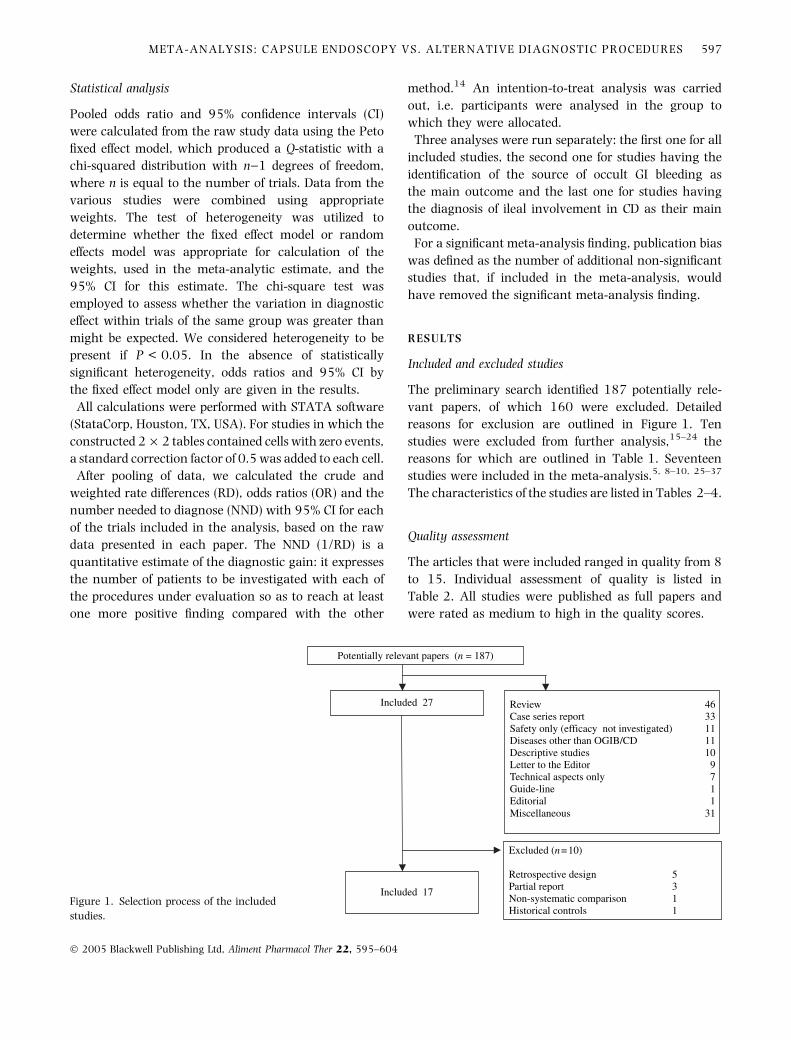
The preliminary search identified 187 potentially rele-
vant papers, of which 160 were excluded. Detailed
reasons for exclusion are outlined in Figure 1. Ten
studies were excluded from further analysis,15–24 the
reasons for which are outlined in Table 1. Seventeen
studies were included in the meta-analysis.5, 8–10, 25–37
The characteristics of the studies are listed in Tables 2–4.
Quality assessment
The articles that were included ranged in quality from 8
to 15. Individual assessment of quality is listed in
Table 2. All studies were published as full papers and
were rated as medium to high in the quality scores.
Review Case series report Safety only (efficacy not investigated) Diseases other than OGIB/CD Descriptive studies Letter to the EditorTechnical aspects onlyGuide-lineEditorialMiscellaneous
Included 27
Potentially relevant papers (n = 187)
Excluded (n = 10)
Retrospective designPartial reportNon-systematic comparisonHistorical controls
Included 17
4633111110
9711
31
5311
Figure 1. Selection process of the included
studies.
META-ANALYSIS: CAPSULE ENDOSCOPY VS. ALTERNATIVE DIAGNOSTIC PROCEDURES 597
� 2005 Blackwell Publishing Ltd, Aliment Pharmacol Ther 22, 595–604

Contraindications
A total of 39 of 604 patients had contraindications to
CE (6.5%) (95% CI 4.6–8.7). CE was contraindicated
in patients with OGIB in only eight of 336 cases
(2.4%) (95% CI 1.0–4.6) for the following reasons:
two major abdominal surgery, two diabetes, two
pacemaker and two small bowel strictures (Table 3).
Contraindications to CE in patients with CD were
present in 31 of 268 cases (11.6%) (95% CI 8.0–
16.0); small bowel stricture at pre-CE radiology in 30
cases and previous major abdominal surgery in one
patient (Table 4).
There was a significantly higher probability of having
a contraindication to CE in patients with CD compared
with obscure bleeders (OR 5.36, 95% CI 2.35–13.72)
(P < 0.001). If we consider only the presence of
stricture as contraindication, this chance in patients
with CD rises dramatically (OR 21.05, 95% CI 5.24–
182.83).
Efficacy
A total of 526 patients were evaluated (289 submitted
to CE for obscure bleeding, 237 submitted to CE for
known or suspected CD). The null hypothesis of
homogeneity was not rejected. The P-value for the
Q-statistic for the outcome that was considered (positive
finding) was 1.0.
The results indicate a clinically relevant and statisti-
cally significant advantage for CE over alternative
modalities in the diagnosis of small bowel diseases.
From a fixed effect model, the pooled RD in all the
studies was 40.8% (95% CI 35.6–45.9) (P < 0.0001).
The plot in Figure 2 shows that CE significantly
increases the probability of a positive finding compared
with controls (OR 4.9, 95% CI 3.9–6.4) (P < 0.001).
The calculated NND for this group was 2 (95% CI 2–3),
that is, for every two patients investigated, CE provides
one more diagnosis compared with controls. Publication
bias was 513. Therefore, the significant meta-analysis
finding for diagnosing small bowel pathology could be
removed by including 513 additional studies with non-
significant results.
Table 2. Characteristics of the study trial
included in the meta-analysisReference Disease investigated
Procedure compared
with CE
Quality
assessment
Adler et al.25 Obscure bleeding Push enteroscopy 11
Buchman et al.10 Known Crohn SBFT 8
Chong Ho et al.26 Known + suspected Crohn Enteroclysis 15
Costamagna et al.8 Small bowel diseases SBFT 10
Eliakim et al.27 Suspected Crohn SBFT 9
Ell et al.28 Obscure bleeding Push enteroscopy 12
Fireman et al.5 Suspected Crohn SBFT 8
Ge et al.29 Suspected Crohn SBFT 8
Hartmann et al.30 Obscure bleeding Push enteroscopy 13
Herrerias et al.31 Suspected Crohn SBFT 12
Lewis and Swain32 Obscure bleeding Push enteroscopy 11
Marmo et al.34 Known Crohn Enteroclysis 15
Mata et al.33 Obscure bleeding Push enteroscopy 12
Mylonaki et al.35 Obscure bleeding Push enteroscopy 13
Saurin et al.36 Obscure bleeding Push enteroscopy 15
Van Gossum et al.9 Obscure bleeding Push enteroscopy 9
Voderholzer et al.37 Known Crohn CT enteroclysis 10
CE, capsule endoscopy; SBFT, small bowel follow through; CT, computed tomography. No
study had randomized design.
Table 1. Reasons for excluding articles
Reference Reasons for exclusion
Mata et al.15 Preliminary results of Mata et al.33
Eliakim et al.16 Preliminary results of Eliakim et al.27
Voderholzer et al.17 Preliminary results of Voderholzer et al.37
May et al.18 Non-systematic comparison
with double balloon enteroscopy
Liangpunsakul et al.19 Retrospective design
Hara et al.20 Retrospective design
Caunedo et al.21 Retrospective design
Rastogi et al.22 Retrospective design
Mow et al.23 Retrospective design
Ge et al.24 Historical controls
598 R. MARMO et al.
� 2005 Blackwell Publishing Ltd, Aliment Pharmacol Ther 22, 595–604

In the subgroup analysis of studies of OGIB (Figure 3),
the pooled RD was 36.9% (95% CI 29.6–44.1)
(P < 0.0001). All the studies but one had push
enteroscopy as comparator investigation. CE significantly
increases the probability of a positive finding in such
patients compared with push enteroscopy (OR 4.3, 95%
CI 3.1–6.0) (P < 0.001). The calculated NND for this
subgroup was 3 (95% CI 3–5). Publication bias was 146.
Table 3. Clinical features of patients with obscure gastrointestinal bleeding
Reference
No. patients with
contraindication
to CE/included
No. patients submitted
to either diagnostic
procedure
Overt
bleeding
Occult
bleeding
Duration
of disease
Mean Hb
level (g/dL)
Adler et al.25 No data 20 11 9 n.d. 7.5 (4.0–12.1)
Ell et al.28 2/65 32 0 32 29 ± 2.4 months 5.9 (±1.4)
Van Gossum et al.9 No data 21 5 16 n.d. 9.8 (5.2–11.2)
Hartmann et al.30 0/33 33 33 0 30 ± 36 months 6.5 (±1.6)
Lewis and Swain32 4/25 21 9 12 36.5 (2–144) months 6.4 (2–8)
Mata et al.33 No data 42 26 16 n.d. 7.8 (±2.1)
Mylonaki et al.35 0/50 50 11 39 4.2 (0.5–20) years n.d.
Saurin et al.36 0/60 60 32 28 24.8 ± 21.7 months 9.4 (±2.5)
Costamagna et al.8 2/20 13 n.d. n.d. n.d. n.d.
CE, capsule endoscopy; n.d., not declared; Hb, haemoglobin. Values are mean (±standard deviation or range). See text for details of contraindi-
cations.
Table 4. Clinical features of patients with
Crohn’s disease
Reference
No. patients with
contraindication
to CE/included
No. patients submitted
to either diagnostic
procedure
Time interval between
diagnosis of CD and
CE examination
Eliakim et al.27 0 35 At initial diagnosis
Fireman et al.5 0 17 At initial diagnosis
Herrerias et al.31 0 21 At initial diagnosis
Ge et al.29 0 20 At initial diagnosis
Voderholzer et al.37 15/56 41 At initial diagnosis*
Buchman et al.10 12/42 30 5.5 ± 6.5 years
Chong Ho et al.26 2/45 43 At initial diagnosis*
Marmo et al.34 2/33 31 11 days
CD, Crohn’s disease; CE, capsule endoscopy. See text for details of contraindications.
* These two studies include patients with suspected CD (CE performed for initial diagnosis) and
also patients with known or surgically treated CD (CE performed not for diagnosis but for
staging).
21/3020/302004Buchmann
21/424/422005Chong Ho
25/4112/412005Voderholzer
22/318/312005Marmo
13/200/202004Ge
9/210/212003Herrerias
12/170/172003Fireman
27/358/352004Eliakim
4/131/132002Costamagna
40/5822/582003Saurin
34/5016/502003Mylonaki
31/428/422004Mata
11/206/202004Lewis
25/337/332003Hartmann
13/2116/212003Van Gossum
21/329/322002Ell
14/204/202004Adler
VCEOther Procedure
YearAuthor
Pooled (fixed effect) 4.992 (3.898 / 6.392)
1/161/64 1/4 1 4 16 64
Better controls Better VCE
141/526 343/526
Figure 2. Meta analysis: capsule enteros-
copy vs. alternative procedures in small
bowel disease (overall population).
META-ANALYSIS: CAPSULE ENDOSCOPY VS. ALTERNATIVE DIAGNOSTIC PROCEDURES 599
� 2005 Blackwell Publishing Ltd, Aliment Pharmacol Ther 22, 595–604

In the subgroup analysis concerning studies performed
in patients with known or suspected CD, there was
significant heterogeneity between the studies (Q ¼17.41, P < 0.01). From a random effect model, pooled
RD was 44.5% (95% CI 30.9–58.0) (P < 0.0001). As
shown in Figure 4, in these patients, CE provided a
significant diagnostic gain over both enteroclysis (OR 5.4,
95% CI 3.0–9.9) and small bowel follow-through (OR
13.0, 95% CI 2.3–71.4) with an overall sevenfold
increase in diagnostic capability against controls (OR
7.2, 95% CI 3.2–16.3) (P < 0.0001). The calculated
NND for this subgroup was 2 (95% CI 2–3). Publication
bias was 105.
Safety
Failure to visualize the caecum. Overall, in 526 patients
submitted to CE, the caecum was not visualized in 68
patients (12.9%, 95% CI 10.2–16.1). Such bad outcome
occurred in 48 patients investigated for OGIB (16.6%,
95% CI 12.5–21.4) (Table 5) and 20 investigated for
CD (8.4%, 95% CI 5.2–12.7) (Table 6). This difference
of 8.2% (95% C.I. 2.8–13.8) was statistically significant
(P < 0.006).
Adverse events. Adverse events were recorded in 29
patients (5.5%) (95% CI 3.7–7.8). In the population with
OGIB, there were 15 adverse events related to CE (5.2%,
95% CI 2.9–8.4) (Table 5). Capsule retention occurred
in two patients (0.7%, 95% CI 0.08–2.5) with one
surgical and one endoscopic removal. In the population
with CD, there were seven adverse events (3%, 95% CI
1.2–6.0), all related to capsule retention. Of these, five
necessitated a surgical removal, one endoscopic retrieval
and in one case there was a spontaneous passage after
3 days of steroid therapy (Table 6). The risk of capsule
retention was higher in the CD subgroup (OR 4.37, 95%
CI 0.82–43.37).
VCE: video capsule enteroscopy; REM: random effect model; FEM: fixed effect model; SBFT: small bowel follow-through
VCE 1/64 1/16 1/4 1 4 16 64Other procedure
YearN. Author
Better other procedures Better VCE
82/12328/123
52/237 150/237
SubGroup(REM)
(FEM)
68/11424/114SubGroup
SBFT
(REM)
(FEM)
Pooled(REM)
(FEM)
21/3020/302004Buchmann
21/424/422005Chong Ho
25/4112/412005Voderholze
22/318/312005Marmo
Enteroclysis
14
15
16
10
11
12
17
13
9/210/21
0/20
2003Herrerias
12/17
13/20
0/172003Fireman
27/358/352004
2004Ge
Eliakim
5.467 (3.014/9.917)
5.092 (2.988/8.678)
7.235 (3.210/16.310)
6.012 (4.132/8.748)
13.006 (2.369/71.412)
7.074 (4.174/11.990)
Figure 4. Meta analysis: capsule enterosco-
py vs. alternative procedures in patients
with suspected small bowel Crohn’s disease.
VCE, video capsule enteroscopy; REM, ran-
dom effect model; FEM, fixed effect model;
SBFT, small bowel follow-through.
VCE: video capsule enteroscopy; REM: random effect model; FEM: fixed effect model; SBFT: small bowel follow-through
4/131/13
4/131/13
89/289 193/289
Better other procedures
2002Costamagna
40/5822/582003Saurin
SubGroup 88/276 189/276
SBFT
(REM)
(FEM)
SubGroup(REM)
(FEM)
Pooled(REM)
(FEM)
34/5016/502003Mylonaki
31/428/422004Mata
11/206/202004Lewis
25/337/332003Hartmann
13/2116/212003Van Gossum
21/329/322002Ell
14/204/202004Adler1
Push enteroscopy
2
3
4
5
6
7
8
9
VCE 1/64 1/16 1/4 1 4 16 64
4.328 (2.399/7.808)
4.331 (3.101/6.048)
3.947 (0.518/30.907)
4.173 (0.616/28.257)
4.314 (2.487/7.483)
4.326 (3.113/6.011)
Better VCE
Other procedure
YearN. Author
Figure 3. Meta analysis: capsule enterosco-
py vs. alternative procedures in patients
with obscure gastrointestinal bleeding. VCE,
video capsule enteroscopy; REM, random
effect model; FEM, fixed effect model; SBFT,
small bowel follow-through.
600 R. MARMO et al.
� 2005 Blackwell Publishing Ltd, Aliment Pharmacol Ther 22, 595–604

DISCUSSION
The small intestine is relatively inaccessible to endo-
scopic examination because of length, location and
tortuosity. Conventional imaging with barium radiol-
ogy fails to detect flat lesions, such as angiodysplasia,
and is an insensitive method of detecting fine mucosal
disease and raised lesions. The clinical need for more
detailed small bowel imaging has been recently
fulfilled by the introduction of wireless capsule
enteroscopy. CE has opened up a new world of
diagnoses and possibilities to the gastroenterologist,
providing him/her with images of small intestinal
abnormalities which were not possible until recently.
It has been particularly successful in finding the cause
of OGIB and evaluating patients with inflammatory
bowel diseases.2–4 Nonetheless, there is a paucity of
data on the ultimate clinical impact of CE in small
Table 6. Negative outcomes in patients
with CDReference
No.
patients Adverse events of CE
CE failure to reach
the caecum
Eliakim et al.27 35 0 No data
Fireman et al.5 17 0 No data
Herrerias et al.31 21 0 0
Ge et al.29 20 3 (capsule retention)* 2
Voderholzer et al.37 41 2 (capsule retention)* 10
Buchman et al.10 30 2 (capsule retention) 0
Chong Hoet al.26 42 0 6
Marmo et al.34 31 0 2
CD, Crohn’s disease; CE, capsule endoscopy.
* One passed 3 days after steroid therapy and one successfully removed by push enteroscopy.
In this subgroup of CD patients enteroclysis could not be completed in six patients because of
intolerance to naso-jejunal tube.
Table 5. Negative outcomes in patients with OGIB
Reference
No.
patients Adverse events of CE
Adverse events of
push enteroscopy
CE failure to
reach the caecum
Adler et al.25 20 0 0 0
Ell et al.28 32 2
Technical defect (1)
Vision obscured by active bleeding (1)
0 n.d.
Van Gossum et al.9 21 1
Capsule blocked in an appendiceal stump
0 1
Hartmann et al.30 33 0 0 22
Lewis and Swain32 20 1
Long oesophageal transit
0 3
Mata et al.33 42 3
Capsule retention (1)
Long oesophageal transit (1)
Capsule malfunction (1)
0 3
Mylonaki et al.35 50 5
Long oesophageal transit (1)
Temporary electrical disconnection (1)
Battery dysfunction (1)
1
No advance beyond
duodenal bulb
16
Saurin et al.36 58 2
Battery dysfunction (1)
Error to transfer data (1)
0 3
Costamagnaet al. 8 13 1
Battery dysfunction
NA 0
OGIB, obscure gastrointestinal bleeding; CE, capsule endoscopy; NA, not applicable as in this study CE was compared with small bowel follow-
through. n.d., not declared.
META-ANALYSIS: CAPSULE ENDOSCOPY VS. ALTERNATIVE DIAGNOSTIC PROCEDURES 601
� 2005 Blackwell Publishing Ltd, Aliment Pharmacol Ther 22, 595–604

bowel disease, especially concerning changes in
management that lead to enduring positive outcomes.
Depending on the indication for which CE is per-
formed, however, a negative capsule study can be
reassuring and might potentially limit further diag-
nostic tests.38 Much of the available literature (mostly
abstracts) indicates that, although the initial diagnos-
tic yield of CE might be high, the proportion of
patients with changes in management is less, and the
proportion of patients with successful long-term
outcomes is even less.35, 39 Compared with push
enteroscopy, CE increases the diagnosis yield in
patients with OGIB and allows modification on
therapy strategy in up to 66% of cases.3, 6, 9, 25, 28, 30
In such patients, who have undergone multiple prior
procedures, even a much lower sustained long-term
success rate of 15–20% might be quite favourable. In
patients with known or suspected CD, CE has a higher
yield than push enteroscopy and enteroclysis when
small bowel mucosal disease is suspected.5, 27, 33, 37
Capsule examination alters the knowledge of the
presence and/or extension of disease in about half of
the patients with CD, leading to a relevant change in
therapeutic strategy.34
Capsule endoscopy is considered a simple, non-inva-
sive procedure, which can be universally performed,
even in out-patients. Nonetheless, our data show that
6.5% of patients who may potentially benefit from this
examination, actually have a contraindication to CE,
mainly related to radiological detection of small bowel
stricture during the pre-CE diagnostic work up.
Although the commercially available M2A device lists
the presence of pacemakers and use in children as
contraindications to its use, some groups including our
own have used the capsule in patients in both of these
settings.
The results of the present meta-analysis indicate that
the diagnostic gain of CE vs. all other procedures in the
assessment of OGIB and CD is both clinically relevant
and statistically significant. In obscure bleeders, CE
provides a fourfold chance of a positive finding. Such
superiority is consistent and homogeneous throughout
the studies and is witnessed by the huge number of void
or negative trials necessary to render meaningless the
meta-analysis. In patients with known or suspected CD,
the chance of a positive finding further rises to
sevenfold, independently of which investigation is
the comparator (from fivefold when compared with
enteroclysis to 13-fold when compared with SBFT).
However, the presence of significant heterogeneity
among studies prevents generalizability of results to
the whole population. Subgroup of patients such as
those with long-standing disease, surgically treated,
with indication to CE to monitor therapeutic response,
may represent the target of additional studies. CE will
likely not provide the answer if the clinician’s question
is preoperative assessment of ileal involvement and/or
extension. Such a question at the moment is still
answered by radiological imaging.
There is little doubt that bowel preparation may affect
sensitivity of CE examination. The optimal bowel
preparation for video CE has yet to be determined.
Given imaging recommends a 10-h fasting without
bowel purge. Data from the literature document that
bowel preparation accelerates small bowel capsule
transit, leads to a higher rate of complete endoscopic
examination and improves visualization of the small
bowel. Several different regimens have been tested: 2 L
polyethylene glycol (PEG) given 16 h before capsule
ingestion,40 ‘colonoscopy-type preparation’ with so-
dium picolax,35 sodium phosphate bowel preparation41 or 4 L PEG-based electrolyte solution (3 L during the
evening and 1 L 3 h before CE).42 Independently of the
type of bowel cleansing, all the authors agree that bowel
preparation before CE improved both the quality of
images and the diagnostic yield of the examination.
For a procedure to be recommended, both efficacy and
safety must be known as every endoscopic procedure
may induce complications. Despite the importance of
this matter, available studies fail to address the safety of
CE satisfactorily. Clinical trials typically assess only a
small number of patients and thus the chances of
detecting rare or delayed adverse events are small. The
present systematic review proved that 4.2% of patients
submitted to CE experience a negative outcome because
of technical problems and that 13% of patients do not
have a complete ileal exploration because of capsule
failure to reach the caecum. Such bad outcome
occurred more frequently in patients with OGIB
(16.6%) than in patients with CD (8.4%). We do not
have a clear explanation for this: alterations in gastric
emptying and/or gut motility, influence of co-prescrip-
tions and/or comorbidities, bowel preparation, etc. may
all be contributing factors. This observation may
attenuate the enthusiastic opinion on the ability of CE
to fully explore the small bowel. Further technical
refinement and or development will have to address this
issue.
602 R. MARMO et al.
� 2005 Blackwell Publishing Ltd, Aliment Pharmacol Ther 22, 595–604

The main complication associated with capsule use is
that is can get stuck in strictures. Capsule retention is
infrequent (0.7–3%) but still happens even in a selected
population. The four times higher risk of capsule
retention in the subgroup of patients with CD is
clinically relevant and does not reach statistical signi-
ficance probably due to a beta error. In the work up of
small bowel disease, because of safety reasons, radiology
cannot initially be replaced if the clinical suspicion is CD
to assess potential contraindication to CE. Nonetheless,
the risk that barium studies also may fail to detect
strictures, especially in early CD, should not be over-
looked. The use of a bio-fragmentable capsule (patency
capsule) may overcome these limitations, but needs to
be validated with proper investigations.
In conclusion, the diagnostic advantage of CE over all
other current diagnostic modalities is such that CE
should be considered as the procedure of choice in the
evaluation of patients with obscure bleeding and
suspected small bowel CD.
ACKNOWLEDGEMENT
The authors are indebted to Gioacchino Leandro, MD,
for his valuable help in statistical calculation.
REFERENCES
1 Iddan G, Meron G, Glukhovsky A, et al. Wireless capsule
endoscopy. Nature 2000; 405: 17.
2 Swain P, Fritscher-Ravens A. Role of video endoscopy in
managing small bowel disease. Gut 2004; 53: 1866–75.
3 Arnott ID, Lo SK. The clinical utility of wireless capsule
endoscopy. Dig Dis Sci 2004; 49: 893–901.
4 Kornbluth A, Legnani P, Lewis BS. Video capsule endoscopy in
inflammatory bowel disease: past, present, and future.
Inflamm Bowel Dis 2004; 10: 278–85.
5 Fireman Z, Mahajna E, Broide E, et al. Diagnosing small bowel
Crohn’s disease with wireless capsule endoscopy. Gut 2003;
52: 390–92.
6 Pennazio M, Santucci R, Rondonotti E, et al. Outcome of
patients with obscure gastrointestinal bleeding after capsule
endoscopy: report of 100 consecutive cases. Gastroenterology
2004; 126: 643–53.
7 Appleyard M, Fireman Z, Glukhovsky A, et al. A randomized
trial comparing wireless capsule endoscopy with push
enteroscopy for the detection of small bowel lesions.
Gastroenterology 2000; 119: 1431–8.
8 Costamagna G, Shah SK, Riccioni ME, et al. A prospective trial
comparing small bowel radiographs and video capsule
endoscopy for suspected small bowel disease.
Gastroenterology 2002; 123: 999–1005.
9 Van Gossum A, Hittelet A, Schmit A, et al. A prospective
comparative study of push and wireless-capsule enteroscopy
in patients with obscure digestive bleeding. Acta Gastroenterol
Belg 2003; 66: 199–205.
10 Buchman AL, Miller FH, Wallin A, et al. Videocapsule
endoscopy versus barium contrast studies for the diagnosis
of Crohn’s disease recurrence involving the small intestine.
Am J Gastroenterol 2004; 99: 2171.
11 Fleischer DE, Van de Mierop F, Eisen GM, et al. A new system for
defining endoscopic complications emphasizing the measure of
importance. Gastrointest Endosc 1997; 45: 128–33.
12 Altman DG, Schulz KF, Moher D, et al. The revised CONSORT
statement for reporting randomized trials: explanation and
elaboration. Ann Intern Med 2001; 134: 663–94.
13 Moher D, Cook DJ, Eastwood S, et al. Improving the quality of
reports of meta-analyses of randomised controlled trials: the
QUORUM statement. Quality of reporting of meta-analyses.
Lancet 1999; 354: 1896–900.
14 Leandro G. Meta-analysis in Medical Research. Oxford:
Blackwell Publishing, 2005.
15 Mata A, Llach J, Bordas JM, et al. Role of capsule endoscopy in
patients with obscure digestive bleeding Gastroenterol Hepatol
2003; 26: 619–23 [article in Spanish].
16 Eliakim R, Fischer D, Suissa A, et al. Wireless capsule video
endoscopy is a superior diagnostic tool in comparison to
barium follow-through and computerized tomography in
patients with suspected Crohn’s disease. Eur J Gastroenterol
Hepatol 2003; 15: 363–7.
17 Voderholzer WA, Ortner M, Rogalla P, et al. Diagnostic yield
of wireless capsule enteroscopy in comparison with
computed tomography enteroclysis. Endoscopy 2003; 35:
1009–14.
18 May A, Nachbar L, Wardak A, et al. Double-balloon
enteroscopy: preliminary experience in patients with obscure
gastrointestinal bleeding or chronic abdominal pain.
Endoscopy 2003; 35: 985–91.
19 Liangpunsakul S, Chadalawada V, Rex DK, et al. Wireless
capsule endoscopy detects small bowel ulcers in patients with
normal results from state of the art enteroclysis. Am J
Gastroenterol 2003; 98: 1295–8.
20 Hara AK, Leighton JA, Sharma VK, et al. Small bowel:
preliminary comparison of capsule endoscopy with barium
study and CT. Radiology 2004; 230: 260–5.
21 Caunedo A, Rodriguez-Tellez M, Garcia-Montes JM, et al.
Usefulness of capsule endoscopy in patients with suspected
small bowel disease. Rev Esp Enferm Dig 2004; 96: 10–21
[article in Spanish].
22 Rastogi A, Schoen RE, Slivka A. Diagnostic yield and clinical
outcomes of capsule endoscopy. Gastrointest Endosc 2004;
60: 959–64.
23 Mow WS, Lo SK, Targan SR, et al. Initial experience with
wireless capsule enteroscopy in the diagnosis and
management of inflammatory bowel disease. Clin
Gastroenterol Hepatol 2004; 2: 31–40.
24 Ge ZZ, Hu YB, Xiao SD. Capsule endoscopy and push
enteroscopy in the diagnosis of obscure gastrointestinal
bleeding. Chin Med J (Engl) 2004; 117: 1045–9.
META-ANALYSIS: CAPSULE ENDOSCOPY VS. ALTERNATIVE DIAGNOSTIC PROCEDURES 603
� 2005 Blackwell Publishing Ltd, Aliment Pharmacol Ther 22, 595–604
25 Adler DG, Knipschield M, Gostout C. A prospective
comparison of capsule endoscopy and push enteroscopy in
patients with GI bleeding of obscure origin. Gastrointest
Endosc 2004; 59: 492–8.
26 Chong Ho A, Taylor A, Miller A, et al. Capsule endoscopy vs.
push enteroscopy and enteroclysis in suspected small-bowel
Crohn’s disease. Gastrointest Endosc 2005; 61: 255–61.
27 Eliakim R, Suissa A, Yassin K, et al. Wireless capsule video
endoscopy compared to barium follow-through and compu-
terised tomography in patients with suspected Crohn’s disease
– final report. Dig Liver Dis 2004; 36: 519–22.
28 Ell C, Remke S, May A, et al. The first prospective controlled
trial comparing wireless capsule endoscopy with push
enteroscopy in chronic gastrointestinal bleeding. Endoscopy
2002; 34: 685–9.
29 Ge ZZ, Hu YB, Xiao SD. Capsule endoscopy in diagnosis of
small bowel Crohn’s disease. World J Gastroenterol 2004; 10:
1349–52.
30 Hartmann D, Schilling D, Bolz G, et al. Capsule endoscopy
versus push enteroscopy in patients with occult
gastrointestinal bleeding. Z Gastroenterol 2003; 41: 377–82.
31 Herrerias JM, Caunedo A, Rodriguez-Tellaz M, et al. Capsule
endoscopy in patients with suspected Crohn’s disease and
negative endoscopy. Endoscopy 2003; 35: 564–8.
32 Lewis BS, Swain P. Capsule endoscopy in the evaluation of
patients with suspected small intestinal bleeding: results of a
pilot study. Gastrointest Endosc 2002; 56: 349–53.
33 Mata A, Bordas JM, Feu F, et al. Wireless capsule endoscopy in
patients with obscure gastrointestinal bleeding: a comparative
study with push enteroscopy. Aliment Pharmacol Ther 2004;
20: 189–94.
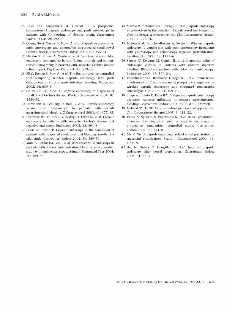
34 Marmo R, Rotondano G, Piscopo R, et al. Capsule endoscopy
vs enteroclysis in the detection of small bowel involvement in
Crohn’s disease: a prospective trial. Clin Gastroenterol Hepatol
2005; 3: 772–76.
35 Mylonaki M, Fritscher-Ravens A, Swain P. Wireless capsule
endoscopy: a comparison with push enteroscopy in patients
with gastroscopy and colonoscopy negative gastrointestinal
bleeding. Gut 2003; 52: 1122–6.
36 Saurin JC, Delvaux M, Gaudin JL, et al. Diagnostic value of
endoscopic capsule in patients with obscure digestive
bleeding: Blinded comparison with video push-enteroscopy.
Endoscopy 2003; 35: 576–84.
37 Voderholzer WA, Beinhoelzl J, Rogalla P, et al. Small bowel
involvement in Crohn’s disease: a prospective comparison of
wireless capsule endoscopy and computed tomography
enteroclysis. Gut 2005; 54: 369–73.
38 Shapira S, Pluta K, Enns RA. A negative capsule enteroscopy
decreases resource utilization in obscure gastrointestinal
bleeding. Gastrointest Endosc 2004; 59: AB166 [abstract].
39 Melmed GY, Lo SK. Capsule endoscopy: practical applications.
Clin Gastroenterol Hepatol 2005; 3: 411–22.
40 Viazis N, Sgouros S, Papaxoinis K, et al. Bowel preparation
increases the diagnostic yield of capsule endoscopy: a
prospective, randomized, controlled study. Gastrointest
Endosc 2004; 60: 534–8.
41 Niv Y, Niv G. Capsule endoscopy: role of bowel preparation in
successful visualization. Scand J Gastroenterol 2004; 39:
1005–9.
42 Dai N, Gubler C, Hengstler P, et al. Improved capsule
endoscopy after bowel preparation. Gastrointest Endosc
2005; 61: 28–31.
604 R. MARMO et al.
� 2005 Blackwell Publishing Ltd, Aliment Pharmacol Ther 22, 595–604