Input from the medial septum regulates adult hippocampal neurogenesis
Upload
independentCategory
view
0download
0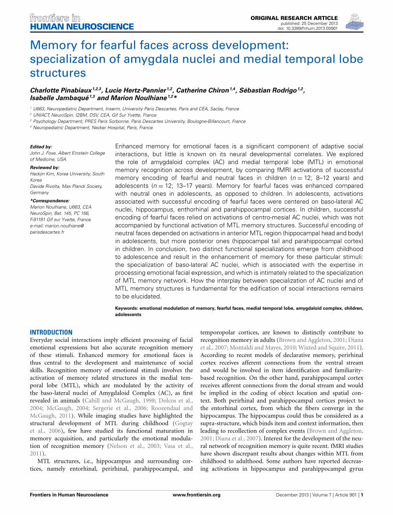
ORIGINAL RESEARCH ARTICLEpublished: 25 December 2013
doi: 10.3389/fnhum.2013.00901
Memory for fearful faces across development:specialization of amygdala nuclei and medial temporal lobestructuresCharlotte Pinabiaux1,2,3, Lucie Hertz-Pannier1,2, Catherine Chiron1,4, Sébastian Rodrigo1,2,
Isabelle Jambaqué1,3 and Marion Noulhiane1,2*
1 U663, Neuropediatric Department, Inserm, University Paris Descartes, Paris and CEA, Saclay, France2 UNIACT, NeuroSpin, I2BM, DSV, CEA, Gif Sur Yvette, France3 Psychology Department, PRES Paris Sorbonne, Paris Descartes University, Boulogne-Billancourt, France4 Neuropediatric Department, Necker Hospital, Paris, France
Edited by:
John J. Foxe, Albert Einstein Collegeof Medicine, USA
Reviewed by:
Hackjin Kim, Korea University, SouthKoreaDavide Rivolta, Max Planck Society,Germany
*Correspondence:
Marion Noulhiane, U663, CEANeuroSpin, Bat. 145, PC 156,F-91191 Gif sur Yvette, Francee-mail: [email protected]
Enhanced memory for emotional faces is a significant component of adaptive socialinteractions, but little is known on its neural developmental correlates. We exploredthe role of amygdaloid complex (AC) and medial temporal lobe (MTL) in emotionalmemory recognition across development, by comparing fMRI activations of successfulmemory encoding of fearful and neutral faces in children (n = 12; 8–12 years) andadolescents (n = 12; 13–17 years). Memory for fearful faces was enhanced comparedwith neutral ones in adolescents, as opposed to children. In adolescents, activationsassociated with successful encoding of fearful faces were centered on baso-lateral ACnuclei, hippocampus, enthorhinal and parahippocampal cortices. In children, successfulencoding of fearful faces relied on activations of centro-mesial AC nuclei, which was notaccompanied by functional activation of MTL memory structures. Successful encoding ofneutral faces depended on activations in anterior MTL region (hippocampal head and body)in adolescents, but more posterior ones (hippocampal tail and parahippocampal cortex)in children. In conclusion, two distinct functional specializations emerge from childhoodto adolescence and result in the enhancement of memory for these particular stimuli:the specialization of baso-lateral AC nuclei, which is associated with the expertise inprocessing emotional facial expression, and which is intimately related to the specializationof MTL memory network. How the interplay between specialization of AC nuclei and ofMTL memory structures is fundamental for the edification of social interactions remainsto be elucidated.
Keywords: emotional modulation of memory, fearful faces, medial temporal lobe, amygdaloid complex, children,
adolescents
INTRODUCTIONEveryday social interactions imply efficient processing of facialemotional expressions but also accurate recognition memoryof these stimuli. Enhanced memory for emotional faces isthus central to the development and maintenance of socialskills. Recognition memory of emotional stimuli involves theactivation of memory related structures in the medial tem-poral lobe (MTL), which are modulated by the activity ofthe baso-lateral nuclei of Amygdaloid Complex (AC), as firstrevealed in animals (Cahill and McGaugh, 1998; Dolcos et al.,2004; McGaugh, 2004; Sergerie et al., 2006; Roozendaal andMcGaugh, 2011). While imaging studies have highlighted thestructural development of MTL during childhood (Gogtayet al., 2006), few have studied its functional maturation inmemory acquisition, and particularly the emotional modula-tion of recognition memory (Nelson et al., 2003; Vasa et al.,2011).
MTL structures, i.e., hippocampus and surrounding cor-tices, namely entorhinal, perirhinal, parahippocampal, and
temporopolar cortices, are known to distinctly contribute torecognition memory in adults (Brown and Aggleton, 2001; Dianaet al., 2007; Montaldi and Mayes, 2010; Wixted and Squire, 2011).According to recent models of declarative memory, perirhinalcortex receives afferent connections from the ventral streamand would be involved in item identification and familiarity-based recognition. On the other hand, parahippocampal cortexreceives afferent connections from the dorsal stream and wouldbe implied in the coding of object location and spatial con-text. Both perirhinal and parahippocampal cortices project tothe entorhinal cortex, from which the fibers converge in thehippocampus. The hippocampus could thus be considered as asupra-structure, which binds item and context information, thenleading to recollection of complex events (Brown and Aggleton,2001; Diana et al., 2007). Interest for the development of the neu-ral network of recognition memory is quite recent. fMRI studieshave shown discrepant results about changes within MTL fromchildhood to adulthood. Some authors have reported decreas-ing activations in hippocampus and parahippocampal gyrus
Frontiers in Human Neuroscience www.frontiersin.org December 2013 | Volume 7 | Article 901 | 1
HUMAN NEUROSCIENCE
Pinabiaux et al. Fearful faces memory across development
(Menon et al., 2005; Maril et al., 2010), whereas others foundno age effect in hippocampus activation (Ofen et al., 2007; Marilet al., 2011). On the other hand, Ghetti et al. (2010) investi-gated the role of hippocampus and parahippocampal gyrus indetail recollection across ages (3 groups: 8 year olds, adoles-cents and adults). The groups were presented with black andwhite line drawings during a scanned encoding phase and laterattempted to recall outside the scanner which color originallybordered the drawings. Correct recall of the surrounding colorwas considered as successful episodic detail recollection. Itemrecognition activated the hippocampus and posterior parahip-pocampal gyrus in 8 year olds, whereas these regions werespecialized in detail recollection in adults. However, the struc-tural maturation of the hippocampus is non-linear, encouragingto consider hippocampal subregions in developmental studies(Gogtay et al., 2006). Only one recent study has consideredseparately anterior and posterior hippocampal subregions in acomparison of 8–11 years old children and adults during sourcememory retrieval (Demaster and Ghetti, 2013). Data, acquiredusing the task of Ghetti et al. (2010), showed an age-relateddissociation of hippocampal activity during successful episodicretrieval, with activity in the anterior hippocampus in adultsbut in the posterior one in children. Such developmental pat-terns of hippocampal function remain to be further exploredto clarify the functional maturation of MTL network, and inparticular the relationships between hippocampus, surround-ing cortices and AC in the context of emotional modulation ofmemory.
The modulatory role of AC on the recognition memory net-work is well known in adults (Dolcos et al., 2004; Kensingerand Corkin, 2004; Sergerie et al., 2006; Murty et al., 2010).The memory modulation hypothesis proposes that amygdalarprojections to the MTL declarative memory system are criti-cal for consolidating memories of emotionally arousing events(McGaugh, 2004). This is favored by anatomical disposition ofAC, anterior to and in continuity with the hippocampus, andby its numerous connections with cortical and sub-cortical areasengaged in memory. AC is a complex structure composed ofseveral nuclei with distinct cytoarchitectony and connectivity: lat-eral nucleus, basal nucleus, central nucleus and cortico-mesialnucleus (Aggleton, 2000; LeDoux, 2007). Rodent studies haveshown that (i) the basal nucleus is more specifically involvedin fear conditioning (Sierra-Mercado et al., 2011); (ii) the lat-eral nucleus is more activated for learning associations betweenaffect and stimuli (Johansen et al., 2011); (iii) the central nucleuswould be at the crossroads of behavioral responses to painfulstimulations (Kalin et al., 2004); and (iv) the mesial nucleuswould be engaged in olfactory associations and sexual behav-ior (Lehman et al., 1980; Bian et al., 2008). The baso-lateralnuclei are involved in the modulation of memory-related brainactivity in animals (Cahill and McGaugh, 1998; McGaugh, 2004;Roozendaal and McGaugh, 2011) and in memory recognitionin human adults when using emotional pictures (Dolcos et al.,2004) and emotional facial expressions, mainly fear (Sergerieet al., 2006). These neuroimaging studies showed that the co-activation of the MTL and AC is critical to emotional memoryformation. The localization of the modulated areas within MTL
differs across studies, sometimes pointing to the hippocampalformation (Hamann et al., 1999; Dolcos et al., 2004; Kensingerand Corkin, 2004; Murty et al., 2009; St. Jacques et al., 2009)or to the entorhinal and perirhinal cortices in anterior MTL(Hamann et al., 1999; Dolcos et al., 2004; Ritchey et al., 2008)and parahippocampal cortex in posterior MTL (Kilpatrick andCahill, 2003). Anterior hippocampus and surrounding corticesindeed have a high density of noradrenergic and glucocorticoidreceptors which are thought to mediate AC’s modulatory roleon declarative memory (Roozendaal et al., 2009). Posterior MTLhas been strongly implicated in contextual fear conditioning inboth rodents (Rudy et al., 2004) and humans (Alvarez et al.,2008). Together, these findings support the memory modula-tion hypothesis. However, the development of neural networkslinking recognition memory and emotional stimuli, especiallythe role of AC nuclei, during childhood, remains poorly under-stood.
The role of MTL in memory of emotional stimuli during ado-lescence has been underlined in two neuropsychological studies.The first one showed that, contrary to healthy adolescents, 11–15years old patients with temporal lobe epilepsy displayed no emo-tional memory enhancement during learning of emotional wordlists or recall of stories (Jambaqué et al., 2009). In the secondstudy, Pinabiaux et al. (2013) compared memory recognitionof emotional and neutral words and faces in a group of 8–18years old patients with temporal lobe resection and a group ofhealthy age-matched participants. They found a deficit in emo-tional enhancement of memory in patients with temporal loberesection for all emotional stimuli but fearful faces. Additionally,two fMRI studies have analyzed emotional modulation effects onthe development of recognition memory networks in adolescents.Nelson et al. (2003) studied successful encoding of fearful, angry,happy and neutral faces in healthy 9–17 years old adolescents andadults. Memory enhancement for emotional faces was similar inadults and adolescents; surprisingly, in both groups, the amyg-dala was engaged bilaterally during successful encoding of neutralfaces but not emotional faces (Nelson et al., 2003). According tothe authors, methodological issues may explain why no activa-tions of AC were found for fearful faces and why no age-relateddifferences were observed. Indeed, there were four emotionalexpression conditions, and despite four presentations with differ-ent rating conditions (passive viewing, two emotional judgments,perceptual judgment) of the stimuli at encoding, the number ofsubsequent “hit” or “miss” trials for each emotional expressionwas reduced. More recently, Vasa et al. (2011) specifically ana-lyzed, within hippocampus and AC, the activations associatedwith encoding of negative, positive and neutral pictures, usinga procedure similar to that of Nelson et al. (2003). Activationswere compared according to age (12–18 years old adolescents vs.adults) and memory accuracy (recalled vs. non-recalled pictures).Right AC was more engaged in adolescents than in adults for posi-tive, but not for fearful, pictures (Vasa et al., 2011). No age-relatedchanges were found in the hippocampus in either study, and nospecific baso-lateral activations in AC were disclosed, in contrastto findings in adults (Dolcos et al., 2004; Sergerie et al., 2006).
In the present study, we sought to further characterize theage-related changes in the neural networks engaged during
Frontiers in Human Neuroscience www.frontiersin.org December 2013 | Volume 7 | Article 901 | 2
Pinabiaux et al. Fearful faces memory across development
successful encoding of fearful and neutral faces in 8–12 yearschildren and 13–17 years adolescents using a detailed analysisof Regions Of Interest (ROIs) in MTL structures (hippocam-pus head, body, and tail) extending to surrounding cortices(temporopolar, perirhinal, entorhinal, parahippocampal) and ACnuclei. We assumed that activations in AC and MTL mem-ory related structures would vary with age when successfullyencoding faces. In particular, we sought to investigate the involve-ment of baso-lateral AC nuclei in fear memory modulationfrom childhood. Considering the functional specialization of thehippocampus and parahippocampal gyrus previously reportedduring childhood, we expected to observe activations of moreanterior regions of the MTL (hippocampal subregions and sur-rounding cortices) in adolescents compared with younger chil-dren. In addition we aimed to further decipher the functionalorganization within the MTL in relationship with amygdalanuclei.
MATERIALS AND METHODSPARTICIPANTSThe study was approved by ethical committee (CPP Ile de FranceVI, Protocole INSERM C08-12, ID RCB: 2008-A00683, Paris,France) and informed consent was obtained from all parents andsubjects.
A total of 24 healthy participants aged 12.87 ± 2.87 years (agerange: 8–16.83 years, males/females: 13/11, 18 right handed) wereincluded. None had history of neurological or psychiatric illness,and all participants completed the protocol. The population wassplit in two groups: children, from 8 to 12 years (n = 12, 5 girls,mean ± SD age = 10.46 years, ± 1.64 years) and adolescents, from13 to 17 years (n = 12, 6 girls, mean age = 15.28 years ± 1.40years).
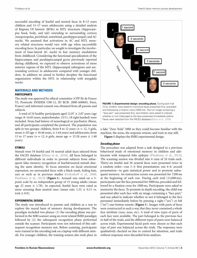
STIMULIStimuli were 54 fearful and 54 neutral adult faces selected fromthe FACES database (Ebner et al., 2010). All faces belonged todifferent individuals in order to prevent subjects from subse-quent false memory recognition of fearful/neutral stimuli shar-ing the same identity. To focus attention on facial emotionalexpression, we surrounded faces with a black mask, hiding hair,ears or neck as in previous studies (Golouboff et al., 2008;Pinabiaux et al., 2013) (Figure 1). Arousal was rated on a 5-point scale by an independent group of 15 young adults (meanage 22 years ± 1.78). As expected, fearful faces were rated asmore arousing than neutral ones (mean rate: 1.52 ± 0.11 vs.4.64 ± 0.9).
EXPERIMENTAL DESIGNThe study was introduced to parents and children as a way toexplore the neural basis of memory during development. Theparadigm included two phases: (1) the encoding phase was per-formed in the MRI scanner using an event related fMRI paradigmfollowed by (2) the subsequent recognition phase performedoutside the scanner. Participants were not informed of this sub-sequent recognition memory test. Before scanning, participantswere trained in the encoding task on a laptop with different stim-uli. For younger children, the training session also took place in
FIGURE 1 | Experimental design: encoding phase. During each trial(12 s), children were asked to memorize faces presented (5 s), precededand followed by a fixation cross (1000 ms). Then an image containing a“face part” was presented (5 s), and children were asked to indicatewhether or not it belonged to the face presented immediately before.Faces were selected from the FACES database (Ebner et al., 2010).
a fake “Zero Tesla” MRI so they could become familiar with themachine, the noise, the response remote, and train to stay still.
Figure 1 displays the fMRI experimental design.
Encoding phaseThe procedure was adapted from a task designed in a previousbehavioral study of emotional memory in children and ado-lescents with temporal lobe epilepsy (Pinabiaux et al., 2013).The scanning session was divided into 6 runs of 24 trials each.Thirty-six fearful and 36 neutral faces were presented twice ina random order—run 1–3: first presentation; run 4–6: secondpresentation—to gain statistical power and to promote subse-quent memory. An instruction screen was presented for 7200 msat the beginning of each run. During each trial (12,000 ms),participants saw the face presented for 5000 ms, preceded and fol-lowed by a fixation cross for 1000 ms. Participants were asked tomemorize the faces. To promote in depth encoding, the child waspresented after each face with an image containing a “face part,”and was asked to indicate whether or not it belonged to the facepresented immediately before by pressing a right (“yes”) or left(“no”) one-button remote (Figure 1). Images with parts of faceswere constructed in such a way that they never contained a wholeface attribute (eyes, nose, etc). A total of nine types of parts ofeach face were available. The part belonged to the previous facein half of the trials, and the different types of parts were balancedacross trials. Experimental facial parts were chosen so that eachtype of part was balanced across the trials. The responses werequalitatively checked on line to control for attention, and trialswithout responses were discarded from analyses.
Frontiers in Human Neuroscience www.frontiersin.org December 2013 | Volume 7 | Article 901 | 3
Pinabiaux et al. Fearful faces memory across development
Recognition phaseAn unexpected recognition memory test was performed outsidethe scanner 30 min after the end of the encoding phase. In thisphase, the 72 previously presented faces and 36 foiled faces (18fearful and 18 neutral) were randomly presented one by one ona computer screen. Faces were counterbalanced across subjectsbetween being targets and foils. Participants judged each item asnew or old using the keyboard, without time constraints.
IMAGING PROCEDUREImages were acquired on a 3-T Siemens Magnetom Trio scan-ner. High resolution 3D IR–prepped T1-weighted anatomic scanswere first acquired (repetition time = 2300 ms, echo time = 3 ms,TI : 900 ms, 256-mm field of view, matrix 240 × 256, 160 sagit-tal slices, 1 × 1 × 1.1 mm3) in 7 min 46 s. During encoding, 123T2∗-weighted EPI images were acquired per run (TR = 2400 ms,TE = 30 ms, 192-mm FOV, 64 × 64 matrix, 81 flip angle, 40 axialslices, 3 mm isotropic, 5 min 2 s). The three initial volumes werediscarded to allow for T1 equilibration.
Data were analyzed using SPM5 (http://www.fil.ion.ucl.ac.uk/spm/). Differences in slice acquisition timing were corrected byresampling all slices in time to match the middle slice. Functionalvolumes were spatially realigned to correct for motion artifacts.Scans with more than 4 mm movement in one direction were dis-carded. Images were then spatially normalized using a pediatrictemplate based on matched reference data of NIH MRI study ofnormal brain development created with the TOM toolbox (Wilkeet al., 2008). This template was automatically generated in a SPM5toolbox using an algorithm including age and gender of each ofour participants. Finally, images were smoothed using a 5-mmisotropic Gaussian filter.
DATA ANALYSISBehavioral dataData were analyzed with Statistica (www.statsoft.com). Percentsof correct and false recognitions were collected and averagedin each age group and a corrected memory accuracy measurewas computed as %hits-%false recognition. Global effects of age(8–12 years vs. 13–17 years) were analyzed using 2 by 2 Mann–Whitney tests. Global effects of emotional expression (fearful vs.neutral) were analyzed within each group separately using 2 by2 Wilcoxon tests. Group x emotional expression interaction wasalso explored using 2 by 2 Wilcoxon tests. Accuracy on the “faceparts” task and interaction between encoding and recognitionaccuracy were also examined using non-parametric comparisonand correlation.
fMRI dataIndividual GLM–based analyses were conducted with SPM5(http://www.fil.ion.ucl.ac.uk/spm/). Trials were categorized infour types according to both emotional expression and recogni-tion status (fearful hits, fearful misses, neutral hits, and neutralmisses) based on individual memory performance and regressorfunctions were constructed for each trial type.
Whole brain analysis. Individual contrasts of interest for fear-ful and neutral faces (Dmfear and Dmneutral) were based on
the differential neural activity on a common memory con-trast defined by Dm = hits-misses (Paller and Wagner, 2002),using a fixed-effects model across the six sessions. Second-levelgroup analyses used a mixed-effects (MFX) model implementedin the DISTANCE toolbox of SPM5 (Mériaux et al., 2006;Roche et al., 2007). This MFX model takes into account theerror measure on blood oxygenation level–dependent (BOLD)contrast in data with high inter-subject variability (Mériauxet al., 2006; Roche et al., 2007). Permutation t-tests (one mil-lion of permutations) were computed to compare Dmfear andDmneutral between 8 and 12 years and 13 and 17 years groups.Permutation analyses were conducted under a non-parametricassumption and corrected for multiple comparisons (Holmeset al., 1996). Whole brain Dmfear and Dmneutral contrast mapswere compared between groups using, at the voxel level, anuncorrected height threshold of p < 0.005, and a cluster sizethreshold of >5 contiguous voxels (de Vanssay-Maigne et al.,2011).
Eight ROIs were manually delineated in the MTL on bothsides of each subject, as described in previous studies (Noulhianeet al., 2006, 2007; de Vanssay-Maigne et al., 2011): AC, hippocam-pus head, body, and tail, entorhinal, perirhinal, parahippocampaland temporopolar cortices. The ROI boundaries were identi-fied using anatomical landmarks (Insausti et al., 1998; Duvernoyand Bourgouin, 1999; Pruessner et al., 2002) taking into accounthippocampus development (Insausti et al., 2010). The protocolconsisted of a volumetric analysis based on histological land-marks reported on T1-MRI, to offer pertinent MRI landmarks.Because AC nuclei are not visible on T1-MRI, we adopted amethodology previously used by Dolcos et al. (2004) consist-ing in dividing AC in four quadrants. To delineate the ROIs, theprotocol implied to progress in a rostro-caudal direction alongthe MTL in a coronal plane (1 mm section) while checking thedelimitation in the other planes (axial, sagittal, 3D). To accountfor age-dependent volume changes, ROIs were separately man-ually drawn on three subjects of each group (the youngest, theoldest and the median). ROIs from these subjects were then nor-malized to create a template. Mean contrast values of fear hits,fear misses, neutral hits and neutral misses were extracted fromROIs in each subject using MarsBar toolbox (http://marsbar/sourceforge.net). For each subject, BOLD response was con-trasted between subsequent recognized faces (Hits) and fixationtrials, and between subsequent non-recognized faces (Misses)and fixation trials, within each ROI. First, global effects of sub-sequent memory (Hits > Misses) were analyzed within eachgroup for fearful and neutral faces separately using 2 by 2Wilcoxon tests. Group (8–12 years vs. 13–17 years) × subse-quent memory (Hits vs. Misses) interaction was then exploredwithin regions showing subsequent memory effects, using 2 by2 Mann–Whitney tests. Due to the multiple ROIs approach(MTL: n = 16; AC nuclei: n = 4), thresholds were corrected formultiple comparisons (Bonferroni’s adjustement). P-values havebeen adjusted in accordance with the number of ROIs separatelyfor MTL and AC analyses. Thus adjusted p-values correspondto p/16 for MTL (8 ROIs × 2 hemispheres) and p/4 for AC(2 ROIs × 2 hemispheres), and were compared to α = 0.05threshold.
Frontiers in Human Neuroscience www.frontiersin.org December 2013 | Volume 7 | Article 901 | 4
Pinabiaux et al. Fearful faces memory across development
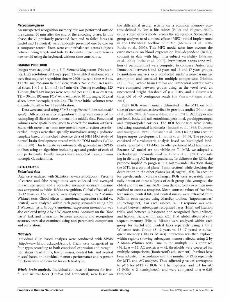
RESULTSBEHAVIORAL RESULTSFigure 2 presents the behavioral performances on the recog-nition memory task of the two age groups. All participantsperformed well above chance level at the ‘face parts’ task dur-ing encoding (accuracy range: min = 72%–max = 89%). Therewere no age-related differences, neither for “face parts” taskfor fearful (Mean13−17 = 0.82 ± 0.08; Mean8−12 = 0.79 ± 0.05;Z = 1.17, ns), nor for neutral faces (Mean13−17 = 0.85 ± 0.08;Mean8−12 = 0.80 ± 06; Z = 1.6, ns). We thus assume that chil-dren and adolescents paid attention to the faces equivalentlyduring the scanning session. There was no significant relation-ship in accuracy between encoding and recognition tasks, whetherfor fearful or neutral faces (rho = −0.088; ns; rho = −0.12; ns,respectively).
Percent of hits was similar in both groups (fearful: Z = 0.72,ns; neutral: Z = −1.22, ns), but age and emotional expres-sion significantly interacted during the recognition test. Fearfulfaces were more accurately recognized than neutral faces in ado-lescents (MeanFear = 0.56 ± 0.13 vs. MeanNeutral = 0.49 ± 0.24,Z = 2.25, p = 0.024), but not in children (MeanFear = 0.59 ±0.10 vs. MeanNeutral = 0.58 ± 0.14; Z = 0.67, ns). In children,there was more false recognition of fearful faces than neutralones (0.41 vs. 0.29, Z = 2.63, p = 0.0086), whereas equivalentrecognition was observed in adolescents (0.23 vs. 0.20, Z = 0.66,ns). Consequently, an interaction between groups and emo-tional expressions was observed on corrected memory accuracymeasure: it was better for fearful than neutral faces in the ado-lescent group (MeanFear = 0.39 ± 0.13 vs. MeanNeutral = 0.30 ±0.14, Z = 2.58, p = 0.0099), whereas no difference was observedin the children group (MeanFear = 0.19 ± 0.17; MeanNeutral =0.30 ± 0.10, Z = 1.27, ns).
FIGURE 2 | Behavioral results: hits, False Recognition (FR) for fearful
and neutral faces in children (8–12 years) and adolescents (13–17
years). Error bars = mean square errors. Significant differences betweenfearful and neutral conditions are indicated as follows: ∗p < 0.05;∗∗p < 0.01. Fearful faces were more accurately recognized than neutralfaces in the adolescents group, but not in the children group. There weremore false recognitions of fearful faces than neutral ones in the childrengroup only.
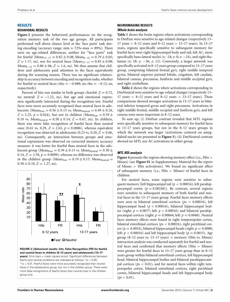
NEUROIMAGING RESULTSWhole brain analysisTable 1 shows the brain regions where activations correspondingto DmFear were sensitive to age-related changes (respectively 13–17 years > 8–12 years and 8–12 years > 13–17 years). In 13–17years, regions specifically sensitive to subsequent memory forfearful faces were right hippocampal body and tail, left AC, morespecifically baso-lateral nuclei (x: -24; y: 0; z: −24), and right tha-lamus (x: 18; y: −36; z: 12). Conversely, a larger network wasspecifically activated in 8–12 years group compared to 13–17 yearsgroup, comprising bilateral frontal gyri, right middle temporalgyrus, bilateral superior parietal lobule, cingulum, left caudate,bilateral cuneus, precuneus, fusiform and middle occipital gyri,and right cerebellum.
Table 2 shows the regions where activations corresponding toDmNeutral were sensitive to age-related changes (respectively 13–17 years > 8–12 years and 8–12 years > 13–17 years). Thesecomparisons showed stronger activations in 13–17 years in bilat-eral inferior temporal gyrus and right precuneus. Activations inright middle frontal, middle occipital and fusiform gyri, and leftcuneus were more important in 8–12 years.
To sum up, (i) DmFear contrast revealed that MTL regionswere specifically sensitive to subsequent memory for fearful facesin 13–17 years groups, but not in the 8–12 years groups inwhich the network was larger (activations centered on amyg-daloid nuclei are presented in Figure 3); (ii) DmNeutral contrastshowed no MTL nor AC activations in either group.
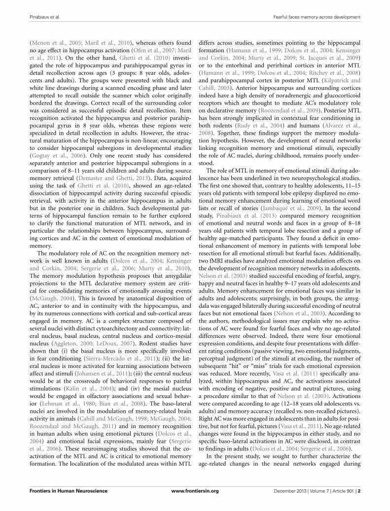
MTL ROI analysisFigure 4 presents the regions showing memory effect (i.e., Hits >
Misses) (see Figure S1 in Supplementary Material for the reportof Misses > Hits activations). We found no significant effectof subsequent memory (i.e., Hits > Misses) of fearful faces inchildren.
For neutral faces, some regions were sensitive to subse-quent memory [left hippocampal tail (p < 0.00016), left parahip-pocampal cortex (p = 0.00248)]. By contrast, several regionswere sensitive to subsequent memory of both fearful and neu-tral faces in the 13–17 years group. Fearful faces memory effectswere seen in bilateral entorhinal cortices (ps < 0.00016), lefthippocampal head (p < 0.00016), bilateral hippocampal bod-ies (right: p = 0.0077; left: p < 0.00016) and bilateral parahip-pocampal cortices (right: p = 0.00064; left: p = 0.0048). Neutralfaces memory effects were found in right temporopolar cortex,bilateral entorhinal cortices (ps < 0.00016), right perirhinal cor-tex (p = 0.0054), bilateral hippocampal heads (right: p = 0.0088;left: p < 0.00016) and left hippocampal body (p = 0.0015). Agegroup (8–12 years vs. 13–17 years) × memory (Hits vs. Misses)interaction analysis was conducted separately for fearful and neu-tral faces and confirmed that memory effects (Hits > Misses)were greater for fearful faces in 13–17 years group than in 8–12years group within bilateral entorhinal cortices, left hippocampalhead, bilateral hippocampal bodies and bilateral parahippocam-pal cortices (ps < 0.01), and for neutral faces within right tem-poropolar cortex, bilateral entorhinal cortices, right perirhinalcortex, bilateral hippocampal heads and left hippocampal body(ps < 0.01).
Frontiers in Human Neuroscience www.frontiersin.org December 2013 | Volume 7 | Article 901 | 5
Pinabiaux et al. Fearful faces memory across development
Table 1 | Neural substrates of subsequent memory effect (Dm) for fearful faces across ages.
Regions X Y Z Cluster size Z -value p
13–17 YEARS > 8–12 YEARS
Left amygdaloid complex(Baso-lateral nuclei) −24 0 −24 6 2.967 0.00151
Right hippocampal body 33 −27 −6 6 3.031 0.00122
Right hippocampal tail 39 −33 −6 5 2.705 0.00342
Right thalamus 18 −36 12 11 2.863 0.00210
8–12 YEARS > 13–17 YEARS
Right amygdaloid complex(Centro-mesial nuclei) 21 0 −15 10 3.487 0.00024
Left amygdaloid complex(Centro-mesial nuclei) −18 −3 −18 11 3.487 0.00024
Right inferior frontal gyrus 45 21 3 5 3.487 0.00024
Left inferior frontal gyrus −42 45 −12 5 3.487 0.00024
Right middle frontal gyrus 24 21 51 13 3.297 0.00049
Left middle frontal gyrus −3 54 −9 13 3.487 0.00024
Right superior frontal gyrus 18 48 3 30 3.297 0.00049
Right middle temporal gyrus 63 −33 −15 33 3.487 0.00024
Right superior parietal lobule 30 −75 48 11 3.297 0.00049
Right fusiform gyrus 30 −48 −18 5 3.297 0.00049
Left fusiform gyrus −36 −57 −18 5 3.182 0.00073
Right precuneus 6 −48 9 7 3.297 0.00049
Left precuneus −15 −81 45 9 3.487 0.00024
Left cuneus −3 −99 12 11 3.487 0.00024
Right lingual gyrus 24 −66 45 6 2.815 0.00244
Left lingual gyrus −6 −72 −9 6 3.297 0.00049
Left inferior occipital gyrus −30 −93 −12 43 3.487 0.00024
Right middle occipital gyrus 33 −81 12 90 3.487 0.00024
Left middle occipital gyrus −45 −72 9 50 3.487 0.00024
Anterior cingulum −3 27 30 6 3.031 0.00122
Middle cingulum 3 −3 36 8 3.182 0.00073
Posterior cingulum 3 36 27 18 3.297 0.00049
Right cerebellum 3 −72 −30 42 3.487 0.00024
Whole brain analysis. Non-parametric group comparison (13–17 years > 8–12 years and 8–12 years >13–17 years) of Dm (Dm = Hits-Misses) for fearful faces
(p < 0.005; 5 contiguous voxels).
Table 2 | Neural substrates of subsequent memory effect (Dm) for neutral faces across ages Whole brain analysis.
Regions X Y Z Cluster size Z -value p
13–17 YEARS > 8–12 YEARS
Right inferior temporal gyrus 54 −63 −15 8 3.487 0.00024
Left inferior temporal gyrus −48 −54 −15 6 3.487 0.00024
Right precuneus 33 −69 36 5 3.487 0.00024
8–12 YEARS > 13–17 YEARS
Right middle frontal gyrus 27 3 51 10 3.297 0.00049
Right fusiform gyrus 36 −57 −12 6 3.297 0.00049
Left cuneus −18 −93 3 5 3.297 0.00049
Right middle occipital gyrus 36 −84 9 8 3.031 0.00122
Non-parametric group comparison (13–17 years > 8–12 years and 8–12 years > 13–17 years) of Dm (Dm = Hits-Misses) for neutral faces (p < 0.005; 5 contiguous
voxels).
Amygdaloid complex ROI analysisFigure 5 presents the mean contrast values with hits and misses inamygdaloïd complex. In the 8–12 years group, a significant mem-ory effect (Hits > Misses) was found in centro-mesial nuclei of
left AC for fearful (p = 0.0027) and neutral faces (p = 0.0034),but no such effect was seen in baso-lateral nuclei. Inversely, in the13–17 years group, baso-lateral nuclei on both sides were specifi-cally sensitive to fearful memory effects (right: p = 0.0034; left:
Frontiers in Human Neuroscience www.frontiersin.org December 2013 | Volume 7 | Article 901 | 6
Pinabiaux et al. Fearful faces memory across development
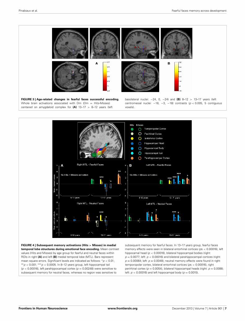
FIGURE 3 | Age-related changes in fearful faces successful encoding.
Whole brain activations associated with Dm (Dm = Hits-Misses)centered on amygdaloid complex for (A) 13–17 > 8–12 years (left
basolateral nuclei: −24, 0, −24) and (B) 8–12 > 13–17 years (leftcentromesial nuclei: −18, −3, −18) contrasts (p < 0.005, 5 contiguousvoxels).
FIGURE 4 | Subsequent memory activations (Hits > Misses) in medial
temporal lobe structures during emotional face encoding. Mean contrastvalues (Hits and Misses) by age group for fearful and neutral faces withinROIs in right (A) and left (B) medial temporal lobe (MTL). Bars representmean square errors. Significant levels are indicated as follows: ∗p < 0.01;∗∗p < 0.001; ∗∗∗p < 0.0005. In 8–12 years group, left hippocampal tail(p < 0.00016), left parahippocampal cortex (p = 0.00248) were sensitive tosubsequent memory for neutral faces, whereas no region was sensitive to
subsequent memory for fearful faces. In 13–17 years group, fearful facesmemory effects were seen in bilateral entorhinal cortices (ps < 0.00016), lefthippocamal head (p < 0.00016), bilateral hippocampal bodies (right:p = 0.0077; left: p < 0.00016) and bilateral parahippocampal cortices (right:p = 0.00064; left: p = 0.0048); neutral memory effects were found in righttemporopolar cortex, bilateral entorhinal cortices (ps < 0.00016), rightperirhinal cortex (p = 0.0054), bilateral hippocampal heads (right: p = 0.0088;left: p < 0.00016) and left hippocampal body (p = 0.0015).
Frontiers in Human Neuroscience www.frontiersin.org December 2013 | Volume 7 | Article 901 | 7
Pinabiaux et al. Fearful faces memory across development
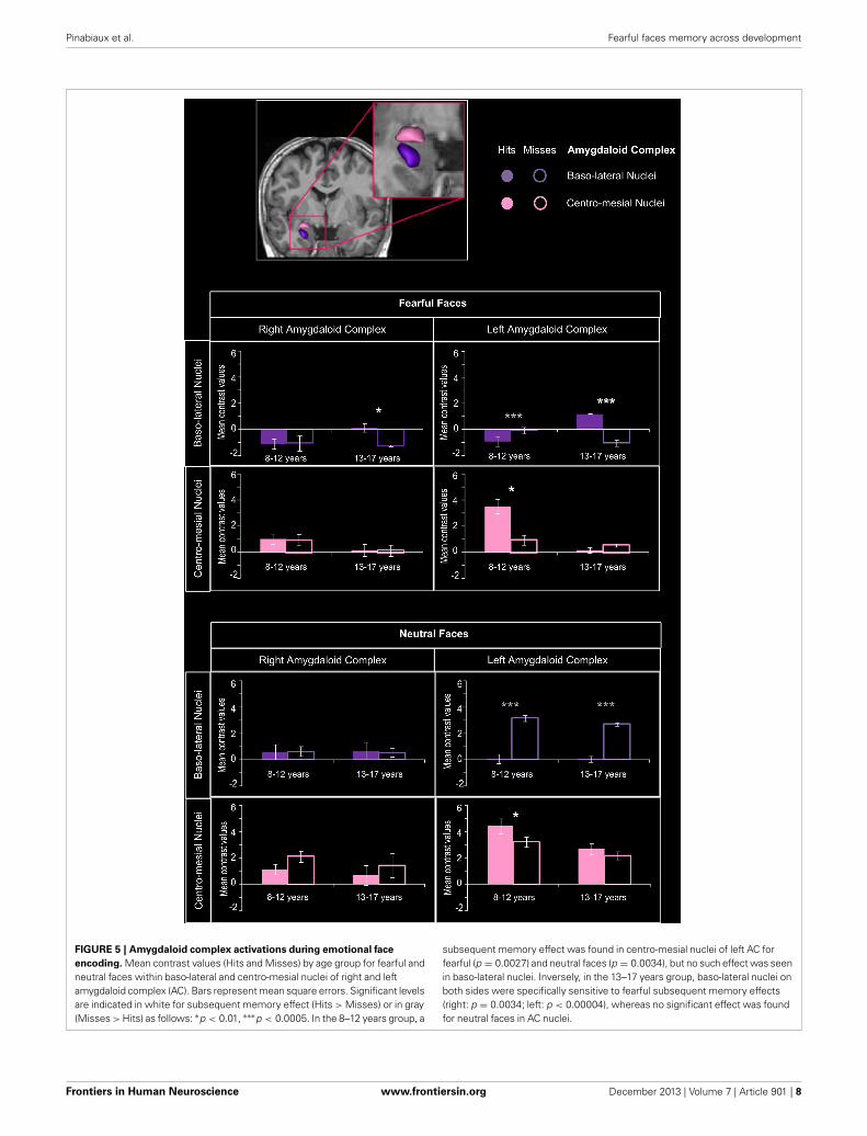
FIGURE 5 | Amygdaloid complex activations during emotional face
encoding. Mean contrast values (Hits and Misses) by age group for fearful andneutral faces within baso-lateral and centro-mesial nuclei of right and leftamygdaloid complex (AC). Bars represent mean square errors. Significant levelsare indicated in white for subsequent memory effect (Hits > Misses) or in gray(Misses > Hits) as follows: ∗p < 0.01, ∗∗∗p < 0.0005. In the 8–12 years group, a
subsequent memory effect was found in centro-mesial nuclei of left AC forfearful (p = 0.0027) and neutral faces (p = 0.0034), but no such effect was seenin baso-lateral nuclei. Inversely, in the 13–17 years group, baso-lateral nuclei onboth sides were specifically sensitive to fearful subsequent memory effects(right: p = 0.0034; left: p < 0.00004), whereas no significant effect was foundfor neutral faces in AC nuclei.
Frontiers in Human Neuroscience www.frontiersin.org December 2013 | Volume 7 | Article 901 | 8
Pinabiaux et al. Fearful faces memory across development
p < 0.00004), whereas no significant Hits > Misses effect wasfound for neutral faces in AC nuclei.
For fearful faces, age group (8–12 vs. 13–17 years) × memory(Hits vs. Misses) interaction analysis revealed that subsequentmemory effects were greater in 13–17 years group than in8–12 years group within right and left baso-lateral nuclei (ps< 0.00004), but greater in 8–12 years group than in 13–17years group within left centro-mesial nuclei (p < 0.00004). Asignificant interaction was found for neutral faces within leftcentro-mesial nuclei, subsequent memory effects of neutral facesbeing more important in 8–12 years than in 13–17 years (p <
0.0024).
DISCUSSIONThe aim of this study was to clarify age-related changes in neu-ral networks involved in encoding memory of fearful faces, andespecially the role of the AC in modulating memories acrossdevelopment. Behavioral data showed that, when compared withmemory of neutral faces, memory for fearful faces was enhancedin adolescents but not in children. This difference was associatedwith two functional developmental specializations: (i) a special-ization of baso-lateral AC nuclei for specific encoding of fearfulfaces in adolescents, but a non-specific involvement of centro-mesial AC nuclei in children; (ii) the specialization of distinctparts of the hippocampus and surrounding cortices in recogni-tion memory processes, depending on the emotional content:whereas MTL activation was associated with encoding of fear-ful faces in adolescents only, a rostro-caudal segregation wasobserved for neutral stimuli in children, with more posterior acti-vations. Finally, with age, extra-MTL regions were less engaged,reflecting an ‘economic’ dynamic of networks across develop-ment, i.e., relying on a smaller number of ultra-specialized struc-tures. Importantly, these functional activity changes reflect thenatural plasticity of neural memory networks during develop-ment rather than a modification in memory accuracy or inattentional engagement during the encoding task between agegroups.
EMOTIONAL SPECIALIZATION OF AC NUCLEI WITH AGEWe demonstrate a developmental switch in the involvement of ACnuclei during successful encoding of faces. Successful encodingof fearful faces specifically relied on activity of left baso-lateralAC nuclei in adolescents, who thus presented an adult-like pat-tern of emotional memory related amygdala activity (Dolcoset al., 2004; Sergerie et al., 2006). In animals and human adults,baso-lateral AC nuclei are especially implicated in emotionalmodulation of memory encoding in hippocampus (LaBar andCabeza, 2006; Roozendaal and McGaugh, 2011). By contrast, weshow that children engaged centro-mesial AC nuclei during suc-cessful encoding of both fearful and neutral faces. In light oftheir anatomical arrangement, cytoarchitecture and connectiv-ity with the hypothalamus, the involvement of centro-mesial ACnuclei in children may reflect the engagement of low level pro-cesses (Lehman et al., 1980; Kalin et al., 2004; Bian et al., 2008).Indeed, while our data suggest that the involvement of baso-lateral AC nuclei in adolescents translates into a higher level ofexpertise in processing emotional facial expression, the activation
of centro-mesial AC nuclei would be correlated with the imma-turity of such social skill (Guyer et al., 2008). That, in 8–12 yearschildren, centro-mesial AC nuclei activation was not specific offearful faces may explain why this age group showed no emo-tional enhancement of memory. Accordingly, stronger activationof AC has been associated with neutral faces in 11 year-old chil-dren, whereas, in adults, AC was more activated when viewingfearful faces (Thomas et al., 2001). More recently, AC was foundto be more activated in adolescents than in adults during passiveand active processing of emotional facial expressions (Passarottiet al., 2009), notably fear (Guyer et al., 2008). However, owingto the anatomical complexity of AC substructures and of theirfunctional segregation, it is surprising that no study so far hasaddressed the role of the activation of AC nuclei. We show here afunctional developmental specialization of AC nuclei related tofearful faces memory. On the one hand, this study brings newevidence that emotional enhancement of fearful faces memoryrelies on the specialization of baso-lateral nuclei of AC in emo-tional memory modulation, which appears during adolescence.This may be particularly true for fearful faces, since AC is espe-cially sensitive to this type of emotional stimuli (McGaugh, 2004).Evidence of baso-lateral AC nuclei activations for other emo-tional stimuli during adolescence is however needed. On the otherhand, the proposition of non-specific activity of centro-mesial ACnuclei in children should be explored in further functional and/oranatomical studies focusing on the distinct role of AC nuclei inemotional faces processing across development.
MEMORY SPECIALIZATION OF MTL WITH AGEWe bring new evidence about the role of MTL structures inencoding fearful and neutral faces during development, thanksto a specific MTL ROIs approach combined with a sensitiveparadigm using facial expressions. Previous developmental fMRIstudies of emotional memories did not elicit age-related dif-ferential hippocampal activity (Nelson et al., 2003; Vasa et al.,2011), while those of recognition memory showed an age-relateddecrease of hippocampal activity (Menon et al., 2005; Marilet al., 2010). Based on anatomical evidence (Gogtay et al., 2006)and on recent functional findings (Demaster and Ghetti, 2013),we looked at age-related differences in hippocampal head, bodyand tail, using a dedicated ROI analysis. Using an fMRI source-memory task, Demaster and Ghetti (2013) have demonstrateda shift of activity during episodic retrieval, from the posteriorhippocampus in 8–11 years old children toward the anterior hip-pocampus in adults. We further demonstrate that age-relatedcontributions of hippocampal sub-regions depend on emotionalexpression. The successful encoding of neutral faces was associ-ated with a stronger activity in the posterior part of the MTL (hip-pocampal tail and parahippocampal cortex) in children, whereasit was related to greater activations in the anterior one (hip-pocampal head and body) in adolescents. This developmentalchange in functional engagement along a longitudinal axis inMTL is thus congruent with DeMaster and Ghetti’s results (2013),although stimuli are intrinsically different (faces vs. drawings)and the tasks did not request the same cognitive resources (encod-ing vs. source memory). Individual longitudinal studies maygreatly help in the description of this developmental shift. When
Frontiers in Human Neuroscience www.frontiersin.org December 2013 | Volume 7 | Article 901 | 9
Pinabiaux et al. Fearful faces memory across development
dealing with fearful faces, age-related MTL activation changeswere different with a “all or nothing” pattern of activations, ratherthan a rostro-caudal age-related segregation. Indeed, no effect ofsubsequent memory for fearful faces was found within MTL inchildren, whereas bilateral hippocampus and parahippocampalgyrus (entorhinal and parahippocampal cortices) were activatedin adolescents. A lack of interplay between centro-mesial ACnuclei and MTL in children would be congruent with the absenceof emotional memory enhancement in behavioral results, and isalso illustrated by the non-specific centro-mesial activation of ACduring successful encoding of both fearful and neutral faces inchildren. Eventually, adolescents thus display a more “adult-like”pattern of behavior and activations, involving baso-lateral AC,hippocampus and parahippocampal gyrus (Dolcos et al., 2004;Sergerie et al., 2006).
RELATIONSHIP BETWEEN SPECIALIZATIONS OF AC NUCLEI AND MTLACROSS DEVELOPMENTThis study shows that the specialized activations of baso-lateralAC nuclei and MTL memory network are associated withthe emergence of emotional memory modulation in adoles-cents. Indeed, the developmental shift from the engagement of
centro-mesial AC nuclei in children to baso-lateral AC nucleiin adolescents could reflect the growing expertise in processingemotional facial expression, which may enhance the memoryfor such stimuli (Figure 6). At least two alternative interpreta-tions can be suggested. The first is a “cascade explanation,” inwhich the specialization of baso-lateral AC nuclei constitutes asubstantial gateway to MTL memory network, resulting in theemergence of emotional memory modulation. Data from Guyeret al. (2008) support this assumption, as these authors observeda co-activation of AC and hippocampus in association with per-ception of fearful faces in 9–17 year-old adolescents. The secondis an “interplay explanation,” in which the specializations ofbaso-lateral AC nuclei and MTL memory network emerge con-comitantly. Neuropsychological studies in adults with temporallobe epilepsy (Glogau et al., 2004; Carvajal et al., 2009) and inadolescents after temporal lobe resection (Pinabiaux et al., 2013)have shown that perception and encoding of emotional faces arecorrelated. Indeed, patients with temporal lobe epilepsy showimpairments in perception of facial expressions, which are moreimportant in early onset epilepsy (Meletti et al., 2003; Golouboffet al., 2008; Sedda et al., 2013). In the “interplay explanation,”enhanced memory for emotional faces would be a determinant
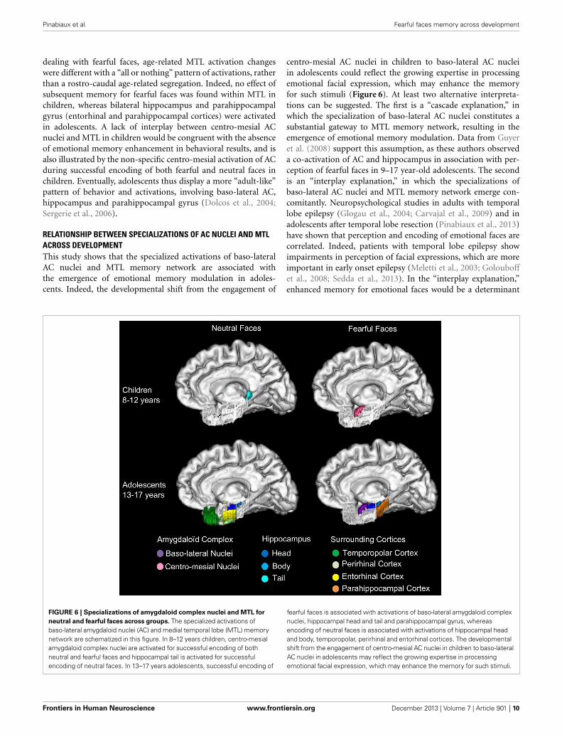
FIGURE 6 | Specializations of amygdaloid complex nuclei and MTL for
neutral and fearful faces across groups. The specialized activations ofbaso-lateral amygdaloid nuclei (AC) and medial temporal lobe (MTL) memorynetwork are schematized in this figure. In 8–12 years children, centro-mesialamygdaloid complex nuclei are activated for successful encoding of bothneutral and fearful faces and hippocampal tail is activated for successfulencoding of neutral faces. In 13–17 years adolescents, successful encoding of
fearful faces is associated with activations of baso-lateral amygdaloid complexnuclei, hippocampal head and tail and parahippocampal gyrus, whereasencoding of neutral faces is associated with activations of hippocampal headand body, temporopolar, perirhinal and entorhinal cortices. The developmentalshift from the engagement of centro-mesial AC nuclei in children to baso-lateralAC nuclei in adolescents may reflect the growing expertise in processingemotional facial expression, which may enhance the memory for such stimuli.
Frontiers in Human Neuroscience www.frontiersin.org December 2013 | Volume 7 | Article 901 | 10
Pinabiaux et al. Fearful faces memory across development
of growing expertise in facial processing. Accordingly, accuratememory for a fearful face would later result in more accurateprocessing of this face. In the case of memory for fearful faces,this agrees with the appraisal theory of emotion (Brosch et al.,2010) which suggests that fear is a more relevant emotion thanhappiness for species survival and social behavior. Future studiesusing other emotional facial expressions are needed to con-firm this assumption about the age-related enrolment of ACnuclei in emotional modulation of memory. Especially, functionalconnectivity analyses would help to better understand how theinterplay between AC and MTL specializations establishes acrossdevelopment.
EXTRA-MTL ACTIVATIONSWhole brain analyses (Supplementary material) showed thatextra-MTL areas were more activated in children than inadolescents. Parieto-occipital cortices were more engaged in chil-dren during successful encoding of fearful faces, possibly reflect-ing the involvement of perceptual systems (Maril et al., 2011).Ventral stream, cuneus and precuneus activations indicate thatsuccessful encoding of fear faces may require more visual atten-tional resources and visuo-spatial imagery in children comparedwith adolescents (Cavanna and Trimble, 2006; Murty et al., 2010).Frontal areas also were more activated in children when encod-ing fearful faces. Again, this may stand for the higher attentionalengagement in younger children, given our encoding task. Thisnetwork may subtend memory along with the centro-mesial ACnuclei until the MTL becomes mature enough to sustain efficientlong-term memory. By contrast, the only regions more activatedin adolescents were in the MTL. It thus seems that adolescenceis truly characterized by a switch toward a network centeredon baso-lateral AC and its interactions with MTL memory sys-tem, like in adults. Alternatively, this greater involvement of theemotional system involving AC may agree with neurobehavioralmodels describing the imbalance between cognitive/regulatorysystems involving frontal areas and sub-cortical emotional sys-tems involving AC as a characteristic of adolescence (Nelson et al.,2005; Ernst et al., 2006; Casey et al., 2008; Guyer et al., 2008;Passarotti et al., 2009). Indeed, prefrontal areas are involved inmemory of emotional faces in adults (Sergerie et al., 2005). Thatwe found no specific frontal activation in adolescents contrary tochildren may thus be a peculiarity of this period of development;if and how frontal involvement evolves between adolescence andadulthood remains, however, unclear.
METHODOLOGY ISSUES AND STUDY LIMITATIONSFirst, we had to deal with some variability of BOLD signal in chil-dren. The use of non-parametric analyses allowed us to computegroup comparisons for small samples without normality assump-tion (Mériaux et al., 2006; Roche et al., 2007). Our definition ofage group was stated a priori, in order to constitute homogeneousgroups in terms of number of subjects (n = 12) and age range(4 years), but also because behavioral studies have indicated thatrapid improvements in episodic memory occur during middleand late childhood (Brainerd et al., 2004; Ghetti and Angelini,2008). This age-group split is congruent with neuroimagingresults obtained by Ghetti et al. (2010) showing that there wasa qualitative change in memory related activations within MTL
between 11 year-old children and 14 year-old adolescents, whopresented a more adult-like pattern of activations. With a largernumber of subjects, however, age could have been considered as acontinuous variable and regression analyses could have been con-ducted without any a priori statement on age-groups definition.Indeed, our results point to age-related changes around 12–13years, but more gradual changes may occur within 9–17 yearsrange. Studies exploring longitudinal intra-individual changes areneeded to demonstrate such progressive changes in the functionalbases of memory, but are exceedingly difficult to conduct.
Second, in a preliminary pilot study, we had used childrenand adolescents faces (5–15 years old) from our own database(Golouboff et al., 2008; Pinabiaux et al., 2013), but we had beenunable to show subsequent memory effects due to an insufficientnumber of item per conditions (fear hits, fear misses, neutral hits,neutral misses). We thus employed adult faces (young, middleage and older) from a larger database (Ebner et al., 2010) ignor-ing the reported own-age bias, in which people recognize facesof people of their own age more accurately than faces of otherages (e.g., Anastasi and Rhodes, 2005). Nevertheless, recent stud-ies have shown that this age effect changes rapidly with age (Hillsand Lewis, 2011) and that the face recognition system is updatedon the basis of recent experience and/or motivation to processfaces (Hills, 2012). We can assume that the use of adults’ facesputs our two experimental groups on an equal level regarding theown-age bias, which might not have been true with a 5–15 yearsrange database.
Finally, a recent study pointed out that the use of adult-sizedhead coils on child-sized heads may lead to underestimation ofthe signal-noise ratio, especially in mesial brain regions (McKoneet al., 2012). Thus, one may say that age-related activations withinMTL may be explained by bigger distance (i.e., smaller heads)between brain and coils in children with respect to adolescents.However, brain growth is largely achieved by the age 7 and verymarginal after age 11, making it unlikely to create significantdifferences between groups (Giedd et al., 2006).
CONCLUSIONMemory for fearful faces is enhanced when compared with thatfor neutral ones in adolescents, but not in children. During ado-lescence, neural networks get similar to that of adults, involvingMTL key structures, namely baso-lateral AC nuclei, hippocam-pus and parahippocampal gyrus. In children, however, encodingof fearful faces relies on large frontal and posterior activationsand on the engagement of centro-mesial AC nuclei, that is notaccompanied by activation of memory structures in MTL or bybehavioral enhancement of memory. The specific activation ofbaso-lateral AC nuclei during adolescence is thought to accom-pany a higher level of expertise in processing emotional stimuli.Whether memory bias for fearful faces results from or in expertisein the processing of fearful expressions remains to be explored.Future effective functional connectivity analysis would be helpfulto investigate this issue, using stimuli and procedure which havebeen validated with the present study.
AUTHORS CONTRIBUTIONSCharlotte Pinabiaux, Isabelle Jambaqué, Lucie Hertz-Pannier,Sébastian Rodrigo, and Catherine Chiron conceived the
Frontiers in Human Neuroscience www.frontiersin.org December 2013 | Volume 7 | Article 901 | 11
Pinabiaux et al. Fearful faces memory across development
experiments. Charlotte Pinabiaux and Marion Noulhianedesigned and performed the experiments and analyzeddata. Marion Noulhiane manually delimited Regions ofInterest. Charlotte Pinabiaux, Marion Noulhiane, LucieHertz-Pannier, Catherine Chiron, and Isabelle Jambaquéco-wrote the paper, discussed results, and commented on themanuscript.
ACKNOWLEDGMENTSThis work was supported by a PhD grant to CharlottePinabiaux and a BQR grant to Pr. I. Jambaqué from the ParisDescartes University. We thank the nurses and technicians ofthe Laboratoire de Recherche Biomédicale (NeuroSpin, CEA)for their help in participant’s recruitment and organization ofMRI exams. We are thankful to Dr. S. Mériaux and Dr. A.-D.Devauchelle for their helpful advices on fMRI statistical analysisand comments.
SUPPLEMENTARY MATERIALThe Supplementary Material for this article can be foundonline at: http://www.frontiersin.org/journal/10.3389/fnhum.2013.00901/abstract.
Figure S1 | Regions showing no subsequent memory effect (Misses >
Hits) in medial temporal lobe during emotional face encoding. Mean
contrast values (Hits and Misses) by age group for fearful and neutral
faces within ROIs in right (A) and left (B) medial temporal lobe (MTL). Bars
represent mean square errors. Significant levels are indicated as follows:∗p < 0.01; ∗∗∗p < 0.0005. Higher activations for Misses than for Hits for
fearful faces were found in bilateral hippocampal heads and left
hippocampal body, bilateral temporopolar and entorhinal cortices, right
perirhinal cortex (ps < 0.00016), and left parahippocampal cortex
(p = 0.0056) and for neutral faces in right entorhinal cortex (p < 0.00016)
and hippocampal body (p = 0.00034). In 13–17 years group, bilateral
parahippocampal cortex (right: p = 0.0062; left: p = 0.0048) and left
hippocampal tail (p < 0.00016) displayed the Misses > Hits pattern for
neutral faces but no regions were concerned for fearful faces.
REFERENCESAggleton, J. P. (2000). The amygdala: a functional analysis. Oxford: Oxford
University Press.Alvarez, R. P., Biggs, A., Chen, G., Pine, D. S., and Grillon, C. (2008). Contextual
fear conditioning in humans: cortical-hippocampal and amygdala contribu-tions. J. Neurosci. 28, 6211–6219. doi: 10.1523/JNEUROSCI.1246-08.2008
Anastasi, J. S., and Rhodes, M. G. (2005). An own-age bias in face recogni-tion for children and older adults. Psychon. Bull. Rev. 12, 1043–1047. doi:10.3758/BF03206441
Bian, X., Yanagawa, Y., Chen, W. R., and Luo, M. (2008). Cortical-like functionalorganization of the pheromone-processing circuits in the medial amygdala.J. Neurophysiol. 99, 77–86. doi: 10.1152/jn.00902.2007
Brainerd, C. J., Holliday, R. E., and Reyna, V. F. (2004). Behavioral measurementof remembering phenomenologies: so simple a child can do it. Child Dev. 75,505–522. doi: 10.1111/j.1467-8624.2004.00689.x
Brosch, T., Pourtois, G., and Sander, D. (2010). The perception and cate-gorisation of emotional stimuli: a review. Cogn. Emot. 24, 377–400. doi:10.1080/02699930902975754
Brown, M. W., and Aggleton, J. P. (2001). Recognition memory: what are the rolesof the perirhinal cortex and hippocampus? Nat. Rev. Neurosci. 2, 51–61. doi:10.1038/35049064
Cahill, L., and McGaugh, J. L. (1998). Mechanisms of emotional arousal andlasting declarative memory. Trends Neurosci. 21, 294–299. doi: 10.1016/S0166-2236(97)01214-9
Carvajal, F., Rubio, S., Martin, P., Serrano, J. M., and Garcia-Sola, R. (2009).Perception and recall of faces and facial expressions following temporal lobec-tomy. Epilepsy Behav. 14, 60–65. doi: 10.1016/j.yebeh.2008.08.016
Casey, B. J., Getz, S., and Galvan, A. (2008). The adolescent brain. Dev. Rev. 28,62–77. doi: 10.1016/j.dr.2007.08.003
Cavanna, A. E., and Trimble, M. R. (2006). The precuneus: a review of itsfunctional anatomy and behavioural correlates. Brain 129, 564–583. doi:10.1093/brain/awl004
Demaster, D. M., and Ghetti, S. (2013). Developmental differences in hippocam-pal and cortical contributions to episodic retrieval. Cortex 49, 1482–1493. doi:10.1016/j.cortex.2012.08.004
de Vanssay-Maigne, A., Noulhiane, M., Devauchelle, A. D., Rodrigo, S., Baudoin-Chial, S., Meder, J. F., et al. (2011). Modulation of encoding and retrievalby recollection and familiarity: mapping the medial temporal lobe networks.Neuroimage 58, 1131–1138. doi: 10.1016/j.neuroimage.2011.06.086
Diana, R. A., Yonelinas, A. P., and Ranganath, C. (2007). Imaging recollection andfamiliarity in the medial temporal lobe: a three-component model. Trends Cogn.Sci. 11, 379–386. doi: 10.1016/j.tics.2007.08.001
Dolcos, F., LaBar, K. S., and Cabeza, R. (2004). Interaction between the amyg-dala and the medial temporal lobe memory system predicts better memory foremotional events. Neuron 42, 855–863. doi: 10.1016/S0896-6273(04)00289-2
Duvernoy, H. M., and Bourgouin, P. (1999). The Human Brain: Surface, Three-Dimensional Sectional Anatomy with MRI, and Blood Supply. New York, NY:Springer. doi: 10.1007/978-3-7091-6792-2
Ebner, N. C., Riediger, M., and Lindenberger, U. (2010). FACES—a database offacial expressions in young, middle-aged, and older women and men: develop-ment and validation. Behav. Res. Meth. 42, 351–362. doi: 10.3758/BRM.42.1.351
Ernst, M., Pine, D. S., and Hardin, M. (2006). Triadic model of the neurobi-ology of motivated behavior in adolescence. Psychol. Med. 36, 299–312. doi:10.1017/S0033291705005891
Ghetti, S., and Angelini, L. (2008). The development of recollection and familiarityin childhood and adolescence: evidence from the dual-process signal detectionmodel. Child Dev. 79, 339–358. doi: 10.1111/j.1467-8624.2007.01129.x
Ghetti, S., DeMaster, D. M., Yonelinas, A. P., and Bunge, S. A. (2010).Developmental differences in medial temporal lobe function duringmemory encoding. J. Neurosci. 30, 9548–9556. doi: 10.1523/JNEUROSCI.3500-09.2010
Giedd, J. N., Clasen, L. S., Lenroot, R., Greenstein, D., Wallace, G. L., Ordaz, S., et al.(2006). Puberty-related influences on brain development. Mol. Cell. Endocrinol.254–255, 154–162. doi: 10.1016/j.mce.2006.04.016
Glogau, S., Ellgring, H., Elger, C. E., and Helmstaedter, C. (2004). Face and facialexpression memory in temporal lobe epilepsy patients: preliminary results.Epilepsy Behav. 5, 106–112. doi: 10.1016/j.yebeh.2003.11.014
Gogtay, N., Nugent, T. F. 3rd., Herman, D. H., Ordonez, A., Greenstein. D.,Hayashi, K. M., et al. (2006). Dynamic mapping of normal human hippocampaldevelopment. Hippocampus 16, 664–672. doi: 10.1002/hipo.20193
Golouboff, N., Fiori, N., Delalande, O., Fohlen, M., Dellatolas, G., andJambaqué, I. (2008). Impaired facial expression recognition in childrenwith temporal lobe epilepsy: impact of early seizure onset on fear recog-nition. Neuropsychologia 46, 1415–1428. doi: 10.1016/j.neuropsychologia.2007.12.019
Guyer, A. E., Monk, C. S., McClure-Tone, E. B., Nelson, E. E., Roberson-Nay, R., Adler, A. D., et al. (2008). A developmental examination of amyg-dala response to facial expressions. J. Cogn. Neurosci. 20, 1565–1582. doi:10.1162/jocn.2008.20114
Hamann, S., Ely, D., Grafton, S. T., and Kilts, C. D. (1999). Amygdala activity relatedto enhanced memory relative to pleasant and aversive stimuli. Nat. Neurosci. 2,289–293. doi: 10.1038/6404
Hills, P. J. (2012). A developmental study of the own-age face recognition bias inchildren. Dev. Psychol. 48, 499–508. doi: 10.1037/a0026524
Hills, P. J., and Lewis, M. B. (2011). The own-age face recognition bias in childrenand adults. Q. J. Exp. Psychol. 64, 17–23. doi: 10.1080/17470218.2010.537926
Holmes, A. P., Blair, R. C., Watson, J. D., and Ford, I. (1996). Nonparametricanalysis of statistic images from functional mapping experiments.J. Cereb. Blood Flow Metab. 16, 7–22. doi: 10.1097/00004647-199601000-00002
Insausti, R., Cebada-Sánchez, S., and Marcos, P. (2010). Postnatal Development ofthe Human Hippocampal Formation. New York, NY: Springer. doi: 10.1007/978-3-642-03661-3
Frontiers in Human Neuroscience www.frontiersin.org December 2013 | Volume 7 | Article 901 | 12
Pinabiaux et al. Fearful faces memory across development
Insausti, R., Juottonen, K., Soininen, H., Insausti, A. M., Partanen, K.,Vainio, P., et al. (1998). MR volumetric analysis of the human entorhi-nal, perirhinal, and temporopolar cortices. Am. J. Neuroradiol. 19,659–671.
Jambaqué, I., Pinabiaux, C., Dubouch, C., Fohlen, M., Bulteau, C., andDelalande, O. (2009). Verbal emotional memory in children and adolescentswith temporal lobe epilepsy: a first study. Epilepsy Behav. 16, 69–75. doi:10.1016/j.yebeh.2009.07.006
Johansen, J. P., Cain, C. K., Ostroff, L. E., and LeDoux, J. E. (2011).Molecular mechanisms of fear learning and memory. Cell 147, 509–524. doi:10.1016/j.cell.2011.10.009
Kalin, N. H., Shelton, S. E., and Davidson, R. J. (2004). The role of the cen-tral nucleus of the amygdala in mediating fear and anxiety in the primate.J. Neurosci. 24, 5506–5515. doi: 10.1523/JNEUROSCI.0292-04.2004
Kensinger, A. E., and Corkin, S. (2004). Two routes to emotional memory: dis-tinct neural processes for valence and arousal. Proc. Natl. Acade. Sci. U.S.A. 101,3310–3315. doi: 10.1073/pnas.0306408101
Kilpatrick, L., and Cahill, L. (2003). Amygdala modulation of parahip-pocampal and frontal regions during emotionally influenced memorystorage. Neuroimage 20, 2091–2099. doi: 10.1016/j.neuroimage.2003.08.006
LaBar, K. S., and Cabeza, R. (2006). Cognitive neuroscience of emotional memory.Nat. Rev. Neurosci. 7, 54–64. doi: 10.1038/nrn1825
LeDoux, J. (2007). The amygdala. Curr. Biol. 17, R868–R874. doi:10.1016/j.cub.2007.08.005
Lehman, M. N., Winans, S. S., and Powers, J. B. (1980). Medial nucleus of the amyg-dala mediates chemosensory control of male hamster sexual behavior. Science210, 557–560. doi: 10.1126/science.7423209
Maril, A., Avital, R., Reggev, N., Zuckerman, M., Sadeh, T., Ben Sira, L.,et al. (2011). Event congruency and episodic encoding: a developmentalfMRI study. Neuropsychologia 49, 3036–3045. doi: 10.1016/j.neuropsychologia.2011.07.004
Maril, A., Davis, P. E., Koo, J. J., Reggev, N., Zuckerman, M., Ehrenfeld, L., et al.(2010). Developmental fMRI study of episodic verbal memory encoding inchildren. Neurology 75, 2110–2116. doi: 10.1212/WNL.0b013e318201526e
McGaugh, J. L. (2004). The Amygdala modulates the consolidation of memo-ries of emotionally arousing experience. Annu. Rev. Neurosci. 27, 1–28. doi:10.1146/annurev.neuro.27.070203.144157
McKone, E., Crookes, K., Jeffery, L., and Dilks, D. D. (2012). A critical reviewof the development of face recognition: experience is less important thanpreviously believed. Cogn. Neuropsychol. 29, 174–212. doi: 10.1080/02643294.2012.660138
Meletti, S., Benuzzi, F., Rubboli, G., Cantalupo, G., Stanzani Maserati, M., Nichelli,P., et al. (2003). Impaired facial emotion recognition in early-onset rightmesial temporal lobe epilepsy. Neurology 60, 426–431. doi: 10.1212/WNL.60.3.426
Menon, V., Boyett-Anderson, J. M., and Reiss, A. L. (2005). Maturation of medialtemporal lobe response and connectivity during memory encoding. Brain Res.25, 379–385. doi: 10.1016/j.cogbrainres.2005.07.007
Mériaux, S., Roche, A., Dehaene-Lambertz, G., Thirion, B., and Poline, J.-B. (2006).Combined permutation test and mixed-effect model for group average analysisin fMRI. Hum. Brain. Mapp. 27, 402–410. doi: 10.1002/hbm.20251
Montaldi, D., and Mayes, A. R. (2010). The role of recollection and familiarity inthe functional differentiation of the medial temporal lobes. Hippocampus 20,1291–1314. doi: 10.1002/hipo.20853
Murty, V. P., Ritchey, M., Adcock, R. A., and LaBar, K. S. (2010). fMRI studiesof successful emotional memory encoding: a quantitative meta-analysis.Neuropsychologia 48, 3459–3469. doi: 10.1016/j.neuropsychologia.2010.07.030
Murty, V. P., Sambataro, F., Das, S., Tan, H.-Y., Callicott, J. H., Goldberg, T.E., et al. (2009). Age-related alterations in simple declarative memory andthe effect of negative stimulus valence. J. Cogn. Neurosci. 21, 1920–1933. doi:10.1162/jocn.2009.21130
Nelson, E. E., Leibenluft, E., McClure, E. B., and Pine, D. S. (2005). Thesocial re-orientation of adolescence: a neuroscience perspective on the pro-cess and its relation to psychopathology. Psychol. Med. 35, 163–174. doi:10.1017/S0033291704003915
Nelson, E. E., McClure, E. B., Monk, C. S., Zarahn, E., Leibenluft, E., Pine, D.S., et al. (2003). Developmental differences in neuronal engagement during
implicit encoding of emotional faces: an event-related fMRI study. J. ChildPsychol. Psychiatry 44, 1015–1024. doi: 10.1111/1469-7610.00186
Noulhiane, M., Piolino, P., Hasboun, D., Clemenceau, S., Baulac, M., and Samson,S. (2007). Autobiographical memory after temporal lobe resection: neu-ropsychological and MRI volumetric findings. Brain 130, 3184–3199. doi:10.1093/brain/awm258
Noulhiane, M., Samson, S., Clémenceau, S., Dormont, D., Baulac, M., andHasboun, D. (2006). A volumetric MRI study of the hippocampus andthe parahippocampal region after unilateral medial temporal lobe resection.J. Neurosci. Meth. 156, 293–304. doi: 10.1016/j.jneumeth.2006.02.021
Ofen, N., Kao, Y. C., Sokol-Hessner, P., Whitfield-Gabrieli, S., and Gabrieli, J. D.E. (2007). Development of the declarative memory system in the human brain.Nat. Neurosci. 10, 1198–1205. doi: 10.1038/nn1950
Paller, K. A., and Wagner, A. D. (2002). Observing the transformation of experienceinto memory. Trends Cogn. Sci. 6, 93–102ú. doi: 10.1016/S1364-6613(00)01845-3
Passarotti, A. M., Sweeney, J. A., and Pavuluri, M. N. (2009). Neural cor-relates of incidental and directed facial emotion processing in adolescentsand adults. Soc. Cogn. Affect. Neurosci. 4, 387–398. doi: 10.1093/scan/nsp029
Pinabiaux, C., Bulteau, C., Fohlen, M., Dorfmüller, G., Chiron, C., Hertz-Pannier,L., et al. (2013). Impaired emotional memory recognition after early tempo-ral lobe epilepsy surgery: the fearful face exception? Cortex 49, 1386–1393. doi:10.1016/j.cortex.2012.06.008
Pruessner, J. C., Köhler, S., Crane, J., Pruessner, M., Lord, C., Byrne, A.,et al. (2002). Volumetry of temporopolar, perirhinal, entorhinal and parahip-pocampal cortex from high-resolution MR images: considering the variabilityof the collateral sulcus. Cereb. Cortex 12, 1342–1353. doi: 10.1093/cercor/12.12.1342
Ritchey, M., Dolcos, F., and Cabeza, R. (2008). Role of amygdala connec-tivity in the persistence of emotional memories over time: an event-related FMRI investigation. Cereb. Cortex 18, 2494–2504. doi: 10.1093/cercor/bhm262
Roche, A., Mériaux, S., Keller, M., and Thirion, B. (2007). Mixed-effectstatistics for group analysis in fMRI: a nonparametric maximum like-lihood approach. Neuroimage 38, 501–510. doi: 10.1016/j.neuroimage.2007.06.043
Roozendaal, B., McEwen, B. S., and Chattarji, S. (2009). Stress, memory and theamygdala. Nat. Rev. Neurosci. 10, 423–433. doi: 10.1038/nrn2651
Roozendaal, B., and McGaugh, J. L. (2011). Memory modulation. Behav. Neurosci.125, 797–824. doi: 10.1037/a0026187
Rudy, J. W., Huff, N. C., and Matus-Amat, P. (2004). Understanding contextualfear conditioning: insights from a two-process model. Neurosci. Biobehav. Rev.28, 675–685. doi: 10.1016/j.neubiorev.2004.09.004
Sedda, A., Rivolta, D., Scarpa, P., Burt, M., Frigerio, E., Zanardi, G., et al. (2013).Ambiguous emotion recognition in temporal lobe epilepsy: the role of expres-sion intensity. Cogn. Affect. Behav. Neurosci. 13, 452–463. doi: 10.3758/s13415-013-0153-y
Sergerie, K., Lepage, M., and Armony, J. L. (2005). A face to remember: emo-tional expression modulates prefrontal activity during memory formation.Neuroimage 24, 580–585. doi: 10.1016/j.neuroimage.2004.08.051
Sergerie, K., Lepage, M., and Armony, J. L. (2006). A process-specific functionaldissociation of the amygdala in emotional memory. J. Cogn. Neurosci. 18,1359–1367. doi: 10.1162/jocn.2006.18.8.1359
Sierra-Mercado, D., Padilla-Coreano, N., and Quirk, G. J. (2011). Dissociableroles of prelimbic and infralimbic cortices, ventral hippocampus, and baso-lateral amygdala in the expression and extinction of conditioned fear.Neuropsychopharmacology 36, 529–538. doi: 10.1038/npp.2010.184
St. Jacques, P. L., Dolcos, F., and Cabeza, R. (2009). Effects of agingon functional connectivity of the amygdala for subsequent memoryof negative pictures: a network analysis of functional magnetic reso-nance imaging data. Psychol. Sci. 20, 74–84. doi: 10.1111/j.1467-9280.2008.02258.x
Thomas, K. M., Drevets, W. C., Whalen, P. J., Eccard, C. H., Dahl, R. E.,Ryan, N. D., et al. (2001). Amygdala response to facial expressions inchildren and adults. Biol. Psychiat. 49, 309–316. doi: 10.1016/S0006-3223(00)01066-0
Vasa, R. A., Pine, D. S., Thorn, J. M., Nelson, T. E., Spinelli, S., Nelson,E., et al. (2011). Enhanced right amygdala activity in adolescents during
Frontiers in Human Neuroscience www.frontiersin.org December 2013 | Volume 7 | Article 901 | 13
Pinabiaux et al. Fearful faces memory across development
encoding of positively valenced pictures. Dev. Cogn. Neurosci. 1, 88–99. doi:10.1016/j.dcn.2010.08.004
Wilke, M., Holland, S. K., Altaye, M., and Gaser, C. (2008).Template-O-Matic: a toolbox for creating customized pediatrictemplates. Neuroimage 41, 903–913. doi: 10.1016/j.neuroimage.2008.02.056
Wixted, J. T., and Squire, L. R. (2011). The medial temporal lobe and the attributesof memory. Trends Cogn. Sci. 15, 210–217. doi: 10.1016/j.tics.2011.03.005
Conflict of Interest Statement: The authors declare that the researchwas conducted in the absence of any commercial or financialrelationships that could be construed as a potential conflict ofinterest.
Received: 18 September 2013; accepted: 10 December 2013; published online: 25December 2013.Citation: Pinabiaux C, Hertz-Pannier L, Chiron C, Rodrigo S, Jambaqué I andNoulhiane M (2013) Memory for fearful faces across development: specialization ofamygdala nuclei and medial temporal lobe structures. Front. Hum. Neurosci. 7:901.doi: 10.3389/fnhum.2013.00901This article was submitted to the journal Frontiers in Human Neuroscience.Copyright © 2013 Pinabiaux, Hertz-Pannier, Chiron, Rodrigo, Jambaqué andNoulhiane. This is an open-access article distributed under the terms of the CreativeCommons Attribution License (CC BY). The use, distribution or reproduction in otherforums is permitted, provided the original author(s) or licensor are credited and thatthe original publication in this journal is cited, in accordance with accepted academicpractice. No use, distribution or reproduction is permitted which does not comply withthese terms.
Frontiers in Human Neuroscience www.frontiersin.org December 2013 | Volume 7 | Article 901 | 14
Copyright © 2022 FDOKUMEN