34. Do we need to reflect the Manufacturer's name ... - ASEAN
Upload
independentCategory
view
0download
0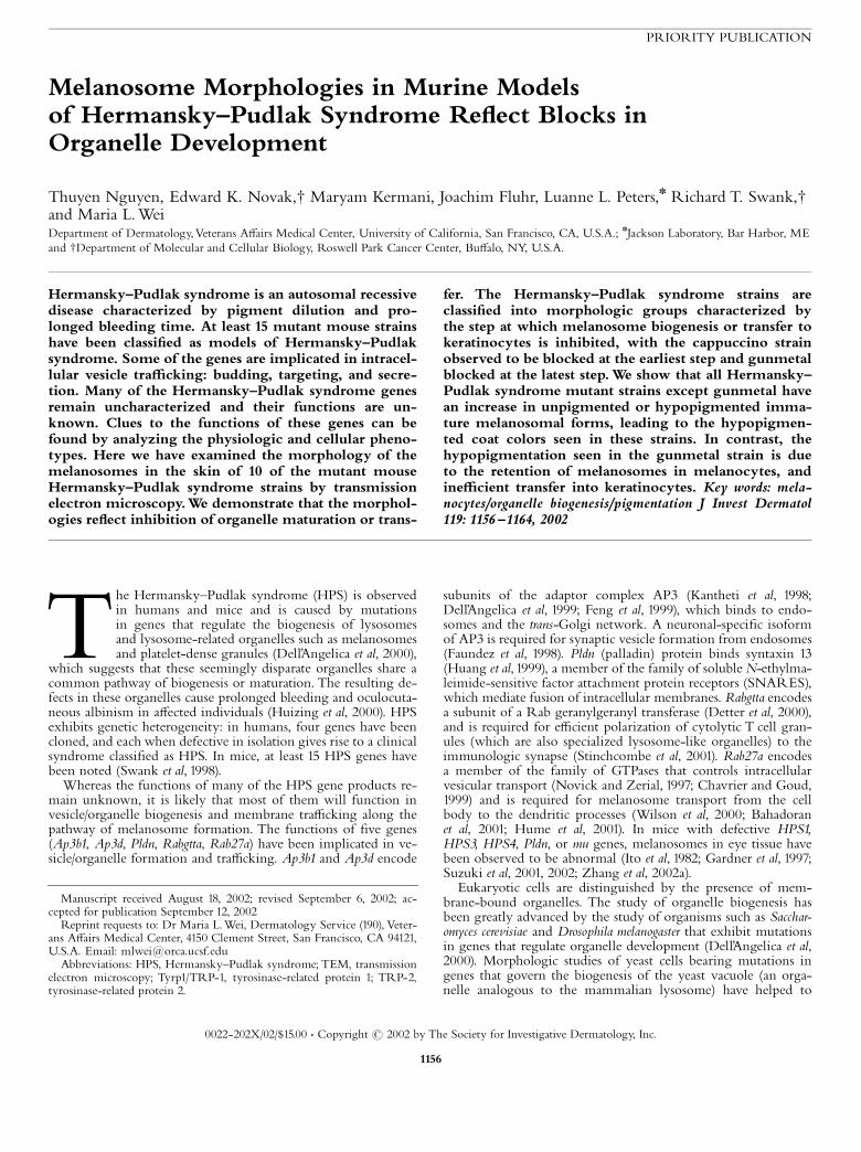
PRIORITY PUBLICATION
Melanosome Morphologies in Murine Modelsof Hermansky^Pudlak Syndrome Re£ect Blocks inOrganelle Development
Thuyen Nguyen, Edward K. Novak,w Maryam Kermani, Joachim Fluhr, Luanne L. Peters,n Richard T. Swank,wand Maria L. WeiDepartment of Dermatology,Veterans A¡airs Medical Center, University of California, San Francisco, CA, U.S.A.; nJackson Laboratory, Bar Harbor, MEand wDepartment of Molecular and Cellular Biology, Roswell Park Cancer Center, Bu¡alo, NY, U.S.A.
Hermansky^Pudlak syndrome is an autosomal recessivedisease characterized by pigment dilution and pro-longed bleeding time. At least 15 mutant mouse strainshave been classi¢ed as models of Hermansky^Pudlaksyndrome. Some of the genes are implicated in intracel-lular vesicle tra⁄cking: budding, targeting, and secre-tion. Many of the Hermansky^Pudlak syndrome genesremain uncharacterized and their functions are un-known. Clues to the functions of these genes can befound by analyzing the physiologic and cellular pheno-types. Here we have examined the morphology of themelanosomes in the skin of 10 of the mutant mouseHermansky^Pudlak syndrome strains by transmissionelectron microscopy.We demonstrate that the morphol-ogies re£ect inhibition of organelle maturation or trans-
fer. The Hermansky^Pudlak syndrome strains areclassi¢ed into morphologic groups characterized bythe step at which melanosome biogenesis or transfer tokeratinocytes is inhibited, with the cappuccino strainobserved to be blocked at the earliest step and gunmetalblocked at the latest step.We show that all Hermansky^Pudlak syndrome mutant strains except gunmetal havean increase in unpigmented or hypopigmented imma-ture melanosomal forms, leading to the hypopigmen-ted coat colors seen in these strains. In contrast, thehypopigmentation seen in the gunmetal strain is dueto the retention of melanosomes in melanocytes, andine⁄cient transfer into keratinocytes. Key words: mela-nocytes/organelle biogenesis/pigmentation J Invest Dermatol119: 1156 ^1164, 2002
The Hermansky^Pudlak syndrome (HPS) is observedin humans and mice and is caused by mutationsin genes that regulate the biogenesis of lysosomesand lysosome-related organelles such as melanosomesand platelet-dense granules (Dell’Angelica et al, 2000),
which suggests that these seemingly disparate organelles share acommon pathway of biogenesis or maturation. The resulting de-fects in these organelles cause prolonged bleeding and oculocuta-neous albinism in a¡ected individuals (Huizing et al, 2000). HPSexhibits genetic heterogeneity: in humans, four genes have beencloned, and each when defective in isolation gives rise to a clinicalsyndrome classi¢ed as HPS. In mice, at least 15 HPS genes havebeen noted (Swank et al, 1998).
Whereas the functions of many of the HPS gene products re-main unknown, it is likely that most of them will function invesicle/organelle biogenesis and membrane tra⁄cking along thepathway of melanosome formation. The functions of ¢ve genes(Ap3b1, Ap3d, Pldn, Rabgtta, Rab27a) have been implicated in ve-sicle/organelle formation and tra⁄cking. Ap3b1 and Ap3d encode
subunits of the adaptor complex AP3 (Kantheti et al, 1998;Dell’Angelica et al, 1999; Feng et al, 1999), which binds to endo-somes and the trans-Golgi network. A neuronal-speci¢c isoformof AP3 is required for synaptic vesicle formation from endosomes(Faundez et al, 1998). Pldn (palladin) protein binds syntaxin 13(Huang et al, 1999), a member of the family of soluble N-ethylma-leimide-sensitive factor attachment protein receptors (SNARES),which mediate fusion of intracellular membranes. Rabgtta encodesa subunit of a Rab geranylgeranyl transferase (Detter et al, 2000),and is required for e⁄cient polarization of cytolytic T cell gran-ules (which are also specialized lysosome-like organelles) to theimmunologic synapse (Stinchcombe et al, 2001). Rab27a encodesa member of the family of GTPases that controls intracellularvesicular transport (Novick and Zerial, 1997; Chavrier and Goud,1999) and is required for melanosome transport from the cellbody to the dendritic processes (Wilson et al, 2000; Bahadoranet al, 2001; Hume et al, 2001). In mice with defective HPS1,HPS3, HPS4, Pldn, or mu genes, melanosomes in eye tissue havebeen observed to be abnormal (Ito et al, 1982; Gardner et al, 1997;Suzuki et al, 2001, 2002; Zhang et al, 2002a).
Eukaryotic cells are distinguished by the presence of mem-brane-bound organelles. The study of organelle biogenesis hasbeen greatly advanced by the study of organisms such as Sacchar-omyces cerevisiae and Drosophila melanogaster that exhibit mutationsin genes that regulate organelle development (Dell’Angelica et al,2000). Morphologic studies of yeast cells bearing mutations ingenes that govern the biogenesis of the yeast vacuole (an orga-nelle analogous to the mammalian lysosome) have helped to
Reprint requests to: Dr Maria L.Wei, Dermatology Service (190), Veter-ans A¡airs Medical Center, 4150 Clement Street, San Francisco, CA 94121,U.S.A. Email: [email protected]
Abbreviations: HPS, Hermansky^Pudlak syndrome; TEM, transmissionelectron microscopy; Tyrp1/TRP-1, tyrosinase-related protein 1; TRP-2,tyrosinase-related protein 2.
Manuscript received August 18, 2002; revised September 6, 2002; ac-cepted for publication September 12, 2002
0022-202X/02/$15.00 � Copyright r 2002 by The Society for Investigative Dermatology, Inc.
1156
identify and classify more than 40 genes that were involved(Banta et al, 1988; Raymond et al, 1992;Wada et al, 1992). Althoughthe human HPS1, HPS3, and HPS4 genes have no sequencehomologs in yeast, the HPS2/Ap3b1 gene has a yeast homologand some of the as yet uncharacterized mouse genes may haveyeast homologs as well. HPS mutations result in mammalian dis-eases due to defects in organelle biogenesis. We have undertakenthe current morphologic study in order to characterize and classi-fy HPS mouse strains as a step towards understanding HPS geneproduct function.
The melanosome is an organelle uniquely suited for morpho-logic examination in tissue samples by transmission electron mi-croscopy (TEM) due to its electron-dense pigmentation. Otherorganelles consistently a¡ected in HPS are not readily amenableto examination by TEM. Platelet-dense granules in HPS losetheir electron density (Witkop et al, 1987) and lysosomes are onlyeasily identi¢ed after internalization of electron-dense material orby antibody labeling, and thus are more di⁄cult to identify intissue samples.
The melanosome has four distinct stages of maturation (Nordlundet al, 1998), stages I^IV. Type I melanosomes have intralumenal vesi-cles (Raposo et al, 2001) and resemble multivesicular bodies, struc-tures found along the endosomal pathway. Type II is characterizedby an elongated, elliptical shape, with intralumenal ¢ne ¢brils giv-ing a striated appearance; type III exhibits pigment deposition alongthe ¢brils, and type IV has dense pigmentation ¢lling the organelleand obscuring the ¢brillar structure (Seiji et al, 1963). In normal mel-anosome development, the type I melanosome is distinguished byits accumulation of the Pmel17/gp100 protein (silver locus product)(Kushimoto et al, 2001; Raposo et al, 2001; Raposo and Marks,2002), which is cleaved and localized to the intralumenal vesicles(Berson et al, 2001). Two enzymes involved in pigment synthesis,Tyrp1/ tyrosinase-related protein (TRP)-1 and dopachrome tauto-merase/TRP-2, may also be in the type I melanosome, but appearto be inactivated in this compartment by proteolytic cleavage (Kush-imoto et al, 2001). Pmel17/gp100 functions in the formation of themelanosomal striations and elongated shape to give rise to the typeII melanosome (Berson et al, 2001). Subsequently, pigment synthesisand deposition along the striations lead to the appearance of type IIIand IV forms.Whereas HPS-a¡ected individuals exhibit pigment di-lution, pigment is not absent, as a functional tyrosinase enzyme, ne-cessary and su⁄cient for pigment formation, is present.
We examined melanosomes in the skin of HPS mice, classi¢edthe mice by the stage of organelle maturity and demonstratedthat the defective genes result in blocks in melanosome develop-ment or transfer to keratinocytes.
MATERIALS AND METHODS
Mice Dorsal back skin samples from 4 wk old mice were harvested. Themutant mice and their corresponding control strain, C57BL/6, C3H/HeJ,or C57BL/10J were analyzed. All of the mutants arose spontaneously, moston the C57BL/6 background; others were bred into the C57BL/6 genotype
(Novak et al, 1980, 1984; Gibb et al, 1981; Zhang et al, 2002b). The cappuccinostrain arose on the C3H/HeJ background (Gwynn et al, 2000). The cocoamouse arose on the C57BL/10J background (Novak et al, 1988). The threecontrol strains had very similar melanosome morphology (data notshown), so C57BL/6 was used as a representative control. The HPS miceexamined and the corresponding human diseases and genes are listed inTable I. All animal procedures were approved by Institutional AnimalCare Use Committee.
Measurement of coat color The dorsal back coat pigmentation (P) wasmeasured using a Mexameter MX 18 (Courage & Khazaka, Cologne,Germany), which gives a calculated value of melanin content based onthe absorption of light emitted at 568 nm and 880 nm. Background wassubtracted by subtracting Ptyr, the coat color measurement for the whitealbino mouse C57BL/6J-Tyrc^2J (TyrG291 T), which is lacking tyrosinaseactivity and has no pigment production. A comparison with the coatpigment of the control strain (Pcon) was done:
Percent pigment ¼ ðP � PtyrÞ=ðPcon � PtyrÞ�100
Electron microscopy Tissue was ¢xed in modi¢ed Karnovsky’s ¢xative(2% paraformaldehyde/2% glutaraldehyde/0.1 M cacodylate bu¡er, pH 7.3/0.06% CaCl2) for 24 h, washed twice in 0.1 M cacodylate bu¡er, post¢xedin reduced osmium (1.5% potassium ferrocyanide/2% osmium tetroxide)for 2 h, rinsed in H2O, dehydrated with increasing ethanol concentrations,and embedded in Epon resin. Grids were stained in 10% uranyl acetate/50% methanol. Cross-sections of melanosomes were measured at� 50,000 magni¢cation for major axis and minor axis, in order to assessrelative size and shape. Images of a calibration grid were taken to controlfor any variability in magni¢cation. Melanocytes were identi¢ed by theirorganization in hair follicles, as well as their lack of keratin ¢laments anddesmosomes (both are present in keratinocytes). Each melanosome wasmeasured and classi¢ed by morphology, i.e.: (i) multiple intralumenalvesicles present, no pigment or striations seen; (ii) striated matrix present,round cross-section; (iii) striated matrix, elliptical cross-section; (iv) fullypigmented, round cross-section; and (v) fully pigmented, elliptical cross-section. Noted also were any melanosomal features unique to anyparticular strain. For each strain at least 100 melanosomes were examinedin each of at least two di¡erent animals. Statistical analysis of ¢ve controlmice revealed no interanimal variance.
Statistical analysis Statistics were calculated with Prism3 (GraphPad,San Diego, CA). The data were tested for normal distribution and in thecase of more than two groups an ANOVA with post-hoc pairwise comparisonwas calculated (Bonferroni or Dunn’s test, respectively).
RESULTS
HPS mouse strains have varying coat colors In HPSpatients, it has been observed that the pigment dilution can havewide variation, and at times can be quite subtle (Huizing et al,2000). The assessment of gene e¡ects in patients can becomplicated by variations in genetic backgrounds. This factorcan be eliminated by studying inbred mice, when mutant strainsshare a common genetic background, as is the case for most ofthe HPS murine strains. The HPS mouse strains were alsoobserved to have a range of pigment dilution. The strains can be
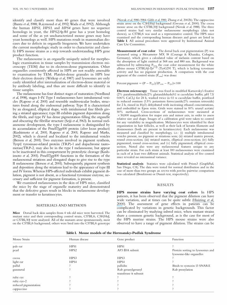
Table I. Mouse models of the Hermansky-Pudlak Syndrome
Mouse Strain Human disease Gene product Function
pale ear HPS1 HPS1 ?pearl HPS2 AP3 �3A submit Protein sorting to lysosomes and
lysosome-like organellescocoa HPS3 HPS3 ?light ear HPS4 HPS4 ?pallid ? pallidin Binds to syntaxin 13 SNAREgunmetal ? Rab geranylgeranyl
transferase a subunitRab prenylation
ruby eye ? ? ?ruby eye-2 ? ? ?reduced pigmentation ? ? ?cappuccino ? ? ?
MELANOSOMES IN HERMANSKY^PUDLAK SYNDROME 1157VOL. 119, NO. 5 NOVEMBER 2002

separated into two groups by coat color. The ¢rst group variesfrom dark to light shades of gray. This group consists ofgunmetal, pearl, pale ear, light ear, cocoa, reduced pigmentation,ruby eye, and ruby eye-2. As noted previously (Swank et al, 1998),the pale ear and light ear strains were identical in coat color andin having hypopigmented ears and tails. It is somewhat di⁄cultto discern cocoa from reduced pigmentation, and ruby eye andruby eye-2 are indistinguishable from one another by coat andeye color. The second group, consisting of pallid andcappuccino, is very hypopigmented, with hues in the o¡-whiteor cream-colored category.
It is possible that the coat color in these strains is an e¡ectivemarker for the severity of the defect in the biogenesis of themelanosome, and of similarly a¡ected organelles such as theplatelet-dense granule and the lysosome. In order to quantitatethe pigment dilution in these HPS strains, the coat color wasmeasured (Fig 1). The relative ranking of the strains byMexameter readings correlates well with the color rankingdiscerned by eye.
Murine HPS strains can be grouped by melanosomalmorphology Melanosomes in follicular melanocytes wereidenti¢ed and examined by TEM (Fig 2). In the parentalC57BL/6 strain (Fig 2C), a mixture of both ellipsoidal andspherical type IV fully pigmented melanosomes predominate.Striated type II and III forms are also readily seen. Very few tono spherical multivesicular bodies are seen.
A strikingly immature melanosomal phenotype was observedin the cappuccino and pallid strains (Fig 2; cno, pa), the twostrains that had the most hypopigmented coat colors. Incappuccino the melanosomes were noticeably smaller than thoseseen in the parental strain, and were markedly abnormal. Veryfew type IV melanosomes were observed, either elliptical orspherical. Pigmentation appeared granular and irregularlydeposited, compared with the ¢ne pigment deposited evenlyalong the striations seen in the parental strain. All structurescontaining pigment were spherical, and often, the pigment wasa small core within a larger limiting membrane. Few striatedstructures were observed, and multivesicular bodies were evidentand abundant. In pallid, an increase in multivesicular forms wasalso observed, and whereas relatively more striated forms were
seen in comparison with cappuccino, these forms were generallymisshapen and had irregular pigmentation.
A second group of mouse strains (cocoa, ruby, ruby eye-2,reduced pigmentation) appeared to have an intermediatematuration phenotype. These strains showed little to noevidence of ellipsoidal type IV melanosomes (Fig 2; coa, ru, ru-2,rp). Almost all fully pigmented forms were spherical. In addition,in some of these strains, an increase in the relative number ofstriated forms was evident, although these were not normallyshaped type II and III melanosomes. In reduced pigmentation,the striated forms were elongated, but most were irregularlyshaped and only approximated ellipsoidal shapes. In cocoa, rubyeye-2, and pallid, the striated forms were predominantly roundin cross-section, suggesting that they remained spherical. Also,spherical multivesicular bodies were observed, in contrast to therelative paucity of such forms in the parental strain.
The pale ear, light ear, pearl, and gunmetal strains exhibitednear normal phenotype by TEM. The melanosomes appearedquite similar to those in the parental strain, in that type IV fullypigmented melanosomes were the predominant form (Fig 2;ep, le, pe, gm). Type II and III forms were seen to a lesser extentthan type IV melanosomes. Pearl, however, was distinguishedfrom gunmetal, pale ear, light ear, and C57BL/6 by an apparentincrease in melanosomal membrane blebbing, suggesting abnormalvesicular tra⁄cking involving these melanosomes.
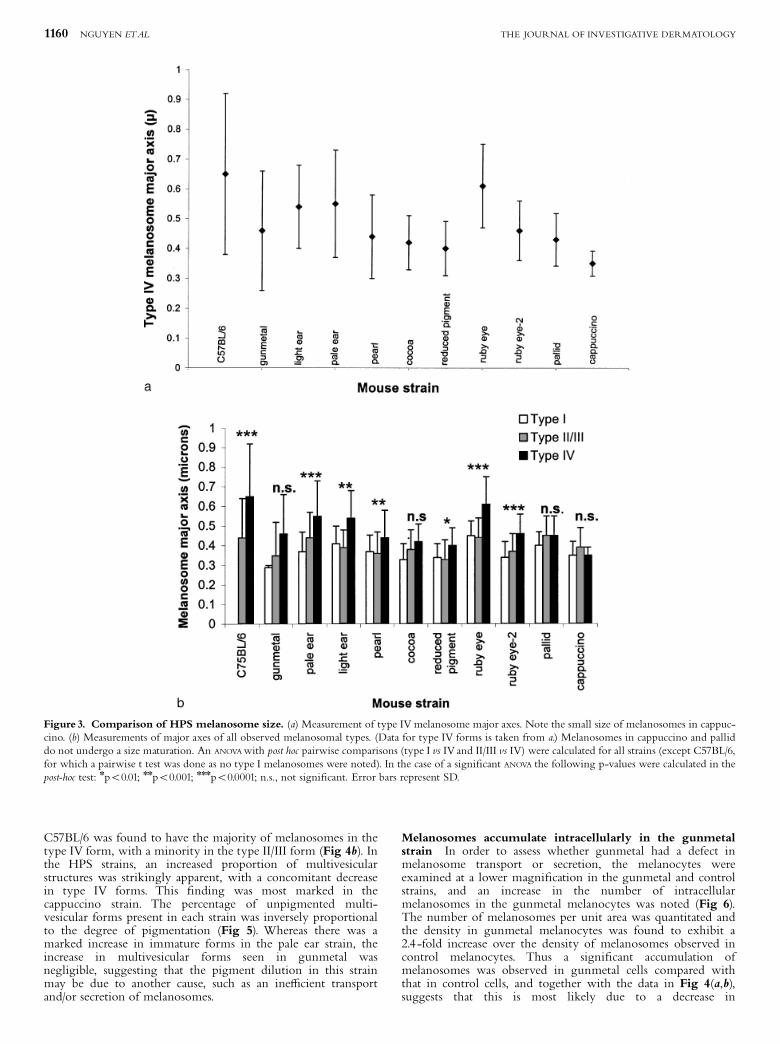
Melanosome biogenesis in the cappuccino and pallid strainsis blocked at an early stage In each of the mouse strains, themajor axis of type IV melanosomes were measured as anindication of mature melanosome size (Fig 3a). The few, poorlyformed type IV melanosomes from the cappuccino mouse werefound to be signi¢cantly smaller than those measured in theparental strain. Most of the other strains exhibited maturemelanosomes that were slightly smaller than the parentalmelanosome size.
The major axis of each of the immature melanosomal formswas also measured to quantitate the sizes of these forms (Fig3b), and to determine if there was an increase in size whenmelanosomes underwent maturation. In the parental cells, thereis a clear progression in size from type II/III melanosomes totype IV. A less marked progression in size from precursor to
Figure1. Coat color variation in HPS strains. Pigmentation was measured using a Mexameter. Tyr-c, tyrosinase activity-negative mouse strain C57BL/6J-Tyrc^2J (TyrG291T) whose melanosomes are devoid of pigment. Error bars represent SD.
1158 NGUYEN ETAL THE JOURNAL OF INVESTIGATIVE DERMATOLOGY

mature melanosome was seen in most of the other strains. Theincrease in size was completely abolished in both the cappuccinoand pallid strains, suggesting that melanosomal development wasarrested at an early stage in these strains. Two additional strains,gunmetal and cocoa, also did not undergo a signi¢cant increasein size from type II/III to type IV forms.
The percentage of immature melanosomal forms isincreased in HPS mouse strains As it appeared that thepallid and cappuccino strains were blocked at an early stage ofmelanosome development, it seemed likely that the remainingstrains could also be blocked or inhibited at a morphologicallydistinct step in biogenesis, so a quantitative assessment ofmelanosomal maturity was done. One indication of degree ofmaturity could be shape, with immature spherical formsprogressing to more mature elliptical forms. Therefore, the shape
distribution of the type IV melanosomes was assessed (Fig 4a). InC57BL/6 cells, there was a slight predominance of spherical vselliptical forms (52% vs 48%). A similar distribution was seen inthe light ear and pearl strains. Several strains (cocoa, reducedpigmentation, ruby eye, ruby eye-2, pallid, cappuccino) hada marked shift of distribution towards a predominance ofspherical forms, suggesting that these strains had an increase inthe percentage of immature forms. Two strains (pale ear andgunmetal) that we had identi¢ed as ‘‘near normal’’ on the basis ofpigment content and overall morphology, had a shift indistribution towards more elliptical forms. The secretion of theseelliptical type IV forms might be a rate-limiting step in thesestrains, or a rate-limiting step at an earlier point of biogenesismight cause an increase in elliptical type II/III forms.
To distinguish between these two possibilities, the relativenumbers of the di¡erent melanosomal forms were quantitated.
Figure 2. HPS mouse strains exhibit di¡erent degrees of aberrant melanosomal morphology. C, C57BL/6; cno, cappuccino; pa, pallid; coa, cocoa;ru, ruby eye; ru-2, ruby eye-2; rp, reduced pigmentation; ep, pale ear; le, light ear; pe, pearl; gm, gunmetal. The cappuccino strain is unique in having verysmall, abnormally pigmented structures. The ¢elds were chosen to show representative forms of all melanosomal types for each strain, so do not necessarilydemonstrate predominance of any speci¢c melanosomal type. Asterisks, mitochondria. White arrowheads, multivesicular bodies; arrows, striated forms; blackarrowhead, blebbing of the limiting membrane in pearl melanosomes. Scale bar¼ 0.5 mm.
MELANOSOMES IN HERMANSKY^PUDLAK SYNDROME 1159VOL. 119, NO. 5 NOVEMBER 2002
C57BL/6 was found to have the majority of melanosomes in thetype IV form, with a minority in the type II/III form (Fig 4b). Inthe HPS strains, an increased proportion of multivesicularstructures was strikingly apparent, with a concomitant decreasein type IV forms. This ¢nding was most marked in thecappuccino strain. The percentage of unpigmented multi-vesicular forms present in each strain was inversely proportionalto the degree of pigmentation (Fig 5). Whereas there was amarked increase in immature forms in the pale ear strain, theincrease in multivesicular forms seen in gunmetal wasnegligible, suggesting that the pigment dilution in this strainmay be due to another cause, such as an ine⁄cient transportand/or secretion of melanosomes.
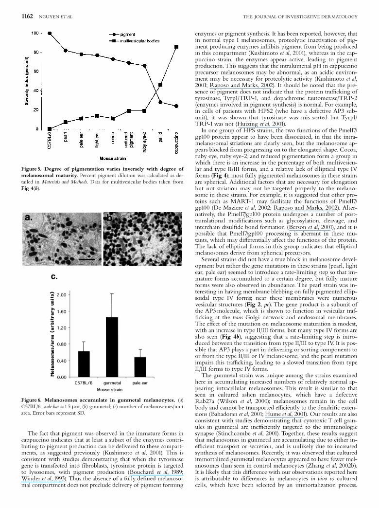
Melanosomes accumulate intracellularly in the gunmetalstrain In order to assess whether gunmetal had a defect inmelanosome transport or secretion, the melanocytes wereexamined at a lower magni¢cation in the gunmetal and controlstrains, and an increase in the number of intracellularmelanosomes in the gunmetal melanocytes was noted (Fig 6).The number of melanosomes per unit area was quantitated andthe density in gunmetal melanocytes was found to exhibit a2.4 -fold increase over the density of melanosomes observed incontrol melanocytes. Thus a signi¢cant accumulation ofmelanosomes was observed in gunmetal cells compared withthat in control cells, and together with the data in Fig 4(a,b),suggests that this is most likely due to a decrease in
Figure 3. Comparison of HPS melanosome size. (a) Measurement of type IV melanosome major axes. Note the small size of melanosomes in cappuc-cino. (b) Measurements of major axes of all observed melanosomal types. (Data for type IV forms is taken from a.) Melanosomes in cappuccino and palliddo not undergo a size maturation. An ANOVA with post hoc pairwise comparisons (type I vs IV and II/III vs IV) were calculated for all strains (except C57BL/6,for which a pairwise t test was done as no type I melanosomes were noted). In the case of a signi¢cant ANOVA the following p-values were calculated in thepost-hoc test: npo0.01; nnpo0.001; nnnpo0.0001; n.s., not signi¢cant. Error bars represent SD.
1160 NGUYEN ETAL THE JOURNAL OF INVESTIGATIVE DERMATOLOGY

melanosomal transport or secretion (see Discussion). In contrast,the number of melanosomes per unit area in pale earmelanocytes was somewhat decreased compared with controlcells, suggesting that no block in melanosome transport exists inthis strain.
DISCUSSION
This study is a direct demonstration that HPS can be character-ized by blocks in organelle development or transport. We ob-served that in many of the strains there was an increase in thepercentage of immature forms relative to mature forms, suggest-ing that a rate-limiting step or block was introduced into thepathway of melanosome biogenesis.
The cappuccino and pallid strains exhibited an inhibition ofmelanosomal development at the earliest step, with cappuccinobeing the most severely a¡ected. The accumulation of multivesi-cular structures in these strains is additional evidence that thesestructures are early precursors in melanosome biogenesis, possiblytype I melanosomes (Raposo et al, 2001); this further con¢rms theclose relationship between melanosomes and lysosomes (Orlow,1995), which also have multivesicular bodies as precursors. As thepredominant melanosome form in cappuccino has multivesicularmorphology, and few striated and elongated shapes are observed,it appears that the Pmel17/gp100 protein may not be functioningnormally in this strain. This may be due to a lack of cleavage ofPmel17/gp100, as it has been suggested that cleavage is required forstriation formation (Raposo and Marks, 2002), or due to aberranttra⁄cking of the protein so that Pmel17/gp100 is not reaching theprecursor melanosome.
Figure 4. Melanosomes are shifted to immature forms in most HPS strains. (a) Type IV melanosomes classi¢ed by shape.White bars, percent of typeIV melanosomes that are spherical. Gray bars, percent of type IV melanosomes that are elliptical. (b) Melanosomes classi¢ed by morphologic stage.White bars,percent of multivesicular bodies. Light gray bars, percent of melanosomes that are type II/III. Dark gray bars, percent of melanosomes that are type IV.
MELANOSOMES IN HERMANSKY^PUDLAK SYNDROME 1161VOL. 119, NO. 5 NOVEMBER 2002
The fact that pigment was observed in the immature forms incappuccino indicates that at least a subset of the enzymes contri-buting to pigment production can be delivered to these compart-ments, as suggested previously (Kushimoto et al, 2001). This isconsistent with studies demonstrating that when the tyrosinasegene is transfected into ¢broblasts, tyrosinase protein is targetedto lysosomes, with pigment production (Bouchard et al, 1989;Winder et al, 1993). Thus the absence of a fully de¢ned melanoso-mal compartment does not preclude delivery of pigment forming
enzymes or pigment synthesis. It has been reported, however, thatin normal type I melanosomes, proteolytic inactivation of pig-ment producing enzymes inhibits pigment from being producedin this compartment (Kushimoto et al, 2001), whereas in the cap-puccino strain, the enzymes appear active, leading to pigmentproduction. This suggests that the intralumenal pH in cappuccinoprecursor melanosomes may be abnormal, as an acidic environ-ment may be necessary for proteolytic activity (Kushimoto et al,2001; Raposo and Marks, 2002). It should be noted that the pre-sence of pigment does not indicate that the protein tra⁄cking oftyrosinase, Tyrp1/TRP-1, and dopachrome tautomerase/TRP-2(enzymes involved in pigment synthesis) is normal. For example,in cells of patients with HPS2 (who have a defective AP3 sub-unit), it was shown that tyrosinase was mis-sorted but Tyrp1/TRP-1 was not (Huizing et al, 2001).
In one group of HPS strains, the two functions of the Pmel17/gp100 protein appear to have been dissociated, in that the intra-melanosomal striations are clearly seen, but the melanosome ap-pears blocked from progressing on to the elongated shape. Cocoa,ruby eye, ruby eye-2, and reduced pigmentation form a group inwhich there is an increase in the percentage of both multivesicu-lar and type II/III forms, and a relative lack of elliptical type IVforms (Fig 4); most fully pigmented melanosomes in these strainsare spherical. Additional factors that are necessary for elongationbut not striation may not be targeted properly to the melano-some in these strains. For example, it is suggested that other pro-teins such as MART-1 may facilitate the functions of Pmel17/gp100 (De Maziere et al, 2002; Raposo and Marks, 2002). Alter-natively, the Pmel17/gp100 protein undergoes a number of post-translational modi¢cations such as glycosylation, cleavage, andinterchain disul¢de bond formation (Berson et al, 2001), and it ispossible that Pmel17/gp100 processing is aberrant in these mu-tants, which may di¡erentially a¡ect the functions of the protein.The lack of elliptical forms in this group indicates that ellipticalmelanosomes derive from spherical precursors.
Several strains did not have a true block in melanosome devel-opment but rather the gene mutations in these strains (pearl, lightear, pale ear) seemed to introduce a rate-limiting step so that im-mature forms accumulated to a certain degree, but fully matureforms were also observed in abundance. The pearl strain was in-teresting in having membrane blebbing on fully pigmented ellip-soidal type IV forms; near these membranes were numerousvesicular structures (Fig 2, pe). The gene product is a subunit ofthe AP3 molecule, which is shown to function in vesicular traf-¢cking at the trans-Golgi network and endosomal membranes.The e¡ect of the mutation on melanosome maturation is modest,with an increase in type II/III forms, but many type IV forms arealso seen (Fig 4b), suggesting that a rate-limiting step is intro-duced between the transition from type II/III to type IV. It is pos-sible that AP3 plays a part in delivering or sorting components toor from the type II/III or IV melanosome, and the pearl mutationimpairs this tra⁄cking, leading to a slowed transition from typeII/III forms to type IV forms.
The gunmetal strain was unique among the strains examinedhere in accumulating increased numbers of relatively normal ap-pearing intracellular melanosomes. This result is similar to thatseen in cultured ashen melanocytes, which have a defectiveRab27a (Wilson et al, 2000); melanosomes remain in the cellbody and cannot be transported e⁄ciently to the dendritic exten-sions (Bahadoran et al, 2001; Hume et al, 2001). Our results are alsoconsistent with studies demonstrating that cytotoxic T cell gran-ules in gunmetal are ine⁄ciently targeted to the immunologicsynapse (Stinchcombe et al, 2001). Together, these results suggestthat melanosomes in gunmetal are accumulating due to either in-e⁄cient transport or secretion, and is unlikely due to increasedsynthesis of melanosomes. Recently, it was observed that culturedimmortalized gunmetal melanocytes appeared to have fewer mel-anosomes than seen in control melanocytes (Zhang et al, 2002b).It is likely that this di¡erence with our observations reported hereis attributable to di¡erences in melanocytes in vivo vs culturedcells, which have been selected by an immortalization process.
Figure 5. Degree of pigmentation varies inversely with degree ofmelanosomal maturity. Percent pigment dilution was calculated as de-tailed in Materials and Methods. Data for multivesicular bodies taken fromFig 4(b).
Figure 6. Melanosomes accumulate in gunmetal melanocytes. (a)C57BL/6, scale bar¼1.5 mm; (b) gunmetal; (c) number of melanosomes/unitarea. Error bars represent SD.
1162 NGUYEN ETAL THE JOURNAL OF INVESTIGATIVE DERMATOLOGY

The mutation in gunmetal not only a¡ects melanosomal trans-port, but also melanosomal size and maturation: the gunmetalmelanosomes were smaller than those in control cells, and didnot undergo a size increase from type II/III to type IV melano-somes. This is possibly due to the defective gene product in gun-metal a¡ecting the prenylation of other rab proteins, in additionto Rab27a.
Two strains, pale ear and light ear, have melanosomes that aresimilar in appearance to one another and to wild-type melano-somes as well. This correlates with the observation that the bodycoat colors of both pale ear and light ear adult mice are dark andindistinguishable from one another (Swank et al, 1998), and thatthe pale ear gene product is not detectable in extracts from lightear cells, suggesting that the pale ear and light ear gene productsfunction together in a complex (Suzuki et al, 2002). Similarly,ruby eye and ruby eye-2 are phenotypically indistinguishable,and the strains have very similar pro¢les of melanosome matur-ity. It does appear, however, that ruby eye can be distinguishedfrom ruby eye-2 on the basis of average melanosome size.
Our ¢ndings of smaller melanosomes in the cocoa, pallid, andcappuccino strains are in agreement with previous studies that ex-amined cultured melanocytes (Suzuki et al, 2001), skin tissue (Itoet al, 1982), and ocular tissue (Gwynn et al, 2000), respectively. An-other study (Gardner et al, 1997) reported that pale ear culturedmelanocytes contained melanosomes that were larger in size thanseen in control melanocytes. This discrepancy with our observa-tions can be explained by several factors. First, there may be dif-ferences due to examination of cultured cells vs melanocytes intissue. Second, this study reports a statistical analysis of a largenumber of melanosomes, whereas the previous study selectivelymeasured the largest melanosomes. It was also noted (Gardneret al, 1997; Suzuki et al, 2002) that pale ear and light ear had en-larged melanosomes in choroid melanocytes. This ¢nding of lar-ger melanosomes could be due to tissue-speci¢c di¡erencesbetween melanocytes in the skin vs in the eye; melanocytes inthe skin are secretory cells, whereas those in the eye are notknown to secrete melanosomes. Alternatively the observationalnature of the previous studies could account for the reported sizedi¡erences; for example, the study of pallid (Ito et al, 1982) notedpredominantly immature melanosomes in melanocytes, but occa-sional melanocytes with megamelanosomes.
The varying degrees to which melanosomal forms were shiftedto immature, less pigmented forms (Fig 4b) suggests a mechan-ism for the varying degrees of hypopigmentation seen in theHPS coat colors. As shown in Fig 5, an increase in the percentageof unpigmented multivesicular forms correlates with coat colordilution. Coat color is determined by the transfer of melano-somes into keratinocytes and subsequent incorporation into thehair shaft. Very little is known about the regulation of melano-some transfer, but we observed that immature melanosomalforms are transferred to keratinocytes (M.W. unpublished obser-vations), indicating that secretion and uptake of melanosomes bykeratinocytes is not limited to type IV forms. Thus, it is possiblethat hypopigmentation of the hair in most of these strains iscaused by a dilution e¡ect due to an increased percentage ofrelatively hypopigmented or unpigmented melanosomes beingtransferred; however, the hypopigmentation seen in gunmetalmice appears mainly due to retention of melanosomes in themelanocyte. Additionally, hypopigmentation may result fromabnormal pigment synthesis, due to altered tra⁄cking of selectedenzymes or other components involved in pigment synthesis.It has been shown that transfection of HPS1 anti-sense cDNAinto human melanoma cells caused mislocalization of tyrosinaseand Tyrp1/TRP-1, and decreased melanin content (Sarangarajanet al, 2001). Also, when tyrosinase is routed to a compartment,such as the lysosome, which does not contain the full comple-ment of proteins found in normal melanosomes, the pigmentsynthesized is pheomelanin, not eumelanin (Winder et al,1993). It is not known if the biochemical composition of pigmentin these HPS strains corresponds to that found in the controlstrains.
In summary, we have examined melanosomes in HPS murinestrains and classi¢ed the strains into categories that re£ect the de-gree of melanosomal maturation. Figure 7 depicts a proposedmodel that places the HPS protein products along the pathwayof melanosomal biogenesis. Study of the murine melanosomes isuseful in several ways. First, this quantitative in vivo assessment ofthe e¡ects of HPS genes on a subcellular organelle shows clearlythat these genes are involved in organelle maturation. Second,comparison of melanosome morphology from suspected HPSpatients with melanosomes from murine strains could facilitatediagnosis of HPS. Currently, it is thought that many HPS pa-tients are unrecognized and are diagnosed as having an unclassi-¢ed bleeding disorder. To diagnose patients with HPS, bleedingtime is measured, an ophthalmologic examination is done, andTEM analysis of platelets is performed. This last test is only avail-able in one lab in the United States (Witkop et al, 1987), whereasTEM of a skin biopsy to examine melanosomes could be doneusing a few specialized university-based pathology laboratorieswith an interest in this area. Third, it is very likely that the HPSmurine genes will be sequenced before the human genes, as themurine strains and corresponding chromosomal regions are al-ready identi¢ed. By comparing patient melanosome morphologywith murine HPS melanosome morphology, it may be possible
Figure 7. The morphologies of melanosomes in HPS strains re£ectblocks in organelle biogenesis.The cappuccino (cno) mutation results ina proximal block (step 1) such that very few striated type II/III forms orfully pigmented forms are observed. The sizes of the organelles remainthe size of precursor organelles, suggesting that pigment is being depositedin these early forms. The pallid (pa) mutation inhibits maturation at thesame step as the cappuccino block. A more distal block (step 2) is predictedfor the cocoa (coa), ruby eye (ru), ruby eye-2 (ru-2) and reduced pigmenta-tion (rp) strains, as striated spherical forms are seen, but few ellipsoidalforms are noted. Accumulation of melanosomes in gunmetal (gm) melano-cytes suggests that melanosomes are retained and ine⁄ciently secreted (step4). Introduction of a rate-limiting step, but not a true block in biogenesis, ispredicted for pale ear and light ear (step 1), and pearl (step 3). Blebbing ofmelanosome limiting membrane in the pearl strain suggests that increasedand/or slowed vesicular activity is occurring. TGN, trans-Golgi network.
MELANOSOMES IN HERMANSKY^PUDLAK SYNDROME 1163VOL. 119, NO. 5 NOVEMBER 2002

to classify human patients, to narrow down which chromosomalregions the defective genes are likely to reside, and to facilitateidenti¢cation of the human HPS genes.
Many thanks to Drs Corey Largman and Regis Kelly for critical reading of the manu-script.We are grateful to Sandra Huling, Debra Crumrine, and Jerelyn Magnusson forexcellent technical assistance.This work was supported by the Department of VeteransAdministration (Research Career Development Award, M.L.W.), also in part by Na-tional Institutes of Health (NIH HL55321, L.L.P.; HL51480, HL31698, EY12104,R.T.S.) and this research utilized core facilities supported in part by RPCI’s NCI-funded Cancer Center Support Grant, CA16056.
REFERENCES
Bahadoran P, Aberdam E, Mantoux F, et al: Rab27a: a key to melanosome transportin human melanocytes. J Cell Biol 152:843^850, 2001
Banta LM, Robinson JS, Klionsky DJ, Emr SD: Organelle assembly in yeast, char-acterization of yeast mutants defective in vacuolar biogenesis and protein sort-ing. J Cell Biol 107:1369^1383, 1988
Berson JF, Harper DC, Tenza D, Raposo G, Marks MS: Pmel17 initiates premelano-some morphogenesis within multivesicular bodies. Mol Biol Cell 12:3451^3464,2001
Bouchard B, Fuller BB, Vijayasaradhi S, Houghton AN: Induction of pigmentationin mouse ¢broblasts by expression of human tyrosinase cDNA. J Exp Med169:2029^2042, 1989
Chavrier P, Goud B: The role of ARF and Rab GTPases in membrane transport.Curr Opin Cell Biol 11:466^475, 1999
De Maziere AM, Muehlethaler K, Van Donselaar E, et al: The melanocytic proteinmelan-A/MART-1 has a subcellular localization distinct from typical melano-somal proteins. Tra⁄c 3:678^693, 2002
Dell’Angelica EC, Shotelersuk V, Aguilar RC, Gahl WA, Bonifacino JS: Altered traf-¢cking of lysosomal proteins in Hermansky^Pudlak syndrome due to muta-tions in the beta 3A subunit of the AP-3 adaptor. Mol Cell 3:11^21, 1999
Dell’Angelica EC, Mullins C, Caplan S, Bonifacino JS: Lysosome-related organelles.FASEB J 14:1265^1278, 2000
Detter JC, Zhang Q, Mules EH, et al: Rab geranylgeranyl transferase alpha mutationin the gunmetal mouse reduces Rab prenylation and platelet synthesis. ProcNatl Acad Sci USA 97:4144^4149, 2000
Faundez V, Horng JT, Kelly RB: A function for the AP3 coat complex in synapticvesicle formation from endosomes. Cell 93:423^432, 1998
Feng L, Seymour AB, Jiang S, et al: The beta3A subunit gene (Ap3b1) of the AP-3adaptor complex is altered in the mouse hypopigmentation mutant pearl, amodel for Hermansky^Pudlak syndrome and night blindness. Hum Mol Genet8:323^330, 1999
Gardner JM, Wildenberg SC, Keiper NM, et al: The mouse pale ear (ep) mutation isthe homologue of human Hermansky^Pudlak syndrome. Proc Natl Acad SciUSA 94:9238^9243, 1997
Gibb S, Hakansson EM, Lundin LG, Shire JG: Reduced pigmentation (rp), a newcoat colour gene with e¡ects on kidney lysosomal glycosidases in the mouse.Genet Res 37:95^103, 1981
Gwynn B, Ciciotte SL, Hunter SJ, et al: Defects in the cappuccino (cno) gene onmouse chromosome 5 and human 4p cause Hermansky^Pudlak syndrome byan AP-3-independent mechanism. Blood 96:4227^4235, 2000
Huang L, Kuo YM, Gitschier J: The pallid gene encodes a novel, syntaxin 13-inter-acting protein involved in platelet storage pool de¢ciency. Nat Genet 23:329^332, 1999
Huizing M, Anikster Y, Gahl WA: Hermansky^Pudlak syndrome and related disor-ders of organelle formation. Tra⁄c 1:823^835, 2000
Huizing M, Sarangarajan R, Strovel E, Zhao Y, Gahl WA, Boissy RE: AP-3 med-iates tyrosinase but not TRP-1 tra⁄cking in human melanocytes. Mol Biol Cell12:2075^2085, 2001
Hume AN, Collinson LM, Rapak A, Gomes AQ, Hopkins CR, Seabra MC: Rab27aregulates the peripheral distribution of melanosomes in melanocytes. J Cell Biol152:795^808, 2001
Ito M, Hashimoto K, Organisciak DT: Ultrastructural, histochemical, and biochem-ical studies of the melanin metabolism in eye and skin of pallid mice. J InvestDermatol 78:414^424, 1982
Kantheti P, Qiao X, Diaz ME, et al: Mutation in AP-3 delta in the mocha mouselinks endosomal transport to storage de¢ciency in platelets, melanosomes,and synaptic vesicles. Neuron 21:111^122, 1998
Kushimoto T, Basrur V, Valencia J, et al: A model for melanosome biogenesis basedon the puri¢cation and analysis of early melanosomes. Proc Natl Acad Sci USA98:10698^10703, 2001
Nordlund JJ, Boissy RE, Hearing VJ, King RA, Ortonne JP: The Pigmentary System.Physiology and Pathophysiology. New York: Oxford University Press, 1998
Novak EK,Wieland F, Jahreis GP, Swank RT: Altered secretion of kidney lysosomalenzymes in the mouse pigment mutants ruby-eye, ruby-eye-2-J, and maroon.Biochem Genet 18:549^561, 1980
Novak EK, Hui SW, Swank RT: Platelet storage pool de¢ciency in mouse pigmentmutations associated with seven distinct genetic loci. Blood 63:536^544, 1984
Novak EK, Sweet HO, Prochazka M, et al: Cocoa: a new mouse model for plateletstorage pool de¢ciency. Br J Haematol 69:371^378, 1988
Novick P, Zerial M:The diversity of Rab proteins in vesicle transport. Curr Opin CellBiol 9:496^504, 1997
Orlow SJ: Melanosomes are specialized members of the lysosomal lineage of orga-nelles. J Invest Dermatol 105:3^7, 1995
Raposo G, Marks MS: The dark side of lysosome-related organelles: specialization ofthe endocytic pathway for melanosome biogenesis.Tra⁄c 3:237^248, 2002
Raposo G, Tenza D, Murphy DM, Berson JF, Marks MS: Distinct protein sortingand localization to premelanosomes, melanosomes, and lysosomes in pigmen-ted melanocytic cells. J Cell Biol 152:809^824, 2001
Raymond CK, Howald-Stevenson I, Vater CA, Stevens TH: Morphological classi¢-cation of the yeast vacuolar protein sorting mutants: evidence for a prevacuolarcompartment in class E vps mutants. Mol Biol Cell 3:1389^1402, 1992
Sarangarajan R, Budev A, ZhaoY, Gahl WA, Boissy RE: Abnormal translocation oftyrosinase and tyrosinase-related protein 1 in cutaneous melanocytes of Her-mansky^Pudlak syndrome and in melanoma cells transfected with anti-senseHPS1 cDNA. J Invest Dermatol 117:641^646, 2001
Seiji M, Fitzpatrick T, Simpson R, Birbeck M: Chemical composition and terminol-ogy of specialized organelles (melanosomes and melanin granules) in mamma-lian melanocytes. Nature 197:1082^1084, 1963
Stinchcombe JC, Barral DC, Mules EH, et al: Rab27a is required for regulated secre-tion in cytotoxic T lymphocytes. J Cell Biol 152:825^834, 2001
Suzuki T, Li W, Zhang Q, et al: The gene mutated in cocoa mice, carrying a defect oforganelle biogenesis, is a homologue of the human Hermansky^Pudlak syn-drome-3 gene. Genomics 78:30^37, 2001
Suzuki T, Li W, Zhang Q, et al: Hermansky^Pudlak syndrome is caused by mutationsin HPS4, the human homolog of the mouse light-ear gene. Nat Genet 30:321^324, 2002
Swank RT, Novak EK, McGarry MP, Rusiniak ME, Feng L: Mouse models of Her-mansky Pudlak syndrome: a review. Pigment Cell Res 11:60^80, 1998
Wada Y, Ohsumi Y, Anraku Y: Genes for directing vacuolar morphogenesis in Sac-charomyces cerevisiae . I. Isolation and characterization of two classes of vam mu-tants. J Biol Chem 267:18665^18670, 1992
Wilson SM,Yip R, Swing DA, et al: A mutation in Rab27a causes the vesicle trans-port defects observed in ashen mice. Proc Natl Acad Sci USA 97:7933^7938, 2000
Winder AJ, Wittbjer A, Rosengren E, Rorsman H: Fibroblasts expressing mouse clocus tyrosinase produce an authentic enzyme and synthesize phaeomelanin. JCell Sci 104:467^475, 1993
Witkop CJ, Krumwiede M, Sedano H,White JG: Reliability of absent platelet densebodies as a diagnostic criterion for Hermansky^Pudlak syndrome. Am J Hema-tol 26:305^311, 1987
Zhang Q, Li W, Novak EK, et al: The gene for the muted (mu) mouse, a model forHermansky^Pudlak syndrome, de¢nes a novel protein which regulates vesicletra⁄cking. Hum Mol Genet 11:697^706, 2002a
Zhang Q, Zhen L, Li W, et al: Cell-speci¢c abnormal prenylation of Rab proteins inplatelets and melanocytes of the gunmetal mouse. Br J Haematol 117:414^423,2002b
1164 NGUYEN ETAL THE JOURNAL OF INVESTIGATIVE DERMATOLOGY
Copyright © 2022 FDOKUMEN