
Medicine, Faculty of
21
International Journal of Systematic and Evolutionary Microbiology (2001), 51, 2145–2165 Printed in Great Britain Reorganization of genera in the families Rickettsiaceae and Anaplasmataceae in the order Rickettsiales : unification of some species of Ehrlichia with Anaplasma, Cowdria with Ehrlichia and Ehrlichia with Neorickettsia, descriptions of six new species combinations and designation of Ehrlichia equi and ‘HGE agent ’ as subjective synonyms of Ehrlichia phagocytophila 1 Division of Medical Microbiology, Department of Pathology, The Johns Hopkins Medical Institutions, Meyer B1-193, 600 North Wolfe St, Baltimore, MD 21287, USA 2 Department of Infectious Diseases, College of Veterinary Medicine, University of Florida, Gainesville, FL, USA 3 Division of Parasitology and Tropical Veterinary Medicine, Faculty of Veterinary Medicine, Utrecht University, Utrecht, The Netherlands 4 Naval Medical Research Center, Silver Spring, MD, USA 5 Department of Veterinary Microbiology and Pathology, Washington State University, Pullman, WA, USA 6 Division of Infectious Diseases, Department of Medicine, The Johns Hopkins Medical Institutions, Baltimore, MD, USA 7 Department of Veterinary Biosciences, College of Veterinary Medicine, The Ohio State University, Columbus, OH, USA J. Stephen Dumler, 1 Anthony F. Barbet, 2 Cornelis P. J. Bekker, 3 Gregory A. Dasch, 4 Guy H. Palmer, 5 Stuart C. Ray, 6 Yasuko Rikihisa 7 and Fred R. Rurangirwa 5 Author for correspondence : J. Stephen Dumler. Tel : ›1 410 955 5077. Fax: ›1 410 614 8087. e-mail : sdumler!jhmi.edu The genera Anaplasma, Ehrlichia, Cowdria, Neorickettsia and Wolbachia encompass a group of obligate intracellular bacteria that reside in vacuoles of eukaryotic cells and were previously placed in taxa based upon morphological, ecological, epidemiological and clinical characteristics. Recent genetic analyses of 16S rRNA genes, groESL and surface protein genes have indicated that the existing taxa designations are flawed. All 16S rRNA gene and groESL sequences deposited in GenBank prior to 2000 and selected sequences deposited thereafter were aligned and phylogenetic trees and bootstrap values were calculated using the neighbour-joining method and compared with trees generated with maximum-probability, maximum-likelihood, majority-rule consensus and parsimony methods. Supported by bootstrap probabilities of at least 54 %, 16S rRNA gene comparisons consistently clustered to yield four distinct clades characterized roughly as Anaplasma (including the Ehrlichia phagocytophila group, Ehrlichia platys and Ehrlichia bovis) with a minimum of 96<1 % similarity, Ehrlichia (including Cowdria ruminantium) with a minimum of 97<7 % similarity, Wolbachia with a minimum of 95<6% similarity and Neorickettsia (including Ehrlichia sennetsu and Ehrlichia risticii ) with a minimum of 94<9 % similarity. Maximum similarity between clades ranged from 87<1 to 94<9 %. Insufficient differences existed among E. phagocytophila, Ehrlichia equi and the human granulocytic ehrlichiosis (HGE) agent to support separate species designations, and this group was at least 98<2 % similar to any Anaplasma species. These 16S rRNA gene analyses are strongly supported by similar groESL clades, as well as biological and antigenic characteristics. It is proposed that all members of the tribes Ehrlichieae and Wolbachieae be transferred to the family Anaplasmataceae and that the tribe structure of the family Rickettsiaceae be eliminated. The genus Anaplasma should be emended to include Anaplasma (Ehrlichia) phagocytophila comb. nov. (which also encompasses the former E. equi and the HGE agent), Anaplasma (Ehrlichia) bovis comb. nov. and Anaplasma (Ehrlichia) platys comb. nov., the genus Ehrlichia should be emended to include Ehrlichia (Cowdria) ruminantium comb. nov. and the genus Neorickettsia should be emended to include Neorickettsia (Ehrlichia) risticii comb. nov. and Neorickettsia (Ehrlichia) sennetsu comb. nov. Keywords : Anaplasmataceae, Ehrlichia, Anaplasma, Neorickettsia, Cowdria ................................................................................................................................................................................................................................................................................................................. Abbreviation : HGE, human granulocytic ehrlichiosis. Details of the similarity values used in construction of the trees are available in IJSEM Online at http ://ijs.sgmjournals.org/ 01661 2145
-
Upload
uamericana -
Category
Documents
-
view
1 -
download
0
Transcript of Medicine, Faculty of

International Journal of Systematic and Evolutionary Microbiology (2001), 51, 2145–2165 Printed in Great Britain
Reorganization of genera in the familiesRickettsiaceae and Anaplasmataceae in theorder Rickettsiales : unification of some speciesof Ehrlichia with Anaplasma, Cowdria withEhrlichia and Ehrlichia with Neorickettsia,descriptions of six new species combinationsand designation of Ehrlichia equi and ‘HGEagent’ as subjective synonyms of Ehrlichiaphagocytophila
1 Division ofMedical Microbiology,Department of Pathology,The Johns Hopkins MedicalInstitutions, Meyer B1-193,600 North Wolfe St,Baltimore, MD 21287, USA
2 Department of InfectiousDiseases, College ofVeterinary Medicine,University of Florida,Gainesville, FL, USA
3 Division of Parasitologyand Tropical VeterinaryMedicine, Faculty ofVeterinary Medicine,Utrecht University, Utrecht,The Netherlands
4 Naval Medical ResearchCenter, Silver Spring, MD,USA
5 Department of VeterinaryMicrobiology andPathology, WashingtonState University, Pullman,WA, USA
6 Division of InfectiousDiseases, Department ofMedicine, The JohnsHopkins MedicalInstitutions, Baltimore,MD, USA
7 Department of VeterinaryBiosciences, College ofVeterinary Medicine, TheOhio State University,Columbus, OH, USA
J. Stephen Dumler,1 Anthony F. Barbet,2 Cornelis P. J. Bekker,3
Gregory A. Dasch,4 Guy H. Palmer,5 Stuart C. Ray,6 Yasuko Rikihisa7
and Fred R. Rurangirwa5
Author for correspondence: J. Stephen Dumler. Tel : 1 410 955 5077. Fax: 1 410 614 8087.e-mail : sdumler!jhmi.edu
The genera Anaplasma, Ehrlichia, Cowdria, Neorickettsia and Wolbachia encompass agroup of obligate intracellular bacteria that reside in vacuoles of eukaryotic cells andwere previously placed in taxa based upon morphological, ecological, epidemiologicaland clinical characteristics. Recent genetic analyses of 16S rRNA genes, groESL andsurface protein genes have indicated that the existing taxa designations are flawed. All16S rRNA gene and groESL sequences deposited in GenBank prior to 2000 and selectedsequences deposited thereafter were aligned and phylogenetic trees and bootstrapvalues were calculated using the neighbour-joining method and compared with treesgenerated with maximum-probability, maximum-likelihood, majority-rule consensusand parsimony methods. Supported by bootstrap probabilities of at least 54%, 16SrRNA gene comparisons consistently clustered to yield four distinct clades characterizedroughly as Anaplasma (including the Ehrlichia phagocytophila group, Ehrlichia platysand Ehrlichia bovis) with a minimum of 96<1% similarity, Ehrlichia (including Cowdriaruminantium) with a minimum of 97<7% similarity, Wolbachia with a minimum of 95<6%similarity and Neorickettsia (including Ehrlichia sennetsu and Ehrlichia risticii ) with aminimum of 94<9% similarity. Maximum similarity between clades ranged from 87<1 to94<9%. Insufficient differences existed among E. phagocytophila, Ehrlichia equi and thehuman granulocytic ehrlichiosis (HGE) agent to support separate species designations,and this group was at least 98<2% similar to any Anaplasma species. These 16S rRNAgene analyses are strongly supported by similar groESL clades, as well as biologicaland antigenic characteristics. It is proposed that all members of the tribes Ehrlichieaeand Wolbachieae be transferred to the family Anaplasmataceae and that the tribestructure of the family Rickettsiaceae be eliminated. The genus Anaplasma should beemended to include Anaplasma (Ehrlichia) phagocytophila comb. nov. (which alsoencompasses the former E. equi and the HGE agent), Anaplasma (Ehrlichia) bovis comb.nov. and Anaplasma (Ehrlichia) platys comb. nov., the genus Ehrlichia should beemended to include Ehrlichia (Cowdria) ruminantium comb. nov. and the genusNeorickettsia should be emended to include Neorickettsia (Ehrlichia) risticii comb. nov.and Neorickettsia (Ehrlichia) sennetsu comb. nov.
Keywords : Anaplasmataceae, Ehrlichia, Anaplasma, Neorickettsia, Cowdria
.................................................................................................................................................................................................................................................................................................................
Abbreviation: HGE, human granulocytic ehrlichiosis.
Details of the similarity values used in construction of the trees are available in IJSEM Online at http://ijs.sgmjournals.org/
01661 2145

J. S. Dumler and others
INTRODUCTION
Recent improvements in molecular technologies havesignificantly advanced our abilities to conduct geneticanalyses and, for the first time, clearly indicated theproper phylogenetic positions of most of the fastidiousbacterial species in the families Rickettsiaceae, Bar-tonellaceae and Anaplasmataceae in the order Ricket-tsiales (Woese et al., 1990; Weisburg et al., 1989;Brenner et al., 1993; Birtles et al., 1995). By 16S rRNAsequencing, Weisburg et al. (1989) demonstrated thatCoxiella burnetii and Wolbachia persica belonged tothe γ-Proteobacteria, while the remaining members ofthe order Rickettsiales that they examined (threespecies of Rickettsia and Ehrlichia risticii) formed atight monophyletic cluster within the α-Proteobacteria.In fact, Wolbachia persica and related tick symbiontsare most closely related to species of Francisella(Forsman et al., 1994; Noda et al., 1997; Niebylski etal., 1997). Subsequently, Anaplasma marginale andCowdria ruminantium were also found to be closelyrelated to Rickettsia and Ehrlichia (Weisburg et al.,1991; van Vliet et al., 1992; Dame et al., 1992). Thesecond major reorganization of the order Rickettsialescame with the removal of the Bartonellaceae from theorder andwith the unificationof the generaGrahamellaand Rochalimaea in the genus Bartonella (Brenner etal., 1993; Birtles et al., 1995). Subsequently, additionalspecies have been removed from the order Rickettsialesas their 16S rRNA sequences were determined. Rick-ettsiella grylli was found to be closely related toCoxiella and Legionella (Roux et al., 1997), while thegenera Haemobartonella and Eperythrozoon were uni-fied in the order Mollicutes (Neimark & Kocan, 1997;Rikihisa et al., 1997). Wolbachia was found to bepolyphyletic, as Wolbachia pipientis belongs to thecluster of rickettsial species in the α-Proteobacteria(O’Neill et al., 1992) while Wolbachia melophagiis actually a species of Bartonella (R. J. Birtles andD. H. Molyneux, unpublished GenBank accessionno. X89110).
We propose here a reorganization of the remainingmembers of the order Rickettsiales in the familiesRickettsiaceae and Anaplasmataceae. We emend theorder by elimination of the tribes Rickettsieae, Ehrl-ichieae, Wolbachieae and Anaplasmataceae because (i)many of the genera contained in each tribe have nophylogenetic affinities and have already been removedfrom the order and (ii), as described further below,the remaining species previously placed in the tribesEhrlichiaeae, Wolbachieae and Anaplasmataceae havemolecular and phenotypic affinities that are moreappropriate to recognition at the family level. Wepropose that the family Rickettsiaceae be composed ofthe closely related genera Rickettsia and Orientia,which was recently split from Rickettsia (Tamura etal., 1995). All of the species in the family Rickettsiaceaeare obligate intracellular bacteria that grow freely inthe cytoplasm of their eukaryotic host cells.
We retain the family Anaplasmataceae, but broaden it
to include all species of the α-Proteobacteria presentlycontained in the genera Ehrlichia, Anaplasma, Cow-dria, Wolbachia and Neorickettsia, as described below.Aegyptianella is also retained provisionally in theAnaplasmataceae, but designated as genus incertaesedis, since its 16S rRNA and other gene sequenceshave not been determined but it has strong phenotypicsimilarities to the species of Anaplasma. All membersof the family Anaplasmataceae are obligate intra-cellular bacteria that replicate while enclosed in aeukaryotic host cell membrane-derived vacuole (Rik-ihisa, 1991a). Except for the genus Wolbachia, eachspecies can replicate in vertebrate hosts, usually withincells derived from mesodermal structures, in particu-lar, mature and immature haematopoietic cells(Rikihisa, 1991a; Barbet, 1995; Logan et al., 1987).Moreover, for each species of these genera for whichsufficient study has been accomplished, an invertebratevector host has been identified, predominantly ticks ortrematodes (Rikihisa, 1991a), except for Wolbachiaspecies, which are highly promiscuous for diverseinvertebrate hosts and are also found in a variety ofhelminths (Werren, 1997; Zhou et al., 1998).
The data generated by 16S rRNA gene sequencingstudies support the prior classification of the speciesand genera in the newly constituted family Ana-plasmataceae (Weisburg et al., 1989; van Vliet et al.,1992; Dame et al., 1992). Based upon 16S rRNA geneand groESL operon sequence results (Sumner et al.,1997; Zhang et al., 1997) and antigenic analyses(Zhang et al., 1997), the data suggest strongly that anaccurate reorganization of these taxa would requirethe reorganization of most members of the existinggenera Anaplasma, Cowdria, Neorickettsia, Wolbachiaand Ehrlichia into four distinct genetic groups. Con-sistent with these genetic groups, which also haveparallel differences in phenotype, we propose thefollowing: (i) that the present genus Anaplasma beexpanded to include Ehrlichia phagocytophila, Ehrl-ichia bovis and Ehrlichia platys and that Anaplasmaphagocytophila comb. nov. will include the subjectivesynonyms Ehrlichia equi and Ehrlichia ‘HGE agent ’ ;(ii) that the species Cowdria ruminantium be placed inthe genus Ehrlichia as Ehrlichia ruminantium comb.nov. with the existing species Ehrlichia canis, Ehrlichiachaffeensis, Ehrlichia ewingii and Ehrlichia muris ; (iii)that the genus Neorickettsia be expanded to includethe species Ehrlichia risticii and Ehrlichia sennetsu ; and(iv) that the species Wolbachia pipientis be provision-ally retained as the sole member of the genus Wol-bachia. Molecular and biological data supporting thistaxonomic reorganization of species and genera in thefamily Anaplasmataceae are presented here.
METHODS
The literature on species in the family Anaplasmataceae,including analysis of nucleic acid sequences, antigenicproperties, their ecology and geographical distribution andpathogenicity, was reviewed in order to determine the most
2146 International Journal of Systematic and Evolutionary Microbiology 51

Reorganization of the Rickettsiaceae and Anaplasmataceae
scientifically supported scheme for classification. Due to thesubjective nature of the clinical and non-microbial pheno-typic parameters used in previous taxonomic associations,accepted standards of phylogenetic analysis based uponidentified gene nucleic acid sequences or protein amino acidsequences of ehrlichiae have been given greater weight in thefinal determination of the positions of proposed taxa.Sequence analyses were conducted by obtaining all 16SrRNA gene and groESL sequences deposited in GenBankthat could be retrieved with a key word search for Ehrlichia,Anaplasma, Cowdria, Wolbachia or Neorickettsia (Tables 1and 2). Because of a paucity of sequences available forAnaplasma species and the absence of sequence data forEhrlichia ovina, additional 16S rRNA gene sequences weredetermined by participating authors, submitted for inclusionin GenBank and included in the final analyses. The methodsand details of these sequences will be presented elsewhere.The 16S rRNA gene sequences of Escherichia coli, Rickettsiaspecies, Chlamydia trachomatis and a variety of otherbacteria with arthropod associations were included forcomparison. Sequences were aligned using version 1.8 (Thompson et al., 1997) and then corrected byhand to preserve codon alignment and conserved proteinmotifs, where relevant. Sites containing gaps or havingambiguous alignment were removed prior to phylogeneticanalysis.
Phylogenetic trees were inferred from nucleotide sequencesusing * (Swofford, 2000). Trees were constructed usingthe maximum-parsimony, minimum-evolution and max-imum-likelihood criteria as implemented in *. The mostparsimonious tree was sought using a heuristic searchprocedure with 100 random addition sequence replicatesand tree bisection–reconnection branch swapping. Fordistance-based methods, the HKY85 two-parameter modelof sequence evolution was applied, with empirical estimationof transition}transversion ratio and base frequency. Theminimum-evolution tree was used as the starting tree formaximum-likelihood analyses. Internal node support wasverified using the bootstrap method (Felsenstein, 1985) with1000 replicates.
RESULTS AND DISCUSSION
Multiple analyses and alignments of the 16S rRNAgene sequences of these organisms have revealed fourdistinct clusters, regardless of method. This phenom-enon was also confirmed by comparing the nucleotidesequences of the groESL operon for organisms wherethose sequences have been described (van Vliet et al.,1992; Dame et al., 1992; Rikihisa et al., 1997; Zhang etal., 1997; Roux & Raoult, 1995, 1999; Drancourt &Raoult, 1994; Anderson et al., 1991; Chen et al.,1994a; Wen et al., 1995a, b; Sumner et al., 1997). Inthe genetic analyses, full-length sequences were notavailable for many 16S rRNA gene entries. Therefore,analysis was performed using the largest fragment thatwas available for most taxa. Thus, a 1292 nt fragment(after gap-stripping) including 87 taxa was used tovalidate subsequent comparisons using a smaller frag-ment so that the remaining taxa could also be assessed.This smaller fragment included the first 455 nt of thelarger fragment, representing 138 taxa. Four groupswere consistently identified (Fig. 1; details of the
similarity values are available as Additional Table 1 inIJSEM Online at http:}}ijs.sgmjournals.org}cgi}content}full}51}6}2145}DC1) in both the large andsmall fragment comparisons, with 16S rRNA genesequence similarities between 82±2 and 100%, butgenerally greater than 91±0% (mean 90±9%). Theseanalyses also revealed the genus Rickettsia to be atleast 80±2% but not more than 86±1% similar to anymember in the genus Ehrlichia, Neorickettsia hel-minthoeca, A. marginale, C. ruminantium and W.pipientis. In the dendrograms, E. phagocytophila, E.equi, the human granulocytic ehrlichiosis (HGE)agent, E. platys and A. marginale (E. phagocytophilagroup) clustered to obtain at least 96±1% similarity,but were at most 94±9% similar to the next closestgrouping, which included E. canis, E. chaffeensis, E.ewingii, E. muris and C. ruminantium (E. canis group).Likewise, members of the E. canis group clustered toobtain at least 97±7% similarity. In contrast, the groupdefined by E. sennetsu (including E. sennetsu, E. risticii,Neorickettsia helminthoeca and the SF agent) was lessthan 88±3% similar to any member of the E. canis or E.phagocytophila groups or to W. pipientis. W. pipientisis an obligate intracellular bacterium that is trans-mitted vertically (maternally) in arthropod and hel-minth hosts. This species seems to occupy an in-termediate phylogenetic position, between 82±3 and90±0% similar to each of the other three geneticclusters. The legitimacy of this grouping analysis wasconfirmed, as very similar results were obtained withnucleotide sequence alignments of groESL (Fig. 2;details of the similarity values are available asAdditional Table 2 in IJSEM Online at http:}}ijs.sgmjournals.org}cgi}content}full}51}6}2145}DC2) and comprehensive analyses of the outer-mem-brane protein genes that are shared among the E.phagocytophila and E. canis groups and with membersof the genus Wolbachia but not among E. sennetsu,E. risticii or N. helminthoeca (Sumner et al., 1997;Zhang et al., 1997; Yu et al., 1999a; Ohashi et al.,1998a, b; Murphy et al., 1998; Dawson et al., 1996a;Lally et al., 1995).
With the 16S rRNA gene and groESL alignments usedas an initial starting template for a genetically basedtaxonomic classification system, further evidence ofvalidity was sought by evaluation of other objectivephenotypic characteristics, especially analyses of theamino acid or nucleotide sequences of outer-mem-brane protein genes, antigenic analyses, biologicalcharacteristics including infected host cell type, po-tential vectors, mammalian hosts with and withoutclinically evident signs of infection and clinical signs ininfected hosts. Progressively less weight was attributedto these characteristics as objectivity decreased.
The E. phagocytophila/Anaplasma group
Within the E. phagocytophila}Anaplasma group clus-ter, three organisms share at least 99±1% nucleotide
International Journal of Systematic and Evolutionary Microbiology 51 2147

J. S. Dumler and others
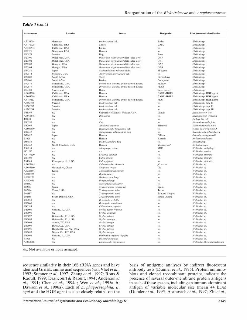
Table 1. 16S rRNA sequences used in the phylogenetic analyses and associated information
Accession no. Location Source Designation Prior taxonomic classification
AF283007 Japan Bovine Japan Anaplasma centrale
AF318944 South Africa Ovine Anaplasma centrale
AF309866 Virginia, USA Bovine Virginia Anaplasma marginale
AF309867 Florida, USA Bovine Florida Anaplasma marginale
AF309868 Idaho, USA Bovine South Idaho Anaplasma marginale
AF309869 Israel Bovine Israel Anaplasma marginale
AF311303 Virginia, USA Bovine Virginia Anaplasma marginale
M60313 Bovine Anaplasma marginale
AF309865 Ovine South Africa Anaplasma ovis
AF318945 Ovine Anaplasma ovis
NKIT36586 South Africa Ovine Sheep 3573}7 Anaplasma ovis
AB001521 Africa Ornithodoros moubata tick Symbiote A Argasid tick ‘symbiote A’
AB001522 Africa Ornithodoros moubata tick Symbiote B Argasid tick ‘symbiote B’
AE001345 Human D}UW-3}CX Chlamydia trachomatis
AF069758 South Africa Ruminant Mara 87}7 Cowdria ruminantium
U03776 South Africa Ruminant Omatjenne Cowdria ruminantium
U03777 South Africa Ruminant Ball3 Cowdria ruminantium
X61659 Zimbabwe Ruminant Crystal Springs Cowdria ruminantium
X62432 Senegal Ruminant Senegal Cowdria ruminantium
D84559 Rhipicephalus sanguineus tick Coxiella sp.
U03775 South Africa Bovine Ehrlichia bovis
AF162860 Guangzhou, China Dog Gzh982 Ehrlichia canis
M73221 Oklahoma, USA Dog OklahomaT Ehrlichia canis
M73226 Florida, USA Dog Florida Ehrlichia canis
U26740 Israel Dog 611 Ehrlichia canis
AF147752 China Amblyomma testudinarium tick Ehrlichia chaffeensis
M73222 Arkansas, USA Human ArkansasT Ehrlichia chaffeensis
U23503 Arkansas, USA Human 91HE17 Ehrlichia chaffeensis
U60476 Oklahoma, USA Human Sapulpa Ehrlichia chaffeensis
U86664 Florida, USA Human Jax Ehrlichia chaffeensis
U86665 Florida}Georgia, USA Human St Vincent Ehrlichia chaffeensis
AF036645 California, USA Horse Alice Ehrlichia equi
AF036646 California, USA Ixodes pacificus tick}horse Atempo Ehrlichia equi
AF036647 California, USA Horse Meretricious Ehrlichia equi
AF172164 California, USA Horse CASOLJ Ehrlichia equi
AF172165 California, USA Horse CAMEBS Ehrlichia equi
AF172166 California, USA Horse CASITL Ehrlichia equi
AF172167 California, USA Horse CAMAWI Ehrlichia equi
M73223 North America Horse Ehrlichia equi
M73227 Oklahoma, USA Dog StillwaterT Ehrlichia ewingii
U96436 North Carolina}Virginia, USA Dog 95E9-TS Ehrlichia ewingii
AB013008 Japan Apodemus speciosus I268 Ehrlichia muris
AB013009 Japan Haemaphysalis flava tick NA1 Ehrlichia muris
U15527 Japan Eothenomys kageus AS145T Ehrlichia muris
AF318946 Turkey Ovine Ehrlichia ovina
M73220 Scotland, UK Sheep Old Sourhope Ehrlichia phagocytophila
M73224 Scotland, UK Goat Feral goat Ehrlichia phagocytophila
AF156784 Guangzhou, China Dog Gzh981 Ehrlichia platys
M82801 North America Dog Ehrlichia platys
AF036648 Oregon, USA Horse Buck Ehrlichia risticii
AF036649 Oregon, USA Horse Bunn Ehrlichia risticii
AF036650 Oregon, USA Horse Danny Ehrlichia risticii
AF036651 California, USA Juga spp. (snail) None Ehrlichia risticii
AF036652 California, USA Juga spp. (snail) DrPepper Ehrlichia risticii
AF036653 Pennsylvania, USA Horse Eclipse Ehrlichia risticii
AF036654 California, USA Juga spp. (snail) Juga}snail Ehrlichia risticii
AF036655 Oregon, USA Juga spp. (snail) Stagnicola Ehrlichia risticii
AF036656 Michigan, USA Horse MostlyMemories Ehrlichia risticii
AF036657 California, USA Juga spp. (snail) MsAnnie Ehrlichia risticii
AF036658 Oregon, USA Juga spp. (snail) Tate Ehrlichia risticii
AF036659 Oregon, USA Juga spp. (snail) Thorenberg Ehrlichia risticii
AF037210 California, USA Juga spp. (snail) SHSN-1 Ehrlichia risticii
AF037211 California, USA Juga spp. (snail) SHSN-2 Ehrlichia risticii
AF170727 California, USA Coyote CATE Ehrlichia risticii
AF170729 California, USA Coyote CAPL Ehrlichia risticii
M21290 Maryland, USA Horse IllinoisT Ehrlichia risticii
M73219 Japan Human MiyaymaT Ehrlichia sennetsu
M73225 Human 11908 Ehrlichia sennetsu
AF012528 France Ixodes ricinus tick EHR62 Ehrlichia sp.
AF057707 Switzerland Horse Ehrlichia sp.
AF069062 California, USA Haliotis cracherodii (abalone) WSA Ehrlichia sp.
AF084907 Switzerland Ixodes ricinus tick Ehrlichia sp.
AF104680 Netherlands Ixodes ricinus tick Schotti variant Ehrlichia sp.
AF136712 Germany Ixodes ricinus tick Frankonia 2 Ehrlichia sp.
AF136713 Germany Ixodes ricinus tick Frankonia 1 Ehrlichia sp.
2148 International Journal of Systematic and Evolutionary Microbiology 51
Reorganization of the Rickettsiaceae and Anaplasmataceae
Table 1 (cont.)
Accession no. Location Source Designation Prior taxonomic classification
AF136714 Germany Ixodes ricinus tick Baden Ehrlichia sp.
AF170728 California, USA Coyote CASC Ehrlichia sp.
AF241532 California, USA Llama Ehrlichia sp.
U02521 Wisconsin, USA Human Ehrlichia sp.
U10873 Sweden Dog Rosa Ehrlichia sp.
U27101 Oklahoma, USA Odocoileus virginianus (white-tailed deer) OK3 Ehrlichia sp.
U27102 Oklahoma, USA Odocoileus virginianus (white-tailed deer) OK1 Ehrlichia sp.
U27103 Georgia, USA Odocoileus virginianus (white-tailed deer) GA2 Ehrlichia sp.
U27104 Georgia, USA Odocoileus virginianus (white-tailed deer) GA4 Ehrlichia sp.
U34280 Japan Stellantchasmus falcatus (fluke) SF agent Ehrlichia sp.
U52514 Missouri, USA Amblyomma americanum tick Ehrlichia sp.
U54805 South Africa Sheep Germishuys Ehrlichia sp.
U54806 South Africa Bovine Omatjenne Ehrlichia sp.
U72878 Minnesota, USA Peromyscus leucopus (white-footed mouse) PL1559 Ehrlichia sp.
U72879 Minnesota, USA Peromyscus leucopus (white-footed mouse) PL505 Ehrlichia sp.
U77389 Switzerland Horse Swiss horse 1 Ehrlichia sp.
AF093788 California, USA Human CAHU-HGE1 Ehrlichia sp. HGE agent
AF093789 California, USA Human CAHU-HGE2 Ehrlichia sp. HGE agent
AF189153 Minnesota, USA Peromyscus leucopus (white-footed mouse) PL59 Ehrlichia sp. HGE agent
AJ242785 Sweden Ixodes ricinus tick Ehrlichia sp. type Ia
AJ242783 Sweden Ixodes ricinus tick Ehrlichia sp. type Ib
AJ242784 Sweden Ixodes ricinus tick Ehrlichia sp. type IIb
U88565 University of Illinois, Urbana, USA Illinois Eperythrozoon suis
AF016546 Bos taurus Eperythrozoon wenyonii
J01859 Escherichia coli
U95297 Cat Haemobartonella felis
U82963 Japan Apodemus argentus Shizuoka Haemobartonella muris
AB001519 Haemaphysalis longicornis tick Ixodid tick ‘symbiote A’
U12457 Nanophyetus salmincola in dog Neorickettsia helminthoeca
D38622 Japan Human Gilliam Orientia tsutsugamushi
L36217 Human R strain Rickettsia rickettsii
D84558 Ixodes scapularis tick Rickettsia sp.
U12463 North Carolina, USA Human WilmingtonT Rickettsia typhi
X89110 Melophagus ovinus MO6 Wolbachia melophagi
M21292 Wolbachia persica
AF179630 Folsomia candida Wolbachia pipientis
U23709 Culex pipiens Wolbachia pipientis
X61768 Champaign, IL, USA Culex pipiens Wolbachia pipientis
AB025965 Callosobruchus chinensis jC strain Wolbachia sp.
AF035160 Guangzhou, China Sitophilus oryzae Ch Wolbachia sp.
AF220604 Korea Thecodiplosis japonensis Wolbachia sp.
AJ010275 Brugia malayi Wolbachia sp.
AJ010276 Onchocerca ochengi Wolbachia sp.
AJ012646 Brugia pahangi Wolbachia sp.
L02882 Muscidifurax uniraptor Wolbachia sp.
L02883 Spain Trichogramma cordubensis Spain Wolbachia sp.
L02884 Texas, USA Trichogramma deion Texas Wolbachia sp.
L02887 Trichogramma deion Bautista Canyon Wolbachia sp.
L02888 South Dakota, USA Trichogramma deion South Dakota Wolbachia sp.
U17059 Drosophila sechellia Wolbachia sp.
U17060 Drosophila mauritiana Wolbachia sp.
U80584 Phlebotomus papatasi Wolbachia sp.
U83090 Urbana, IL, USA Gryllus pennsylvanicus Wolbachia sp.
U83091 Gryllus assimilis Wolbachia sp.
U83092 Gainesville, FL, USA Gryllus rubens Wolbachia sp.
U83093 Gainesville, FL, USA Gryllus ovisopis Wolbachia sp.
U83094 Austin, TX, USA Gryllus integer Wolbachia sp.
U83095 Davis, CA, USA Gryllus integer Wolbachia sp.
U83096 Humboldt Co., NV, USA Gryllus integer Wolbachia sp.
U83097 Wayne Co., UT, USA Gryllus integer Wolbachia sp.
U83098 Urbana, IL, USA Diabrotica virgifera virgifera Wolbachia sp.
Z49261 Dirofilaria immitis Wolbachia sp.
AF069068 Litomosoides sigmodontis Wolbachia-like endobacterium
, Not available or none assigned.
sequence similarity in their 16S rRNA genes and haveidentical GroEL amino acid sequences (van Vliet et al.,1992; Sumner et al., 1997; Zhang et al., 1997; Roux &Raoult, 1999; Drancourt & Raoult, 1994; Anderson etal., 1991; Chen et al., 1994a; Wen et al., 1995a, b;Dawson et al., 1996a). Each of E. phagocytophila, E.equi and the HGE agent is also closely related on the
basis of antigenic analyses by indirect fluorescentantibody tests (Dumler et al., 1995). Protein immuno-blots and cloned recombinant proteins indicate thepresence of several outer-membrane protein antigensin each of these species, including an immunodominantantigen of variable molecular size (mean 44 kDa)(Dumler et al., 1995; Asanovich et al., 1997; Zhi et al.,
International Journal of Systematic and Evolutionary Microbiology 51 2149

J. S. Dumler and others
Table 2. groESL operon sequences used in the phylogenetic analyses and associated information
Accession no. Location Source Designation Prior taxonomic classification
AF165812 North America Bovine Anaplasma marginale
M98257 South America Human ATCC 35685 Bartonella bacilliformis
AF008210 Schizaphis graminum Buchnera aphidicola
AE001285 Human D}UW-3}CX Chlamydia trachomatis
U13638 South Africa Bovine WelgevondenT Cowdria ruminantium
U96731 Florida, USA Dog Florida Ehrlichia canis
L10917 Arkansas, USA Human ArkansasT Ehrlichia chaffeensis
AF172158 California, USA Horse CASOLJ Ehrlichia equi
AF172160 California, USA Horse CAMAWI Ehrlichia equi
AF172162 California, USA Horse CASITL Ehrlichia equi
U96727 California, USA Horse California horse Ehrlichia equi
U96729 Scotland, UK Goat Feral goat Ehrlichia phagocytophila
U96730 Scotland, UK Sheep Old Sourhope Ehrlichia phagocytophila
U96735 Switzerland Horse Swiss horse Ehrlichia phagocytophila
U24396 Horse 90-12 Ehrlichia risticii
U96732 Maryland, USA Horse IllinoisT Ehrlichia risticii
U88092 Japan Human Japan Ehrlichia sennetsu
AF033101 Slovenia Human Ehrlichia sp. HGE agent
AF172159 California, USA Human CAHU-HGE2 Ehrlichia sp. HGE agent
AF172163 California, USA Human CAHU-HGE1 Ehrlichia sp. HGE agent
U96728 New York, USA Human HGE agent Ehrlichia sp. HGE agent
X07850 Escherichia coli
U64996 Human MS11-A Neisseria gonorrhoeae
M31887 Human Karp Orientia tsutsugamushi
AJ235272 Human Madrid E Rickettsia prowazekii
U96733 Montana, USA R Rickettsia rickettsii
AF075440 North Carolina, USA Human WilmingtonT Rickettsia typhi
AB002286 Teleogryllus taiwanemma Group B Wolbachia sp.
NA, Not available.
1997). The gene encoding this 44 kDa immuno-dominant protein is one of a multigene family com-prising multiple distinct genes (Murphy et al., 1998;Zhi et al., 1998; IJdo et al., 1998) that also encodeproteins with significant amino acid similarity to (i) the36 kDa antigen called major surface protein 2 (MSP2)and the precursor of the 31 kDa antigen of A.marginale called MSP4 (Murphy et al., 1998; Zhi et al.,1998; IJdo et al., 1998), (ii) the C. ruminantium 28 kDamajor antigenic protein 1 (MAP1) (Jongejan & Thiel-emans, 1989; Jongejan et al., 1993; Ohashi et al.,1998b; Yu et al., 1999a), (iii) the E. chaffeensis and E.canis P28 and P30 protein families (Yu et al., 1999a;Ohashi et al., 1998a, b; Reddy et al., 1998) and (iv)Wolbachia spp. outer-surface protein precursors (Yuet al., 1999a; Ohashi et al., 1998b). This complex ofouter-membrane proteins is encoded in the HGEagent, A. marginale, E. chaffeensis, E. canis, E. muris,C. ruminantium and potentially in other Ehrlichiaspecies by polymorphic multigene families that aresuspected to contribute to immune evasion or per-sistence in reservoir hosts (Reddy et al., 1998; Allemanet al., 1997; French et al., 1998; Reddy & Streck,1999).
A gene encoding a protein antigen of approximately150–160 kDa that has repeated ankyrin motifs on theamino terminus, ankA, has been cloned from the HGEagent (Storey et al., 1998; Caturegli et al., 2000). Thefunction of this protein is unknown and it is a uniquebut relatively minor antigen among the HGE agent, E.equi and E. phagocytophila. Comparison of the nucleo-tide sequence of a 444 bp region of the ankyrin repeatregion from five Wisconsin strains and one New Yorkstrain designated as HGE agent by 16S rRNA genesequence revealed 100% similarity, whereas the se-quence of the MRK strain of E. equi is 99±6% similarto that of the HGE agent. Similarly, the sequence ofankA of the HGE agent is between 95±5 and 96±8%similar to those of Swedish and Spanish strains of E.phagocytophila from cattle and goats, respectively(Caturegli et al., 2000). These data are confirmed byfull gene sequences of a larger number of E. phago-cytophila-group organisms from various geographicalregions (Massung et al., 2000).
Biologically, A. marginale, E. phagocytophila, E. equi,E. platys, E. bovis and the HGE agent are most oftendetected in cells in the peripheral blood that are derived
2150 International Journal of Systematic and Evolutionary Microbiology 51

Reorganization of the Rickettsiaceae and Anaplasmataceae
from bone marrow precursors. E. phagocytophila, E.equi and the HGE agent are capable of growth in vitroin undifferentiated HL-60 promyelocytic cells, HL-60cells differentiated into neutrophil-like cells and po-tentially in precursors of the myelomonocytic lineage,as well as in embryonic Ixodes scapularis tick cell lines(Goodman et al., 1996; Heimer et al., 1997; Klein etal., 1997; Feng, 1997; Munderloh et al., 1996a). TheHGE agent and E. equi do not propagate in HL-60cells differentiated into mature macrophages. Thissituation resembles that in vivo, since each of thesespecies is detected most often in neutrophils or bandneutrophils in the blood of infected animals andhumans. A. marginale infects predominantly erythro-cytes in vivo and a suitable equivalent mammalian cellline for propagation has not been identified. A.marginale can be grown in embryonic tick cells in vitroand short-term propagation in erythrocyte culture andendothelial}erythrocyte co-cultures has also beenachieved (Munderloh et al., 1996b; Kessler et al.,1979; Waghela et al., 1997). E. platys infects canineplatelets in vivo and E. bovis infects bovine monocytes ;neither has been cultivated in vitro. Although the hostcell ligand is not known for E. platys or E. bovis, theHGE agent, a member of the E. phagocytophila group,adheres to platelet glycoprotein selectin ligand-1(PGSL-1; Herron et al., 2000), a sialic acid-bearingsurface protein molecule that shares many chemicalcharacteristics, such as sensitivity to neuraminidaseand chymotrypsin, with the erythrocyte ligand of A.marginale (McGarey & Allred, 1994).
Ticks transmit all of these species, but transovarialtransmission in ticks does not occur for those inves-tigated. E. phagocytophila, E. equi and the HGE agentare each transmitted by members of the Ixodespersulcatus complex, whereas A. marginale is trans-mitted by Dermacentor spp. ticks in temperate regionsof North America and by Boophilus spp. or othergenera in other geographical regions (Kuttler, 1984;Eriks et al., 1993; Kocan et al., 1992; Telford et al.,1996; Richter et al., 1996; Walls et al., 1997; Gordon etal., 1932; MacLeod & Gordon, 1933). Except for E.platys and E. bovis, the life cycles of these agents arepartially known. A. marginale and the closely relatedAnaplasma centrale and Anaplasma ovis are usuallymaintained by persistent subclinical infection of ru-minants, including wild ruminants such as deer(Kuttler, 1984; Eriks et al., 1993). A role exists fortransmission by male ticks among multiple animals ina single herd and mechanical transmission via bitingflies provides a potential alternative transmissionvehicle (Kocan et al., 1992). The HGE agent ismaintained, at least in part, by infection of smallmammal species such as the white-footed mouse,Peromyscus leucopus, or the dusky-footed wood rat,Neotoma fuscipes, in which occasional persistent infec-tions may be detected (Telford et al., 1996; Walls et al.,1997; Nicholson et al., 1999). E. phagocytophila mayestablish persistent infections in ruminants undernatural and experimental circumstances (Gordon et
al., 1932; MacLeod & Gordon, 1933; Hudson, 1950;Foggie, 1951; Foster & Cameron, 1970; McDiarmid,1965) and mounting evidence suggests that both E.equi and the HGE agent establish subclinical persistentinfections in domestic and wild ruminants, includingdeer (Foley et al., 1998; Belongia et al., 1997; Walls etal., 1998; Magnarelli et al., 1999). The HGE agentproduces disease typical of E. equi infection in horsesand induces protective immunity to challenge with E.equi (Madigan et al., 1995; Barlough et al., 1995).Likewise, E. equi-like bacteria have caused infection inhumans that is indistinguishable from HGE (Foley etal., 1999). Clinical manifestations, even in typicalmammalian hosts, are highly variable for each of E.phagocytophila, E. equi and the HGE agent ; clinicalfeatures therefore provide a lower degree of certaintyabout classification, since these are likely to be at leastin part host-dependent (Gordon et al., 1932; MacLeod& Gordon, 1933; Hudson, 1950; Foggie, 1951; Mad-igan, 1993; Reubel et al., 1998b; Bakken et al., 1994,1996, 1998; Aguero-Rosenfeld et al., 1996).
These common features are expected of organismswith a high degree of relatedness and indicate thatthese bacteria should be unified within a single genus.Moreover, the data indicate that sufficient similarityexists among E. phagocytophila, E. equi and the HGEagent for them to be classified as a single species. A.marginale is sufficiently divergent to be considered aseparate species, but the 16S rRNA gene sequencesof strains of A. marginale, A. ovis and A. centrale,excepting a Japanese strain, are nearly identical (mini-mum 99±1% similarity), suggesting the possibility thatthese also represent variants of a single species, asdenoted initially by Theiler (1911). The existence ofa strain of A. centrale that has 1±8% nucleotidedifference from other phenotypically characterizedstrains of A. centrale indicates the polygenic nature ofthis designation and casts some doubt upon theclassical morphological taxonomic methods for thisspecies and genus. Overall, a close grouping of erythro-cytic anaplasmas is supported by other genetic, pheno-typic and antigenic characteristics that also indicate aclose grouping with A. marginale (McGuire et al.,1984; Palmer et al., 1988, 1998; Visser et al., 1992). Infact, all species of Anaplasma are known to shareantigens that reside on 19, 36 and 105 kDa proteins,data that strengthen the close relationship based uponhost cell type and morphological characteristics (Pal-mer et al., 1988; Visser et al., 1992).
A large genetic distance (minimum 74±3% similarity)in groESL sequences was noted between the E.phagocytophila group members and A. marginale,which is in part explained by the paucity of groESLsequences examined. All members of the E. phago-cytophila group were at least 98±8% similar and noother sequence representatives (E. platys, E. bovis etc.)of the Anaplasma}E. phagocytophila group were avail-able. Similarly large genetic distances (minimum86±31% similarity) were observed for groESL se-quences between E. canis and C. ruminantium, which
International Journal of Systematic and Evolutionary Microbiology 51 2151
J. S. Dumler and others
.................................................................................................................................................................................................................................................................................................................
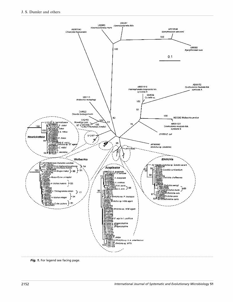
Fig. 1. For legend see facing page.
2152 International Journal of Systematic and Evolutionary Microbiology 51

Reorganization of the Rickettsiaceae and Anaplasmataceae
.................................................................................................................................................................................................................................................................................................................
Fig. 2. Phylogenetic tree inferred from groESL gene sequences of Ehrlichia, Anaplasma, Neorickettsia and Wolbachiaspecies, including 1077 sites after removal of sites containing a gap in any sequence. The sequence from Chlamydiatrachomatis (accession no. AE001285) was used as an outgroup. Numbers above internal nodes indicate the percentageof 1000 bootstrap replicates that supported the branch. All bootstrap values are included for clades that wereconsistently observed using the phylogenetic methods applied (maximum parsimony, minimum evolution, maximumlikelihood and majority-rule bootstrap analysis of neighbour-joining trees). The maximum-likelihood tree is shown. Bar,estimated number of substitutions per site; scale for the figure and insets are the same.
also appear to be clearly related on the basis of 16SrRNA gene sequences and phenotypic findings. Over-all, the groESL sequences support the divisions asindicated by 16S rRNA gene sequences and provideevidence of polymorphisms that may be random ormay represent subtleties of evolutionary selection.Thus, despite these ambiguous differences, insufficientgenetic distance and biological differences exist amongthe Anaplasma species, the E. phagocytophila group,E. bovis and the E. platys clade to designate them intoseparate genera. This is supported further by the lack
Fig. 1. Phylogenetic tree inferred from small subunit (16S) rRNA gene sequences of Ehrlichia, Anaplasma, Neorickettsiaand Wolbachia species, including 455 sites after removal of sites containing a gap in any sequence. The sequence fromChlamydia trachomatis (accession no. AE001345) was used as an outgroup. Numbers above internal nodes indicate thepercentage of 1000 bootstrap replicates that supported the branch. All bootstrap values are included for clades that wereconsistently observed using the phylogenetic methods applied (maximum parsimony, minimum evolution, maximumlikelihood and majority-rule bootstrap analysis of neighbour-joining trees). The maximum-likelihood tree is shown. Bars,estimated number of substitutions per site; the scale for the figure and insets are the same.
of bootstrap support for the clear separation of thetwo major arms of this clade and by the inconsistentpresence of E. bovis in either the Anaplasma or E.phagocytophila clades in the various phylogeneticanalyses. Additional sequence analyses of conservedand semi-conserved genes (e.g. gltA), whole genomeanalysis, as well as analysis of additional strains mayfurther identify taxonomic divisions or support thecurrent analyses of 16S rRNA and groESL genes.
Little is known about the antigenic characteristics of
International Journal of Systematic and Evolutionary Microbiology 51 2153

J. S. Dumler and others
either E. platys or E. bovis ; their taxonomic positionsmust therefore be assigned on the basis of what isknown about their genetic characteristics (Anderson etal., 1992). For some previously described agents, suchas ‘Cytoecetes microti ’ (Tyzzer, 1938), no isolates orgenetic information are available for analysis and theirrelationships to other named species cannot be as-sessed objectively. Of interest is the identification ofseveral 16S rRNA gene sequences from the blood ofwhite-tailed deer (Odocoileus virginianus) from Okla-homa and Georgia in the USA (Dawson et al., 1996c),from an Amblyomma americanum tick in Missouri(USA) and from the blood of sheep in South Africa(Allsopp et al., 1997), each of which is most similar toE. platys. A definitive bacterial morphology has neverbeen identified for any of these sequences; theirtaxonomic positions can therefore only be judged onthe basis of the 16S rRNA gene sequences.
The E. canis/Cowdria group
The second genetic cluster includes E. canis, E.chaffeensis, E. ewingii, E. muris and C. ruminantium, allof which are at least 97±7% similar in 16S rRNA genesequences (van Vliet et al., 1992; Dame et al., 1992;Rikihisa et al., 1997; Zhang et al., 1997; Roux &Raoult, 1995, 1999; Drancourt & Raoult, 1994;Anderson et al., 1991; Wen et al., 1995a, b; Shibata etal., 2000). E. canis, E. chaffeensis and E. muris aredetected mostly in macrophages and monocytes in vivoand can be propagated in vitro, most effectivelyin macrophage cell lines (Dawson et al., 1991a, b;Barnewell & Rikihisa, 1994; Heimer et al., 1998). C.ruminantium is most often found in endothelial cells,neutrophils or macrophages in vivo and can also bepropagated in cell lines derived from both endothelialcells and macrophages (Cowdry, 1926; Logan et al.,1987; Bezuidenhout et al., 1985; Sahu, 1986; Prozesky& Du Plessis, 1987). E. ewingii is the exception in thatit is detected most frequently in peripheral bloodneutrophils and it has not been grown in long-termculture (Ewing et al., 1971). E. canis is best recognizedas a pathogen of canids (Huxsoll, 1976; Woody &Hoskins, 1991), but can infect humans and may infectfelines (Perez et al., 1996; Bouloy et al., 1994), whereasE. chaffeensis causes symptomatic infection in humansand subclinical persistent infections in deer and canids(Fishbein et al., 1994; Ewing et al., 1995, Lockhart etal., 1997; Dawson et al., 1996b; Dawson & Ewing,1992). E. ewingii causes low-grade infections of canidsthat are sometimes characterized by lameness due topolyarthritis (Ewing et al., 1971) and has recently beenimplicated as a human pathogen (Buller et al., 1999).C. ruminantium is best known as the cause of heart-water in African and Caribbean ruminants (Cowdry,1926; Uilenberg, 1983; Camus et al., 1993). Each ofthese species is known to be transmitted and main-tained in a tick vector reservoir, including Amblyommaspp. for C. ruminantium (Bezuidenhout, 1987),Amblyomma americanum for E. chaffeensis and E.ewingii (Ewing et al., 1995; Anziani et al., 1990) and
Rhipicephalus sanguineus for E. canis (Groves et al.,1975). Transovarial transmission is ineffective for E.canis and C. ruminantium, the only species studiedsufficiently (Bezuidenhout, 1987; Groves et al., 1975).
Polyclonal antibodies to these organisms have a highdegree of cross-reactivity by immunofluorescence, aresult consistent with a close genetic relationship. Low-level antigenic cross-reactivity is also recognized be-tween C. ruminantium and E. phagocytophila andbetween E. phagocytophila, E. equi, the HGE agentand E. chaffeensis, E. canis or E. ewingii (Dumler et al.,1995; Dawson et al., 1991a; Jongejan et al., 1989;Buller et al., 1999; Brouqui et al., 1992; Rikihisa et al.,1992). The antigens of these organisms have beenstudied in some detail by Western blotting, whichreveals the presence of cross-reactive immuno-dominant antigens of similar molecular size but with adegree of diversity when detected with monoclonalantibodies (Dumler et al., 1995; Asanovich et al.,1997; Zhi et al., 1997; Visser et al., 1992; Palmer et al.,1985, 1998; Brouqui et al., 1992, 1994; Rikihisa et al.,1992, 1994; Yu et al., 1993; Chen et al., 1994b, 1996;Kim & Rikihisa, 1998; Ravyn et al., 1999; Adams etal., 1986; Vidotto et al., 1994; Alleman & Barbet,1996; Barbet et al., 1994; Rossouw et al., 1990; Mahanet al., 1993, 1994; Bowie et al., 1999; Kelly et al., 1994).A group of antigens that range between 27 and 32 kDais common among these organisms and is sharedbetween these different species when analysed byimmunoblotting methods (Rikihisa, 1991a; Rikihisaet al., 1992, 1994; Iqbal et al., 1994; Wen et al., 1995a;Ohashi et al., 1998a, b; Jongejan et al., 1993). Mono-clonal antibodies reactive with proteins in this mol-ecular size range that are raised against one isolate donot always react with other isolates (Chen et al., 1996,1997). These proteins are encoded by polymorphicgenes and are called MAP1 in C. ruminantium, MAP1homologue, p28 and p30 in E. canis and Omp1 or p28in E. chaffeensis, but have yet to be described in E.ewingii ; a homologous gene has been identified inother Ehrlichia species (Ohashi et al., 1998a, b; Reddyet al., 1998; Yu et al., 1999b; McBride et al., 1999; vanVliet et al., 1994). In fact, a high degree of amino acidsimilarity exists between these proteins and the MSP4of A. marginale, further clarifying the basis for priorevidence of serological cross-reactions obtained byimmunofluorescence studies (Ohashi et al., 1998a, b;Yu et al., 1999b; McBride et al., 1999; van Vliet et al.,1994).
The data on the tick-transmitted ehrlichiae in theAnaplasma}E. phagocytophila and E. canis}Cowdriagroups argue convincingly for the unification of thesespecies within either one or two separate genera.However, the large degree of internal genetic similarity(Fig. 1), the extent of shared amino acid sequences inmajor outer-membrane proteins, the similarity in hostcells and similarity in serological cross-reactions arguefor consolidation of the species of the E. phago-cytophila complex in a genus that contains only A.marginale, E. platys and E. bovis. Moreover, the
2154 International Journal of Systematic and Evolutionary Microbiology 51
Reorganization of the Rickettsiaceae and Anaplasmataceae
repeated genetic clustering of members of the E.canis}Cowdria group to the exclusion of members ofthe E. phagocytophila}Anaplasma group suggests thatthe establishment of two separate genera for thesegroups is the best way to emphasize the degree ofbiological difference between these clades. However,should a large number of apparently ancestral types toboth these groups be found, like the ‘Schotti variant ’(Fig. 1), future consolidation of these two closelyrelated groups may be warranted.
The E. sennetsu/Neorickettsia group
The third and most divergent genetic cluster of theehrlichiae includes E. sennetsu, E. risticii (van Vliet etal., 1992; Dame et al., 1992; Rikihisa et al., 1997;Zhang et al., 1997; Roux & Raoult, 1995, 1999;Drancourt & Raoult, 1994; Anderson et al., 1991;Chen et al., 1994a; Wen et al., 1995a, b), N. helmin-thoeca and an ehrlichia-like bacterium present in themetacercarial stage of the fluke Stellantchasmus fal-catus (SF), all of which exhibit between 94±9 and100±0% similarity in 16S rRNA gene sequences (Wenet al., 1996; Barlough et al., 1998; Pretzman et al.,1995; Chaichanasiriwithaya et al., 1994). However,individual isolates of E. risticii may diverge in 16SrRNA gene sequence by as many as 15 nucleotides(Wen et al., 1995b; Barlough et al., 1998). These dataunderscore the phylogenetic heterogeneity of thisclade. In spite of these observations, fluorescentantibody and protein immunoblot studies show a highdegree of antigenic similarity among E. sennetsu, E.risticii, N. helminthoeca and the SF agent, but not toother species of Ehrlichia (Rikihisa, 1991b; Rikihisaet al., 1988; Dumler et al., 1995; Wen et al., 1996;Holland et al., 1985a, b; Ristic et al., 1986; Shank-arappa et al., 1992). Each of these species infectspredominantly mononuclear phagocytes in vivo andcan be propagated in vitro most efficiently in cell linesderived from macrophages (Zhang et al., 1997; Wen etal., 1996; Shankarappa et al., 1992; Rikihisa et al.,1991, 1995). Ticks have never been implicated intransmission of these agents, whereas transmission viainfected metacercariae or cercariae of flukes that infesteither snails, fish or aquatic insects has been shown forN. helminthoeca and E. risticii and is strongly suspectedfor E. sennetsu (Rikihisa, 1991a; Barlough et al., 1998;Madigan et al., 2000). While no naturally existingmammalian infection with the SF agent has beenrecognized, its presence in flukes and pathogenicity inmice is consistent with the above observations in otherE. sennetsu-group organisms (Rikihisa, 1991a; Wen etal., 1996; Fukuda & Yamamoto, 1981).
E. sennetsu is best known as the agent of sennetsufever, a mononucleosis-like illness described only inJapan and Malaysia (Misao & Kobayashi, 1955;Rapmund, 1984). Early epidemiological studies sug-gested that individuals who consumed uncooked fishfrom certain areas of Japan were at risk (Rikihisa,1991a; Tachibana et al., 1976). Although not proven,
this epidemiology has long suggested the possibility ofenteral ingestion of fish contaminated with ehrlichia-infected flukes as the mechanism for transmission. E.sennetsu causes a fatal infection in mice and producesno clinical signs in horses, but protects horses againstchallenge by E. risticii (Tachibana & Kobayashi, 1975;Rikihisa et al., 1988). E. risticii causes Potomac horsefever, also known as equine monocytic ehrlichiosis or‘Shasta River crud’ (Holland et al., 1985a; Rikihisa &Perry, 1985; Madigan et al., 1997). Presumably, theagent is either ingested when horses feed upon snail-ridden grasses or by ingestion of infected metacercaria-containing aquatic insects (Reubel et al., 1998a;Barlough et al., 1998; Madigan et al., 2000). Thepresentation is that of a febrile illness with profusewatery diarrhoea. N. helminthoeca is acquired byingestion of fluke-infested fish by dogs and causes afebrile infection called salmon poisoning disease(Rikihisa, 1991a).
The degree of 16S rRNA gene sequence similarity ofthe E. sennetsu group to those in the E. phagocytophilaand E. canis groups is not more than exists between theE. sennetsu group and Rickettsia species (Wen et al.,1995a, b). Although minor serological cross-reactivityhas been described in some studies (Holland et al.,1985a; Ristic et al., 1981), no firm similarities in outer-membrane protein amino acid sequences have beenestablished and there appear to be no haematophagousarthropod vectors such as ticks involved in the lifecycle. However, the common infected host cells aresimilar to those of other Ehrlichia species, although theclinical manifestations of enteric involvement are morepronounced. The significant genetic, antigenic andecological traits of the species of the E. sennetsu groupsuggest that it is a distinct clade deserving of des-ignation as a separate genus.
Wolbachia species
The sole remaining named species of the genusWolbachia is W. pipientis, an obligate intracellularbacterium that resides within cytoplasmic vacuoles,predominantly in the ovaries of many species ofarthropods and increasingly identified in helminths(Werren, 1997; Popov et al., 1998; O’Neill et al., 1992;Dobson et al., 1992; Bandi et al., 1998). Analysis offtsZ gene amplicons of arthropod and filarial wol-bachiae indicates the existence of at least two distincthost-associated clades (Bandi et al., 1998; Vande-kerckhove et al., 1999). However, by 16S rRNA genesequence analysis, W. pipientis and the Wolbachia spp.occupy a position intermediate between the two tick-transmitted groups (E. canis}C. ruminantium and E.phagocytophila}Anaplasma) and the helminth-borneE. sennetsu}Neorickettsia group (Roux & Raoult,1995; Wen et al., 1995b; O’Neill et al., 1992). Deducedamino acid sequences of Wolbachia spp. outer-mem-brane protein genes exhibit similarity to those of themajor outer-membrane proteins of A. marginale, theE. phagocytophila complex, E. chaffeensis, E. canis and
International Journal of Systematic and Evolutionary Microbiology 51 2155

J. S. Dumler and others
C. ruminantium, thus corroborating the phylogeneticposition of W. pipientis (Yu et al., 1999a; Ohashi et al.,1998b; Zhou et al., 1998). However, W. pipientis is notrecognized as a vertebrate pathogen, since mammalianinfection has never been documented.
Although there are significant morphological, geneticand amino acid sequence similarities between W.pipientis and the other Ehrlichia}Cowdria}Anaplasmagroups, the significant degree of differences in 16SrRNA and groESL gene sequences, the lack of asignificant vertebrate host phase, the promiscuousinvertebrate host associations and its highly efficienttransovarial transmission adequately differentiate W.pipientis and related organisms from species found inthe genera Ehrlichia, Cowdria, Anaplasma and Neo-rickettsia.
Historical precedents
The historical precedent for naming species in theentire group of ehrlichiae is A. marginale, which wasfirst described and named by Theiler (1910). Theorganism currently denoted C. ruminantium was de-scribed initially by Cowdry (1925) and given the genusdesignation Cowdria by Moshkovski (1947). Gordonfirst clearly differentiated tick-borne fever from loup-ing ill in goats in 1932 and suggested that the diseasewas caused by a rickettsia, an assertion that wasaffirmed in 1940 by Foggie (Gordon et al., 1932;Foggie, 1951). The genus designation Ehrlichia wasfirst coined in 1945 to honour Paul Ehrlich (Mosh-kovski, 1945; Silverstein, 1998), 2 years before thedesignation of C. ruminantium ; however, the typespecies, E. canis, was first described as Rickettsia canisby Donatien & Lestoquard (1935) and, in 1936, thesame authors described E. bovis as Rickettsia bovis(Donatien & Lestoquard, 1936). Hertig first describedrickettsia-like organisms in insects in 1936 and thesewere placed in the genus Wolbachia in honour of S.Burt Wolbach, who demonstrated the presence ofrickettsiae in pathological lesions in the vasculotropicrickettsioses (Hertig, 1936). The designation ‘Cyto-ecetes microti ’ was created to describe a micro-organism with morphological features similar to theorganism now called E. phagocytophila (Tyzzer, 1938) ;however, original materials and isolates no longer existfor verification of its identity (Ristic & Huxsoll, 1984).Subsequently, other designations were made into thegenus Ehrlichia (in North America) or ‘Cytoecetes ’ (inEurope and Asia) for organisms that were recognizedto be pathogenic for mammals (Ristic & Huxsoll,1984; Moshkovski, 1945). N. helminthoeca was de-scribed in 1953, E. sennetsu in 1954, E. equi in 1969, E.platys in 1982, E. risticii in 1984, E. chaffeensis in 1991,E. ewingii in 1993 and E. muris in 1995 (Wen et al.,1995a; Anderson et al., 1992; Misao & Kobayashi,1955; Philip et al., 1953; Stannard et al., 1969; Gribble,1969).
Convincing phylogenetic data now show that a seriesof significant flaws exists in the taxonomic structure of
the families Anaplasmataceae and Rickettsiaceae in theorder Rickettsiales. Similar phylogenetic studies led toa significant taxonomic modification of the formergenera Rochalimaea and Grahamella (Brenner et al.,1993; Birtles et al., 1995). It is now clear that adistinction between some members of the familiesRickettsiaceae and Anaplasmataceae is not supported.Moreover, some members of the family Anaplas-mataceae, the genera Eperythrozoon and Haemo-bartonella, are clearly not related to the genus Ana-plasma and should be removed and reassigned withinthe family Mycoplasmataceae (Rikihisa et al., 1997).While no classification system fits all criteria perfectly,genetic data have become the objective standards and,when evaluated carefully, often closely predict similarbiological and clinical behaviours. Thus, the datacompiled here indicate that a sufficient genotypic andphenotypic relationship exists among the genera Ana-plasma, Cowdria, Wolbachia and Ehrlichia, excludingN. helminthoeca, E. sennetsu and E. risticii, to meritunification into two separate genera. Since the validlypublished names Anaplasma and A. marginale andEhrlichia and E. canis predate Cowdria and Wolbachia,Anaplasma should be retained for the unified genusthat encompasses the existing Anaplasma species, theE. phagocytophila group, E. bovis and E. platys, whilethe genus Ehrlichia should be retained and used todescribe members of the Ehrlichia canis group, in-cluding C. ruminantium. This change further neces-sitates accommodation of the members of the E.sennetsu group within a single genus, Neorickettsia.Thus, a revised classification may be formulated thatdifferentiates organisms in the order Rickettsiales intotwo families, Rickettsiaceae, which contains the rick-ettsiae (Rickettsia, Orientia) that occupy an intra-cytoplasmic compartment, and Anaplasmataceae,which contains the ehrlichiae (Neorickettsia, Wol-bachia, Ehrlichia, Anaplasma) that occupy an intra-vacuolar compartment within infected host cells.Consequently, new combinations for the multiplegenera and species that are involved must also becreated.
Emended description of Rickettsiales(Gieszczykiewicz 1939) Weiss and Moulder 1984
It is proposed that the tribes Rickettsieae, Ehrlichieaeand Wolbachieae should be abolished. Furthermore,all species formerly within the tribes Ehrlichieae andWolbachieae are transferred into the family Anaplas-mataceae.
Emended description of Rickettsiaceae (Pinkerton1936) Weiss and Moulder 1984
It is proposed that the genera Ehrlichia, Cowdria,Neorickettsia and Wolbachia be transferred from thefamily Rickettsiaceae to the family Anaplasmataceae, achange that results in the elimination of all tribeswithin the family Rickettsiaceae. It is also proposedthat the genera Haemobartonella and Eperythrozoon
2156 International Journal of Systematic and Evolutionary Microbiology 51

Reorganization of the Rickettsiaceae and Anaplasmataceae
should be transferred from the family Anaplas-mataceae to the order Mycoplasmatales and thatCoxiella, Rickettsiella, Francisella (Wolbachia) persicaand Wolbachia melophagi (Weisburg et al., 1989; Rouxet al., 1997) should be removed from the familyRickettsiaceae. This proposal also requires emendationof the description of the family Rickettsiaceae tospecify that organisms infect host cells within thecytoplasm or nucleus and are not bounded by avacuole. The family Rickettsiaceae includes only thegenera Rickettsia and Orientia.
Emended description of Anaplasmataceae (Philip1957) Ristic and Kreier 1984
It is proposed that the family Anaplasmataceae beemended to include species in the genera Wolbachia,Ehrlichia, Cowdria and Neorickettsia and to retainspecies in the genera Anaplasma and Aegyptianella.This requires emendation of the description of theAnaplasmataceae to specify infection within a cyto-plasmic vacuole of host cells that include erythrocytes,reticuloendothelial cells, bone marrow-derived phago-cytic cells, endothelial cells and cells of insect, helminthand arthropod reproductive tissues. Aegyptianella isretained as genus incertae sedis.
Emended description of Anaplasma (Theiler 1910)Ristic and Kreier 1984
It is proposed that members of the E. phagocytophilagroup, including E. phagocytophila, E. equi, the HGEagent, as well as E. bovis and E. platys, should beunited with the genus Anaplasma. This change requiresemendation of the description of the genus Anaplasma(Ristic & Kreier, 1984) by integrating it with somedescriptions of the genera Ehrlichia and new data forAnaplasma and Ehrlichia, as follows.
Gram-negative, small, often pleomorphic, coccoid toellipsoidal organisms that reside within cytoplasmicvacuoles, either singly and more often in compactinclusions (morulae) present in mature or immaturehaematopoietic cells, particularly myeloid cells andneutrophils and including erythrocytes, in peripheralblood or in tissues, usually mononuclear phagocyteorgans (spleen, liver, bone marrow) of mammalianhosts. By ultrastructure, two morphological forms areobserved, including larger reticulate cells and smallerforms with condensed protoplasm called dense-coreforms (Popov et al., 1998). Vectors, where known, areticks. Organisms grow in tick vectors. Non-motile.Not cultivable in cell-free media or chicken embryos.Some species are cultivable in neutrophils, myelo-monocytic cell lines, promyelocytic cell lines, eryth-rocytes and tick cell lines. Aetiological agents ofdiseases of dogs and other canids, humans andruminants such as cattle, goats, sheep and llamas.Variably pathogenic or non-pathogenic infections insome ruminants such as cattle, goats, sheep and deer,horses and rodents. The estimated GC content of
the DNA varies between approximately 30 and 56mol%. The type species is Anaplasma marginale(Theiler, 1910).
Emended description of Ehrlichia (Moshkovski 1945)Ristic and Huxsoll 1984
Gram-negative, small, often pleomorphic, coccoid toellipsoidal organisms that reside within cytoplasmicvacuoles, either singly and more often in compactinclusions (morulae) present in mature or immaturehaematopoietic cells, especially mononuclear phago-cytes such as monocytes and macrophages and forsome species in myeloid cells such as neutrophils, inperipheral blood or in tissues, usually mononuclearphagocyte organs (spleen, liver, bone marrow, lymphnode) of mammalian hosts. By ultrastructure, twomorphological forms are observed, including largerreticulate cells and smaller forms with condensedprotoplasm (dense-core forms) (Popov et al., 1998).Vectors, where known, are ticks. Organisms grow intick vectors. Non-motile. Not cultivable in cell-freemedia or chicken embryos. Some species cultivable inblood monocytes, monocytic or macrophage cellslines, myelomonocytic cell lines, endothelial cell linesand tick cell lines. Aetiological agents of diseases ofdogs and other canids, rodents and humans. Variablypathogenic or non-pathogenic infections in someruminants such as deer and some rodents. The GCcontent of the DNA varies between approximately 30and 56 mol%. The type species is Ehrlichia canis(Donatien and Lestoquard 1935) Moshkovski 1945.
Emended description of Neorickettsia (Philip, Hadlowand Hughes 1953)
It is proposed that some descriptions of the genusEhrlichia be united with the genus Neorickettsia(Pretzman et al., 1995). This requires emendation ofthe description of the genus Neorickettsia by inte-gration with some descriptions of the genus Ehrlichiaand new data, as follows.
Small, coccoid, often pleomorphic, intracytoplasmicbacteria that occur primarily in vacuoles of monocytesin the blood and macrophages of lymphoid or othertissues of dogs, horses and humans. Certain tissues ofmature fluke vectors, all other fluke stages, eggs,rediae, cercariae and metacercariae have been proveninfectious by injection into susceptible vertebratehosts, as have mature stages of aquatic insects, whichconfirms that the infectious cycle includes transovarialand trans-stadial transmission in the vectors (Reubelet al., 1998a; Barlough et al., 1998). Gram-negative.Non-motile. Not cultivable in cell-free media or inchicken embryos. Sensitive to tetracycline antibiotics.The GC content of the DNA is not known. The typespecies is Neorickettsia helminthoeca (Philip et al.,1953).
International Journal of Systematic and Evolutionary Microbiology 51 2157
J. S. Dumler and others
Description of Anaplasma phagocytophila comb. nov.
The most recent description of E. phagocytophila isthat of Ristic & Huxsoll (1984). It is proposed that thespecies E. equi (Ristic & Huxsoll, 1984; Stannard et al.,1969) and the unnamed HGE agent (Chen et al.,1994a; Bakken et al., 1994) be united within the singlespecies designation E. phagocytophila and transferredinto the genus Anaplasma. This requires emendation ofthe species description for E. phagocytophila by inte-grating portions of the description of the species E.equi (Ristic & Huxsoll, 1984) and new data for E. equiand the HGE agent as follows.
Gram-negative, coccoid to ellipsoidal, often pleo-morphic, intracytoplasmic bacteria that infect cells ofmammalian bone marrow derivation, predominantlycells in the myeloid lineage. Two ultrastructuralmorphologies are observed, including a larger re-ticulate form and a smaller dense-core form thatcontains condensed protoplasm. Tick vectors includespecies of the Ixodes persulcatus complex (Telford etal., 1996; Richter et al., 1996; MacLeod & Gordon,1933; Foggie, 1951). In mammalian cells, morulae areusually 1±5–2±5 µm in diameter, but may be as large as6 µm (Popov et al., 1998). Individual bacterial cells areof two types, dense-core and reticulate, both present inthe same vacuole ; both may undergo equal or unequalbinary fission. Individual cells may be as large as 2 µmin diameter. Empty vesicles may be present in thevacuolar space, but fibrillar matrix is lacking. Abun-dant cytoplasmic membrane may be present, formingprotrusions into the periplasmic space or invaginationsinto the bacterial protoplasm. Mitochondria do notcontact with or cluster around morulae. Causativeagent of tick-borne fever of ruminants (Gordon et al.,1932; Hudson, 1950; Foggie, 1951). Equine granulo-cytic ehrlichiosis (Madigan, 1993; Stannard et al.,1969; Gribble, 1969), a type of canine granulocyticehrlichiosis that lacks lameness as a significant sign(Greig et al., 1996; Pusterla et al., 1997) and humangranulocytic ehrlichiosis (Chen et al., 1994a; Good-man et al., 1996; Bakken et al., 1994) are caused byvariants of A. phagocytophila, previously known as E.equi and the HGE agent, respectively. Tick-bornefever is chiefly reported as a febrile disease of goats,sheep and cattle in the UK, The Netherlands, Scan-dinavia, Spain, France, Germany and Switzerland.Clinical signs vary from no detectable illness to severefebrile disease associated with opportunistic infections,haemorrhage and abortions. Equine granulocytic ehrl-ichiosis and a form of canine granulocytic ehrlichiosishave been described broadly across the USA, Canada,Brazil, Venezuela and Northern Europe. Equine andcanine diseases are characterized by fever, depression,anorexia, leukopaenia and thrombocytopaenia;equine infection also frequently results in limb oedemaand ataxia and may lead to opportunistic infections.Human granulocytic ehrlichiosis has also been de-scribed in many of the same geographical areas ofCalifornia, Wisconsin, Minnesota and the NewEngland states in the USA and in Slovenia, Norway,
Switzerland and Sweden in Europe; serological evi-dence of human infection in the absence of overthuman disease has been described in the USA, UK,Switzerland, Norway, Sweden, Denmark, Germanyand Bulgaria. Human disease is characterized by fever,headache, myalgia and malaise and by the presence ofleukopaenia, thrombocytopaenia and evidence of hep-atic injury (Bakken et al., 1996; Aguero-Rosenfeld etal., 1996). The case fatality rate in humans is less than1%, but is associated with severe opportunistic infec-tions (Walker & Dumler, 1997). Although A. phago-cytophila has a broad geographical distribution and allisolates appear to have significant serological cross-reactivity, a minor degree of variation in the nucleotidesequence of up to 5 bp (" 99±5% identity) in the16S rRNA gene and & 99±0% identity in groESL isdetected. The organism shares significant antigenswith E. canis, E. chaffeensis and E. (Cowdria) rumin-antium comb. nov. The major constitutively producedprotein antigens are encoded by a multigene family,vary between 42 and 49 kDa in molecular size and areexpressed on the outer membrane (Murphy et al.,1998; Dumler et al., 1995; Asanovich et al., 1997; Zhiet al., 1997, 1998). The amino acid sequences of themajor outer-membrane proteins are similar to those ofA. marginale, E. (Cowdria) ruminantium, E. canis, E.chaffeensis and Wolbachia species. The genome size isapproximately 1500 kbp (Rydkina et al., 1999). TheGC content of the DNA estimated from sequencedgenes is 41 mol%. The 16S rRNA gene sequence of theA. phagocytophila type strain WebsterT is the same asdeposited in GenBank under the accession numberU02521.
Description of Anaplasma bovis comb. nov.
The most recent description of Ehrlichia bovis is that ofScott (1994). In addition, A. bovis is a Gram-negative,coccoid to coccobacillary and often pleomorphicobligate intravacuolar bacterium that infects cattleand perhaps other mammals. Mononuclear cells aremost often infected but are infrequently identified inperipheral blood. African tick vectors include Hy-alomma excavatum, Rhipicephalus appendiculatus,Amblyomma variegatum and possibly Amblyommacajennense in Brazil. Serological cross-reactions withE. ruminantium have been reported (Du Plessis et al.,1987). The 16S rRNA gene sequence of A. bovis isdeposited in GenBank under the accession numberU03775.
Description of Ehrlichia ruminantium comb. nov.
The most recent description of Cowdria ruminantiumis that of van Vliet et al. (1992). In addition, E.ruminantium is a Gram-negative, coccoid to ellipsoidal,often pleomorphic, intracytoplasmic bacteria thatinfects cattle, sheep, goats and occasionally murineendothelial cells as well as cells of bone marrowderivation, predominantly cells in the myeloid andmonocytic lineages. Various species of wild Africanruminants are reservoir hosts (Neitz, 1933, 1935; Peter
2158 International Journal of Systematic and Evolutionary Microbiology 51

Reorganization of the Rickettsiaceae and Anaplasmataceae
et al., 1998, 1999). Tick vectors include at least10 species of the genus Amblyomma. The major con-stitutionally produced protein antigens (MAP1) areencoded by a multigene family, vary between 31 and 32kDa in molecular size and are expressed on the outermembrane. The amino acid sequences of this majorouter-membrane protein are similar to those of A.marginale, A. phagocytophila, E. chaffeensis, E. canisand W. pipientis. The 16S rRNA gene sequence of thetype strain, WelgevondenT, is the same as that for theCrystal Springs strain deposited in GenBank underaccession no. X61659 (M. T. Allsopp, personal com-munication).
Emended description of Ehrlichia canis
The most recent description of E. canis is that of Ristic& Huxsoll (1984). In addition, cells are Gram-negative,coccoid to ellipsoidal, often pleomorphic, intra-cytoplasmic bacteria that infect canid and perhapshuman cells of bone marrow derivation (Perez et al.,1996), predominantly cells in the monocytic lineage.The predominant tick vector is Rhipicephalus sanguin-eus. The major constitutionally produced proteinantigen varies between 28 and 32 kDa in molecularsize and is expressed on the outer membrane (Yu et al.,1999a; Ohashi et al., 1998b; Reddy et al., 1998;McBride et al., 1999). The amino acid sequences of themajor outer-membrane proteins are similar to those ofE. chaffeensis, E. ruminantium, A. marginale, A. phago-cytophila and W. pipientis. The 16S rRNA genesequence of the type strain OklahomaT is deposited inGenBank under accession no. M73221.
Emended description of Ehrlichia chaffeensis
With the following additions, the description is thesame as that given previously (Anderson et al., 1991).The tick vector is Amblyomma americanum. The typestrain is strain ArkansasT. The major constitutionallyproduced protein antigens are encoded by a multigenefamily, vary between 28 and 32 kDa in molecular sizeand are expressed on the outer membrane (Yu et al.,1999a; Ohashi et al., 1998a; Reddy et al., 1998). Theamino acid sequences of the major outer-membraneproteins are similar to those of E. canis, E. rumin-antium, A. phagocytophila, A. marginale and Wol-bachia species. The genome size is approximately1250 kbp (Rydkina et al., 1999). The 16S rRNA genesequence of the type strain, ArkansasT, is deposited inGenBank under accession no. M73222.
Emended description of Ehrlichia ewingii
With the following additions, the description is thesame as that given previously (Anderson et al., 1992).The tick vector is Amblyomma americanum. The typestrain is strain StillwaterT. Aetiological agent of canineand human disease (Ewing et al., 1971; Buller et al.,1999). The 16S rRNA gene sequence is deposited inGenBank under accession no. M73227.
Emended description of Ehrlichia muris
With the following additions, the description is thesame as that given previously (Wen et al., 1995a).Haemaphysalis flava ticks may be naturally infected,but a role as a vector has not been established(Kawahara et al., 1999). The 16S rRNA gene sequenceof the type strain, AS145T, is deposited in GenBankunder accession no. U15527.
Description of Anaplasma platys comb. nov.
With the following additions, the description is thesame as that given previously for E. platys (Ristic &Huxsoll, 1984; Anderson et al., 1992). A tick vector issuspected, but has not been established. The 16SrRNA gene sequence is deposited in GenBank underaccession no. M82801.
Description of Neorickettsia sennetsu comb. nov.
With the following additions, the description is thesame as that given previously for E. sennetsu (Ristic &Huxsoll, 1984). The organism shares antigens with N.(Ehrlichia) risticii. Not pathogenic for the horse but,after infection, horses are protected from infectionwith N. risticii. The mode of transmission is notknown, although a fish parasite is suspected. Mice arehighly susceptible to infection. The genome size isapproximately 880 kbp (Rydkina et al., 1999). Thetype strain is MiyayamaT, for which the 16S rRNAgene sequence is deposited in GenBank under ac-cession no. M73225 (Anderson et al., 1991).
Description of Neorickettsia risticii comb. nov.
With the following additions, the description is thesame as that given previously for E. risticii (Holland etal., 1985b). Causative agent of Potomac horse fever,also called equine monocytic ehrlichiosis. Transmittedby the ingestion of fresh-water snail species or insectsinfested with infected trematodes or metacercariae(Reubel et al., 1998a; Madigan et al., 2000). Sharesantigens with N. sennetsu, N. helminthoeca and the SFagent bacterium. The organism is found to infectperipheral blood monocytes, intestinal epithelial cellsand equine mast cells. The 16S rRNA gene may varyby up to 15 bases in nucleic acid sequences. Theapproximate genome size is 880 kbp (Rydkina et al.,1999). The type strain is IllinoisT, for which the 16SrRNA gene sequence is deposited in GenBank underaccession no. M21290.
ACKNOWLEDGEMENTS
The authors thank Frans Jongejan, Philippe Brouqui, DidierRaoult, B. A. Allsopp, M. Allsopp, Sam R. Telford III,Roger Stich, David H. Walker, Xuijue Yu, Gere McBride,Kathy Kocan, William Nicholson, John Sumner, RobMassung and the members of the Combined TaxonomicCommittee of the American Society for Rickettsiology andthe European Working Group for Rickettsiology for helpfuldiscussions.
International Journal of Systematic and Evolutionary Microbiology 51 2159

J. S. Dumler and others
REFERENCES
Adams, J. H., Smith, R. D. & Kuhlenschmidt, M. S. (1986).Identification of antigens of two isolates of Anaplasma mar-ginale, using a western blot technique. Am J Vet Res 47,501–506.
Aguero-Rosenfeld, M. E., Horowitz, H. W., Wormser, G. P., Mc-Kenna, D. F., Nowakowski, J., Munoz, J. & Dumler, J. S. (1996).Human granulocytic ehrlichiosis : a case series from a medicalcenter in New York State. Ann Intern Med 125, 904–908.
Alleman, A. R. & Barbet, A. F. (1996). Evaluation of Anaplasmamarginale major surface protein 3 (MSP3) as a diagnostic testantigen. J Clin Microbiol 34, 270–276.
Alleman, A. R., Palmer, G. H., McGuire, T. C., McElwain, T. F.,Perryman, L. E. & Barbet, A. F. (1997). Anaplasma marginalemajor surface protein 3 is encoded by a polymorphic, multigenefamily. Infect Immun 65, 156–163.
Allsopp, M. T. E. P., Visser, E. S., du Plessis, J. L., Vogel, S. W. &Allsopp, B. A. (1997). Different organisms associated withheartwater as shown by analysis of 16S ribosomal RNA genesequences. Vet Parasitol 71, 283–300.
Anderson, B. E., Dawson, J. E., Jones, D. C. & Wilson, K. H. (1991).Ehrlichia chaffeensis, a new species associated with humanehrlichiosis. J Clin Microbiol 29, 2838–2842.
Anderson, B. E., Greene, C. E., Jones, D. C. & Dawson, J. E. (1992).Ehrlichia ewingii, sp. nov., the etiologic agent of caninegranulocytic ehrlichiosis. Int J Syst Bacteriol 42, 299–302.
Anziani, O. S., Ewing, S. A. & Barker, R. W. (1990). Experimentaltransmission of a granulocytic form of the tribe Ehrlichieae byDermacentor variabilis and Amblyomma americanum to dogs.Am J Vet Res 51, 929–931.
Asanovich, K. M., Bakken, J. S., Madigan, J. E., Aguero-Rosen-feld, M., Wormser, G. P. & Dumler, J. S. (1997). Antigenicdiversity of granulocytic Ehrlichia isolates from humans inWisconsin and New York and a horse in California. J Infect Dis176, 1029–1034.
Bakken, J. S., Dumler, J. S., Chen, S. M., Eckman, M. R., Van Etta,L. L. & Walker, D. H. (1994). Human granulocytic ehrlichiosis inthe upper midwest United States. A new species emerging? J AmMed Assoc 272, 212–218.
Bakken, J. S., Krueth, J., Wilson-Nordskog, C., Tilden, R. L.,Asanovich, K. & Dumler, J. S. (1996). Clinical and laboratorycharacteristics of human granulocytic ehrlichiosis. J Am MedAssoc 275, 199–205.
Bakken, J. S., Goellner, P., Van Etten, M. & 8 other authors (1998).Seroprevalence of human granulocytic ehrlichiosis amongpermanent residents of northwestern Wisconsin. Clin Infect Dis27, 1491–1496.
Bandi, C., Anderson, T. J., Genchi, C. & Blaxter, M. L. (1998).Phylogeny of Wolbachia in filarial nematodes. Proc R Soc LondB Biol Sci 265, 2407–2413.
Barbet, A. F. (1995). Recent developments in the molecularbiology of anaplasmosis. Vet Parasitol 57, 43–49.
Barbet, A. F., Semu, S. M., Chigagure, N., Kelly, P. J., Jongejan, F.& Mahan, S. M. (1994). Size variation of the major immuno-dominant protein of Cowdria ruminantium. Clin Diagn LabImmunol 1, 744–746.
Barlough, J. E., Madigan, J. E., DeRock, E., Dumler, J. S. & Bakken,J. S. (1995). Protection against Ehrlichia equi is conferred byprior infection with the human granulocytotropic ehrlichia(HGE agent). J Clin Microbiol 33, 3333–3334.
Barlough, J. E., Reubel, G. H., Madigan, J. E., Vredevoe, L. K.,Miller, P. E. & Rikihisa, Y. (1998). Detection of Ehrlichia risticii,the agent of Potomac horse fever, in freshwater stream snails(Pleuroceridae: Juga spp.) from northern California. ApplEnviron Microbiol 64, 2888–2893.
Barnewell, R. E. & Rikihisa, Y. (1994). Abrogation of gammainterferon-induced inhibition of Ehrlichia chaffeensis infectionin human monocytes with iron-transferrin. Infect Immun 62,4804–4810.
Belongia, E. A., Reed, K. D., Mitchell, P. D., Kolbert, C. P., Persing,D. H., Gill, J. S. & Kazmierczak, J. J. (1997). Prevalence ofgranulocytic Ehrlichia infection among white-tailed deer inWisconsin. J Clin Microbiol 35, 1465–1468.
Bezuidenhout, J. D. (1987). Natural transmission of heartwater.Onderstepoort J Vet Res 54, 349–351.
Bezuidenhout, J. D., Paterson, C. L. & Barnard, B. J. (1985). Invitro cultivation of Cowdria ruminantium. Onderstepoort J VetRes 52, 113–120.
Birtles, R. J., Harrison, T. G., Saunders, N. A. & Molyneux, D. H.(1995). Proposals to unify the generaGrahamella and Bartonella,with descriptions of Bartonella talpae comb. nov., Bartonellaperomysci comb. nov., and three new species, Bartonellagrahamii sp. nov., Bartonella taylorii sp. nov., and Bartonelladoshiae sp. nov. Int J Syst Bacteriol 45, 1–8.
Bouloy, R. P., Lappin, M. R., Holland, C. H., Thrall, M. A., Baker, D.& O’Neil, S. (1994). Clinical ehrlichiosis in a cat. J Am Vet MedAssoc 204, 1475–1478.
Bowie, M. V., Reddy, G. R., Semu, S. M., Mahan, S. M. & Barbet,A. F. (1999). Potential value of major antigenic protein 2 forserological diagnosis of heartwater and related ehrlichialinfections. Clin Diagn Lab Immunol 6, 209–215.
Brenner, D. J., O’Connor, S. P., Winkler, H. H. & Steigerwalt, A. G.(1993). Proposals to unify the genera Bartonella andRochalimaea, with descriptions of Bartonella quintana comb.nov., Bartonella vinsonii comb. nov., Bartonella henselae comb.nov., and Bartonella elizabethae comb. nov., and to remove thefamily Bartonellaceae from the order Rickettsiales. Int J SystBacteriol 43, 777–786.
Brouqui, P., Dumler, J. S., Raoult, D. & Walker, D. H. (1992).Antigenic characterization of ehrlichiae : protein immuno-blotting of Ehrlichia canis, Ehrlichia sennetsu, and Ehrlichiaristicii. J Clin Microbiol 30, 1062–1066.
Brouqui, P., Lecam, C., Olson, J. G. & Raoult, D. (1994). Serologicdiagnosis of human monocytic ehrlichiosis by immunoblotanalysis. Clin Diagn Lab Immunol 1, 645–649.
Buller, R. S., Arens, M., Hmiel, S. P. & 9 other authors (1999).Ehrlichia ewingii, a newly recognized agent of human ehrl-ichiosis. N Engl J Med 341, 148–155.
Camus, E., Martinez, D., Beauperthuy, L. & 18 other authors(1993). Heartwater in Guadeloupe and in the Caribbean. RevElev Med Vet Pays Trop 46, 109–114.
Caturegli, P., Asanovich, K. M., Walls, J. J., Bakken, J. S., Madigan,J. E., Popov, V. L. & Dumler, J. S. (2000). ankA: an Ehlrichiaphagocytophila group gene encoding a cytoplasmic proteinantigen with ankyrin repeats. Infect Immun 68, 5277–5283.
Chaichanasiriwithaya, W., Rikihisa, Y., Yamamoto, S., Reed,S. M., Crawford, T. B., Perryman, L. E. & Palmer, G. H. (1994).Antigenic, morphologic, and molecular characterization of newEhrlichia risticii isolates. J Clin Microbiol 32, 3026–3033.
Chen, S. M., Dumler, J. S., Bakken, J. S. & Walker, D. H. (1994a).Identification of a granulocytotropic Ehrlichia species as theetiologic agent of human disease. J Clin Microbiol 32, 589–595.
2160 International Journal of Systematic and Evolutionary Microbiology 51
Reorganization of the Rickettsiaceae and Anaplasmataceae
Chen, S. M., Dumler, J. S., Feng, H. M. & Walker, D. H. (1994b).Identification of the antigenic constituents of Ehrlichia chaf-feensis. Am J Trop Med Hyg 50, 52–58.
Chen, S.-M., Popov, V. L., Feng, H.-M. & Walker, D. H. (1996).Analysis and ultrastructural localization of Ehrlichia chaffeensisproteins with monoclonal antibodies. Am J Trop Med Hyg 54,405–412.
Chen, S. M., Yu, X. J., Popov, V. L., Westerman, E. L., Hamilton,F. G. & Walker, D. H. (1997). Genetic and antigenic diversity ofEhrlichia chaffeensis : comparative analysis of a novel humanstrain from Oklahoma and previously isolated strains. J InfectDis 175, 856–863.
Cowdry, E. V. (1925). Studies on the aetiology of heartwater.I. Observation of a rickettsia, Rickettsia ruminantium (n. sp.) inthe tissues of infected animals. J Exp Med 44, 803–814.
Cowdry, E. V. (1926). Studies on the aetiology of heartwater :III. The multiplication of Rickettsia ruminantium within theendothelial cells of infected animals and their discharge into thecirculation. J Exp Med 44, 803–814.
Dame, J. B., Mahan, S. M. & Yowell, C. A. (1992). Phylogeneticrelationship of Cowdria ruminantium, agent of heartwater, toAnaplasma marginale and other members of the order Rick-ettsiales determined on the basis of 16S rRNA sequence. Int JSyst Bacteriol 42, 270–274.
Dawson, J. E. & Ewing, S. A. (1992). Susceptibility of dogs toinfection with Ehrlichia chaffeensis, causative agent of humanehrlichiosis. Am J Vet Res 53, 1322–1327.
Dawson, J. E., Rikihisa, Y., Ewing, S. A. & Fishbein, D. B. (1991a).Serologic diagnosis of human ehrlichiosis using two Ehrlichiacanis isolates. J Infect Dis 163, 564–567.
Dawson, J. E., Anderson, B. E., Fishbein, D. B., Sanchez, J. L.,Goldsmith, C. S., Wilson, K. H. & Duntley, C. W. (1991b). Isolationand characterization of an Ehrlichia sp. from a patient diag-nosed with human ehrlichiosis. J Clin Microbiol 29, 2741–2745.
Dawson, J. E., Warner, C. K., Standaert, S. & Olson, J. G. (1996a).The interface between research and the diagnosis of an emergingtick-borne disease, human ehrlichiosis due to Ehrlichia chaf-feensis. Arch Intern Med 156, 137–142.
Dawson, J. E., Biggie, K. L., Warner, C. K., Cookson, K., Jenkins,S., Levine, J. F. & Olson, J. G. (1996b). Polymerase chain reactionevidence of Ehrlichia chaffeensis, an etiologic agent of humanehrlichiosis, in dogs from southeast Virginia. Am J Vet Res 57,1175–1179.
Dawson, J. E., Warner, C. K., Baker, V., Ewing, S. A., Stallknecht,D. E., Davidson, W. R., Kocan, A. A., Lockhart, J. M. & Olson, J. G.(1996c). Ehrlichia-like 16S rDNA sequence from wild white-tailed deer (Odocoileus virginianus). J Parasitol 82, 52–58.
Dobson, S. L., Bourtzis, K., Braig, H. R., Jones, B. F., Zhou, W.,Rousset, F. & O’Neill, S. L. (1999). Wolbachia infections aredistributed throughout insect somatic and germ line tissues.Insect Biochem Mol Biol 29, 153–160.
Donatien, A. & Lestoquard, F. (1935). Existence en Alge! rie d’uneRickettsia du chien. Bull Soc Pathol Exot 28, 418–419.
Donatien, A. & Lestoquard, F. (1936). Rickettsia bovis. Nouvelleespe' ce pathoge' ne pour le boeuf. Bull Soc Pathol Exot 29,1057–1061.
Drancourt, M. & Raoult, D. (1994). Taxonomic position of therickettsiae : current knowledge. FEMS Microbiol Rev 13, 13–24.
Dumler, J. S., Asanovich, K. M., Bakken, J. S., Richter, P., Kimsey,R. & Madigan, J. E. (1995). Serologic cross-reactions among
Ehrlichia equi, Ehrlichia phagocytophila, and the human granu-locytic ehrlichia. J Clin Microbiol 33, 1098–1103.
Du Plessis, J. L., Camus, E., Oberem, P. T. & Malan, L. (1987).Heartwater serology: some problems with the interpretations ofresults. Onderstepoort J Vet Res 54, 327–329.
Eriks, I. S., Stiller, D. & Palmer, G. H. (1993). Impact of persistentAnaplasma marginale rickettsemia on tick infection and trans-mission. J Clin Microbiol 31, 2091–2096.
Ewing, S. A., Roberson, W. R., Buckner, R. G. & Hayat, C. S.(1971). A new strain of Ehrlichia canis. J Am Vet Med Assoc 159,1771–1774.
Ewing, S. A., Dawson, J. E., Kocan, A. A., Barker, R. W., Warner,C. K., Panciera, R. J., Fox, J. C., Kocan, K. M. & Blouin, E. F. (1995).Experimental transmission of Ehrlichia chaffeensis (Ricket-tsiales : Ehrlichieae) among white-tailed deer by Amblyommaamericanum (Acari : Ixodidae). J Med Entomol 32, 368–374.
Felsenstein, J. (1985). Confidence limits on phylogenies : anapproach using the bootstrap. Evolution 39, 783–791.
Feng, H.-M. (1997). Host cell defense factors in the growth ofEhrlichia chaffeensis in different monocyte cell lines. In Programand Abstracts of the 13th Sesquiannual Meeting of the AmericanSociety for Rickettsiology, Sept 21–24 1997, Champion, PA,USA. Abstract 111.
Fishbein, D. B., Dawson, J. E. & Robinson, L. E. (1994). Humanehrlichiosis in the United States, 1985 to 1990. Ann Intern Med120, 736–743.
Foggie, A. (1951). Studies on the infectious agent of tick-bornefever in sheep. J Pathol Bacteriol 63, 1–15.
Foley, J. E., Barlough, J. E., Kimsey, R. B., Madigan, J. E., DeRock,E. & Poland, A. (1998). Ehrlichia spp. in cervids from California.J Wildl Dis 34, 731–737.
Foley, J. E., Crawford-Miksza, L., Dumler, J. S. & 7 other authors(1999). Human granulocytic ehrlichiosis in northern California :two case descriptions with genetic analysis of the ehrlichiae.Clin Infect Dis 29, 388–392.
Forsman, M., Sandstro$ m, G. & Sjo$ stedt, A. (1994). Analysis of 16Sribosomal DNA sequences of Francisella strains and utilizationfor determination of the phylogeny of the genus and foridentification of strains by PCR. Int J Syst Bacteriol 44, 38–46.
Foster, W. N. M. & Cameron, A. E. (1970). Observations on ovinestrains of tick-borne fever. J Comp Pathol 80, 429–436.
French, D. M., McElwain, T. F., McGuire, T. C. & Palmer, G. H.(1998). Expression of Anaplasma marginale major surfaceprotein 2 variants during persistent cyclic rickettsemia. InfectImmun 66, 1200–1207.
Fukuda, T. & Yamamoto, S. (1981). Neorickettsia-like organismisolated from metacercaria of a fluke, Stellantchasmus falcatus.Jpn J Med Sci Biol 34, 103–107.
Goodman, J. L., Nelson, C., Vitale, B., Madigan, J. E., Dumler, J. S.,Kurtti, T. J. & Munderloh, U. G. (1996). Direct cultivation of thecausative agent of human granulocytic ehrlichiosis. N Engl JMed 334, 209–215.
Gordon, W. S., Brownlee, A., Wilson, D. R. & MacLeod, J. (1932).‘Tick-borne fever ’. (A hitherto undescribed disease of sheep). JComp Pathol Therap 65, 301–307.
Greig, B., Asanovich, K. M., Armstrong, P. J. & Dumler, J. S.(1996). Geographic, clinical, serologic, and molecular evidenceof granulocytic ehrlichiosis, a likely zoonotic disease, inMinnesota and Wisconsin dogs. J Clin Microbiol 34, 44–48.
Gribble, D. H. (1969). Equine ehrlichiosis. J Am Vet Med Assoc155, 462–469.
International Journal of Systematic and Evolutionary Microbiology 51 2161

J. S. Dumler and others
Groves, M. G., Dennis, G. L., Amyx, H. L. & Huxsoll, D. L. (1975).Transmission of Ehrlichia canis to dogs by ticks (Rhipicephalussanguineus). Am J Vet Res 36, 937–940.
Heimer, R., Van Andel, A., Wormser, G. P. & Wilson, M. L. (1997).Propagation of granulocytic Ehrlichia spp. from human andequine sources in HL-60 cells induced to differentiate intofunctional granulocytes. J Clin Microbiol 35, 923–927.
Heimer, R., Tisdale, D. & Dawson, J. E. (1998). A single tissueculture system for the propagation of the agents of the humanehrlichioses. Am J Trop Med Hyg 58, 812–815.
Herron, M. J., Nelson, C. M., Larson, J., Snapp, K. R., Kansas, G. S.& Goodman, J. L. (2000). Intracellular parasitism by the humangranulocytic ehrlichiosis bacterium through the P-selectinligand, PSGL-1. Science 288, 1653–1656.
Hertig, M. (1936). The rickettsia, Wolbachia pipientis (gen. et sp.n.) and associated inclusions in the mosquito, Culex pipiens.Parasitology 28, 453–486.
Holland, C. J., Ristic, M., Cole, A. I., Johnson, P., Baker, G. & Goetz,T. (1985a). Isolation, experimental transmission, and charac-terization of causative agent of Potomac horse fever. Science227, 522–524.
Holland, C. J., Weiss, E., Burgdorfer, W., Cole, A. I. & Kakoma, I.(1985b). Ehrlichia risticii sp. nov. : etiological agent of equinemonocytic ehrlichiosis (synonym, Potomac horse fever). Int JSyst Bacteriol 35, 524–526.
Hudson, J. R. (1950). The recognition of tick-borne fever as adisease of cattle. Br Vet J 106, 3–17.
Huxsoll, D. L. (1976). Canine ehrlichiosis (tropical canine pan-cytopenia) : a review. Vet Parasitol 2, 49–60.
IJdo, J. W., Sun, W., Zhang, Y., Magnarelli, L. A. & Fikrig, E. (1998).Cloning of the gene encoding the 44-kilodalton antigen of theagent of human granulocytic ehrlichiosis and characterizationof the humoral response. Infect Immun 66, 3264–3269.
Ina, Y. (1998). Estimation of the transition}transversion ratio. JMol Evol 46, 521–533.
Iqbal, Z., Chaichanasiriwithaya, W. & Rikihisa, Y. (1994). Com-parison of PCR with other tests for early diagnosis of canineehrlichiosis. J Clin Microbiol 32, 1658–1662.
Jongejan, F. & Thielemans, M. J. C. (1989). Identification of animmunodominant antigenically conserved 32-kilodalton pro-tein from Cowdria ruminantium. Infect Immun 57, 3243–3246.
Jongejan, F., Wassink, L. A., Thielemans, M. J., Perie, N. M. &Uilenberg, G. (1989). Serotypes in Cowdria ruminantium andtheir relationship with Ehrlichia phagocytophila determined byimmunofluorescence. Vet Microbiol 21, 31–40.
Jongejan, F., de Vries, N., Nieuwenhuijs, J., van Vliet, A. H. M. &Wassink, L. A. (1993). The immunodominant 32-kilodaltonprotein of Cowdria ruminantium is conserved with the genusEhrlichia. Rev Elev Med Vet Pays Trop 46, 145–152.
Kawahara, M., Ito, T., Suto, C., Shibata, S., Rikihisa, Y., Hata, K. &Hirai, K. (1999). Comparison of Ehrlichia muris strains isolatedfrom wild mice and ticks and serologic survey of humans andanimals with E. muris as antigen. J Clin Microbiol 37,1123–1129.
Kelly, P. J., Matthewman, L. A., Mahan, S. M., Semu, S., Peter, T.,Mason, P. R., Brouqui, P. & Raoult, D. (1994). Serological evidencefor antigenic relationships between Ehrlichia canis and Cowdriaruminantium. Res Vet Sci 56, 170–174.
Kessler, R. H., Ristic, M., Sells, D. M. & Carson, C. A. (1979). Invitro cultivation of Anaplasma marginale : growth pattern andmorphologic appearance. Am J Vet Res 40, 1767–1773.
Kim, H. Y. & Rikihisa, Y. (1998). Characterization of monoclonalantibodies to the 44-kilodalton major outer membrane proteinof the human granulocytic ehrlichiosis agent. J Clin Microbiol36, 3278–3284.
Klein, M. B., Miller, J. S., Nelson, C. M. & Goodman, J. L. (1997).Primary bone marrow progenitors of both granulocytic andmonocytic lineages are susceptible to infection with the agent ofhuman granulocytic ehrlichiosis. J Infect Dis 176, 1405–1409.
Kocan, K. M., Stiller, D., Goff, W. L., Claypool, P. L., Edwards, W.,Ewing, S. A., McGuire, T. C., Hair, J. A. & Barron, S. J. (1992).Development of Anaplasma marginale in male Dermacentorandersoni transferred from parasitemic to susceptible cattle. AmJ Vet Res 53, 499–507.
Kuttler, K. L. (1984). Anaplasma infections in wild and domesticruminants : a review. J Wildl Dis 20, 12–20.
Lally, N. C., Nicoll, S., Paxton, E. A., Cary, C. M. & Sumption, K. J.(1995). The Cowdria ruminantium groE operon. Microbiology141, 2091–2100.
Lockhart, J. M., Davidson, W. R., Stallknecht, D. E., Dawson, J. E.& Howerth, E. W. (1997). Isolation of Ehrlichia chaffeensis fromwild white-tailed deer (Odocoileus virginianus) confirms theirrole as natural reservoir hosts. J Clin Microbiol 35, 1681–1686.
Logan, L. L., Whyard, T. L., Quintero, J. C. & Mebus, C. A. (1987).The development of Cowdria ruminantium in neutrophils.Onderstepoort J Vet Res 54, 197–204.
McBride, J. W., Yu, X. J. & Walker, D. H. (1999). Molecularcloning of the gene for a conserved major immunoreactive 28-kilodalton protein of Ehrlichia canis : a potential serodiagnosticantigen. Clin Diagn Lab Immunol 6, 392–399.
McDiarmid, A. (1965). Modern trends in animal health andhusbandry. Some infectious diseases of free-living wildlife.Parasitology 121, 245–257.
McGarey, D. J. & Allred, D. R. (1994). Characterization ofhemagglutinating components on the Anaplasma marginaleinitial body surface and identification of possible adhesins.Infect Immun 62, 4587–4593.
McGuire, T. C., Palmer, G. H., Goff, W. L., Johnson, M. I. & Davis,W. C. (1984). Common and isolate-restricted antigens of Ana-plasma marginale detected with monoclonal antibodies. InfectImmun 45, 697–700.
MacLeod, J. R. & Gordon, W. S. (1933). Studies in tick-bornefever of sheep. I. Transmission by the tick, Ixodes ricinus, witha description of the disease produced. Parasitology 25, 273–285.
Madigan, J. E. (1993). Equine ehrlichiosis. Vet Clin North AmEquine Pract 9, 423–428.
Madigan, J. E., Richter, P. J., Jr, Kimsey, R. B., Barlough, J. E.,Bakken, J. S. & Dumler, J. S. (1995). Transmission and passage inhorses of the agent of human granulocytic ehrlichiosis. J InfectDis 172, 1141–1144.
Madigan, J. E., Barlough, J. E., Rikihisa, Y., Wen, B., Miller, P. E. &Sampson, T. J. (1997). Identification of an enzootic diarrhea(‘Shasta River Crud’) in Northern California as Potomac horsefever. J Equine Vet Sci 17, 270–272.
Madigan, J. E., Pusterla, N., Johnson, E., Chae, J. S., Pusterla, J. B.,Derock, E. & Lawler, S. P. (2000). Transmission of Ehrlichiaristicii, the agent of Potomac horse fever, using naturallyinfected aquatic insects and helminth vectors : preliminaryreport. Equine Vet J 32, 275–279.
Magnarelli, L. A., Ijdo, J. W., Stafford, K. C., III & Fikrig, E. (1999).Infections of granulocytic ehrlichiae and Borrelia burgdorferi inwhite-tailed deer in Connecticut. J Wildl Dis 35, 266–274.
2162 International Journal of Systematic and Evolutionary Microbiology 51
Reorganization of the Rickettsiaceae and Anaplasmataceae
Mahan, S. M., Tebele, N., Mukwedeya, D., Semu, S., Nyathi, C. B.,Wassink, L. A., Kelly, P. J., Peter, T. & Barbet, A. F. (1993). Animmunoblotting diagnostic assay for heartwater based on theimmunodominant 32-kilodalton protein of Cowdria rumin-antium detects false positives in field sera. J Clin Microbiol 31,2729–2737.
Mahan, S. M., McGuire, T. C., Semu, S. M., Bowie, M. V., Jonge-jan, F., Rurangirwa, F. R. & Barbet, A. F. (1994). Molecularcloning of a gene encoding the immunogenic 21 kDa protein ofCowdria ruminantium. Microbiology 140, 2135–2142.
Massung, R. F., Owens, J. H., Ross, D., Reed, K. D., Petrovec, M.,Bjoersdorff, A., Coughlin, R. T., Beltz, G. A. & Murphy, C. I. (2000).Sequence analysis of the ank gene of granulocytic ehtlichiae. JClin Microbiol 38, 2917–2922.
Misao, T. & Kobayashi, Y. (1955). Studies on infectious mono-nucleosis (glandular fever). I. Isolation of etiologic agent fromblood, bone marrow and lymph node of a patientwith infectiousmononucleosis by using mice. Kyushu J Med Sci 6, 145–152.
Moshkovski, S. D. (1945). Cytotropic inducers of infection andthe classification of the Rickettsiae with Chlamydozoa. Adv ModBiol (Moscow) 19, 1–44.
Moshkovski, S. D. (1947). Comments by readers. Science 106, 62.
Munderloh, U. G., Madigan, J. E., Dumler, J. S., Goodman, J. L.,Hayes, S. F., Barlough, J. E., Nelson, C. M. & Kurtti, T. J. (1996a).Isolation of the equine granulocytic ehrlichiosis agent, Ehrlichiaequi, in tick cell culture. J Clin Microbiol 34, 664–670.
Munderloh, U. G., Blouin, E. F., Kocan, K. M., Ge, N. L., Edwards,W. L. & Kurtti, T. J. (1996b). Establishment of the tick (Acari :Ixodidae)-borne cattle pathogen Anaplasma marginale (Ricket-tsiales : Anaplasmataceae) in tick cell culture. J Med Entomol 33,656–664.
Murphy, C. I., Storey, J. R., Recchia, J., Doros-Richert, L. A.,Gingrich-Baker, C., Munroe, K., Bakken, J. S., Coughlin, R. T. &Beltz, G. A. (1998). Major antigenic proteins of the agent ofhuman granulocytic ehrlichiosis are encoded by members of amultigene family. Infect Immun 66, 3711–3718.
Neimark, H. & Kocan, K. M. (1997). The cell wall-less rickettsiaEperythrozoon wenyonii is a Mycoplasma. FEMS Microbiol Lett156, 287–291.
Neitz, W. O. (1933). The blesbuck (Damaliscus albifrons) as acarrier of heartwater and blue tongue. J S Afr Vet Med Assoc 4,24–26.
Neitz, W. O. (1935). The blesbuck (Damaliscus albifrons) and theblack wildebeest as carriers of heartwater. Onderstepoort J VetSci Anim Indus 5, 35–40.
Nicholson, W. L., Castro, M. B., Kramer, V. L., Sumner, J. W. &Childs, J. E. (1999). Dusky-footed wood rats (Neotoma fuscipes)as reservoirs of granulocytic ehrlichiae (Rickettsiales :Ehrlichieae) in northern California. J Clin Microbiol 37,3323–3327.
Niebylski, M. L., Peacock, M. G., Fischer, E. R., Porcella, S. F. &Schwan, T. G. (1997). Characterization of an endosymbiontinfecting wood ticks, Dermacentor andersoni, as a member ofthe genus Francisella. Appl Environ Microbiol 63, 3933–3940.
Noda, H., Munderloh, U. G. & Kurtti, T. J. (1997). Endosymbiontsof ticks and their relationship to Wolbachia spp. and tick-bornepathogens of humans and animals. Appl Environ Microbiol 63,3926–3932.
Ohashi, N., Zhi, N., Zhang, Y. & Rikihisa, Y. (1998a). Immuno-dominant major outer membrane proteins of Ehrlichia chaf-feensis are encoded by a polymorphic multigene family. InfectImmun 66, 132–139.
Ohashi, N., Unver, A., Zhi, N. & Rikihisa, Y. (1998b). Cloning andcharacterization of multigenes encoding the immunodominant30-kilodalton major outer membrane proteins of Ehrlichia canisand application of the recombinant protein for serodiagnosis.J Clin Microbiol 36, 2671–2680.
O’Neill, S. L., Giordano, R., Colbert, A. M., Karr, T. L. & Robertson,H. M. (1992). 16S rRNA phylogenetic analysis of the bacterialendosymbionts associated with cytoplasmic incompatibility ininsects. Proc Natl Acad Sci USA 89, 2699–2702.
Palmer, G. H., Kocan, K. M., Barron, S. J., Hair, J. A., Barbet, A. F.,Davis, W. C. & McGuire, T. C. (1985). Presence of commonantigens, including major surface protein epitopes, between thecattle (intraerythrocytic) and tick stages of Anaplasma mar-ginale. Infect Immun 50, 881–886.
Palmer, G. H., Barbet, A. F., Musoke, A. J., Katende, J. M.,Rurangirwa, F., Shkap, V., Pipano, E., Davis, W. C. & McGuire,T. C. (1988). Recognition of conserved surface protein epitopeson Anaplasma centrale and Anaplasma marginale isolates fromIsrael, Kenya and the United States. Int J Parasitol 18, 33–38.
Palmer, G. H., Abbott, J. R., French, D. M. & McElwain, T. F.(1998). Persistence of Anaplasma ovis infection and conservationof the msp-2 and msp-3 multigene families within the genusAnaplasma. Infect Immun 66, 6035–6039.
Perez, M., Rikihisa, Y. & Wen, B. (1996). Ehrlichia canis-like agentisolated from a man in Venezuela: antigenic and geneticcharacterization. J Clin Microbiol 34, 2133–2139.
Peter, T. F., Anderson, E. C., Burridge, M. J. & Mahan, S. M.(1998). Demonstration of a carrier state for Cowdria rumin-antium in wild ruminants from Africa. J Wildl Dis 34, 567–575.
Peter, T. F., Bryson, N. R., Perry, B. D. & 7 other authors (1999).Cowdria ruminantium infection in ticks in the Kruger NationalPark. Vet Rec 145, 304–307.
Philip, C. B., Hadlow, W. J. & Hughes, L. E. (1953). Neorickettsiahelminthoeca, a new rickettsia-like disease agent in dogs inwestern United States transmitted by an helminth. RiassCommun VI Congr Int Microbiol, Roma 2, 256–257.
Popov, V. L., Han, V. C., Chen, S.-M., Dumler, J. S., Feng, H.-M.,Andreadis, T. G., Tesh, R. B. & Walker, D. H. (1998). Ultra-structural differentiation of the genogroups in the genusEhrlichia. J Med Microbiol 47, 235–251.
Pretzman, C., Ralph, D., Stothard, D. R., Fuerst, P. A. & Rikihisa, Y.(1995). 16S rRNA gene sequence of Neorickettsia helminthoecaand its phylogenetic alignment with members of the genusEhrlichia. Int J Syst Bacteriol 45, 207–211.
Prozesky, L. & Du Plessis, J. L. (1987). Heartwater. The de-velopment and life cycle of Cowdria ruminantium in thevertebrate host, ticks and cultured endothelial cells. Onder-stepoort J Vet Res 54, 193–196.
Pusterla, N., Huder, J., Wolfensberger, C., Litschi, B., Parvis, A. &Lutz, H. (1997). Granulocytic ehrlichiosis in two dogs inSwitzerland. J Clin Microbiol 35, 2307–2309.
Rapmund, G. (1984). Rickettsial diseases of the Far East : newperspectives. J Infect Dis 149, 330–338.
Ravyn, M. D., Lamb, L. J., Jemmerson, R., Goodman, J. L. &Johnson, R. C. (1999). Characterization of monoclonal anti-bodies to an immunodominant protein of the etiologic agent ofhuman granulocytic ehrlichiosis. Am J Trop Med Hyg 61,171–176.
Reddy, G. R. & Streck, C. P. (1999). Variability in the 28-kDasurface antigenic protein multigene locus of isolates of theemerging disease agent Ehrlichia chaffeensis suggests that it
International Journal of Systematic and Evolutionary Microbiology 51 2163
J. S. Dumler and others
plays a role in immune evasion. Mol Cell Biol Res Commun 1,167–175.
Reddy, G. R., Sulsona, C. R., Barbet, A. F., Mahan, S. M., Burridge,M. J. & Alleman, A. R. (1998). Molecular characterization of a28 kDa surface antigen gene family of the tribe Ehrlichiae.Biochem Biophys Res Commun 247, 636–643.
Reubel, G. H., Barlough, J. E. & Madigan, J. E. (1998a). Productionand characterization of Ehrlichia risticii, the agent of Potomachorse fever, from snails (Pleuroceridae: Juga spp.) in aquariumculture and genetic comparison to equine strains. J ClinMicrobiol 36, 1501–1511.
Reubel, G. H., Kimsey, R. B., Barlough, J. E. & Madigan, J. E.(1998b). Experimental transmission of Ehrlichia equi to horsesthrough naturally infected ticks (Ixodes pacificus) from northernCalifornia. J Clin Microbiol 36, 2131–2134.
Richter, P. J., Kimsey, R. B., Madigan, J. E., Barlough, J. E., Dumler,J. S. & Brooks, D. L. (1996). Ixodes pacificus as a vector ofEhrlichia equi. J Med Entomol 33, 1–5.
Rikihisa, Y. (1991a). The tribe Ehrlichieae and ehrlichial diseases.Clin Microbiol Rev 4, 286–308.
Rikihisa, Y. (1991b). Cross-reacting antigens between Neo-rickettsia helminthoeca and Ehrlichia species shown by immuno-fluorescence and Western immunoblotting. J Clin Microbiol 29,2024–2029.
Rikihisa, Y. & Perry, B. D. (1985). Causative ehrlichial organismsin Potomac horse fever. Infect Immun 49, 513–517.
Rikihisa, Y., Pretzman, C. I., Johnson, G. C., Reed, S. M., Yama-moto, S. & Andrews, F. (1988). Clinical, histopathological, andimmunological responses of ponies to Ehrlichia sennetsu andsubsequent Ehrlichia risticii challenge. Infect Immun 56,2960–2966.
Rikihisa, Y., Stills, H. & Zimmerman, G. (1991). Isolation andcontinuous culture of Neorickettsia helminthoeca in a macro-phage cell line. J Clin Microbiol 29, 1928–1933.
Rikihisa, Y., Ewing, S. A., Fox, J. C., Siregar, A. G., Pasaribu, F. H.& Malole, M. B. (1992). Analyses of Ehrlichia canis and a caninegranulocytic Ehrlichia infection. J Clin Microbiol 30, 143–148.
Rikihisa, Y., Ewing, S. A. & Fox, J. C. (1994). Western immunoblotanalysis of Ehrlichia chaffeensis, E. canis, or E. ewingii infectionsin dogs and humans. J Clin Microbiol 32, 2107–2112.
Rikihisa, Y., Zhang, Y. & Park, J. (1995). Role of Ca#+ andcalmodulin in ehrlichial infection in macrophages. Infect Immun63, 2310–2316.
Rikihisa, Y., Kawahara, M., Wen, B., Kociba, G., Fuerst, P.,Kawamori, F., Suto, C., Shibata, S. & Futohashi, M. (1997).Western immunoblot analysis of Haemobartonella muris andcomparison of 16S rRNA gene sequences of H. muris, H. felis,and Eperythrozoon suis. J Clin Microbiol 35, 823–829.
Ristic, M. & Huxsoll, D. L. (1984). Tribe II. Ehrlichieae Philip1957, 948AL. In Bergey’s Manual of Systematic Bacteriology,vol. 1, pp. 704–711. Edited by N. R. Krieg & J. G. Holt.Baltimore: Williams & Wilkins.
Ristic, M. & Kreier, J. P. (1984). Family III. AnaplasmataceaePhilip 1957, 980AL. In Bergey’s Manual of Systematic Bac-teriology, vol. 1, pp. 719–729. Edited by N. R. Krieg & J. G.Holt. Baltimore: Williams & Wilkins.
Ristic, M., Huxsoll, D. L., Tachibana, N. & Rapmund, G. (1981).Evidence of a serologic relationship between Ehrlichia canis andRickettsia sennetsu. Am J Trop Med Hyg 30, 1324–1328.
Ristic, M., Holland, C. J., Dawson, J. E., Sessions, J. & Palmer, J.(1986). Diagnosis of equine monocytic ehrlichiosis (Potomac
horse fever) by indirect immunofluorescence. J Am Vet MedAssoc 189, 39–46.
Rossouw, M., Neitz, A. W., de Waal, D. T., du Plessis, J. L., vanGas, L. & Brett, S. (1990). Identification of the antigenic proteinsof Cowdria ruminantium. Onderstepoort J Vet Res 57, 215–221.
Roux, V. & Raoult, D. (1995). Phylogenetic analysis of the genusRickettsia by 16S rDNA sequencing. Res Microbiol 146,385–396.
Roux, V. & Raoult, D. (1999). Phylogenetic analysis and taxo-nomic relationships among the genus Rickettsia. In Rickettsiaeand Rickettsial Diseases at the Turn of the Third Millennium, pp.52–66. Edited by D. Raoult & P. Brouqui. Paris : Elsevier.
Roux, V., Bergoin, M., Lamaze, N. & Raoult, D. (1997). Reassess-ment of the taxonomic position of Rickettsiella grylli. Int J SystBacteriol 47, 1255–1257.
Rydkina, E., Roux, V. & Raoult, D. (1999). Determination of thegenome size of Ehrlichia spp., using pulsed field gel electro-phoresis. FEMS Microbiol Lett 176, 73–78.
Sahu, S. P. (1986). Fluorescent antibody technique to detectCowdria ruminantium in in vitro-cultured macrophages andbuffy coats from cattle, sheep, and goats. Am J Vet Res 47,1253–1257.
Scott, G. R. (1994). Lesser-known rickettsias infecting livestock.In Infectious Diseases of Livestock with Special Reference toSouthern Africa, pp. 371–377. Edited by J. A. W. Coetzer, G. R.Thomson & R. C. Tustin. Oxford: Oxford University Press.
Shankarappa, B., Dutta, S. K. & Mattingly-Napier, B. L. (1992).Antigenic and genomic relatedness among Ehrlichia risticii,Ehrlichia sennetsu, and Ehrlichia canis. Int J Syst Bacteriol 42,127–132.
Shibata, S., Kawahara, M., Rikihisa, Y., Fujita, H., Watanabe, Y.,Suto, C. & Ito, T. (2000). New Ehrlichia species closely related toEhrlichia chaffeensis isolated from Ixodes ovatus ticks in Japan.J Clin Microbiol 38, 1331–1338.
Silverstein, A. M. (1998). On the naming of rickettsiae after PaulEhrlich. Bull Hist Med 72, 731–733.
Stannard, A. A., Gribble, D. H. & Smith, R. S. (1969). Equineehrlichiosis : a disease with similarities to tick-borne fever andbovine petechial fever. Vet Rec 84, 149–150.
Storey, J. R., Doros-Richert, L. A., Gingrich-Baker, C., Munroe, K.,Mather, T. N., Couglin, R. T., Beltz, G. A. & Murphy, C. I. (1998).Molecular cloning and sequencing of three granulocytic Ehrl-ichia genes encoding high-molecular-weight immunoreactiveproteins. Infect Immun 66, 1356–1363.
Sumner, J. W., Nicholson, W. L. & Massung, R. F. (1997). PCRamplification and comparison of nucleotide sequences from thegroESL heat shock operon of Ehrlichia species. J Clin Microbiol35, 2087–2092.
Swofford, D. L. (2000). *, phylogenetic analysis usingparsimony (*and other methods). Version 4.0b4a. Sunderland,MA: Sinauer.
Tachibana, N. & Kobayashi, V. (1975). Effect of cyclo-phosphamide on the growth of Rickettsia sennetsu in exper-imentally infected mice. Infect Immun 12, 625–629.
Tachibana, N., Kusaba, T., Matsumoto, I. & Kobayashi, Y. (1976).Purification of complement-fixing antigens of Rickettsia sen-netsu by ether treatment. Infect Immun 13, 1030–1036.
Tamura, A., Ohashi, N., Urakami, H. & Miyamura, S. (1995).Classification of Rickettsia tsutsugamushi in a new genus,Orientia gen. nov., as Orientia tsutsugamushi comb. nov. Int JSyst Bacteriol 45, 589–591.
2164 International Journal of Systematic and Evolutionary Microbiology 51

Reorganization of the Rickettsiaceae and Anaplasmataceae
Telford, S. R., III, Dawson, J. E., Katavolos, P., Warner, C. K.,Kolbert, C. P. & Persing, D. H. (1996). Perpetuation of the agentof human granulocytic ehrlichiosis in a deer tick–rodent cycle.Proc Natl Acad Sci USA 93, 6209–6214.
Theiler, A. (1910). Anaplasma marginale (gen. and spec. nov.).The marginal points in the blood of cattle suffering from aspecific disease. In Report to the Government, Transvaal, SouthAfrica. Veterinary Bacteriology, Dept of Agriculture 1908–9(1910), pp. 7–64.
Theiler, A. (1911). Further investigations into anaplasmosis ofSouth African cattle. In First Report of the Director ofVeterinary Research, Union of South Africa, pp. 7–46.
Thompson, J. D., Gibson, T. J., Plewniak, F., Jeanmougin, F. &Higgins, D. G. (1997). The j windows interface : flexiblestrategies for multiple sequence alignment aided by qualityanalysis tools. Nucleic Acids Res 24, 4876–4882.
Tyzzer, E. E. (1938). Cytoecetes microti n. gen. n. sp. A parasitedeveloping in granulocytes and infection in small rodents.Parasitology 30, 242–257.
Uilenberg, G. (1983). Heartwater (Cowdria ruminantium in-fection) : current status. Adv Vet Sci Comp Med 27, 428–455.
Vandekerckhove, T. T., Watteyne, S., Willems, A., Swings, J. G.,Mertens, J. & Gillis, M. (1999). Phylogenetic analysis of the 16SrDNA of the cytoplasmic bacterium Wolbachia from the novelhost Folsomia candida (Hexapoda, Collembola) and its impli-cations for wolbachial taxonomy. FEMS Microbiol Lett 180,279–286.
Vidotto, M. C., McGuire, T. C., McElwain, T. F., Palmer, G. H. &Knowles, D. P., Jr (1994). Intermolecular relationships of majorsurface proteins of Anaplasma marginale. Infect Immun 62,2940–2946.
Visser, E. S., McGuire, T. C., Palmer, G. H., Davis, W. C., Shkap, V.,Pipano, E. & Knowles, D. P., Jr (1992). The Anaplasma marginalemsp5 gene encodes a 19-kilodalton protein conserved in allrecognized Anaplasma species. Infect Immun 60, 5139–5144.
van Vliet, A. H. M., Jongejan, F. & van der Zeijst, B. A. M. (1992).Phylogenetic position of Cowdria ruminantium (Rickettsiales)determined by analysis of amplified 16S ribosomal DNAsequences. Int J Syst Bacteriol 42, 494–498.
van Vliet, A. H., Jongejan, F., van Kleef, M. & van der Zeijst, B. A.(1994). Molecular cloning, sequence analysis, and expression ofthe gene encoding the immunodominant 32-kilodalton proteinof Cowdria ruminantium. Infect Immun 62, 1451–1456.
Waghela, S. D., Cruz, D., Droleskey, R. E., DeLoach, J. R. &Wagner, G. G. (1997). In vitro cultivation of Anaplasma mar-ginale in bovine erythrocytes co-cultured with endothelial cells.Vet Parasitol 73, 43–52.
Walker, D. H. & Dumler, J. S. (1997). Human monocytic andgranulocytic ehrlichioses. Discovery and diagnosis of emergingtick-borne infections and the critical role of the pathologist.Arch Pathol Lab Med 121, 785–791.
Walls, J. J., Greig, B., Neitzel, D. F. & Dumler, J. S. (1997). Naturalinfection of small mammal species in Minnesota with theagent of human granulocytic ehrlichiosis. J Clin Microbiol 35,853–855.
Walls, J. J., Asanovich, K. M., Bakken, J. S. & Dumler, J. S. (1998).Serologic evidence of a natural infection of white-tailed deerwith the agent of human granulocytic ehrlichiosis in Wisconsinand Maryland. Clin Diagn Lab Immunol 5, 762–765.
Weisburg, W. G., Dobson, M. E., Samuel, J. E. & 7 other authors(1989). Phylogenetic diversity of the Rickettsiae. J Bacteriol 171,4202–4206.
Weisburg, W. G., Barns, S. M., Pelletier, D. A. & Lane, D. J. (1991).16S ribosomal DNA amplification for phylogenetic study. JBacteriol 173, 697–703.
Wen, B., Rikihisa, Y., Mott, J., Fuerst, P. A., Kawahara, M. & Suto,C. (1995a). Ehrlichia muris sp. nov., identified on the basis of 16SrRNA base sequences and serological, morphological, andbiological characteristics. Int J Syst Bacteriol 45, 250–254.
Wen, B., Rikihisa, Y., Fuerst, P. A. & Chaichanasiriwithaya, W.(1995b). Diversity of 16S rRNA genes of new Ehrlichia strainsisolated from horses with clinical signs of Potomac horse fever.Int J Syst Bacteriol 45, 315–318.
Wen, B., Rikihisa, Y., Yamamoto, S., Kawabata, N. & Fuerst, P. A.(1996). Characterization of the SF agent, an Ehrlichia sp.isolated from the fluke Stellantchasmus falcatus, by 16S rRNAbase sequence, serological, and morphological analyses. Int JSyst Bacteriol 46, 149–154.
Werren, J. H. (1997). Wolbachia run amok. Proc Natl Acad SciUSA 94, 11154–11155.
Woese, C. R., Kandler, O. & Wheelis, M. (1990). Towards anatural system of organisms: proposal for the domains Archaea,Bacteria, and Eukarya. Proc Natl Acad Sci USA 87, 4576–4579.
Woody, B. J. & Hoskins, J. D. (1991). Ehrlichial diseases of dogs.Vet Clin North Am Small Anim Pract 21, 75–98.
Yu, X., Brouqui, P., Dumler, J. S. & Raoult, D. (1993). Detection ofEhrlichia chaffeensis in human tissue by using species specificmonoclonal antibody. J Clin Microbiol 31, 3284–3288.
Yu, X., McBride, J. W. & Walker, D. H. (1999a). Characterizationof the genus-common outer membrane proteins in Ehrlichia. InRickettsiae and Rickettsial Diseases at the Turn of the ThirdMillennium, pp. 103–107. Edited by D. Raoult & P. Brouqui.Paris : Elsevier.
Yu, X. J., McBride, J. W. & Walker, D. H. (1999b). Genetic diversityof the 28-kilodalton outer membrane protein gene in humanisolates of Ehrlichia chaffeensis. J Clin Microbiol 37, 1137–1143.
Zhang, Y., Ohashi, N., Lee, E. H., Tamura, A. & Rikihisa, Y. (1997).Ehrlichia sennetsu groE operon and antigenic properties of theGroEL homolog. FEMS Immunol Med Microbiol 18, 39–46.
Zhi, N., Rikihisa, Y., Kim, H. Y., Wormser, G. P. & Horowitz, H. W.(1997). Comparison of major antigenic proteins of six strains ofthe human granulocytic ehrlichiosis agent by western immuno-blot analysis. J Clin Microbiol 35, 2606–2611.
Zhi, N., Ohashi, N., Rikihisa, Y., Horowitz, H. W., Wormser, G. P.& Hechemy, K. (1998). Cloning and expression of the 44-kilodalton major outer membrane protein gene of the humangranulocytic ehrlichiosis agent and application of the recom-binant protein to serodiagnosis. J Clin Microbiol 36, 1666–1673.
Zhou, W., Rousset, F. & O’Neil, S. (1998). Phylogeny and PCR-based classification of Wolbachia strains using wsp genesequences. Proc R Soc Lond B Biol Sci 265, 509–515.
International Journal of Systematic and Evolutionary Microbiology 51 2165


![[Experiences in the implementation of a faculty development program for teching for professors of a university of medicine in Chile]](https://static.fdokumen.com/doc/165x107/63338391b94d623842023c84/experiences-in-the-implementation-of-a-faculty-development-program-for-teching.jpg)