Congenital Posterior Mediastinal Teratoma with Intraspinal Extension
Upload
independentCategory
view
0download
0
www.elsevier.com/locate/humpath
Human Pathology (2010) 41, 832–837
Original contribution
Mediastinal germ cell tumors with an angiosarcomatouscomponent: a report of 12 casesAlejandro Luiña Contreras MDa, Metin Punar MDa, Pheroze Tamboli MDa,Shi-Ming Tu MDb, Louis Pisters MD c, Cesar Moran MDa,Bogdan A. Czerniak MD, PhDa, Charles C. Guo MDa,⁎
aDepartment of Pathology, The University of Texas M. D. Anderson Cancer Center, Houston, TX 77030-4009, USAbDepartment of Genitourinary Oncology, The University of Texas M. D. Anderson Cancer Center, Houston,TX 77030-4009, USAcDepartment of Urology, The University of Texas M. D. Anderson Cancer Center, Houston, TX 77030-4009, USA
Received 14 July 2009; revised 6 November 2009; accepted 6 November 2009
0d
Keywords:Germ cell tumor;Mediastinum;Sarcomatoidtransformation;
Angiosarcoma
Summary The development of an angiosarcomatous component in germ cell tumors is rare. Here westudied 12 cases of mediastinal germ cell tumors with an angiosarcomatous component. All patientswere men with a mean age of 34 years (range, 24-49 years). No patient had a documented testiculargerm cell tumor. The mean size of mediastinal tumors was 12.9 cm (range, 5.5-16.0 cm). Grossly, thetumors were cystic with variegated hemorrhagic, mucinous, and fleshy solid areas. Microscopically, alltumors were composed of germ cell tumor. The most common germ cell tumor component was teratoma(n = 10); and other germ cell tumor components included seminoma (n = 3), yolk sac tumor (n = 3),embryonal carcinoma (n = 2), and choriocarcinoma (n = 1). The angiosarcomatous component waspresent in primary mediastinal tumors (n = 6), metastasis (n = 3), or both primary mediastinal tumor andmetastasis (n = 3). The angiosarcomatous component accounted for an average of 30% (range, 5%-95%)of the primary mediastinal tumor. In addition, other non–germ cell components, includingrhabdomyosarcoma (n = 3), leiomyosarcoma (n = 1), and poorly differentiated carcinoma (n = 1),were also present in the tumors. Of the 10 patients with follow-up available, all patients developedmetastasis (n = 8) or local recurrence (n = 2); 7 died of disease at a mean of 33 months (range, 21-75months), and 3 patients were alive at a mean of 75 months (range, 5-120 months). Our findings suggestthat the presence of an angiosarcomatous component in mediastinal germ cell tumor, even in a smallamount, is associated with a poor clinical outcome.© 2010 Elsevier Inc. All rights reserved.
extragonadal GCTs are concentrated in the midline of the
1. IntroductionGerm cell tumors (GCTs) are the most common tumors inmen aged 15 to 35 years. Although most GCTs arise from thegonads, some GCTs develop outside the gonads. Most
⁎ Corresponding author.E-mail address: [email protected] (C. C. Guo).
046-8177/$ – see front matter © 2010 Elsevier Inc. All rights reserved.oi:10.1016/j.humpath.2009.11.008
body, including the mediastinum, retroperitoneum, pinealgland, and sacrococcygeal region [1-3]. Of these locations,the mediastinum is the most common for extragonadal GCTs.In fact, GCTs account for approximately 15% and 25% ofmediastinal tumors in adults and children, respectively [4].Like gonadal GCTs, mediastinal GCTs demonstrate a widespectrum of histologic phenotypes. At the current time,
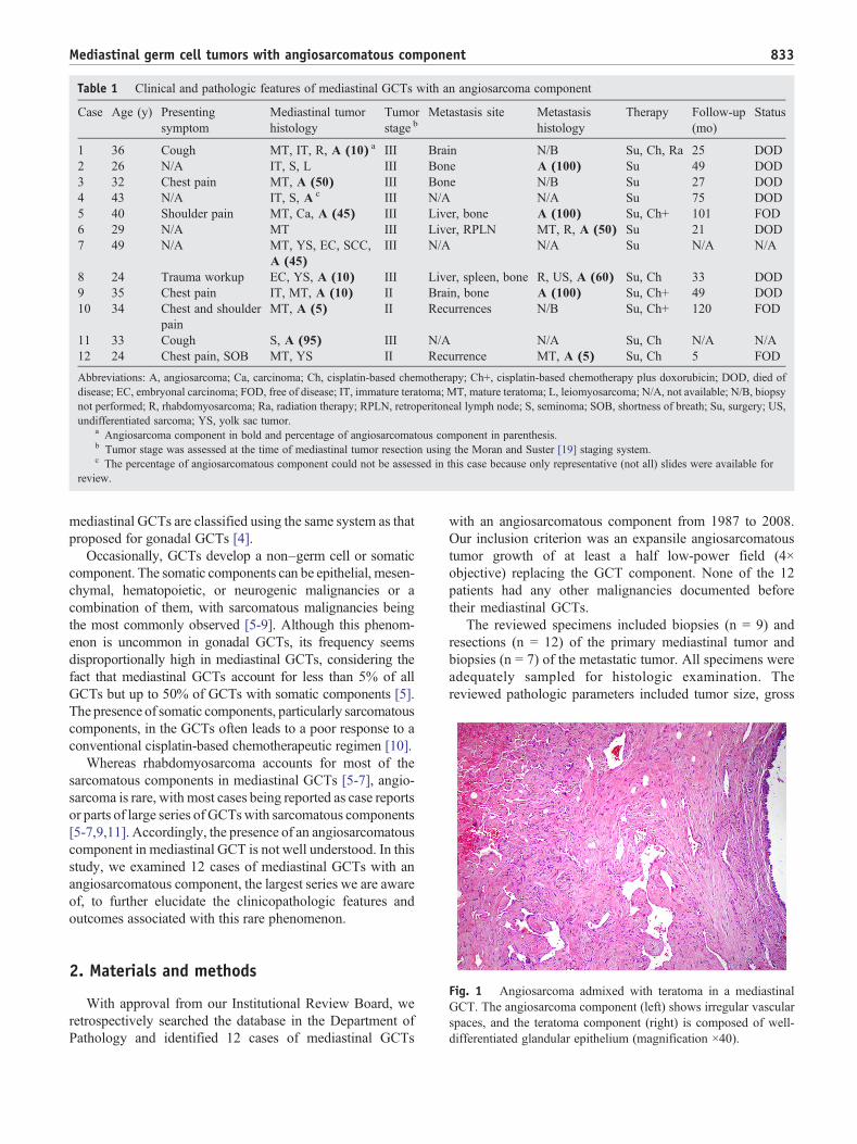
Table 1 Clinical and pathologic features of mediastinal GCTs with an angiosarcoma component
Case Age (y) Presentingsymptom
Mediastinal tumorhistology
Tumorstage b
Metastasis site Metastasishistology
Therapy Follow-up(mo)
Status
1 36 Cough MT, IT, R, A (10) a III Brain N/B Su, Ch, Ra 25 DOD2 26 N/A IT, S, L III Bone A (100) Su 49 DOD3 32 Chest pain MT, A (50) III Bone N/B Su 27 DOD4 43 N/A IT, S, A c III N/A N/A Su 75 DOD5 40 Shoulder pain MT, Ca, A (45) III Liver, bone A (100) Su, Ch+ 101 FOD6 29 N/A MT III Liver, RPLN MT, R, A (50) Su 21 DOD7 49 N/A MT, YS, EC, SCC,
A (45)III N/A N/A Su N/A N/A
8 24 Trauma workup EC, YS, A (10) III Liver, spleen, bone R, US, A (60) Su, Ch 33 DOD9 35 Chest pain IT, MT, A (10) II Brain, bone A (100) Su, Ch+ 49 DOD10 34 Chest and shoulder
painMT, A (5) II Recurrences N/B Su, Ch+ 120 FOD
11 33 Cough S, A (95) III N/A N/A Su, Ch N/A N/A12 24 Chest pain, SOB MT, YS II Recurrence MT, A (5) Su, Ch 5 FOD
Abbreviations: A, angiosarcoma; Ca, carcinoma; Ch, cisplatin-based chemotherapy; Ch+, cisplatin-based chemotherapy plus doxorubicin; DOD, died ofdisease; EC, embryonal carcinoma; FOD, free of disease; IT, immature teratoma; MT, mature teratoma; L, leiomyosarcoma; N/A, not available; N/B, biopsynot performed; R, rhabdomyosarcoma; Ra, radiation therapy; RPLN, retroperitoneal lymph node; S, seminoma; SOB, shortness of breath; Su, surgery; US,undifferentiated sarcoma; YS, yolk sac tumor.
a Angiosarcoma component in bold and percentage of angiosarcomatous component in parenthesis.b Tumor stage was assessed at the time of mediastinal tumor resection using the Moran and Suster [19] staging system.c The percentage of angiosarcomatous component could not be assessed in this case because only representative (not all) slides were available for
review.
833Mediastinal germ cell tumors with angiosarcomatous component
mediastinal GCTs are classified using the same system as thatproposed for gonadal GCTs [4].
Occasionally, GCTs develop a non–germ cell or somaticcomponent. The somatic components can be epithelial, mesen-chymal, hematopoietic, or neurogenic malignancies or acombination of them, with sarcomatous malignancies beingthe most commonly observed [5-9]. Although this phenom-enon is uncommon in gonadal GCTs, its frequency seemsdisproportionally high in mediastinal GCTs, considering thefact that mediastinal GCTs account for less than 5% of allGCTs but up to 50% of GCTs with somatic components [5].The presence of somatic components, particularly sarcomatouscomponents, in the GCTs often leads to a poor response to aconventional cisplatin-based chemotherapeutic regimen [10].
Whereas rhabdomyosarcoma accounts for most of thesarcomatous components in mediastinal GCTs [5-7], angio-sarcoma is rare, with most cases being reported as case reportsor parts of large series of GCTswith sarcomatous components[5-7,9,11]. Accordingly, the presence of an angiosarcomatouscomponent in mediastinal GCT is not well understood. In thisstudy, we examined 12 cases of mediastinal GCTs with anangiosarcomatous component, the largest series we are awareof, to further elucidate the clinicopathologic features andoutcomes associated with this rare phenomenon.
Fig. 1 Angiosarcoma admixed with teratoma in a mediastinalGCT. The angiosarcoma component (left) shows irregular vascularspaces, and the teratoma component (right) is composed of well-differentiated glandular epithelium (magnification ×40).
2. Materials and methods
With approval from our Institutional Review Board, weretrospectively searched the database in the Department ofPathology and identified 12 cases of mediastinal GCTs
with an angiosarcomatous component from 1987 to 2008.Our inclusion criterion was an expansile angiosarcomatoustumor growth of at least a half low-power field (4×objective) replacing the GCT component. None of the 12patients had any other malignancies documented beforetheir mediastinal GCTs.
The reviewed specimens included biopsies (n = 9) andresections (n = 12) of the primary mediastinal tumor andbiopsies (n = 7) of the metastatic tumor. All specimens wereadequately sampled for histologic examination. Thereviewed pathologic parameters included tumor size, gross

834 A. L. Contreras et al.
appearance, stage, GCT components, angiosarcomatouscomponents, and other somatic components. In some cases,immunohistochemical staining was performed using theavidin-biotin-peroxidase complex method in a Dako Auto-Stainer (Dako, Carpinteria, CA). The primary antibodiesused were antibodies to CD31 (Dako, 1:20 dilution), CD34(Dako, 1:50 dilution), and factor VIII (Dako, 1:100 dilution).Clinical data, including demographics, presenting symp-toms, radiographic studies, treatment, and follow-up infor-mation, were obtained from the medical records. Overallsurvival times were measured in months from the date ofinitial diagnosis to death or last available follow-up.
3. Results
All 12 patients were men with a mean age of 34 years(median, 34 years; range, 24-49 years) (Table 1). No patienthad a documented testicular GCT. Presenting symptomswere available for 8 patients and included chest pain (n = 4),shoulder pain (n = 2), cough (n = 2), and shortness of breath(n = 1). In 1 patient, a mediastinal tumor was found
Fig. 2 Angiosarcoma showing irregular vascular spaces that anastomocells are immunoreactive for CD 31 (B), CD 34 (C), and factor VIII (D)
incidentally during a workup after an automobile accident. Inall patients, a mediastinal tumor was demonstrated byradiographic studies. Nine patients underwent core needlebiopsies, which demonstrated GCT only (no sarcomatouscomponent, n = 6), GCT with an angiosarcomatouscomponent (n = 1), angiosarcoma only (n = 1), andundifferentiated sarcoma only (n = 1).
In all patients, the mediastinal tumor was resected. Themean size of tumor was 12.9 cm in the largest dimension(median, 14.5 cm; range, 5.5-16.0 cm). Grossly, the tumorswere cystic with variegated hemorrhagic, mucinous, andfleshy solid areas. Microscopically, the most common GCTcomponents were mature (n = 8) and immature (n = 6)teratomas. Other GCT components, including seminoma (n =3), yolk sac tumor (n = 3), and embryonal carcinoma (n = 2),were also observed (Table 1). The angiosarcomatouscomponent was present in the primary mediastinal tumor(n = 9), in metastasis (n = 3), and in both the primarymediastinal tumor and metastasis (n = 3). In the primarymediastinal tumor, the angiosarcomatous componentaccounted for an average of 30% (range, 5%-95%) of thetumor and was often admixed with GCT components (Fig. 1).The angiosarcomatous components varied in appearance,
se and branch in a sinusoidal fashion (A). The atypical endothelial(magnification ×200).

Fig. 3 Angiosarcoma showing high-grade cellular atypia withpleomorphic and hyperchromatic nuclei (magnification ×200).
835Mediastinal germ cell tumors with angiosarcomatous component
ranging from clearly vasoformative to poorly differentiatedareas in which the vascular nature was not readily recogniz-able. In most areas, the angiosarcoma components werecharacterized by irregular vascular spaces that freely anasto-mosed and branched in a sinusoidal fashion (Fig. 2A). Thecells lining the vascular spaces ranged from spindle toepithelioid and often displayed high-grade cellular atypia withpleomorphic and hyperchromatic nuclei (Fig. 3). On immu-nostaining (n = 5), the lining cells were positive for endothelialmarkers CD 31 (Fig. 2B), CD 34 (Fig. 2C), and factor VIII(Fig. 2D). In focal areas, the angiosarcoma was composed ofclosely packed epithelioid cells with poorly formed vascularspaces (Fig. 4). In addition, focal areas of rhabdomyosarcoma(n = 1), poorly differentiated carcinoma (n = 1), orleiomyosarcoma (n = 1) were also observed in 3 cases.
Follow-up information was available for 10 patients witha mean follow-up of 51 months (median, 41 months; range,
Fig. 4 Angiosarcoma showing various differentiations. The well-differentiated area shows easily recognizable vascular spaces linedby spindle endothelial cells. The poorly differentiated area iscomposed of closely packed epithelioid cells with rudimentaryformation of vascular spaces (magnification ×200).
5-120 months) (Table 1). Seven patients received cisplatin-based chemotherapy for GCTs; and 3 of them also receiveddoxorubicin, a sarcoma-oriented drug. In addition, 1 patientreceived radiation therapy. Despite multimodality therapies,patients developed metastasis (n = 8) or local recurrence (n =2). The most common metastatic sites were the bones (n = 5),liver (n = 3), and brain (n = 2). In 6 cases, the tumor alsoinvolved the lungs, as a result of direct tumor extension ormetastasis. A biopsy was performed on the metastatic tumorin 6 cases, and angiosarcoma was present in all cases. Sevenpatients died of disease at a mean of 33 months after thediagnosis (median, 40 months; range, 21-75 months). Theother 3 patients were alive at a mean of 75 months (median,101 months; range, 5-120 months); 2 of them had receiveddoxorubicin. The clinical outcome was related to the tumorstage, but it was not associated with the amount ofangiosarcomatous component in the mediastinal GCT.
4. Discussion
The origin of mediastinal GCTs remains unclear.Although a small mediastinal GCT may be entirely encasedin the thymus, there is no histogenetic relationship betweenmediastinal GCT and true thymoma [12]. Recent studieshave suggested that a mediastinal GCT may arise fromresidual primordial germ cells in the dorsal midline of thebody [13,14]. During embryogenesis, most primordial germcells migrate laterally to the genital ridges to becomespermatogenetic or ovogenetic stem cells; the cells remain-ing in the midline usually undergo apoptosis [13]. However,this apoptotic process may be disrupted when the ectopicgerm cells are exposed to carcinogenic agents. A recent studyfound that the absence of Bax, a proapoptotic Bcl-2 familymember, prevented germ cells from undergoing apoptosis,resulting in large numbers of extragonadal germ cells [14].
Mediastinal GCTs demonstrate histologic and serologiccharacteristics similar to those of gonadal GCTs [15]. Inaddition, most mediastinal GCTs possess an isochromosomeof 12p, a characteristic cytogenetic alteration that isconsistently present in testicular GCTs [16,17]. Therefore,the possibility of the mediastinal GCT representing ametastasis from a gonadal primary GCT should beconsidered. Whereas the presence of a single tumor in theanterior upper mediastinum with no retroperitoneal involve-ment would favor a mediastinal primary, the presence of acoexisting or preexisting gonadal GCT would suggest thatthe mediastinal GCT represents a metastasis. However, itmay be difficult to recognize metastasis from a “burnt-out”gonadal GCT, in which the primary tumor has regressed inthe gonadal gland [18].
The staging system for mediastinal GCT largely followsthe tumor-node-metastasis classification system used for softtissue tumors [4]. Recently, Moran and Suster [19] proposeda different clinical staging system in which mediastinal

836 A. L. Contreras et al.
GCTs are divided into 3 stages: in stage I, tumors areconfined to the mediastinum; in stage II, tumors invade thesurrounding structures, such as the pleura, pericardium, orlarge vessels; and in stage III, tumors show evidence ofintrathoracic or extrathoracic metastases [19]. According tothis classification system, 9 patients in our study had a stageIII disease; and 3 patients had a stage II disease. Six of the 9patients at stage III died within 21 to 75 months, 1 patientsurvived, and 2 were lost to follow-up. Two of the 3 patientsat stage II were alive at 5 and 120 months, and 1 died at 75months. Our results suggest that this staging system is alsouseful for predicting the clinical outcome of mediastinalGCTs with an angiosarcomatous component.
There are several hypotheses regarding the origin of anangiosarcomatous component in GCTs [9]. In our study, mostGCTs (10/12) contained a teratomatous component, suggest-ing that the angiosarcomatous component may represent amalignant transformation of teratomatous component. None-theless, 2 cases of GCT lacked a teratomatous component,raising the possibility that the angiosarcomatous componentmay also develop from the dedifferentiation of nonteratoma-tous GCT components. Cytogenetic studies have demon-strated the presence of i(12p) abnormalities in somaticcomponents [7]. Because i(12p) is a specific genetic markerpresent in 80% of GCTs [16], its prevalence in the somaticcomponents supports the idea that the somatic componentarises from GCT cells. Others have suggested that theangiosarcomatous component may also develop in themediastinal GCTs as a consequence of radiation therapy[9]. In our study, only 1 patient had a documented history ofradiation therapy; but he had an angiosarcomatous compo-nent in the mediastinal tumor before radiation therapy.Therefore, the development of the angiosarcomatous com-ponent was not related to radiation therapy in this patient.
The angiosarcomatous component in mediastinal GCT ischaracterized by freely anastomosing vascular spaces linedby atypical endothelial cells. Interestingly, there are alsosome atypical cells in the stroma that do not line the vascularspaces. These atypical stromal cells lack immunoreactivityfor endothelial markers (CD 31, CD 34, or factor VIII).When the atypical stromal cells are scattered and do not forma pure nodule, we consider them to be a part of the stromathat is associated with a teratoma. Only when the atypicalstromal cells form a pure nodule of substantial size (largerthan a half field viewed with a 4× objective) are theyconsidered to represent a sarcomatoid transformation. Usingthis definition, we found that 2 patients in the current studyalso developed other sarcomatoid transformations, includingrhabdomyosarcoma and leiomyosarcoma; and 1 patientdeveloped a poorly differentiated carcinoma.
The angiosarcomatous component may develop in aprimary mediastinal GCT or in metastasis. In this study, 9patients showed an angiosarcomatous component in theprimary mediastinal GCT, whereas 6 patients had anangiosarcomatous component in metastasis. Interestingly, 3patients did not have an angiosarcomatous component in the
primary mediastinal tumor, but subsequently developed anangiosarcomatous component in metastasis. Although thisdiscrepancy could be due to inadequate sampling of theprimary mediastinal tumors, an alternative explanation is theemergence of an angiosarcomatous component in metastasisdue to drug selection. Patients with advanced mediastinalGCT often receive cisplatin-based chemotherapy, which ishighly effective against conventional GCT components.However, sarcomatous components are notoriously resistantto cisplatin-based chemotherapy, which allows a growthadvantage of sarcomatous components over conventionalGCT components.
Previous studies have found that the presence of asarcomatous component in a mediastinal GCT carries adismal prognosis [5-7]. In the 25 cases of mediastinal GCTwith sarcomatous components that Malagón et al [5].reported, 16 patients died of tumor at a mean of 13 months,3 patients were alive at a mean of 25 months, and 5 patientswere lost in the follow-up after diagnosis. Likewise, patientswith an angiosarcomatous component in mediastinal GCTsalso have a poor clinical outcome. Manivel et al [6] reported3 cases of mediastinal GCT with angiosarcomatous compo-nents, and all patients died of disease within 9 months. In thecurrent study, despite surgical treatment and chemotherapy,all 10 patients who had follow-up information developedeither metastasis or local recurrence; and 7 died of disease.Of the 7 patients who died, 4 had a biopsy of the metastasis.An angiosarcomatous component was present in all 4 cases.In addition, other components, including mature teratoma,rhabdomyosarcoma, and undifferentiated sarcoma, were alsopresent in 2 of these cases. Although the angiosarcomatouscomponent is a plausible cause of death in the patients, itcannot be ruled out that some patients might die of othersarcomas or even progressive GCT of the usual type. Thepoor clinical outcome is indicated whether the angiosarco-matous component is present in a primary mediastinal GCTor in its metastasis. Furthermore, the same outcome isobserved in mediastinal GCTs with even a small amount ofangiosarcomatous component, such as 5% of the tumor.
The presence of a somatic component in GCT is a majorchallenge in disease management. Although most GCTs arehighly sensitive to cisplatin-based chemotherapy, somatic,particularly sarcomatous, components are resistant to thistreatment. Recent studies found that patients with a singlehistologic type of somatic component could achieve long-term survival when they were treated with a chemothera-peutic regimen based on the specific histologic type ofsomatic component [20,21]. In our study, 3 patients receivedchemotherapy containing doxorubicin, a drug that has beensuccessfully used to treat sarcomas [22]; and 2 achievedlong-term survival. Therefore, our limited retrospectivestudy suggests that patients with an angiosarcomatouscomponent may benefit from sarcoma-oriented drugs,although this should be verified in large studies.
In summary, our study represents the largest series ofmediastinal GCTs with an angiosarcomatous component to

837Mediastinal germ cell tumors with angiosarcomatous component
date. The angiosarcomatous component may develop in aprimary mediastinal GCT or in its metastasis. The highprevalence of a teratomatous component in the mediastinalGCTs with an angiosarcomatous component suggests thatthe angiosarcomatous component may arise from malignanttransformation of a teratomatous component, althoughalternative origins are also possible. The presence of anangiosarcomatous component in mediastinal GCT, even in asmall amount, is associated with metastasis and localrecurrence and carries a poor clinical outcome.
References
[1] Bokemeyer C, Nichols CR, Droz JP, et al. Extragonadal germ celltumors of the mediastinum and retroperitoneum: results from aninternational analysis. J Clin Oncol 2002;20:1864-73.
[2] Gonzales-Crussi F, Winkler RF, Mirkin DL. Sacrococcygeal teratomasin infants and children. Arch Pathol Lab Med 1978;102:420-5.
[3] Parwani AV, Baisden BL, Erozan YS, et al. Pineal gland lesions: acytopathologic study of 20 specimens. Cancer 2005;105:80-6.
[4] Travis WD, Brambilla E, Muller-Hermelink HK, et al, editors. WorldHealth Organization classification of tumours, pathology and genetics:tumours of the lung, pleura, thymus and heart. Lyon, France: IARCPress; 2004.
[5] Malagón HD, Valdez AM, Moran CA, et al. Germ cell tumors withsarcomatous components: a clinicopathologic and immunohistochem-ical study of 46 cases. Am J Surg Pathol 2007;31:1356-62.
[6] Manivel C, Wick MR, Abenoza P, et al. The occurrence ofsarcomatous components in primary mediastinal germ cell tumors.Am J Surg Pathol 1986;10:711-7.
[7] Motzer RJ, Amsterdam A, Prieto V, et al. Teratoma with malignanttransformation: diverse malignant histologies arising in men with germcell tumors. J Urol 1998;159:133-8.
[8] True LD, Otis CN, Delprado W, et al. Spermatocytic seminoma oftestis with sarcomatous transformation. A report of five cases. Am JSurg Pathol 1988;12:75-82.
[9] Ulbright TM, Clark SA, Einhorn LH. Angiosarcoma associated withgerm cell tumors. HUM PATHOL 1985;16:268-72.
[10] Gonzalez-Vela JL, Savage PD, Manivel JC, et al. Poor prognosis ofmediastinal germ cell cancers containing sarcomatous components.Cancer 1990;66:1114-6.
[11] Saito A, Watanabe K, Kusakabe T, et al. Mediastinal mature teratomawith coexistence of angiosarcoma, granulocytic sarcoma and ahematopoietic region in the tumor: a rare case of association betweenhematological malignancy and mediastinal germ cell tumor. Pathol Int1998;48:749-53.
[12] Rosai J, Parkash V, Reuter VE. The origin of mediastinal germ celltumors in men. Int J Surg Pathol 1994;2:73-8.
[13] Molyneaux KA, Stallock J, Schaible K, et al. Time-lapse analysis ofliving mouse germ cell migration. Dev Biol 2001;240:488-98.
[14] Runyan C, Gu Y, Shoemaker A, et al. The distribution and behavior ofextragonadal primordial germ cells in Bax mutant mice suggest a novelorigin for sacrococcygeal germ cell tumors. Int J Dev Biol 2008;52:333-44.
[15] Malagón HD, Montiel DP. Mediastinal germ cell tumors. Semin DiagnPath 2005;22:230-40.
[16] Bosl GJ, Ilson DH, Rodriguez E, et al. Clinical relevance of the i(12p)marker chromosome in germ cell tumors. J Natl Cancer Inst 1994;86:349-55.
[17] Schneider DT, Schuster AE, Fritsch MK, et al. Genetic analysis ofmediastinal nonseminomatous germ cell tumors in children andadolescents. Genes Chromosomes Cancer 2002;34:115-25.
[18] Balzer BL, Ulbright TM. Spontaneous regression of testicular germcell tumors: an analysis of 42 cases. Am J Surg Pathol 2006;30:858-65.
[19] Moran CA, Suster S. Primary germ cell tumors of the mediastinum: I.Analysis of 322 cases with special emphasis on teratomatous lesionsand a proposal for histopathologic classification and clinical staging.Cancer 1997;80:681-90.
[20] Donadio AC, Motzer RJ, Bajorin DF, et al. Chemotherapy for teratomawith malignant transformation. J Clin Oncol 2003;21:4285-91.
[21] El Mesbahi O, Terrier-Lacombe MJ, Rebischung C, et al. Chemo-therapy in patients with teratoma with malignant transformation. EurUrol 2007;51:1306-11.
[22] Sarcoma Meta-analysis Collaboration. Adjuvant chemotherapy forlocalised resectable soft-tissue sarcoma of adults: meta-analysis ofindividual data. Lancet 1997;350:1647-54.
Copyright © 2022 FDOKUMEN